www.atmos-chem-phys.net/16/14079/2016/ doi:10.5194/acp-16-14079-2016
© Author(s) 2016. CC Attribution 3.0 License.
In vitro exposure to isoprene-derived secondary organic aerosol by
direct deposition and its effects on
COX-2
and
IL-8
gene expression
Maiko Arashiro1, Ying-Hsuan Lin1,6, Kenneth G. Sexton1, Zhenfa Zhang1, Ilona Jaspers1,2,3,4,5, Rebecca C. Fry1,3, William G. Vizuete1, Avram Gold1, and Jason D. Surratt1
1Department of Environmental Sciences and Engineering, Gillings School of Global Public Health,
University of North Carolina at Chapel Hill, Chapel Hill, NC 27599, USA
2Center for Environmental Medicine, Asthma, and Lung Biology, School of Medicine, University of North Carolina
at Chapel Hill, Chapel Hill, NC 27599, USA
3Curriculum in Toxicology, University of North Carolina at Chapel Hill, Chapel Hill, NC 27599, USA
4Department of Pediatrics, School of Medicine, University of North Carolina at Chapel Hill, Chapel Hill, NC 27599, USA 5Department of Microbiology and Immunology, School of Medicine, University of North Carolina at Chapel Hill,
Chapel Hill, NC 27599, USA
6Michigan Society of Fellows, Department of Chemistry, University of Michigan, Ann Arbor, MI 48109, USA
Correspondence to:Jason D. Surratt ([email protected])
Received: 13 May 2016 – Published in Atmos. Chem. Phys. Discuss.: 17 May 2016 Revised: 4 October 2016 – Accepted: 18 October 2016 – Published: 15 November 2016
Abstract.Atmospheric oxidation of isoprene, the most abun-dant non-methane hydrocarbon emitted into Earth’s atmo-sphere primarily from terrestrial vegetation, is now recog-nized as a major contributor to the global secondary or-ganic aerosol (SOA) burden. Anthropogenic pollutants sig-nificantly enhance isoprene SOA formation through acid-catalyzed heterogeneous chemistry of epoxide products. Since isoprene SOA formation as a source of fine aerosol is a relatively recent discovery, research is lacking on evalu-ating its potential adverse effects on human health. The ob-jective of this study was to examine the effect of isoprene-derived SOA on inflammation-associated gene expression in human lung cells using a direct deposition exposure method. We assessed altered expression of inflammation-related genes in human bronchial epithelial cells (BEAS-2B) exposed to isoprene-derived SOA generated in an out-door chamber facility. Measurements of gene expression of known inflammatory biomarkers interleukin 8 (IL-8) and cyclooxygenase 2 (COX-2) in exposed cells, together with complementary chemical measurements, showed that a dose of 0.067 µg cm−2 of SOA from isoprene photooxidation leads to statistically significant increases inIL-8and COX-2mRNA levels. Resuspension exposures using aerosol filter extracts corroborated these findings, supporting the
conclu-sion that isoprene-derived SOA constituents induce the ob-served changes in mRNA levels. The present study is an at-tempt to examine the early biological responses of isoprene SOA exposure in human lung cells.
1 Introduction
Recent work has shown that isoprene (2-methyl-1,3-butadiene) is an important precursor of secondary or-ganic aerosol (SOA), which has potential impacts on cli-mate change and public health (Lin et al., 2013b, 2016; Rohr, 2013). Current understanding of isoprene SOA for-mation is based on laboratory studies showing that gas-phase photooxidation of isoprene generates key SOA pre-cursors, including isomeric isoprene epoxydiols (IEPOX), methacrylic acid epoxide (MAE), hydroxymethyl-methyl-α-lactone (HMML), and isoprene hydroxyhydroperoxides (ISOPOOH) (Paulot et al., 2009; Surratt et al., 2010; Lin et al., 2012, 2013b; Nguyen et al., 2015; Krechmer et al., 2015; Liu et al., 2016; Riva et al., 2016). The formation of SOA from these precursors is influenced by controllable anthro-pogenic emissions such as oxides of nitrogen (NOx)and
to particle acidity, which enhances isoprene SOA formation through acid-catalyzed reactive uptake and multiphase chem-istry of IEPOX and MAE (Surratt et al., 2007, 2010; Lin et al., 2012; Gaston et al., 2014; Riedel et al., 2015), while NOx
determines whether the oxidation pathway leading to IEPOX or MAE/HMML predominates (Lin et al., 2013b; Surratt et al., 2010; Nguyen et al., 2015). Isoprene SOA comprises a large portion of global atmospheric fine particles (PM2.5,
aerosol with aerodynamic diameters ≤2.5 µm) (Carlton et al., 2009; Henze et al., 2008), but few studies have focused on its health implications (Lin et al., 2016). Evaluating the health effects of SOA from isoprene oxidation is important from a public health perspective, not only because of its atmospheric abundance but also because the anthropogenic contribution is the only component amenable to control (Pye et al., 2013; Gaston et al., 2014; Xu et al., 2015; Riedel et al., 2015).
Many studies have shown that particulate matter is closely linked to health effects ranging from exacerbation of asthma symptoms to mortality associated with lung cancer and car-diopulmonary disease (Dockery et al., 1993; Schwartz et al., 1993; Samet et al., 2000). PM2.5, in particular, has been
linked to negative health outcomes with an estimated con-tribution of 3.2 million premature deaths worldwide as re-ported in the Global Burden of Disease Study 2010 (Lim et al., 2012). Despite evidence that particle composition af-fects toxicity, fewer studies focus on the link between chem-ical composition and health/biologchem-ical outcomes (Kelly and Fussell, 2012). Prior work on complex air mixtures has shown that gaseous volatile organic compounds (VOCs) al-ter the composition and ultimately the toxicity of particles (Ebersviller et al., 2012a, b). SOA resulting from natural and anthropogenic gaseous precursors, such asα-pinene and 1,3,5-trimethylbenzene, has been shown to affect cellular function (Gaschen et al., 2010; Jang et al., 2006) and recently isoprene SOA formed from the reactive uptake of epoxides has been shown to induce the expression of oxidative stress genes (Lin et al., 2016).
The objective of this study is to generate atmospheri-cally relevant isoprene-derived SOA and examine its tox-icity through in vitro exposures using a direct deposition device. Compared to exposure of cells in culture media to resuspended particles, direct particle deposition likely pro-vides a more biologically relevant exposure model and en-hances sensitivity of cells to air pollution particle exposures (Volckens et al., 2009; Lichtveld et al., 2012; Hawley et al., 2014a, b; Zavala et al., 2014; Hawley and Volckens, 2013). The Electrostatic Aerosol in Vitro Exposure System (EAVES) used in this study deposits particles, generated in our outdoor photochemical chamber, directly onto lung cells by electrostatic precipitation (de Bruijne et al., 2009). Sim-ilar techniques and devices have been used to expose cells to diesel exhaust particles (Lichtveld et al., 2012; Hawley et al., 2014b), but our study is the first to utilize the EAVES to explore the potential adverse effects of isoprene SOA on
human lung cells. Additionally, for a more atmospherically relevant exposure, isoprene SOA was photochemically gen-erated in an outdoor chamber to mimic its formation in the atmosphere.
We have recently demonstrated through a chemical assay that isoprene-derived SOA has the potential for inducing re-active oxygen species (ROS) (Kramer et al., 2016), which are linked to oxidative stress and inflammation (Reuter et al., 2010; Li et al., 2003). An in vitro study that followed supported the potential for isoprene SOA to affect the levels of oxidative stress genes (Lin et al., 2016). In this study we chose to examine the gene expression levels of interleukin-8 (IL-8) and cyclooxygenase-2 (COX-2), not only for their links to inflammation and oxidative stress (Kunkel et al., 1991; Uchida, 2008) but also because both have been ex-amined in previous studies using the EAVES for fresh and aged diesel exhaust (Lichtveld et al., 2012). Other studies on air pollution mixtures have also examinedIL-8as a bi-ological endpoint due to its involvement with inflammation (Zavala et al., 2014; Ebersviller et al., 2012a, b; Doyle et al., 2004, 2007). We compared the gene expression levels in cells exposed to SOA generated in an outdoor chamber from photochemical oxidation of isoprene in the presence of NO and acidified sulfate seed aerosol to cells exposed to a dark control mixture of isoprene, NO, and acidified sul-fate seed aerosol to isolate the effects of the isoprene-derived SOA on the cells using the EAVES. In addition, we collected SOA onto filters for subsequent resuspension exposure to en-sure that effects observed from EAVES expoen-sures were at-tributable to particle-phase organic products.
2 Experimental section
2.1 Generation of SOA in the outdoor chamber facility
SOA was generated by photochemically oxidizing a mixture of acidified sulfate seed aerosol, isoprene, and NO injected into an outdoor smog chamber facility. The outdoor chamber is a 120 m3triangular cross-section Teflon chamber located on the roof of the Gillings School of Global Public Health, University of North Carolina at Chapel Hill. The chamber facility has been described in detail elsewhere by Lichtveld et al. (2012). The outdoor chamber facility is equipped with sampling lines that allow direct deposition exposure of cells, online chemical measurements, and filter collection for of-fline chemical analysis. Sampling lines run from the un-derside of the chamber directly to the chemistry lab below, where online measurement instruments and the direct depo-sition exposure device are located. Injection ports are also located on the underside of the chamber.
nebuliz-ing an aqueous solution containnebuliz-ing 0.06 M MgSO4+0.06 M
H2SO4 into the chamber to a particle concentration of
ap-proximately 170 µg m−3, which was allowed to stabilize for
30 min to ensure a well-mixed condition. After stabiliza-tion, 3.5 ppmv isoprene (Sigma-Aldrich, 99 %) and 200 ppbv NO (AirGas, 1.00 %) were injected into the chamber. Photo-chemical aging was allowed for approximately 1 h to reach the desired exposure conditions of 30–40 µg m−3 growth of isoprene-derived SOA on the pre-existing 170 µg m−3of acidified sulfate aerosol. This chamber experiment was repli-cated on three separate sunny days with temperatures ranging from 24.9 to 26.8◦C with a RH of approximately 70 % in the chamber.
2.2 Control chamber experiments
As a dark chamber control, to isolate the effect of SOA on exposed cells, mixtures of isoprene, NO, and 170 µg m−3of
acidified sulfate seed aerosol were injected into the cham-ber in the dark (after sunset). Conducting the chamcham-ber ex-periments in the dark ensured no photochemical oxidation of isoprene. The dark control was replicated on three different nights. Except for the absence of solar radiation (no SOA), all chamber operations and exposure conditions were similarly maintained.
As an added control to ensure that the device itself and the cell handling had no significant effect on cell cytotoxi-city, cells were exposed in the EAVES to a clean chamber and compared to unexposed cells kept in an incubator for the same duration as the exposure. The cytotoxicity results en-sured that there is no effect of chamber conditions and device operation on the cells.
2.3 Cell culture
Human bronchial epithelial (BEAS-2B) cells were main-tained in keratinocyte growth medium (KGM BulletKit; Lonza), a serum-free keratinocyte basal medium (KBM) supplemented with 0.004 % of bovine pituitary extract and 0.001 % of human epidermal growth factor, insulin, hydro-cortisone, and GA-1000 (gentamicin, amphotericin B), and passaged weekly. Passage number for photochemical expo-sures and dark control expoexpo-sures varied between 52 and 60. Because BEAS-2B are an immortalized line of human bronchial epithelium, there are limitations with its use such as it being genetically homogeneous, being a single cell type, and being SV-40-transformed (Reddel et al., 1988). However, BEAS-2B is a stable, proliferative cell line shown to be use-ful in airway inflammation studies such as ours (Devlin et al., 1994).
2.4 Direct deposition exposure
In preparation for air–liquid interface exposures, cells were seeded onto collagen-coated Millicell cell culture inserts (30 mm diameter, 0.4 µm pore size, 4.2 cm2filter area;
Milli-pore, Cambridge, MA) at a density of 2.0×10⁵cells per well 24 h prior to exposure. At the time of exposure, cells reached ∼80 % confluence, confirmed through microscopy. Immedi-ately before exposure, cell medium was removed from the apical and basolateral sides of two seeded Millicell cell cul-ture inserts. One insert was transferred to a titanium dish con-taining 1.5 mL of keratinocyte basal medium (KBM; Lonza), supplying cells with nutrients from the basolateral side and constant moisture while allowing exposure to be performed at an air–liquid interface. The other insert was transferred into a six-well plate with 2 mL of KBM and placed in the incubator as an unexposed control.
Cells were exposed to chamber-generated isoprene SOA using the EAVES located in the laboratory directly beneath the outdoor chamber (de Bruijne et al., 2009; Lichtveld et al., 2012). The EAVES, located in an incubator at 37◦C, sam-pled chamber air at 1 L min−1. The target RH in the chamber
during EAVES exposures was approximately 70 %. Exposure time was 1 h commencing when target exposure conditions were achieved in the outdoor chamber for both photochemi-cal and dark control experiments. Detailed description of the EAVES can be found in de Bruijne et al. (2009).
Following exposure, the cell culture insert was transferred to a six-well tissue culture plate containing 2 mL of fresh KBM. The control Millicell was also transferred to 2 mL of fresh KBM. Nine hours post-exposure, extracellular medium was collected and total RNA was isolated using Trizol (Life Technologies), consistent with past studies (de Bruijne et al., 2009). Extracellular medium and the extracted RNA samples were stored at−20 and−80◦C, respectively, until further analysis. For quality assurance purposes, the RNA concen-tration and integrity were assessed using a Nanodrop 2000c spectrophotometer (Thermo Scientific) and a Bioanalyzer 2100 (Agilent Technologies) over the period of storage. No changes were observed under the given storage conditions.
2.5 Filter resuspension exposure
Chamber particles were collected, concurrently with EAVES sampling, onto Teflon membrane filters (47 mm diameter, 1.0 µm pore size; Pall Life Science) for photochemical (light) and dark chamber experiments to be used for chemical anal-ysis and resuspension exposures. The resuspension experi-ments served as a control for possible effects of gaseous com-ponents such as ozone (O3)and NOxpresent in the direct
The particles collected on Teflon filter membranes for resus-pension cell exposure were extracted by sonication in high-purity methanol (LC/MS CHROMASOLV, Sigma-Aldrich). Filter samples from multiple experiments were combined and the combined filter extract was dried under a gentle stream of nitrogen (N2). KBM medium was then added into
the extraction vials to re-dissolve SOA constituents.
In preparation for filter resuspension exposures, cells were seeded in 24-well plates at a density of 2.5×10⁴ cells per well in 250 µL of KGM 2 days prior to exposure. At the time of exposure when cells reached ∼80 % conflu-ence, cells were washed twice with phosphate-buffered saline (PBS) buffer, and then exposed to KBM containing 0.01 and 0.1 mg mL−1isoprene SOA extract from photochemical ex-periment and seed particles from dark control exex-periments.
Following a 9 h exposure, extracellular medium was col-lected and total RNA was isolated using Trizol (Life Tech-nologies) and stored alongside samples from direct deposi-tion exposures until further analysis.
2.6 Chemical and physical characterization of exposures
Online and offline techniques were used to characterize the SOA generated in the chamber. The online techniques mea-sured the gas-phase species (NO, NOx, O3) and the physical
properties of the aerosol continuously throughout the cham-ber experiments. Offline techniques measured aerosol-phase species collected onto Teflon membrane filters (47 mm diam-eter, 1.0 µm pore size; Pall Life Science) from photochemical and dark chamber experiments. Filter samples were stored in 20 mL scintillation vials protected from light at−20◦C until analyses.
Real-time aerosol size distributions were measured using a differential mobility analyzer (DMA, Brechtel Manufactur-ing Inc.) coupled to a mixManufactur-ing condensation particle counter (MCPC, model 1710, Brechtel Manufacturing Inc.) located in the laboratory directly underneath the chamber. O3 and
NOxwere measured with a ML 9811 series ozone
photome-ter (Teledyne Monitor Labs, Englewood, CO) and ML 9841 series NOxanalyzer (American Ecotech, Warren RI),
respec-tively. Data were collected at 1 min intervals using a data acquisition system (ChartScan/1400) interfaced to a com-puter. The presence of isoprene in the chamber was con-firmed and quantified using a Varian 3800 gas chromatograph (GC) equipped with a flame ionization detector (FID).
Chemical characterization of SOA constituents was con-ducted offline from extracts of filters collected from chamber experiments by a gas chromatograph interfaced with an elec-tron ionization quadrupole mass spectrometer (GC/EI-MS) or by an ultra-performance liquid chromatograph interfaced with a high-resolution quadrupole time-of-flight mass spec-trometer equipped with electrospray ionization (UPLC/ESI-HR-QTOFMS). Detailed operating conditions for the GC/EI-MS and UPLC/ESI-HR-QTOFGC/EI-MS analyses as well as
de-tailed filter extraction protocols have been described previ-ously by Lin et al. (2012). For GC/EI-MS analysis, filter ex-tracts were dried under a gentle stream of N2and
trimethylsi-lylated by the addition of 100 µL of BSTFA+TMCS (99 : 1 v/v, Supelco) and 50 µL of pyridine (anhydrous, 99.8 %, Sigma-Aldrich) and heated at 70◦C for 1 h. For UPLC/ESI-HR-QTOFMS analysis, residues of filter extracts were re-constituted with 150 µL of a 50 : 50 (v/v) solvent mixture of high-purity water and methanol.
The isoprene-derived SOA markers – 2-methyltetrols, iso-meric 3-methyltetrahydrofurans-3,4-diols (3-MeTHF-3,4-diols), and 2-methylglyceric acid, synthesized according to the published procedures (Lin et al., 2013b; Zhang et al., 2012) – were available in-house as authentic standards to quantify the major components of isoprene SOA. 2-Methyltetrol organosulfates, synthesized as a mixture of tetrabutylammonium salts, were also available as a stan-dard. Purity was determined to be>99 % by1H NMR and UPLC/ESI-QTOFMS analysis (Budisulistiorini et al., 2015). The C5-alkene triols and IEPOX dimer were quantified using
the response factor obtained for the synthetic 2-methyltetrols. A representative ambient PM2.5sample collected from the
rural southeastern US (Yorkville, GA) (Lin et al., 2013a) dur-ing the summer of 2010 was analyzed in an identical manner to confirm atmospheric relevance of the chamber-generated SOA constituents.
2.7 Cytotoxicity assay
Cytotoxicity was assessed through measurement of lactate dehydrogenase (LDH) released into the extracellular medium from damaged cells using the LDH cytotoxicity detection kit (Takara). To ensure that the EAVES device itself and opera-tion procedure had no effect on cytotoxicity, the LDH release from cells exposed to clean chamber air was measured. LDH release by cells exposed via the EAVES to the photochemi-cally aged (light) and non-photochemiphotochemi-cally aged (dark) par-ticles was compared to release from unexposed cells main-tained in the incubator for the same duration. For the resus-pension exposures, LDH release by cells exposed to SOA through resuspended extract of photochemically aged and non-photochemically aged particles was compared to release by cells maintained in KBM only. Additionally, LDH release from the light exposures, dark control, and resuspension posures was compared to release by positive control cells ex-posed to 1 % Triton X-100 to ensure that cell death would not affect gene expression results.
2.8 Gene expression analysis
have also used mRNA transcripts as a proxy for cytokine production (Hawley et al., 2014a, b; Hawley and Volck-ens, 2013; Volckens et al., 2009; Lichtveld et al., 2012). Changes in IL-8 and COX-2 mRNA levels were mea-sured using a QuantiTect SYBR Green RT-PCR kit (Qi-agen) and QuantiTect primer assays for Hs_ACTB_1_SG (catalog no. QT00095431), Hs_PTGS2_1_SG (catalog no. QT00040586), and Hs_CXCL8_1_SG (catalog no. QT00000322) for one-step RT-PCR analysis. All mRNA lev-els were normalized againstβ-actin mRNA, which was used as a housekeeping gene. The relative expression levels (i.e., fold change) ofIL-8 andCOX-2 were calculated using the comparative cycle threshold (2−11CT) method (Livak and Schmittgen, 2001). For EAVES exposures, changes inIL-8
andCOX-2from isoprene-derived SOA exposed cells were compared to cells exposed to the dark controls. Similarly, for resuspension exposures, changes inIL-8andCOX-2from isoprene-derived SOA exposed cells were compared to cells exposed to particles collected under dark conditions. 2.9 Statistical analysis
The software package GraphPad Prism 4 (GraphPad) was used for all statistical analyses. All data were expressed as mean±SEM (standard error of means). Comparisons be-tween data sets for cytotoxicity and gene expression analy-sis were made using unpairedttest with Welch’s correction. Significance was defined asp <0.05.
3 Results and discussion
3.1 Physical and chemical characterization of exposure
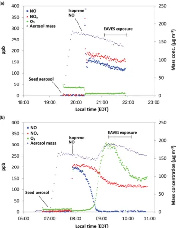
Figure 1 shows the change in particle mass concentration and gas (NO, NOx, O3) concentration over time during
typi-cal photochemitypi-cal and dark control experiments. Under dark control conditions (Fig. 1a) there is no increase in aerosol mass concentration following isoprene injection. Average total aerosol mass concentration was 155.0±2.69 µg m−3 (1 standard deviation) with no particle mass attributable to organic material.
In contrast, Fig. 1b shows an increase in aerosol mass concentration 1 h after isoprene injection, which can be at-tributed to the photochemical oxidation of isoprene and subsequent production and reactive uptake of its oxida-tion products. The average increase in aerosol mass con-centration attributable to SOA formation for three day-light chamber experiments conducted on separate days was 44.5±5.7 µg m−3. Average total aerosol mass concentration during particle exposure was 173.1±4.2 µg m−3.
O3and NOxconcentrations measured during EAVES
ex-posure were approximately 270 and 120 ppb for photochemi-cal experiments. For dark control experiments (e.g., Fig. 1a), the O3and NOx concentrations were approximately 15 and
180 ppb. Previous studies characterizing the EAVES device
Figure 1.Aerosol mass concentration and gas-phase product con-centrations over time for (a) dark control chamber experiment and(b)photochemically produced isoprene-derived SOA exposure chamber experiment.
show definitively that gas-phase products do not induce cell response (de Bruijne et al., 2009). However, resuspension exposures were conducted in addition to EAVES exposure to ensure that biological effects were attributable to only particle-phase constituents and not gas-phase products such as O3and NOx.
The chemical composition of aerosol, collected onto filters concurrently with cell exposure and characterized by GC/EI-MS and UPLC/ESI-HR-QTOFGC/EI-MS, are shown in Fig. 2. No isoprene SOA tracers were observed in the filters collected from dark control experiments. The dominant particle-phase products of the isoprene SOA collected from photochemi-cal experiments are derived from the low-NO channel, where IEPOX reactive uptake onto acidic sulfate aerosol domi-nates, including 2-methyltetrols, C5-alkene triols, isomeric
Figure 2. (a)GC/EI-MS total ion chromatograms (TICs) and(b)UPLC/ESI-HR-QTOFMS base peak chromatograms (BPCs) from a (1) dark control chamber experiment, (2) isoprene-derived SOA exposure chamber experiment, and (3) PM2.5sample collected from Yorkville, GA,
during summer 2010.
formed from the reactive uptake of IEPOX onto acidic sul-fate aerosols. As demonstrated in Fig. 2, all the same particle-phase products are measured in the PM2.5sample collected
in Yorkville, GA (a typical low-NO region), demonstrating that the composition of the chamber-generated SOA is atmo-spherically relevant. Recent SOA tracer measurements from the Southern Oxidant and Aerosol Study (SOAS) campaign at Look Rock, TN; Centerville, AL; and Birmingham, AL, also support the atmospheric relevance of IEPOX-derived SOA constituents that dominate the isoprene SOA mass in summer in the southeastern US (Budisulistiorini et al., 2016; Rattanavaraha et al., 2016).
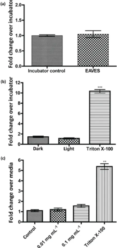
3.2 Cytotoxicity
LDH release for cells exposed using the EAVES device is expressed as a fold change relative to the unexposed incu-bator control. For resuspension exposures, LDH release is expressed as fold change relative to cells exposed to KBM only. Results shown in Fig. 3a confirm that there is no ef-fect of chamber conditions and device operation on the cells when comparing LDH release from cells exposed to a clean air chamber and cells unexposed in an incubator. Addition-ally, LDH release from all exposure conditions in EAVES ex-posed cells (Fig. 3b) and resuspension exex-posed cells (Fig. 3c) is negligible relative to positive controls exposed to 1 % Tri-ton X-100, confirming that the exposure concentration of isoprene-derived SOA utilized in this study was not
cyto-toxic. All cytotoxicity results ensured that exposure condi-tions were not adversely affecting the cells or their gene ex-pression.
3.3 Pro-inflammatory gene expression
Changes in the mRNA levels of IL-8 and COX-2 from cells exposed to isoprene-derived SOA using the EAVES are shown as fold changes relative to dark controls in Fig. 4. This comparison, as well as the results of the resuspension experiment discussed below, ensures that all effects seen in the cells are attributable to the isoprene-derived SOA and no other factors. A 1 h exposure to a mass concentration of approximately 45 µg m−3of organic material was sufficient to significantly alter gene expression of the inflammatory biomarkers in bronchial epithelial cells. Based on deposition efficiency characterized by de Bruijne et al. (2009), the es-timated dose was 0.29 µg cm−2 of total particle mass with 23 % attributable to organic material formed from isoprene photooxidation (0.067 µg cm−2of SOA).
Figure 3. LDH release for (a) clean air controls; (b) EAVES exposures, normalized to incubator control; and (c)resuspension exposures, normalized to KBM-only control. ∗∗ p <0.005 and ∗∗∗
p <0.0005.
0.1 mg mL−1. The statistically significant increase in mRNA expression from the resuspension exposure at 0.1 mg mL−1 confirms that similar fold changes observed for both IL-8
andCOX-2from the EAVES exposures are not attributable to gaseous photooxidation products, such as O3, and support
the characterization of the EAVES as a particle exposure de-vice (de Bruijne et al., 2009).
The similar fold change observed in both the EAVES exposure and resuspension exposure, in addition to con-firming that the biological effects can be attributed to the particle-phase photochemical products (isoprene-derived SOA), suggests that exposure by resuspension is appropriate for isoprene-derived SOA and may yield results similar to di-rect deposition exposures. Unlike diesel particulate extracts, which agglomerate during resuspension exposures, isoprene-derived SOA constituents are water-soluble based on reverse-phase LC separations (Surratt et al., 2006; Lin et al., 2012) and remain well mixed in the cell medium used for expo-sure. Therefore, resuspension exposures do not appear to be a limitation for toxicological assessments of isoprene SOA.
Figure 4.IL-8 and COX-2mRNA expression induced by expo-sure to isoprene-derived SOA using EAVES device all normalized to dark control experiments and against the housekeeping gene,β -actin. All experiments conducted in triplicate.∗∗∗p <0.0005.
Figure 5. IL-8 and COX-2 expression induced by exposure to isoprene-derived SOA using resuspension method all normalized to dark control experiments and against the housekeeping gene,
β-actin. All experiments conducted in triplicate. ∗ p <0.05 and ∗∗
p <0.005.
3.4 Biological implications
The goal of this study was to initially identify potential biological response associated with exposure to isoprene-derived SOA by using a direct exposure device as a model that has both atmospheric and physiological relevance. With this model, a dose of 0.067 µg cm−2 of isoprene SOA in-duced statistically significant increases in IL-8andCOX-2
Our findings are consistent with other studies showing that photochemical oxidation of similar chemical mixtures in-creases toxicity in cell culture models and elevates expres-sion of inflammatory biomarker genes (Lichtveld et al., 2012; Rager et al., 2011). Previous in vitro studies using a gas-phase-only exposure system have shown that gas-phase prod-ucts of isoprene photooxidation significantly enhance cyto-toxicity andIL-8expression (Doyle et al., 2004, 2007).
By choosingIL-8andCOX-2as our genes of interest, we are able to compare our results to other studies of known harmful particle exposures. In a similar study using the EAVES, normal human bronchial epithelial (NHBE) cells exposed to 1.10 µg cm−2 diesel particulate matter showed less than a 2-fold change over controls in bothIL-8and COX-2mRNA expression (Hawley et al., 2014b). In another study, A549 human lung epithelial cells were exposed by direct de-position for 1 h to photochemically aged diesel exhaust par-ticulates at a dose of 2.65 µg cm−2 from a 1980 Mercedes
or a 2006 Volkswagen (Lichtveld et al., 2012). Exposure to aged Mercedes particulates induced a 4-fold change in IL-8 and∼2-fold change in COX-2mRNA expression, while exposure to aged Volkswagen particulates induced a change of ∼1.5-fold in IL-8and 2-fold in COX-2 mRNA expres-sion (Lichtveld et al., 2012). Although the differences in cell types preclude direct comparisons, the finding of significant increases inCOX-2andIL-8expression at doses much lower than reported for comparable increases in gene expression levels induced by photochemically aged diesel particulates is notable.
IL-8andCOX-2are both linked to inflammation and ox-idative stress (Kunkel et al., 1991; Uchida, 2008). IL-8is a potent neutrophil chemotactic factor in the lung and its ex-pression by various cells plays a crucial role in neutrophil re-cruitment leading to lung inflammation (Kunkel et al., 1991).
COX-2 is the inducible form of the cyclooxygenase en-zyme, regulated by cytokines and mitogens, and is responsi-ble for prostaglandin synthesis associated with inflammation (FitzGerald, 2003). Consistent with the reports thatIL-8and
COX-2play important roles in lung inflammation (Nocker et al., 1996; Li et al., 2013), in vivo studies have shown that isoprene oxidation products cause airflow limitation and sen-sory irritation in mice (Rohr et al., 2003). In humans, the role of IL-8andCOX-2in lung inflammation can be associated with diseases such as chronic obstructive pulmonary disease and asthma (Nocker et al., 1996; Peng et al., 2008; Fong et al., 2000).
The mechanism by which isoprene SOA causes elevation of the inflammatory markersIL-8andCOX-2is not yet fully understood. However, recent work from our laboratory us-ing the acellular dithiothreitol (DTT) assay demonstrated that isoprene-derived SOA has significant ROS generation poten-tial (Kramer et al., 2016). High levels of ROS in cells can overwhelm the antioxidant defense and lead to cellular ox-idative stress (Sies, 1991; Bowler and Crapo, 2002; Li et al., 2003). Following the discovery of the potential importance of
isoprene SOA in generating ROS, Lin et al. (2016) showed that isoprene SOA formed from the reactive uptake of epox-ides alters levels of oxidative stress-associated genes, includ-ingCOX-2in human lung cells. Oxidative stress caused by ROS plays a major role in lung inflammation and the induc-tion of oxidative stress can lead toIL-8expression (Tao et al., 2003; Yan et al., 2015). Specifically, oxidants can activate the transcription factor NF-κB, which regulates a wide range of inflammatory genes includingIL-8 and COX-2 (Barnes and Adcock, 1997; Schreck et al., 1992). Therefore, isoprene SOA may cause increases in bothIL-8andCOX-2primarily through an oxidative stress response. Additionally, the rela-tionship betweenIL-8andCOX-2can also explain the ob-served increase inIL-8gene expression as the production of
IL-8 can be stimulated through aCOX-2-dependent mecha-nism in airway epithelial cells (Peng et al., 2008).
In vitro studies such as this one using a direct deposition model cannot fully elucidate mechanisms of lung inflam-mation and potential pathogenesis but serve as a necessary part of hazard characterization, particularly for a complex air mixture that has not been fully studied (Hayashi, 2005; Paur et al., 2011). Ozone exposure studies have shown that com-parable dose and effect measurements forIL-8andCOX-2
can be found between in vivo and in vitro exposures, which adds promise to extrapolating effects seen in vitro to effects in vivo (Hatch et al., 2014). In vivo effects associated with isoprene SOA exposure in vitro cannot be inferred as it is a different system from ozone, so further in vitro studies ex-ploring the health implication of the elevation ofIL-8 and
COX-2due specifically to isoprene SOA exposure are nec-essary and may in turn justify further extension to in vivo work.
4 Conclusions
al., 2016) and in other isoprene-rich environments (Hu et al., 2015). The results of this study show that, because of its abundance, isoprene SOA may be a public health con-cern warranting further toxicological investigation through in vitro or in vivo work.
5 Data availability
Data can be made available upon request to the correspond-ing author (Surratt). Data sets that could be made avail-able include aerosol size distributions, gas-phase constituent concentrations (NOx, O3), characterization of isoprene SOA
tracers from GC/EI-MS and UPLC/ESI-HR-QTOFMS, cy-totoxicity data, and RT-PCR data.
Acknowledgements. Research described in this article was con-ducted under contract to the Health Effects Institute (HEI), an organization jointly funded by the United States Environmental Protection Agency (EPA) (Assistance Award No. R-82811201), and certain motor vehicle and engine manufacturers. The contents of this article do not necessarily reflect the views of HEI, or its spon-sors, nor do they necessarily reflect the views and policies of the EPA or motor vehicle and engine manufacturers. Maiko Arashiro was supported by a graduate fellowship provided by the National Science Foundation (DGE-0646083), from the Center for Faculty Excellence, University of North Carolina at Chapel Hill, and in part by a grant from the National Institute of Environmental Health Sciences (T32-ES007018).
Edited by: Y. Rudich
Reviewed by: X. Qin and two anonymous referees
References
Barnes, P. J. and Adcock, I. M.: NF-kB: a pivotal role in asthma and a new target for therapy, Am. J. Physiol, 265, 577–506, 1997. Bowler, R. P. and Crapo, J. D.: Oxidative stress in allergic
respiratory diseases, J. Allergy Clin. Immun., 110, 349–356, doi:10.1067/mai.2002.126780, 2002.
Budisulistiorini, S. H., Canagaratna, M. R., Croteau, P. L., Marth, W. J., Baumann, K., Edgerton, E. S., Shaw, S. L., Knipping, E. M., Worsnop, D. R., Jayne, J. T., Gold, A., and Surratt, J. D.: Real-Time Continuous Characterization of Secondary Or-ganic Aerosol Derived from Isoprene Epoxydiols in Down-town Atlanta, Georgia, Using the Aerodyne Aerosol Chemi-cal Speciation Monitor, Environ. Sci. Technol., 47, 5686–5694, doi:10.1021/es400023n, 2013.
Budisulistiorini, S. H., Li, X., Bairai, S. T., Renfro, J., Liu, Y., Liu, Y. J., McKinney, K. A., Martin, S. T., McNeill, V. F., Pye, H. O. T., Nenes, A., Neff, M. E., Stone, E. A., Mueller, S., Knote, C., Shaw, S. L., Zhang, Z., Gold, A., and Surratt, J. D.: Examining the effects of anthropogenic emissions on isoprene-derived sec-ondary organic aerosol formation during the 2013 Southern Ox-idant and Aerosol Study (SOAS) at the Look Rock, Tennessee ground site, Atmos. Chem. Phys., 15, 8871–8888, 10.5194/acp-15-8871-2015, 2015.
Budisulistiorini, S. H., Baumann, K., Edgerton, E. S., Bairai, S. T., Mueller, S., Shaw, S. L., Knipping, E. M., Gold, A., and Sur-ratt, J. D.: Seasonal characterization of submicron aerosol chem-ical composition and organic aerosol sources in the southeast-ern United States: Atlanta, Georgia,and Look Rock, Tennessee, Atmos. Chem. Phys., 16, 5171–5189, doi:10.5194/acp-16-5171-2016, 2016.
Carlton, A. G., Wiedinmyer, C., and Kroll, J. H.: A review of Sec-ondary Organic Aerosol (SOA) formation from isoprene, At-mos. Chem. Phys., 9, 4987–5005, doi:10.5194/acp-9-4987-2009, 2009.
de Bruijne, K., Ebersviller, S., Sexton, K. G., Lake, S., Leith, D., Goodman, R., Jetters, J., Walters, G. W., Doyle-Eisele, M., Woodside, R., Jeffries, H. E., and Jaspers, I.: Design and Testing of Electrostatic Aerosol In Vitro Exposure System (EAVES): An Alternative Exposure System for Particles, Inhal. Toxicol., 21, 91–101, doi:10.1080/08958370802166035, 2009.
Devlin, R. B., McKinnon, K. P., Noah, T., Becker, S., and Koren, H. S.: Ozone-induced release of cytokines and fibronectin by alve-olar macrophages and airway epithelial cells, Am. J. Physiol.-Lung C., 266, L612–L619, 1994.
Dockery, D. W., Pope, C. A., Xu, X. P., Spengler, J. D., Ware, J. H., Fay, M. E., Ferris, B. G., and Speizer, F. E.: An Association between Air-Pollution and Mortality in 6 United-States Cities, New Engl. J. Med., 329, 1753–1759, 10.1056/NEJM199312093292401, 1993.
Doyle, M., Sexton, K. G., Jeffries, H., Bridge, K., and Jaspers, I.: Ef-fects of 1,3-butadiene, isoprene, and their photochemical degra-dation products on human lung cells, Environ. Health Persp., 112, 1488–1495, doi:10.1289/ehp.7022, 2004.
Doyle, M., Sexton, K. G., Jeffries, H., and Jaspers, I.: Atmospheric photochemical transformations enhance 1,3-butadiene-induced inflammatory responses in human epithelial cells: The role of ozone and other photochemical degradation products, Chem.-Biol. Interact., 166, 163–169, doi:10.1016/j.cbi.2006.05.016, 2007.
Ebersviller, S., Lichtveld, K., Sexton, K. G., Zavala, J., Lin, Y.-H., Jaspers, I., and Jeffries, H. E.: Gaseous VOCs rapidly mod-ify particulate matter and its biological effects – Part 1: Simple VOCs and model PM, Atmos. Chem. Phys., 12, 12277–12292, doi:10.5194/acp-12-12277-2012, 2012a.
Ebersviller, S., Lichtveld, K., Sexton, K. G., Zavala, J., Lin, Y.-H., Jaspers, I., and Jeffries, H. E.: Gaseous VOCs rapidly modify particulate matter and its biological effects – Part 2: Complex urban VOCs and model PM, Atmos. Chem. Phys., 12, 12293-12312, doi:10.5194/acp-12-12293-2012, 2012b.
FitzGerald, G. A.: COX-2 and beyond: approaches to prostaglandin inhibition in human disease, Nat. Rev. Drug. Discov., 2, 879–890, 2003.
Fong, C. Y., Pang, L., Holland, E., and Knox, A. J.: TGF-β1 stim-ulates IL-8 release, COX-2 expression, and PGE2release in hu-man airway smooth muscle cells, Am. J. Physiol.-Lung C., 279, L201–L207, 2000.
Gaschen, A., Lang, D., Kalberer, M., Savi, M., Geiser, T., Gazd-har, A., Lehr, C.-M., Bur, M., Dommen, J., Baltensperger, U., and Geiser, M.: Cellular Responses after Exposure of Lung Cell Cultures to Secondary Organic Aerosol Particles, Environ. Sci. Technol., 44, 1424–1430, doi:10.1021/es902261m, 2010. Gaston, C. J., Riedel, T. P., Zhang, Z. F., Gold, A., Surratt, J. D., and
Thornton, J. A.: Reactive Uptake of an Isoprene-Derived Epoxy-diol to Submicron Aerosol Particles, Environ. Sci. Technol., 48, 11178–11186, 10.1021/es5034266, 2014.
Hatch, G. E., Duncan, K. E., Diaz-Sanchez, D., Schmitt, M. T., Ghio, A. J., Carraway, M. S., McKee, J., Dailey, L. A., Berntsen, J., and Devlin, R. B.: Progress in Assessing Air Pol-lutant Risks from In Vitro Exposures: Matching Ozone Dose and Effect in Human Airway Cells, Toxicol. Sci., 141, 198–205, doi:10.1093/toxsci/kfu115, 2014.
Hawley, B. and Volckens, J.: Proinflammatory effects of cookstove emissions on human bronchial epithelial cells, Indoor Air, 23, 4–13, doi:10.1111/j.1600-0668.2012.00790.x, 2013.
Hawley, B., L’Orange, C., Olsen, D. B., Marchese, A. J., and Volck-ens, J.: Oxidative Stress and Aromatic Hydrocarbon Response of Human Bronchial Epithelial Cells Exposed to Petro- or Biodiesel Exhaust Treated with a Diesel Particulate Filter, Toxicol. Sci., 141, 505–514, doi:10.1093/toxsci/kfu147, 2014a.
Hawley, B., McKenna, D., Marchese, A., and Volckens, J.: Time course of bronchial cell inflammation following exposure to diesel particulate matter using a modified EAVES, Toxicol. in Vitro, 28, 829–837, doi:10.1016/j.tiv.2014.03.001, 2014b. Hayashi, Y.: Designing in vitro assay systems for hazard
characteri-zation. Basic strategies and related technical issues, Exp. Toxicol. Pathol., 57, 227–232, doi:10.1016/j.etp.2005.05.012, 2005. Henze, D. K., Seinfeld, J. H., Ng, N. L., Kroll, J. H., Fu, T.-M.,
Jacob, D. J., and Heald, C. L.: Global modeling of secondary organic aerosol formation from aromatic hydrocarbons: high-vs. low-yield pathways, Atmos. Chem. Phys., 8, 2405–2420, doi:10.5194/acp-8-2405-2008, 2008.
Hu, W. W., Campuzano-Jost, P., Palm, B. B., Day, D. A., Ortega, A. M., Hayes, P. L., Krechmer, J. E., Chen, Q., Kuwata, M., Liu, Y. J., de Sá, S. S., McKinney, K., Martin, S. T., Hu, M., Budisulistiorini, S. H., Riva, M., Surratt, J. D., St. Clair, J. M., Isaacman-Van Wertz, G., Yee, L. D., Goldstein, A. H., Carbone, S., Brito, J., Artaxo, P., de Gouw, J. A., Koss, A., Wisthaler, A., Mikoviny, T., Karl, T., Kaser, L., Jud, W., Hansel, A., Docherty, K. S., Alexander, M. L., Robinson, N. H., Coe, H., Allan, J. D., Canagaratna, M. R., Paulot, F., and Jimenez, J. L.: Characteri-zation of a real-time tracer for isoprene epoxydiols-derived sec-ondary organic aerosol (IEPOX-SOA) from aerosol mass spec-trometer measurements, Atmos. Chem. Phys., 15, 11807–11833, doi:10.5194/acp-15-11807-2015, 2015.
Jang, M., Ghio, A. J., and Cao, G.: Exposure of BEAS-2B Cells to Secondary Organic Aerosol Coated on Magnetic Nanoparticles, Chem. Res. Toxicol., 19, 1044–1050, doi:10.1021/tx0503597, 2006.
Kelly, F. J. and Fussell, J. C.: Size, source and chemi-cal composition as determinants of toxicity attributable to ambient particulate matter, Atmos. Environ., 60, 504–526, doi:10.1016/j.atmosenv.2012.06.039, 2012.
Kramer, A. J., Rattanavaraha, W., Zhang, Z., Gold, A., Surratt, J. D., and Lin, Y.-H.: Assessing the oxidative potential of
isoprene-derived epoxides and secondary organic aerosol, Atmos. Envi-ron., 130, 211–218, doi:10.1016/j.atmosenv.2015.10.018, 2016. Krechmer, J. E., Coggon, M. M., Massoli, P., Nguyen, T. B.,
Crounse, J. D., Hu, W., Day, D. A., Tyndall, G. S., Henze, D. K., Rivera-Rios, J. C., Nowak, J. B., Kimmel, J. R., Mauldin, R. L., Stark, H., Jayne, J. T., Sipilä, M., Junninen, H., Clair, J. M. S., Zhang, X., Feiner, P. A., Zhang, L., Miller, D. O., Brune, W. H., Keutsch, F. N., Wennberg, P. O., Seinfeld, J. H., Worsnop, D. R., Jimenez, J. L., and Canagaratna, M. R.: Formation of Low Volatility Organic Compounds and Sec-ondary Organic Aerosol from Isoprene Hydroxyhydroperoxide Low-NO Oxidation, Environ. Sci. Technol., 49, 10330–10339, doi:10.1021/acs.est.5b02031, 2015.
Kroll, J. H., Ng, N. L., Murphy, S. M., Flagan, R. C., and Se-infeld, J. H.: Secondary organic aerosol formation from iso-prene photooxidation, Environ. Sci. Technol., 40, 1869–1877, doi:10.1021/es0524301, 2006.
Kunkel, S. L., Standiford, T., Kasahara, K., and Strieter, R. M.: Interleukin-8 (Il-8) – the Major Neutrophil Chemo-tactic Factor in the Lung, Exp. Lung Res., 17, 17–23, doi:10.3109/01902149109063278, 1991.
Li, H., Edin, M. L., Bradbury, J. A., Graves, J. P., DeGraff, L. M., Gruzdev, A., Cheng, J., Dackor, R. T., Wang, P. M., Bortner, C. D., Garantziotis, S., Jetten, A. M., and Zeldin, D. C.: Cyclooxygenase-2 Inhibits T Helper Cell Type 9 Dif-ferentiation during Allergic Lung Inflammation via Down-regulation of IL-17RB, Am. J. Resp. Crit. Care, 187, 812–822, doi:10.1164/rccm.201211-2073OC, 2013.
Li, N., Hao, M., Phalen, R. F., Hinds, W. C., and Nel, A. E.: Partic-ulate air pollutants and asthma: A paradigm for the role of oxida-tive stress in PM-induced adverse health effects, Cl. Immunol., 109, 250–265, doi:10.1016/j.clim.2003.08.006, 2003.
Lichtveld, K. M., Ebersviller, S. M., Sexton, K. G., Vizuete, W., Jaspers, I., and Jeffries, H. E.: In Vitro Exposures in Diesel Exhaust Atmospheres: Resuspension of PM from Filters versus Direct Deposition of PM from Air, Environ. Sci. Technol., 46, 9062–9070, doi:10.1021/es301431s, 2012.
Leasher, J. L., Leigh, J., Li, Y., Lin, J. K., Lipshultz, S. E., Lon-don, S., Lozano, R., Lu, Y., Mak, J., Malekzadeh, R., Mallinger, L., Marcenes, W., March, L., Marks, R., Martin, R., McGale, P., McGrath, J., Mehta, S., Memish, Z. A., Mensah, G. A., Merri-man, T. R., Micha, R., Michaud, C., Mishra, V., Hanafiah, K. M., Mokdad, A. A., Morawska, L., Mozaffarian, D., Murphy, T., Naghavi, M., Neal, B., Nelson, P. K., Nolla, J. M., Norman, R., Olives, C., Omer, S. B., Orchard, J., Osborne, R., Ostro, B., Page, A., Pandey, K. D., Parry, C. D. H., Passmore, E., Patra, J., Pearce, N., Pelizzari, P. M., Petzold, M., Phillips, M. R., Pope, D., Pope Iii, C. A., Powles, J., Rao, M., Razavi, H., Rehfuess, E. A., Rehm, J. T., Ritz, B., Rivara, F. P., Roberts, T., Robinson, C., Rodriguez-Portales, J. A., Romieu, I., Room, R., Rosenfeld, L. C., Roy, A., Rushton, L., Salomon, J. A., Sampson, U., Sanchez-Riera, L., Sanman, E., Sapkota, A., Seedat, S., Shi, P., Shield, K., Shivakoti, R., Singh, G. M., Sleet, D. A., Smith, E., Smith, K. R., Stapelberg, N. J. C., Steenland, K., Stöckl, H., Stovner, L. J., Straif, K., Straney, L., Thurston, G. D., Tran, J. H., Van Din-genen, R., van Donkelaar, A., Veerman, J. L., Vijayakumar, L., Weintraub, R., Weissman, M. M., White, R. A., Whiteford, H., Wiersma, S. T., Wilkinson, J. D., Williams, H. C., Williams, W., Wilson, N., Woolf, A. D., Yip, P., Zielinski, J. M., Lopez, A. D., Murray, C. J. L., and Ezzati, M.: A comparative risk assessment of burden of disease and injury attributable to 67 risk factors and risk factor clusters in 21 regions, 1990–2010: a systematic anal-ysis for the Global Burden of Disease Study 2010, The Lancet, 380, 2224–2260, doi:10.1016/S0140-6736(12)61766-8, 2012. Lin, Y.-H., Zhang, Z., Docherty, K. S., Zhang, H.,
Budisulistior-ini, S. H., Rubitschun, C. L., Shaw, S. L., Knipping, E. M., Edgerton, E. S., Kleindienst, T. E., Gold, A., and Surratt, J. D.: Isoprene Epoxydiols as Precursors to Secondary Organic Aerosol Formation: Acid-Catalyzed Reactive Uptake Studies with Authentic Compounds, Environ. Sci. Technol., 46, 250– 258, doi:10.1021/es202554c, 2012.
Lin, Y.-H., Knipping, E. M., Edgerton, E. S., Shaw, S. L., and Sur-ratt, J. D.: Investigating the influences of SO2and NH3levels on
isoprene-derived secondary organic aerosol formation using con-ditional sampling approaches, Atmos. Chem. Phys., 13, 8457– 8470, doi:10.5194/acp-13-8457-2013, 2013a.
Lin, Y.-H., Zhang, H., Pye, H. O. T., Zhang, Z., Marth, W. J., Park, S., Arashiro, M., Cui, T., Budisulistiorini, S. H., Sexton, K. G., Vizuete, W., Xie, Y., Luecken, D. J., Piletic, I. R., Edney, E. O., Bartolotti, L. J., Gold, A., and Surratt, J. D.: Epoxide as a precur-sor to secondary organic aerosol formation from isoprene pho-tooxidation in the presence of nitrogen oxides, P. Natl. Acad. Sci. USA, 110, 6718–6723, doi:10.1073/pnas.1221150110, 2013b. Lin, Y.-H., Arashiro, M., Martin, E., Chen, Y., Zhang, Z., Sexton, K.
G., Gold, A., Jaspers, I., Fry, R. C., and Surratt, J. D.: Isoprene-Derived Secondary Organic Aerosol Induces the Expression of Oxidative Stress Response Genes in Human Lung Cells, Environ. Sci. Technol. Lett., 3, 250–254, doi:10.1021/acs.estlett.6b00151, 2016.
Liu, J., D’Ambro, E. L., Lee, B. H., Lopez-Hilfiker, F. D., Zaveri, R. A., Rivera-Rios, J. C., Keutsch, F. N., Iyer, S., Kurten, T., Zhang, Z., Gold, A., Surratt, J. D., Shilling, J. E., and Thornton, J. A.: Efficient Isoprene Secondary Organic Aerosol Formation from a Non-IEPOX Pathway, Environ. Sci. Technol., 50, 9872–9880, 2016.
Livak, K. J. and Schmittgen, T. D.: Analysis of Rela-tive Gene Expression Data Using Real-Time QuantitaRela-tive PCR and the 2–11CT Method, Methods, 25, 402–408, doi:10.1006/meth.2001.1262, 2001.
Nguyen, T. B., Bates, K. H., Crounse, J. D., Schwantes, R. H., Zhang, X., Kjaergaard, H. G., Surratt, J. D., Lin, P., Laskin, A., and Seinfeld, J. H.: Mechanism of the hydroxyl radical oxidation of methacryloyl peroxynitrate (MPAN) and its pathway toward secondary organic aerosol formation in the atmosphere, Phys. Chem. Chem. Phys., 17, 17914–17926, 2015.
Nocker, R. E. T., Schoonbrood, D. F. M., vandeGraaf, E. A., Hack, C. E., Lutter, R., Jansen, H. M., and Out, T. A.: Interleukin-8 in airway inflammation in patients with asthma and chronic ob-structive pulmonary disease, Int. Arch. Allergy Imm., 109, 183– 191, 1996.
Paulot, F., Crounse, J. D., Kjaergaard, H. G., Kuerten, A., St Clair, J. M., Seinfeld, J. H., and Wennberg, P. O.: Unexpected Epoxide Formation in the Gas-Phase Photooxidation of Isoprene, Science, 325, 730–733, doi:10.1126/science.1172910, 2009.
Paur, H.-R., Cassee, F. R., Teeguarden, J., Fissan, H., Diabate, S., Aufderheide, M., Kreyling, W. G., Hänninen, O., Kasper, G., Riediker, M., Rothen-Rutishauser, B., and Schmid, O.: In-vitro cell exposure studies for the assessment of nanoparticle toxicity in the lung – A dialog between aerosol science and biology, J. Aerosol Sci., 42, 668–692, doi:10.1016/j.jaerosci.2011.06.005, 2011.
Peng, H., Chen, P., Cai, Y., Chen, Y., Wu, Q.-H., Li, Y., Zhou, R., and Fang, X.: Endothelin-1 increases expres-sion of cyclooxygenase-2 and production of interlukin-8 in hunan pulmonary epithelial cells, Peptides, 29, 419–424, doi:10.1016/j.peptides.2007.11.015, 2008.
Pye, H. O. T., Pinder, R. W., Piletic, I. R., Xie, Y., Capps, S. L., Lin, Y.-H., Surratt, J. D., Zhang, Z., Gold, A., Luecken, D. J., Hutzell, W. T., Jaoui, M., Offenberg, J. H., Kleindienst, T. E., Lewandowski, M., and Edney, E. O.: Epoxide Pathways Im-prove Model Predictions of Isoprene Markers and Reveal Key Role of Acidity in Aerosol Formation, Environ. Sci. Technol., 47, 11056–11064, doi:10.1021/es402106h, 2013.
Rager, J. E., Lichtveld, K., Ebersviller, S., Smeester, L., Jaspers, I., Sexton, K. G., and Fry, R. C.: A Toxicogenomic Com-parison of Primary and Photochemically Altered Air Pol-lutant Mixtures, Environ. Health Persp., 119, 1583–1589, doi:10.1289/ehp.1003323, 2011.
Rattanavaraha, W., Chu, K., Budisulistiorini, S. H., Riva, M., Lin, Y.-H., Edgerton, E. S., Baumann, K., Shaw, S. L., Guo, H., King, L., Weber, R. J., Neff, M. E., Stone, E. A., Offenberg, J. H., Zhang, Z., Gold, A., and Surratt, J. D.: Assessing the im-pact of anthropogenic pollution on isoprene-derived secondary organic aerosol formation in PM2.5 collected from the
Birm-ingham, Alabama, ground site during the 2013 Southern Oxi-dant and Aerosol Study, Atmos. Chem. Phys., 16, 4897–4914, doi:10.5194/acp-16-4897-2016, 2016.
Reuter, S., Gupta, S. C., Chaturvedi, M. M., and Aggar-wal, B. B.: Oxidative stress, inflammation, and cancer: How are they linked?, Free Radical Bio. Med., 49, 1603–1616, doi:10.1016/j.freeradbiomed.2010.09.006, 2010.
Riedel, T. P., Lin, Y.-H., Budisulistiorini, S. H., Gaston, C. J., Thornton, J. A., Zhang, Z., Vizuete, W., Gold, A., and Surratt, J. D.: Heterogeneous Reactions of Isoprene-Derived Epoxides: Reaction Probabilities and Molar Secondary Organic Aerosol Yield Estimates, Environ. Sci. Technol. Lett., 2, 38–42, doi:10.1021/ez500406f, 2015.
Riedel, T. P., Lin, Y.-H., Zhang, Z., Chu, K., Thornton, J. A., Vizuete, W., Gold, A., and Surratt, J. D.: Constrain-ing condensed-phase formation kinetics of secondary organic aerosol components from isoprene epoxydiols, Atmos. Chem. Phys., 16, 1245–1254, doi:10.5194/acp-16-1245-2016, 2016. Riva, M., Budisulistiorini, S. H., Chen, Y., Zhang, Z., D’Ambro,
E. L., Zhang, X., Gold, A., Turpin, B. J., Thornton, J. A., Cana-garatna, M. R., and Surratt, J. D.: Chemical Characterization of Secondary Organic Aerosol from Oxidation of Isoprene Hydrox-yhydroperoxides, Environ. Sci. Technol., 50, 9889–9899, 2016. Rohr, A. C.: The health significance of gas- and particle-phase
ter-pene oxidation products: A review, Environ. Int., 60, 145–162, doi:10.1016/j.envint.2013.08.002, 2013.
Rohr, A. C., Shore, S. A., and Spengler, J. D.: Repeated exposure to isoprene oxidation products causes enhanced respiratory tract ef-fects in multiple murine strains, Inhal. Toxicol., 15, 1191–1207, doi:10.1080/08958370390229870, 2003.
Samet, J. M., Dominici, F., Curriero, F. C., Coursac, I., and Zeger, S. L.: Fine particulate air pollution and mortality in 20 US Cities, 1987–1994, New England Journal of Medicine, 343, 1742–1749, doi:10.1056/NEJM200012143432401, 2000.
Schreck, R., Albermann, K., and Baeuerle, P. A.: Nuclear Factor Kb: An Oxidative Stress-Responsive Transcription Factor of Eu-karyotic Cells (A Review), Free Radical Res. Comm., 17, 221– 237, doi:10.3109/10715769209079515, 1992.
Schwartz, J., Slater, D., Larson, T. V., Pierson, W. E., and Koenig, J. Q.: Particulate Air-Pollution and Hospital Emergency Room Visits for Asthma in Seattle, Am. Rev. Respir. Dis., 147, 826– 831, 1993.
Seagrave, J.: Mechanisms and implications of air pollution particle associations with chemokines, Toxicol. Appl. Pharm., 232, 469– 477, doi:10.1016/j.taap.2008.08.001, 2008.
Sies, H.: Oxidants And Antioxidants: Pathophysiologic Deter-minants and Therapeutic AgentsOxidative stress: From basic research to clinical application, Am. J. Med., 91, S31–S38, doi:10.1016/0002-9343(91)90281-2, 1991.
Surratt, J. D., Murphy, S. M., Kroll, J. H., Ng, N. L., Hildebrandt, L., Sorooshian, A., Szmigielski, R., Vermeylen, R., Maenhaut, W., and Claeys, M.: Chemical composition of secondary organic aerosol formed from the photooxidation of isoprene, J. Phys. Chem. A, 110, 9665–9690, 2006.
Surratt, J. D., Kroll, J. H., Kleindienst, T. E., Edney, E. O., Claeys, M., Sorooshian, A., Ng, N. L., Offenberg, J. H., Lewandowski, M., Jaoui, M., Flagan, R. C., and Seinfeld, J. H.: Evidence for organosulfates in secondary organic aerosol, Environ. Sci. Tech-nol., 41, 517–527, doi:10.1021/es062081q, 2007.
Surratt, J. D., Chan, A. W. H., Eddingsaas, N. C., Chan, M., Loza, C. L., Kwan, A. J., Hersey, S. P., Flagan, R. C., Wennberg, P. O., and Seinfeld, J. H.: Reactive intermediates revealed in secondary organic aerosol formation from isoprene, P. Natl. Acad Sci. USA, 107, 6640–6645, doi:10.1073/pnas.0911114107, 2010.
Tao, F., Gonzalez-Flecha, B., and Kobzik, L.: Reactive oxy-gen species in pulmonary inflammation by ambient particu-lates, Free Radical Bio. Med., 35, 327–340, doi:10.1016/S0891-5849(03)00280-6, 2003.
Uchida, K.: A Lipid-derived Endogenous Inducer of COX-2: a Bridge Between Inflammation and Oxidative Stress, Mol. Cells, 25, 347–351, 2008.
Volckens, J., Dailey, L., Walters, G., and Devlin, R. B.: Direct Particle-to-Cell Deposition of Coarse Ambient Particulate Matter Increases the Production of Inflammatory Mediators from Cul-tured Human Airway Epithelial Cells, Environ. Sci. Technol., 43, 4595–4599, doi:10.1021/es900698a, 2009.
Xu, L., Guo, H. Y., Boyd, C. M., Klein, M., Bougiatioti, A., Cerully, K. M., Hite, J. R., Isaacman-VanWertz, G., Kreisberg, N. M., Knote, C., Olson, K., Koss, A., Goldstein, A. H., Hering, S. V., de Gouw, J., Baumann, K., Lee, S. H., Nenes, A., Weber, R. J., and Ng, N. L.: Effects of anthropogenic emissions on aerosol formation from isoprene and monoterpenes in the south-eastern United States, P. Natl. Acad. Sci. USA, 112, 37–42, doi:10.1073/pnas.1417609112, 2015.
Yan, Z., Wang, J., Li, J., Jiang, N., Zhang, R., Yang, W., Yao, W., and Wu, W.: Oxidative stress and endocytosis are involved in upregulation of interleukin-8 expression in airway cells exposed to PM2.5, Environ. Toxicol., doi:10.1002/tox.22188, online first,
2015.
Zavala, J., Lichtveld, K., Ebersviller, S., Carson, J. L., Walters, G. W., Jaspers, I., Jeffries, H. E., Sexton, K. G., and Vizuete, W.: The Gillings Sampler – An electrostatic air sampler as an alter-native method for aerosol in vitro exposure studies, Chem.-Biol. Interact., 220, 158–168, doi:10.1016/j.cbi.2014.06.026, 2014. Zhang, Z., Lin, Y.-H., Zhang, H., Surratt, J. D., Ball, L. M., and