FLAVOR-DEPENDENT EFFECTS OF E-CIGARETTE LIQUIDS AND THEIR CHEMICAL CONSTITUENTS ON LUNG EPITHELIAL TOXICITY AND CELL Ca2+ SIGNALING
Temperance Rebecca Rowell
A dissertation submitted to the faculty at the University of North Carolina at Chapel Hill in partial fulfillment of the requirements for the degree of Doctor of Philosophy in the Department of
Cell Biology and Physiology in the School of Medicine.
Chapel Hill 2018
© 2018
ABSTRACT
Temperance Rebecca Rowell: Flavor-Dependent Effects of E-cigarette Liquids and their Chemical Constituents on Lung Epithelial Toxicity and Cell Ca2+ Signaling
(Under the direction of Robert Tarran)
E-cigarettes (cigs) are a cigarette alternative that do not contain tobacco or tar, and whose e-liquids are available in over 7,000 flavors. While marketed as a safer smoking alternative, the basic health effects of vaping commercially available e-liquids and their constituent components (i.e., propylene glycol (PG), vegetable glycerin (VG), nicotine, flavors) are not well understood. However, inhalation of diacetyl (butter flavor) has been shown to cause bronchiolitis obliterans (‘Popcorn Lung’). In this dissertation, we sought to better understand the biological effects of flavored e-liquids and their chemical constituents on airway cells.
We first screened 13 flavored e-liquids, PG/VG, and nicotine for toxicity in a pulmonary epithelial cell line. We demonstrated that the responses were both dose- and flavor-dependent by exposing cells to either the unheated or heated (aerosolized) e-liquids. 4 of 13 flavored e-liquids had more pronounced toxicity effects than the PG/VG base or other flavors, and did not share a flavoring chemical between them that might be universally toxic. We then demonstrated that the toxic effects that we were measuring were either due to (1) direct cytotoxicity or (2) inhibiting cell proliferation. We further explored the ability of flavored e-liquids to inhibit cell proliferation by characterizing their effects on intracellular Ca2+ signaling, which can affect cell proliferation and apoptosis. Specifically, we
the presence of common flavorings (e.g., ethyl vanillin, vanillin, and ethyl maltol) with the cytosolic Ca2+ responses. Additional testing confirmed that those flavors caused dose-dependent Ca2+ signaling. Altered Ca2+ homeostasis can have profound effects on airway physiology and has been implicated in disease (i.e., cancer, autoimmune diseases, inflammation). Therefore, these products should be further assessed for their flavor-dependent effects on airway physiology (e.g., Ca2+ homeostasis) and potential
ACKNOWLEDGEMENTS
I want to first and foremost thank my advisor, Dr. Rob Tarran, for the opportunity to work in his lab and specifically, on this project. Through it, I was afforded the independence to lay the scientific foundation for a new and important research focus in our laboratory that also resulted in timely
publications informing consumer health and regulatory agency rulings. I would also like to thank each and every member of my dissertation committee (Drs. Kathleen Caron, Keith Burridge, Ilona Jaspers, and Mehmet Kesimer). My committee members offered sound and supportive guidance that I greatly
appreciated and was instrumental in completing my degree. I also greatly appreciate their efforts to ensure that I was actively supported in my career development at the committee level.
I would also like to briefly thank all of the members of the Tarran lab, Marsico Lung Institute, SoM TCORS, and the Department of Cell Biology and Physiology. While they are too numerous to individually thank here, I appreciated all of the camaraderie and scientific support from a number of students, post-docs, faculty, and staff from all of these groups who were instrumental in me earning my degree. Specifically, I would like to thank Dr. Tongde Wu for her neverending scientific, professional, and personal encouragement. I really could not have done this without her!
During my time at UNC, I have changed my career focus and embraced my new passions in big part thanks to the TIBBS/BBSP office staff, a number of UNC administrators involved in professional development, and members of the 2017-2018 GPSF Cabinet and Executive Branch. As I look to
navigate the ups and downs of grad school together with biscuits from Neal’s Deli, nights out dancing, love of zoos/aquariums, and our annual trips to the Outer Banks. Additionally, I want to acknowledge my long-distance partner-in-crime, Kelly Masuda. Our friendship has lasted for almost a decade already and even with 3,000+ miles between us, you are always one of my most constant and often my biggest advocate for anything in my life.
Lastly, I would like to thank the undying support, love, and patience of my family (Scott, Michelle, Stephanie, John, Amy, and Ian). Specifically, I want to thank my parents for continued patience in following and engaging in my academic pursuits. They have always been proud parents and excited to share my research and achievements with all of their friends and coworkers. I also want to thank my siblings for their years of support. I have been lucky to have a special relationship with each one of you that has only grown with time, and each of you inspires me to pursue my goals daily.
TABLE OF CONTENTS
LIST OF TABLES ... ix
LIST OF FIGURES ... x
LIST OF ABBREVIATIONS ... xii
Chapter 1: Introduction - Will Chronic E-cigarette Use Cause Lung Disease? ... 1
1.1. Overview ... 1
1.1. Lung Anatomy & Physiology ... 2
1.2. E-cigarettes (E-cigs) ... 2
1.3. Health Effects of Nicotine and Tobacco Use ... 3
1.4. Components of Neat and Heated Flavored E-liquids... 6
1.5. E-liquid Thermal Decomposition (Pyrolysis) ... 8
1.6. E-cig Topography ... 10
1.7. Will Nicotine and Chemical Constituents in E-liquids/E-cigs Alter Airway Physiology?... 11
1.8. Effects of E-liquids and E-cig Aerosols on Cultured Cells from the Lungs ... 15
1.9. Effects of E-cigs on the Murine Lung ... 17
1.10. Effects of E-cig Exposure in Humans ... 19
1.11. Conclusions and Future Directions ... 20
Chapter 2: Flavored E-cigarette Liquids Reduce Proliferation and Viability in the CALU3 Airway Epithelial Cell Line ... 25
2.1. Overview ... 25
2.2. Introduction ... 26
2.3. Methods ... 28
2.5. Discussion ... 38
Chapter 3: Banana Pudding-Flavored E-liquid Activates Phospholipase C and Store- Operated Ca2+ Entry in Lung Epithelia ... 56
3.1. Overview ... 56
3.2. Introduction ... 57
3.3. Methods ... 58
3.4. Results... 63
3.5. Discussion ... 68
Chapter 4: Flavoring Chemicals Identified in E-liquids Elicit Dose-Dependent Cytosolic and Store Operated-Ca2+ Entry in Lung Epithelia ... 89
4.1. Overview ... 89
4.2. Introduction ... 90
4.3. Methods ... 91
4.4. Results... 94
4.5. Discussion ... 97
Chapter 5: Conclusions and Future Directions ... 113
5.1. Overview ... 113
5.2. Flavor-Dependent E-liquid Toxicity ... 114
5.3. Flavor-Dependent E-liquid Ca2+ Signaling... 115
5.4. Role of Individual Flavoring Chemicals in Ca2+ Signaling and Toxicity ... 115
5.5. Future Directions ... 116
LIST OF TABLES
Table 2.1. List of Log10(IC50)/Log10(EC50) and IC50/EC50 values for dose response
curves in Figures 2.1 and 2.2 ... 43 Table 2.2. List of chemical constituents identified and compared using Venn diagrams
in Figure 2.8A-B ... 44 Table 2.3. List of unique chemical constituents identified from Venn diagram comparisons
in Figure 2.8C ... 45 Table 3.1. List of taste receptor mRNA expression in primary and immortalized airway cells ... 73 Table 4.1. List of flavored e-liquids used in 3% cytosolic Ca2+ screen in Fig. 4.2 ... 101 Table 4.2. List of EC50 and IC50 values for flavoring chemicals dose response curves in
Figures 4.3C and 4.5 ... 104 Table 5.1. List of % inhibition values of Banana Pudding (BP) or PG/VG e-liquid per
receptor subtype ... 122 Table 5.2. List of kinase phosphorylation responses in pulmonary epithelial cultures
LIST OF FIGURES
Figure 1.1. 1st-3rd generation e-cig device schematics ... 22
Figure 1.2. GPCR signaling pathway causes IP3-mediated ER Ca2+ release and SOCE... 23
Figure 1.3. Summary of the effects of e-cig and e-liquid exposures in pulmonary cell types ... 24
Figure 2.1. Flavored e-liquids cause dose-dependent decreases in cell proliferation and viability ... 46
Figure 2.2. Nicotine alone decreases cell proliferation and cytotoxicity that is independent of nAChR stimulation ... 48
Figure 2.3. E-liquids decrease cell number/viability in sub-confluent CALU3 cultures ... 49
Figure 2.4. Confluent CALU3 cultures show cytotoxicity after Hot Cinnamon Candies and Menthol Tobacco flavor exposure ... 50
Figure 2.5. E-cigarette aerosols dose-dependently decrease cell number/viability in sub-confluent CALU3 cultures ... 51
Figure 2.6. Gas chromatography-mass spectrometry (GC-MS) identified individual chemical constituents in the 13 different e-liquids ... 53
Figure 2.7. Heat map compares individual chemical constituent profiles from 13 different e-liquid flavors tested ... 54
Figure 2.8. Flavor profiles for the 4 flavors of interest compared to identify potential constituents responsible for either cytotoxicity or cell proliferation inhibition ... 55
Figure 3.1. Banana Pudding (BP)-flavored e-liquid acutely elevates cytoplasmic Ca2+ in primary and immortalized epithelia ... 74
Figure 3.2. BP-flavored e-liquid retains similar cytoplasmic Ca2+ and toxicity responses between two purchased lots (batches) ... 75
Figure 3.3. BP e-liquid elevates cytoplasmic Ca2+ independent of nicotine. ... 76
Figure 3.4. BP-flavored aerosol elicits acute dose-dependent increases in cytoplasmic Ca2+ in airway epithelia ... 77
Figure 3.5. BP e-liquid Ca2+ responses are ER and SOCE-dependent and do not involve mitochondrial Ca2+ ... 78
Figure 3.6. BP exposure induces STIM1/Orai1 puncta formation and protein kinase Cα (PKCα) phosphorylation ... 79
Figure 3.7. BP stimulates inositol phosphate accumulation and the BP response is sensitive to IP3R antagonist 2-APB ... 81
Figure 3.9. Endogenous taste receptor mRNA expression very low or not detected in primary HBECs .. 83 Figure 3.10. BP autofluorescence imaging shows e-liquid internalization in lung epithelia ... 84 Figure 3.11. Pre-treatment with BP e-liquid attenuates thapsigargin-induced Ca2+ release ... 86 Figure 3.12. 3 h BP pre-treatment inhibits both ER and SOCE thapsigargin-induced Ca2+ release ... 88 Figure 4.1. Similar signal transduction pathway of sweet and bitter taste receptors
with different downstream effects in (1) airway epithelia, (2) airway smooth muscle,
|(3) airway solitary chemosensory, and gustatory cell types... 105 Figure 4.2. Multiple flavored e-liquids elevate cytosolic Ca2+ levels ... 106 Figure 4.3. Common chemical constituents found in Ca2+-eliciting e-liquids dose-
dependently elevate cytosolic Ca2+ ... 108 Figure 4.4. Ethyl vanillin stimulates STIM1 puncta formation, activating SOCE ... 109 Figure 4.5. 24 h exposure to vanillin and ethyl vanillin dose-dependently decreases
cell viability/proliferation ... 110 Figure 4.6. Vanillin and ethyl vanillin quantified in Ca2+-eliciting banana-flavored e-liquids ... 111 Figure 4.7. Additive effects of ethyl vanillin (EV) and vanillin (V) elicit cytosolic Ca2+
signal in CALU3 cells ... 112 Figure 5.1. Chronic exposure to Kola-flavored e-liquid found to inhibit thapsigargin-
induced Ca2+ response, similar to BP ... 125 Figure 5.2. Select kinases show decreased phosphorylation with exposure to either BP
LIST OF ABBREVIATIONS
2-APB 2-aminoethoxydiphenyl borate AFU Arbitrary fluorescence units AGC Protein kinases A, G, and C ATP Adenosine triphosphate BALF Broncho-alveolar lavage fluid BEGM Bronchial epithelial growth medium
BP Banana pudding
Ca2+ Calcium
CAMKIV Calcium/calmodulin-dependent protein kinase IV cAMP Cyclic adenosine monophosphate
CCCP Carbonyl cyanide m-chlorophenyl hydrazine
Cd Cadmium
CDC Centers for disease control
CFTR Cystic fibrosis transmembrane conductance regulator CHRNA Cholinergic receptor, nicotinic, alpha
CHRNB Cholinergic receptor, nicotine, beta
Cr Chromium
CREB cAMP response element binding COPD Chronic obstructive pulmonary disease DAG Diacylglycerol
DAPI 4’6-diamidino-2-phenylindole DMSO Dimethyl sulfoxide
EGTA Thylene glycol-bis(2-aminoethylether)-N,N,N’,N’-tetraacetic acid ENDS Electronic nicotine delivery systems
ER Endoplasmic reticulum FBS Fetal bovine serum
FDA Food and drug administration
GAPDH Glycerldehyde 3-phosphate dehydrogenase GC-MS Gas chromatography-mass spectometry GPCR G protein-coupled receptor
GRAS Generally recognized as safe HASMC Human airway smooth muscle cells HBEC Human bronchial epithelial culture
Hg Mercury
IC50 Half maximal inhibitory concentration
IL Interleukin
InsP Inositol phosphate
IP Immunoprecipitation
IP3 Inositol 1,4,5-triphosphate
IP3R Inositol ,1,4,5-triphosphate receptor
K+ Potassium
LDH Lactate dehydrogenase
MCP-1 Monocyte chemoattractant protein-1 MEM Minimal essential media
MTT 3-(4,5-Dimenthylthiazol-2-yl)-2,5-diphenyltetrazolium bromide
Na+ Sodium
NA Numerical aperture
ND Not determined
NT Not tested
NNK Nicotine-derived nitrosamine ketone
NO Nitric oxide
NS Not significant
OX/ROS Oxidants/reactive oxygen species P2Y2R Purinergic receptor P2Y2
PBS Phosphate-buffered saline
PFA Paraformaldehyde
PG Propylene glycol
PKA Protein kinase A PKC Protein kinase C
PIP2 Phosphatidylinositol 4,5-bisphosphate
PLC Phospholipase C
PREP Potential to reduced exposure products p-Tyr Phosphorylated tyrosine
RTK Receptor tyrosine kinase
RT-PCR Real time polymerase chain reaction SCC Solitary chemosensory cells
SD Standard deviation
SDS-PAGE Sodium dodecyl sulfate polyacrylamide gel electrophoresis SEM Standard error of the mean
SERCA Sarco/endoplasmic reticulum calcium transport ATPase SPLUNC1 Short palate, lung, and nasal epithelium clone 1
TAS1R/T1R Taste 1 receptor member/sweet taste receptor TAS2R/T2R Taste 2 receptor member/bitter taste receptor TRPA Transient receptor potential ankyrin
TRPM Transient receptor potential menthol TRPV Transient receptor potential vanilloid UTP Uridine triphosphate
VG Vegetable glycerin
Chapter 1: Introduction - Will Chronic E-cigarette Use Cause Lung Disease?
1.1. Overview
Chronic tobacco smoking is a major cause of preventable morbidity and mortality worldwide. In the lung, tobacco smoking increases the risk of lung cancer, and also causes chronic obstructive
pulmonary disease (COPD), which encompasses both emphysema and chronic bronchitis. E-cigarettes (e-cigs), or electronic nicotine delivery systems (ENDS), were developed over a decade ago and are designed to deliver nicotine without combusting tobacco. Although tobacco smoking has declined since the 1950s, e-cig usage has increased, attracting both former tobacco smokers and never smokers. E-cig liquids (e-liquids) contain nicotine in a glycerol/propylene glycol vehicle with flavorings, which are vaporized and inhaled. To date, neither e-cig devices, nor e-liquids, are regulated by the Food and Drug Administration (FDA). The FDA has proposed a deeming rule, which aims to initiate legislation to regulate e-cigs, but the timeline to take effect is uncertain. Proponents of e-cigs say that they are safe and should not be regulated. Opposition is varied, with some opponents proposing that e-cig usage will introduce a new generation to nicotine addiction, reversing the decline seen with tobacco smoking, or that e-cigs generally may not be safe and will trigger diseases like tobacco. In this review, we shall discuss what is known about the effects of e-cigs on the mammalian lung and isolated lung cells in vitro. We hope that collating this data will help illustrate gaps in the knowledge of this burgeoning field, directing researchers toward answering whether or not e-cigs are capable of causing disease.
1.1. Lung Anatomy & Physiology
The lung is a complex organ that is partitioned into two zones: the conducting zone and the respiratory zone. As air is inhaled, it passes down the trachea into a system of branching bronchi and bronchioles which comprise the conducting zone. It is also in the conducting zone that air is warmed, moistened, and passed through defenses to prevent infection. Air then flows into the respiratory zone at the end of the small airways where alveolar sacs (~480 million alveoli/lung) are enriched with their own blood supply (177). The lung’s primary function is to exchange oxygen for carbon dioxide into the blood supply via alveolar capillary beds (262). It is also at the alveolar sacs that blood pH can be regulated with gas exchange. Specifically, carbon dioxide concentration can increase or decrease the blood pH and is regulated through ventilation rate.
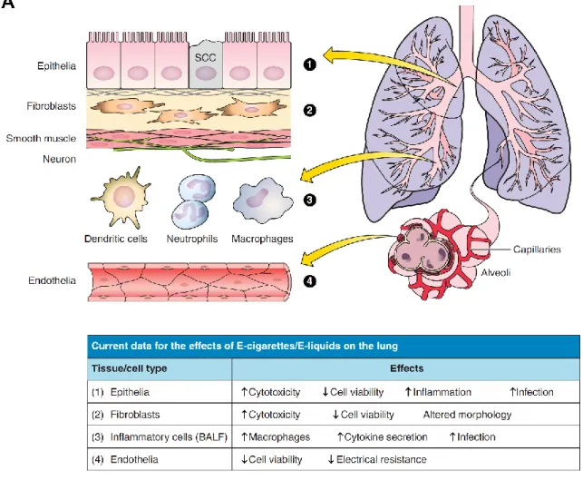
The lung is one of the first lines of defense against inhaled allergens, environmental pollutants, and pathogens. Many different cell types comprise the lung and are important to providing defenses (i.e., physical barrier, local inflammatory responses, mucociliary clearance) (264). These cell types are summarized in Figure 1.3 and include the pulmonary epithelia that line the conducting zones and contain both ciliated and secretory (goblet) cells. Beneath the epithelia are layers of fibroblasts and connective tissue, as well as airway smooth muscle cells that are responsible for constricting and dilating the airways. Immune cells are present in the airways (e.g., resident macrophages) or are recruited by inflammatory signaling (e.g., neutrophils, dendritic cells). Lastly, the respiratory zone is rich in alveolar type 1 and 2 cells in the terminal sacs that exchange gas with the endothelial cells of the capillary beds nearby (193, 264). Prominent airway diseases such as COPD (emphysema and chronic bronchitis) or asthma involve the destruction and/or dysregulation of different lung cell types (i.e., bronchiolar alveoli and epithelia, airway smooth muscle cells), suggesting that they are all susceptible to chronic e-cig exposure. 1.2. E-cigarettes (E-cigs)
tobacco, they will not expose the lung to the same toxic chemicals as regular smoked tobacco and so will not cause the lung disease that is frequently associated with chronic tobacco inhalation, including lung cancer and COPD. E-cig users are a fast-growing subset of nicotine users who are described as vapers rather than smokers, since e-cigs heat and generate aerosols but do not burn e-liquids. There is considerable controversy regarding the disease risk and toxicity of e-cigs (178, 186, 209). However, because e-cigs do not currently fall under the auspices of the Food and Drug Administration (FDA), they have not undergone the typical toxicological evaluation, followed by human clinical trials that are required of other inhaled products (e.g., inhaled therapeutic agents), and, as such, no safety data exist from either humans or animals. Because of this, it is hard to predict whether these products will be benign when chronically inhaled, possibly over a lifetime, or whether they will induce tobacco-like disease or other types of lung disease such as bronchiolitis obliterans, a disease that has been caused by the inhalation of the buttery-tasting flavor diacetyl (124). The clinical evaluation of biomarkers of harm (e.g., inflammatory and cytotoxic markers) is required to inform the FDA and for ensuring safety and proper regulation. However, these studies are only just beginning in what can at best be described as “investigator-initiated trials” rather than formal clinical trials. A further confounder is that many e-cig users have switched after chronically smoking tobacco products, making it difficult to differentiate between the previous effects of tobacco vs. the effects of the e-cigs (58). To date, there are currently 1,273 e-cig articles on Pubmed, of which 135 are reviews and only 85 include the terms “e-cigarette” and “lung.” In contrast, “tobacco” and “lung” yields 9,769 hits, indicating the lack of maturity of this field. In this review, we shall list and evaluate what is known about the effects of e-cig exposure on
lungs/airways in vivo and in vitro (Fig. 1.3). 1.3. Health Effects of Nicotine and Tobacco Use
of delivering nicotine into the bloodstream. Nicotine is a weak base that can be absorbed across the lung in its unionized form into the bloodstream (25). The effects of nicotine on the brain are complex and only just beginning to be understood. Importantly, its effects on the adolescent brain are markedly different compared with the adult brain and can affect neural development. For example, exposure to nicotine in adolescent rats led to an increased sensitivity to nicotine in these rats as adults, even if a smoking cessation period was introduced, suggesting that e-cig inhalation (i.e., vaping) by adolescents may have serious consequences later in life (59).
companies began developing “safe cigarettes.” In the 1980s, “low-tar” cigarettes were developed, which were purportedly safer than regular cigarettes because they exposed users to less of the tar phase from cigarettes. This premise was based on flawed science. In part, the reduced tar output was generated by putting holes at the base of the cigarette, which served to reduce airflow through the cigarette. However, these cigarettes were not safer than regular cigarettes. In fact, users learned to compensate by either covering up the holes with their fingers or taking larger puffs, thus negating the “low-tar effects” (96, 187). Furthermore, the gas phase of cigarette smoke is also highly toxic and was not addressed in “low-tar cigarettes” (176, 205). Additional types of safe cigarettes have been developed, including “heat not burn” types (e.g., the “Eclipse”), which heats a rod in the middle of the cigarette to give off tobacco smoke without burning it. This style of cigarette was not successful commercially (8), and there is no evidence that they are actually safer (101).
1.4. Components of Neat and Heated Flavored E-liquids
Cigarette smoke is a complex and highly reactive mixture that includes metals (e.g., Cr, Cd, Hg), aldehydes (e.g., 4-aminobiphenyl, acrolein, formaldehyde), carbon monoxide, free radicals, and, of course, nicotine (82, 245). Adverse effects can be caused by adduct formation, especially of aldehydes, with DNA or proteins (183), or due to excessive oxidative stress (119). Aldehydes and heavy metals have been shown to have a number of cytotoxic effects on epithelia, including adduct formation to DNA (43, 237, 258). Additionally, tobacco smoke, as well as aldehydes, cadmium, and oxidative stress, also affect plasma membrane proteins such as the cystic fibrosis transmembrane conductance regulator (CFTR) (36, 49, 100, 230), which is required for fluid secretion in the lung (51, 87). In contrast, e-liquids (the flavored liquids that are heated to form the e-cig vapor) are thought to be much simpler and ostensibly contain nicotine (~6–18 mg/ml) in a liquid vehicle (typically propylene glycol and/or vegetable glycerin), along with sweeteners and flavorings (94).
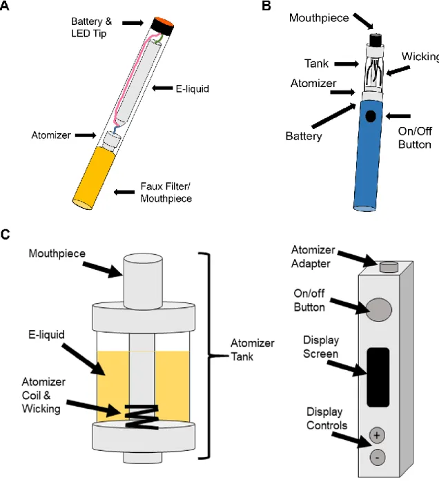
To date, over 400 different brands of e-cigs have been produced (276). Unlike disposable e-cigs, second- and third-generation e-cigs contain a refillable tank to which the e-liquid is added, a battery-powered atomizer that generates the aerosol vapor from the e-liquid, and a mouthpiece that collects and delivers the aerosol (Fig. 1.1). This function is usually controlled by a microchip, which may activate a light-emitting diode at the tip of the e-cig during inhalation for aesthetics. The amount of aerosol that is generated is directly proportional to the power of the battery, which has led some users to modify their e-cig to increase battery power to get a greater nicotine “hit.” This is not without risks, however, since there is a chance of battery explosion, which can lead to injury (34). Although the number of fires and explosions from e-cig devices has increased since inception, interestingly, many of these instances occurred while the device was being charged and are still considered rare
Parameters of e-cig emission, such as aerosol size, mass output, and chemical composition, vary by device and e-liquid types and are predicted to impact the user’s exposure to the e-cig aerosol. For example, aerosol size strongly affects how much of an aerosol is delivered to different regions of the lung and how much is retained in the oral cavity (23, 123).
There are currently over 7,000 different flavored e-liquids that are commercially available (276). Because these e-liquids are not FDA regulated, the vendors do not have to list their e-liquid ingredients, perform any safety testing before they reach the market, nor generate these products under Good
Manufacturing Practice-type conditions. For example, the reported amount of nicotine has been found to vary by up to 20% from what is reported on the e-liquid label and has even been found in purportedly nicotine-free e-liquids (55, 56, 91). It is likely that many of the compounds used in e-liquids fall within the FDA’s Generally Regarded as Safe (GRAS) list
[http://www.accessdata.fda.gov/scripts/fdcc/?set_SCOGS]. The typical vehicle for e-liquids contains a mix of propylene glycol and glycerol, which are both GRAS products (34). To date, most GRAS
nonasthmatic users who vape frequently could experience, at the very least, respiratory irritation although to date no long-term data are available regarding chronic propylene glycol or flavorant exposure in humans.
1.5. E-liquid Thermal Decomposition (Pyrolysis)
E-cig aerosols are typically generated at temperatures of 100–250°C, which is predicted to cause pyrolysis of the e-liquid vehicle (274) and may also induce breakdown of other e-liquid constituents. Recently, formaldehyde has been detected in e-cig emissions (114). However, these data have been disputed (80). Part of the problem lies in deciding which temperature the e-liquid is heated to during the experiment vs. what occurs during actual vaping. For example, Jensen et al. found significant amounts of formaldehyde (~380 µg/10 puffs) in the emission from a tank-style e-cig device when the battery voltage was set at 5.0 V, with no formaldehyde being detected when a lower voltage (3.3 V) was used (114). Because the power consumption/electrical resistance of the coil was not quoted by Jensen et al., it will be hard to see how this observation transfers to other e-cig devices. That is, the power generated by the heating coil cannot be determined purely by the quoted voltage since it also depends on the current, and the temperature reached by the e-liquid is dependent on the power output of the heating element. Thus, for reproducibility, it may be useful for researchers to quote the power output of their e-cig device in addition to the puff profile used. Farsalinos et al. have reported that e-cig users do not use this higher voltage setting, and they also proposed that e-cigs only produce formaldehyde in “dry puff” conditions (80), where a dry puff refers to the scenario where there is little liquid on the atomizer coil and
temperatures get higher than would be seen with sufficient liquid, leading to the potential for increased pyrolysis. However, acrolein and other carbonyls have also been found by other investigators both in neat e-liquids and in e-cig aerosols that were generated by unmodified e-cig devices (222), suggesting that the occurrence/production of these compounds may be more common than originally suspected.
even to 180°C, which is close to temperatures reported for e-cigs (130–350°C) (246). For example, the acrid smell that occurs when oil is burned on a stove is from acrolein (20, 40). Similarly, the chemical decomposition of sugars also causes the release of aldehydes, including acrolein (244).
It has been proposed that e-cig users tend to avoid the bitter taste that is associated with release of aldehydes during overheating/dry puffing and that, in actual e-cig users, aldehyde exposure never actually happens (80). However, during the aforementioned practice of dripping, where the e-liquids are placed directly on the coil, it is possible that significant pyrolysis occurs. Certainly, cigarettes can produce a harsh taste that is concomitant with the production of significant amounts of acrolein, formaldehyde, and other aldehydes, along with many other toxicants (244). However, this relatively unpleasant taste is soon overcome in new smokers due to the power of the nicotine drive (229) and due to cross-desensitization of transient receptor potential ankyrin subtype 1 (TRPA1) channels in sensory neurons (29). Therefore, it is also possible that e-cig users will “learn” to overcome any unpleasant taste due to increased aldehyde production if the nicotine drive is great enough. It is also worth pointing out at this point that many flavors are themselves aldehydes, including anisaldehyde (sweet), cinnamaldehyde (cinnamon), and isovaleraldehyde (nutty). The effects of these flavors on pulmonary surfaces are not known. However, their potential inclusion in e-liquids may increase overall aldehyde exposure to the lung. Indeed, cinnamaldehyde is present in some e-liquids (21) and activates TRPA1 (167), suggesting that they may exert effects on the lung. Similarly, activation of this ion channel in sensory neurons in the airways of rodents by unsaturated aldehydes has previously been shown to trigger neurogenic inflammation (9) and to inhibit the CFTR ion channel (4), suggesting that a higher aldehyde burden may indeed be toxic to the lung. However, the degree of adverse effects will likely depend on dose ranging and whether aldehydes are actually generated in sufficient quantities during real vaping conditions to trigger these responses.
with regular cigarette smoke, different biological effects are seen with freshly produced vs. aged smoke, with aged smoke often being less biologically potent, which has previously been attributed to the decline in OX/ROS over time (108). Furthermore, because OX/ROS are highly reactive, they may also react with other components in the e-cig aerosol, further changing its chemical composition. Indeed, Sussan et al. demonstrated that e-cigs contain 1011 free radicals/puff, which is about 100 times less than is seen in regular cigarettes (240), but still likely to exert significant biological effects (66).
1.6. E-cig Topography
delivering nicotine to maintain sufficient plasma nicotine levels. Indeed, data suggested that users were able to maintain constant nicotine uptake, despite switching brands (22). Importantly, until a greater consensus is reached, these data suggest that a modified Canadian Intense profile may be a suitable parameter for studying e-cig aerosol generation.
1.7. Will Nicotine and Chemical Constituents in E-liquids/E-cigs Alter Airway Physiology?
Nicotine is a highly addictive substance that is a major component of both cigarette smoke and e-cig aerosols that can cause physiological changes to users through nAChRs expressed throughout the body (52). Traditionally, nAChRs were primarily studied as part of the acetylcholine neurotransmitter signaling system in the central and peripheral nervous system. However, nAChR expression has been characterized in the airways as well (52, 145, 155, 277). These ligand-gated ion channels are permeable to both Na+ and divalent cations and are physiologically stimulated by acetylcholine. nAChRs contain five subunits of which different subtypes exist (e.g., α, β, γ and δ) (3, 166). For example, the (α4)3, (β2)2 nAChR subunit configuration is the most common type in the brain while the (α7)5 or α3, α5, and β4 subunits are more common in the lung (247). Lee et al. found that inhaled nicotine from cigarette smoke caused airway irritation and a cough reflex via nAChRs expressed in pulmonary afferent neurons (132).
Interestingly, nAChRs regulate cell proliferation and inhibit apoptosis (69). For instance,
Maouche et al. found that α7 nAChRs were enriched in basal lung epithelia and that, during development, α7 regulated basal cell proliferation (149), which is important for the maintenance of epithelial cell turnover and differentiation. It is well established that smoking is linked to lung cancer, and a hallmark of lung cancer is uncontrolled cell proliferation. West et al. reported that both nicotine and its metabolite (nicotine-derived nitrosamine ketone) stimulated Akt signal transduction downstream of nAChR
activation, which altered cell proliferation and apoptosis in bronchial epithelia (263). Specifically, α3, α5, and β4 were identified as candidate genes for a potential role in lung cancer from genome-wide
compared before and after removal of nicotine. Interestingly, exposing HBECs briefly upregulated nAChR α1, α5, and α7 expression at 72 h that returned to baseline levels after removal of nicotine. While all classes of nAChRs are capable of desensitization through chronic agonist exposure, there are definite immediate effects of nicotine on nAChRs in a subunit-dependent manner. Although it is currently unknown whether chronic exposure of nAChR to nicotine via e-cigs can cause lung cancer, the role of nAChR α7 in contributing to nonsmall cell lung cancer by altering cell proliferation and apoptotic resistance has been reported (128, 180).
Many inflammatory cells contribute to COPD pathogenesis, including, but not limited to, dendritic cells, T and B lymphocytes, monocytes, macrophages, and neutrophils (18, 106). Of note, monocytes, macrophages, and neutrophils, which are impacted by inhaling cigarette smoke in the lungs, also express nAChRs. The effects of noncholinergic signaling in airway inflammatory cells have been described (93). Nicotine suppressed inflammation in human monocytes and in mouse macrophages (154, 272). Neutrophil influx occurs in COPD, and indeed neutrophils present in smokers have upregulated nAChR expression and display a reduced ability to undergo apoptosis (11, 54). Likely, these neutrophils are more sensitive to inhaled nicotine and have extended life spans, which may serve to prolong
inflammation in the lungs. Taken together, these data indicate that nicotine has a proinflammatory effect on neutrophils. However, nicotine also has an anti-inflammatory effect on monocytes/macrophages, which may be negated in the case of cigarette smoke due to the inhalation of other proinflammatory products such as the tar phase. This dualism has curious implications for the chronic inhalation of nicotine from e-cig aerosols, since many of the cigarette tobacco and tar byproducts that contribute to inflammation are not present in e-cig aerosols. It is possible that the anti-inflammatory effects of nicotine, in the absence of proinflammatory constituents, could suppress the user’s immune system. Certainly, it is reasonable to assume that high nicotine exposure from e-cigs will be a major
Despite nicotine’s known addictive and airway irritant properties, it is also known to be bitter tasting. Because of this, e-cigs and their e-liquids present a novel mix of chemical constituents that not only contain nicotine but also flavors, sweeteners, and other chemicals, many of which have not been studied in the lung. Many of these chemicals are present to mask the bitter nicotine taste. Thus, while nicotine has been shown to alter many aspects of airway physiology, the potential exists for salty, sweet-, and bitter-flavored constituents from e-liquids to stimulate taste receptor signaling pathways that could alter airway physiology with chronic use. To date, however, there is no current literature on the effects of e-cigs and chronic vaping in pulmonary physiology of nAChRs or taste receptors. nAChRs are ligand-gated ion channels, similar to ion channels that regulate salty taste transduction (e.g., epithelial sodium channel) (103). However, sweet and bitter taste receptors are G protein-coupled receptors (GPCRs). GPCRs typically act through Gα proteins to activate phospholipase Cβ (PLCβ) that cleaves membrane-bound PIP2 to generate inositol 1,4,5-triphosphate (IP3) (64, 75, 233). IP3 molecules act on ER IP3 receptors (IP3Rs), eliciting ER Ca2+ release. The ER transmembrane protein stromal interaction molecule 1 (STIM1) senses ER Ca2+ depletion and causes puncta formation to interact with the membrane Ca2+ channel Orai1 to initiate store-operated Ca2+ entry (SOCE) (Fig. 1.2A-C) (141, 189). Ca2+ continues to initiate signaling downstream by the phosphorylating proteins such as protein kinase Cα (PKCα) (65, 73). Of note, membrane receptor tyrosine kinases (RTKs) also have the ability to trigger a similar signaling mechanism, though RTKs are not known to detect tastants/odorants (161, 173).
suppressed by a component of cigarette smoke. Yet, a correlation between T2R expression and age was present in the nonsmoker group and absent in the smoker group, suggesting that starting smoking earlier in life could suppress T2R gene expression and contribute to nicotine addiction.
Many T2Rs have been identified in the upper and lower airway epithelia as well as airway smooth muscle cells (50, 63, 223, 253). Interestingly, T2R38 polymorphisms have also been linked to increased susceptibility of upper respiratory infections (135). Although an endogenous ligand is still unknown, known bitter agonists activate these T2Rs and increase intracellular Ca2+, stimulating ciliary beat frequency. Thus, they play a role in detecting noxious inhalants and expelling them from the airways due to increased rates of mucociliary clearance. Nasal mucosa have been reported to express both sweet receptors (T1Rs) as well as T2Rs in special nonciliated epithelial cells called solitary chemosensory cells (SCCs) (133). SCCs in the nasal epithelium harbor these receptors along with known components of the taste receptor signaling pathway and trigeminal nerve innervation. Tizzano et al. characterized the presence of SCCs with T2Rs and the T2Rs’ ability to detect known bitter agonists and acyl-homoserine lactones (253), which are intercellular chemical signaling compounds secreted by Gram-negative bacteria, providing more evidence for T2R roles in innate immunity. Furthermore, Lee et al. found that T1Rs and T2Rs in nasal epithelium converge to arbitrate innate immunity (134), that is, when T1Rs are activated (e.g., hyperglycemia, chronic rhinosinusitis), they can block the antimicrobial effects of T2Rs, causing persistent airway infections. Together, these data suggest that taste reception in the airways is important to innate immunity.
pathways (e.g., Ca2+ as a common second messenger) that regulate cellular responses to nicotine. For example, triggering Ca2+ influx from the activation of one nAChR subunit can attenuate the response of a second subunit through desensitization of the stimuli or prolong increases in intracellular Ca2+ (86).
As mentioned previously, Ca2+ is a common second messenger that acts downstream of not only nAChRs but also transient receptor potential (TRP) membrane ion channels. TRP channels are non-selective cation channels that have the ability to increase cytosolic Ca2+ concentrations upon activation. Several subtypes have been discovered and some natural odorant molecules have been reported as targets (190). These TRP channel subtypes include TRP vanilloid (TRPV), TRP ankyrin (TRPA), and TRP melastatin (TRPM). For example, menthol is able to activate TRPM8 (160) and has been used in menthol cigarettes where the TRPM8 activation triggers a cooling sensation that suppresses the bitter nicotine flavor for smokers (182). Cinnamaldehyde is the common cinnamon flavoring used in food and now e-cig products and is capable of activating TRPA1 (16, 117). Lastly, vanillin is a common vanilla flavoring capable of activating TRPV1 and TRPV3 (143, 269).
1.8. Effects of E-liquids and E-cig Aerosols on Cultured Cells from the Lungs
Tobacco smoke is highly proinflammatory and has been shown to trigger the release of inflammatory cytokines from endothelia, epithelia, and leukocytes (30, 95, 131). These cytokines can then trigger additional changes, including goblet cell metaplasia and neutrophil influx (127).
gene expression and DNA methylation in both the whole lung and in airway epithelia (99, 184, 257), macrophages (68), and endothelia (273). Many of these assays have been established as outcome
In addition to directly studying the effects of e-liquids, they can be heated/aerosolized and then studied. Cervellati et al. exposed A549 (lung epithelial) and HaCaT (keratinocytes) cells to whole cigarette smoke or e-cig vapor from three combinations of e-cigs (nicotine; nicotine + flavor; no flavor, no nicotine) (42). After 50 min of smoke or aerosol exposure, cultures were then left for 24 h, and LDH release and cell viability were studied. No information was given regarding whether these cells were polarized or not. However, they found that, under these conditions, e-cigs with nicotine and/or flavor induced similar cytotoxicity (increased LDH release and decreased cell viability) as standard cigarettes while nicotine and flavor-free e-cigs did not have any effect. E-cig aerosols (generated using a 4 s/35 ml pulse) also caused an increase in IL-6 and IL-8 secretion, which in the case of one flavor (cinnamon roll) was greater than the IL-8 secretion seen with cigarette smoke extract addition (137).
The effects of e-cig exposure have also been studied on the lung’s microvasculature. For
example, Schweitzer et al. found that e-cigs decreased the electrical resistance of endothelial cells derived from mice, rats, and humans, and exerted significant effects on cell viability and 3-(4,5-dimethylthiazol-2-yl)-2,5-diphenyltetrazolium bromide production that were associated with changes in cell signaling (activation of p38 mitogen-activated protein kinase) (222). Interestingly, these changes were similar to those observed after exposure to cigarette smoke extract (222). They also detected increased
phosphorylation of myosin light chain and Rho kinase following e-cig exposure, which may have been due to activation of sphingolipids (222). Changes in the permeability in the lung’s microvasculature may induce edema and/or increase the number of leukocytes that can enter the lung, thus increasing
inflammation, as described elsewhere (126). 1.9. Effects of E-cigs on the Murine Lung
Although little is known about the effects of e-cigs on humans, some studies have been performed in mice. E-cig exposure has been shown to elicit neuropharmacological effects, including upregulation of nAChR, in different areas of the brain and also caused signs of addiction and increased serum and
days and examined the mice 1 day later. They found that several cytokines were increased in the bronchoalveolar lavage of these mice, including IL-1α, IL-6, IL-13, and monocyte chemoattractant protein-1 (MCP-1) (2015). Of note, MCP-1 recruits macrophages to the lung, IL-6 is a proinflammatory cytokine, and IL-13 induces cellular remodeling and goblet cell hyperplasia. Schweitzer et al. found that an e-cig exposure regimen, which was equivalent in dose to exposure to smoke from two cigarettes, caused a significant increase in 8-oxo-2’-deoxyguanosine (8-oxo-dG) in both plasma and bronchoalveolar lavage (222). 8-oxo-dG is a marker of systemic oxidative stress and is indicative of DNA damage (169). They also detected increased nitrotyrosine levels in plasma. Nitrotyrosine can be formed following exposure to reactive nitrogen species such as peroxynitrite anion and nitrogen dioxide and is also a marker of cell stress/damage (204). A 2-wk exposure to e-cigs smoked under relatively standard conditions (2-s 35-ml puff) caused a significant increase in the number of macrophages in murine lungs and actually decreased IL-6 (222). The differences in IL-6 levels observed between the two experiments are most likely due to differences in smoking (vaping) regimens since one was acute and the other was chronic (137, 222). Mouse strain differences and/or differences in e-cig device/e-liquid may also have been factors.
After infection with Streptococcus pneumonia, e-cig-exposed mice were less able to clear this infection, suggesting that innate defense was impaired (240). They also found that H1N1 influenza virus infection also was poorly cleared. Although these data will need to be repeated by other groups, it is the first report that e-cig exposure leads to increased susceptibility to infection, which has important
implications for the safety of e-cig users. Interestingly, increased susceptibility to pathogens is a hallmark of tobacco exposure and is seen following both viral and bacterial infections (84, 175, 197). In addition to these aerosol exposures, a 50-fold-diluted e-cig liquid has been shown to increase IL-4, IL-5, and IL-13 in allergen-sensitized mice when tracheally instilled (140), again suggesting that e-liquids can still have adverse effects even before they are vaporized.
development of lung disease later in life (e.g., asthma) (1, 90). Whereas most published studies to date have focused on adult mice, it has been demonstrated that e-cig exposure also adversely effects neonatal mice and leads to impaired development, including decreased weight gain and reduced cell proliferation in the lungs, suggesting that secondhand vaping may also potentially be a cause for concern and could adversely affect lung development (158).
1.10. Effects of E-cig Exposure in Humans
Currently, many adult e-cig users are former smokers and have a significant history of tobacco usage before using e-cigs and/or continue to be mixed tobacco/e-cig users (89, 192). This will make studying the chronic effects of e-cigs difficult since the airways/lung retain a significant memory of smoking history/exposure even after smoking cessation. For example, Rager et al. found significant evidence of DNA methylation in the nasal epithelia of ex-smokers (194). Thus, for any observed effects on e-cig smokers, the previous and/or current tobacco smoking history must be taken into account. That said, the largest and fastest-growing population of e-cig users who have never smoked tobacco is adolescents. For example, in North Carolina, 15% of high school students have vaped e-cigs, and 60% thought that e-cigs were safe. In contrast, among the same group, 24% had smoked cigarettes (7). This trend is reflected nationally (156).
It has been shown that short-term (e.g., 5 min) e-cig inhalation leads to comparable plasma cotinine levels as regular tobacco smoking and exerts rapid physiological effects on the cardiovascular system, including elevated heart rate (255). These data indicate that modern e-cigs are delivering significant amounts of nicotine to the bloodstream. Although to date no studies have been performed to look at the adverse effects of e-cigs on pulmonary health (e.g., inflammation, etc.), Vardavas et al. looked at the effects of 5 min e-cig exposure on pulmonary function using standard spirometry (256).
to greater changes in resistance. They also found that this 5-min exposure caused a significant decrease in exhaled nitric oxide (NO) levels. NO has a number of functions in the lung, and changes in NO levels can affect ciliary beating, transcription, inflammation, ion transport, and airway smooth muscle tone (31). NO is altered in many diseases, including asthma (increased), cystic fibrosis (decreased), primary ciliary dyskinesia (decreased), and COPD (may be suppressed or mildly increased) (275). Thus, it is possible that e-cig exposure could cause different lung disease to COPD.
1.11. Conclusions and Future Directions
Figure 1.2. GPCR signaling pathway causes IP3-mediated ER Ca2+ release and SOCE. A: At basal
conditions, Ca2+ concentrations are low within the cytosol and high in the extracellular space and within the ER. B: GPCR activation elicits PLCβ cleavage of PIP2 to generate IP3, which acts on ER IP3Rs to
Chapter 2: Flavored E-cigarette Liquids Reduce Proliferation and Viability in the CALU3 Airway
Epithelial Cell Line
2.1. Overview
E-cigarettes are generally thought of as a safer smoking alternative to traditional cigarettes. However, little is known about the effects of e-cigarette liquids (e-liquids) on the lung. Since over 7,000 unique flavors have been identified for purchase in the United States, our goal was to conduct a screen that would test whether different flavored e-liquids exhibited different toxicant profiles. We tested the effects of 13 different flavored e-liquids [with nicotine and propylene glycol/vegetable glycerin (PG/VG) serving as controls] on a lung epithelial cell line (CALU3). Using the 3-(4,5-dimethylthiazol-2-yl)-2,5-diphenyltetrazolium bromide (MTT) assay as an indicator of cell proliferation/viability, we demonstrated a dose-dependent decrease of MTT metabolism by all flavors tested. However, a group of four flavors consistently showed significantly greater toxicity compared with the PG/VG control, indicating the potential for some flavors to elicit more harmful effects than others. We also tested the aerosolized vapor from select e-liquids on cells and found similar dose-dependent trends, suggesting that direct e-liquid exposures are a justifiable first-pass screening approach for determining relative e-liquid toxicity. We then identified individual chemical constituents for all 13 flavors using gas chromatography-mass spectrometry. These data revealed that beyond nicotine and PG/VG, the 13 flavored e-liquids have diverse chemical constituents. Since all of the flavors exhibited some degree of toxicity and a diverse
array of chemical constituents with little inhalation toxicity available, we conclude that flavored e-liquids should be extensively tested on a case-by-case basis to determine the potential for toxicity in the lung and elsewhere.
2.2. Introduction
E-cigarettes (e-cigs) have been growing in popularity since their debut in 2007 and are estimated to become a $50 billion global market by 2025 (202). E-cigs differ from tobacco cigarettes in that they do not contain tobacco, have varied nicotine concentrations (0–36 mg/ml), and produce an inhalable aerosol (vapor) that is generated without combustion. Instead, an e-cig liquid (e-liquid) is drawn and heated over a battery-operated coil as the user inhales. E-liquids are usually composed of a vehicle with varying ratios of propylene glycol (PG) and vegetable glycerin (VG) that contain nicotine and chemical flavors. Recently, the Food and Drug Administration (FDA) introduced rules to regulate e-cig products
(https://www.fda.gov/TobaccoProducts/Labeling/ProductsIngredientsComponents/ucm456610.htm) (206, 209). Despite this legislation, there continues to be much debate over the safety and efficacy of these products. E-cigs have commonly been marketed as a safer smoking alternative because they lack the carcinogens from tobacco and presumably fewer of the pyrolysis products from combusting tobacco that are associated with smoking-related diseases. However, some e-cig devices are capable of producing pyrolysis products (i.e., reactive aldehydes) and oxidant species similar to traditional cigarettes (137, 218, 240), but the conditions under which users would actually be exposed to disease-causing levels of these products remain a source of controversy.
more than a quarter of middle and high students had tried e-cigs (254). Furthermore, the availability of over 7,000 unique flavors in the United States (276) alone may contribute to their popularity in
adolescents (6).
It is currently unknown whether or not long-term e-cig use will cause respiratory diseases similar to cigarette smoke, none at all, or something entirely different. For example, bronchiolitis obliterans or “Popcorn Workers’ Lung” is scarring of the small airways that can range from mild and reversible to severe and irreversible. Prolonged inhalation of diacetyl, a buttery-flavored chemical used in microwave popcorn manufacturing and elsewhere, can and has caused this disease in some workers at microwave popcorn manufacturing plants (17, 124). Although diacetyl is safe to eat and thus found on the “generally recognized as safe” list, it is clearly not safe to inhale. Diacetyl and many other flavorings have only been tested and approved for ingestion and have not been tested for inhalation toxicology. Despite the known link between diacetyl and bronchiolitis obliterans, Allen et al. reported that either diacetyl or 2 other prominent butter-flavored chemicals (2,3-pentanedione and acetoin) were detected in 47 of 51 flavored e-liquid aerosols tested (5). Thus there is the potential for e-e-liquid flavors to have as yet unknown and possibly negative effects on the lung, as has recently been discussed (19).
2.3. Methods
Flavored e-cig liquids. All flavored e-cig liquids (e-liquids) were purchased from The Vapor Girl (https://www.thevaporgirl.com/). The tested flavors were Captain Black Cigar, Peanut Butter Cookie, T-bone, Popcorn, Black Licorice, Energon (orange energy drink), Vanilla Tobacco, Banana Pudding
(Southern Style), Kola, Hot Cinnamon Candies, Menthol Tobacco, and Solid Menthol. All e-liquids were ordered to contain 12 mg/ml nicotine. An additional 0 mg/ml nicotine Captain Black Cigar was
purchased as a nicotine-free control. At the time of purchase, the vehicle liquid was advertised as a 70/30 ratio of PG to VG. Thus a vehicle control was made in our laboratory using 70 PG/30 VG. For all aerosol experiments, additional Peanut Butter Cookies, Banana Pudding, and Hot Cinnamon Candies e-liquids were purchased from The Vapor Girl. All three additional e-e-liquids were ordered to contain 12 mg/ml nicotine and a 55/45 ratio of PG/VG. Therefore, we made an additional 55 PG/45 VG control for the aerosol experiments.
Chemicals and reagents. PG, VG, DMSO, probenecid, and methanol were purchased from Sigma-Aldrich. DAPI, calcein (AM), MitoTracker Red (CMXRos), fluo-4 (AM), and the Vybrant MTT Cell Proliferation Assay Kit were purchased from Life Technologies. Nicotine was purchased from Alfa Aesar. The Cytotoxicity Detection KitPLUS (LDH) was purchased from Roche. DAPI, calcein (AM), MitoTracker Red (CMXRos), and fluo-4 (AM) were reconstituted using DMSO and applied to cells in experiments where the final DMSO concentration was ≤0.1%.
Cell culture. CALU3 cells were cultured in MEM alpha with 10% FBS and
penicillin/streptomycin (GIBCO) as described (49). For the
performed. After aerosol exposures, cells were incubated for 24 h before aerosol-exposed media were removed and assays were performed.
Cell proliferation. The MTT assay was performed as instructed by the manufacturer after cells were treated for 24 h with either PBS, 70 PG/30 VG, nicotine, or flavored e-liquids. Cells were allowed to proliferate for 4 h after removal of the treatments. Data were calculated as percent absorbance of each treatment compared with the average of the 0% e-liquid (media control) treatments in each plate. Nonlinear regression curves were fit to each flavor or nicotine ± PG/VG dose responses in the MTT assays using GraphPad Prism to calculate IC50 values where appropriate.
Cell number and viability. Total cell number was measured at the end of the 24-h e-liquid and aerosol exposures. Cultures were rinsed with PBS and fixed with 100% methanol. After fixation, cells were rinsed again with PBS and stained with DAPI for 10 min. Cultures were rinsed following staining, and DAPI fluorescence intensity was measured using the Tecan Infinite Pro plate reader [excitation (ex): 360 ± 5 nm; emission (em): 460 ± 5 nm]. Total cell number was calculated as percent fluorescence of the e-liquid- or aerosol-treated cells compared with the average of the 0% e-liquid or 0 puff (media control) wells in each plate. Cell/mitochondrial viability was assessed using calcein and MitoTracker Red fluorescent indicators. After 24-h e-liquid or aerosol exposures, treated media were exchanged for fresh media containing either 3 µM calcein or 125 nM MitoTracker Red. Cultures were incubated for 30 min at 37°C. Cultures were then rinsed, media were replaced with a standard Ringer’s solution, and
fluorescence intensities were read using the Tecan Infinite Pro plate reader for calcein (ex: 495 ± 5 nm; em: 516 ± 5 nm) or MitoTracker Red (ex: 579 ± 5 nm; em: 599 ± 5 nm), respectively. Cell/mitochondrial viability was calculated as a percent fluorescence of e-liquid or aerosol-treated cells compared with the average of the 0% e-liquid or 0 puff (media control) wells in each plate.
Data were calculated as percent LDH release compared with a lysed control and reported as %LDH release, where
%𝐿𝐷𝐻 𝑟𝑒𝑙𝑒𝑎𝑠𝑒 =𝑒𝑥𝑝𝑒𝑟𝑖𝑚𝑒𝑛𝑡𝑎𝑙 𝑣𝑎𝑙𝑢𝑒 − 𝑙𝑜𝑤 𝑐𝑜𝑛𝑡𝑟𝑜𝑙 ℎ𝑖𝑔ℎ 𝑐𝑜𝑛𝑡𝑟𝑜𝑙 − 𝑙𝑜𝑤 𝑐𝑜𝑛𝑡𝑟𝑜𝑙 𝑥 100
Ca2+signaling. Changes in cytosolic Ca2+ concentration were measured using 8 µM fluo-4 dye loaded into cells in the presence of 1 mM probenecid for 40 min at 37°C. Cultures were then rinsed, and media were replaced with a standard Ringer’s solution and fluorescence intensities were read every 15 or 30 s using a Tecan Infinite Pro plate reader for fluo-4 (ex: 494 ± 5 nm; em: 516 ± 5 nm). A fluorescent baseline was established before the nicotine doses were added to the wells, and changes in fluorescence were normalized to the baseline (F/F0). The peak change in F/F0 was measured for each dose, and a nonlinear regression curve was fit using GraphPad Prism to calculate the EC50.
RNA extraction, cDNA synthesis, and quantitative RT-PCR. RNA was extracted from untreated CALU3 cells using the Qiagen RNeasy kit following the manufacturer’s protocol. cDNA was
synthesized using the Bio-Rad iScript cDNA synthesis kit following the manufacturer’s protocol. Gene expression was measured using Taqman gene expression assays from Applied Biosystems for Orai1,
Scnn1A, P2Y2R, GAPDH, CHRNA4, CHRNA5, CHRNA6, CHRNA7, CHRNB1, CHRNB2, and CHRNB3
using human primers. Genes of interest were normalized to GAPDH and fold change was calculated using ∆∆CT method relative to Orai1.
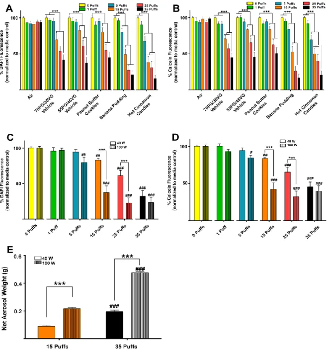
Aerosol exposures. E-liquids were heated to generate aerosols using Uwell Crown tanks with 0.25-Ω dual-coils and a Sigelei Fuchai 200W device. The power output was set to either 40 or 100 W, as indicated. Filter pads (GE Healthcare Life Sciences) with a 2-µm pore size were used to collect
once using a three-dimensional printed acrylic six-channel manifold. After aerosol exposures, the cells were incubated for an additional 24 h before measuring total cell number or viability. These were calculated as percent fluorescence of the aerosol-treated cells compared with the average of the 0 puff (media control) wells per plate, which were covered with silicone strips to avoid aerosol exposures.
Mass spectrometry. Samples of e-liquids were diluted 10- or 50-fold in methanol and analyzed by gas chromatography-mass spectrometry (GC-MS) using an Agilent 6890 GC with an Agilent MSD mass spectrometer. One-microliter volumes were introduced by manual injection and were separated on an Agilent DB-5 column with helium carrier gas. The temperature was ramped from 60 to 300°C at a rate of 20°C/min. GC-MS spectra were analyzed using NIST AMDIS software coupled to the NIST 2008 mass spectral database for automated database searching. Constituent profiles of flavors were compared between all 13 e-liquid flavors using peak areas under the curve from GC-MS data that were discretized into a value of 0 (absent) or 1 (present) and compared using R software.
For Fig. 2.2A, we fit a linear model to each log-transformed outcome with a fixed effect for gene type. Log transformations were used to ensure adherence to the modeling assumptions. If an overall test for the effect of gene type was found to be significant, we tested for pair-wise differences between each gene and Orai1. In Fig. 2.5E we fit a linear model with main effects for puff number and wattage and a puff number by wattage interaction term. Significant differences in puff numbers within each wattage level and significant differences in wattage levels within each puff number were identified using contrasts. Statistical modeling was performed using R statistical software (250) with the nlme package (185). Statistically significant relationships in all figures were reported (#,*P ≤ 0.05; ##,**P ≤0.01; ###,***P ≤ 0.001).
2.4. Results
E-liquid exposures inhibit cell proliferation/viability in a dose-dependent manner. To compare the effects of different flavored e-liquids on airway epithelia, we selected a range of flavors that covered not only traditional menthol and tobacco cigarette flavors but a variety of foods and beverages. We also purchased the Captain Black Cigar flavor with and without nicotine to control for the effects of nicotine. Cells were exposed to a range of e-liquid dilutions directly into the culture media over 24 h to assess cell viability and proliferation after treatment, with 70 PG/30 VG serving as the vehicle control. To control for the possible effects of diluting the media, we also provided a PBS control group where we serially diluted media with PBS. We used the MTT assay to indirectly assess the number of viable cells and their ability to proliferate in each treatment. We found dose-dependent decreases in each e-liquid flavor tested, irrespective of nicotine, as well as our 70 PG/30 VG vehicle. These effects were not due to dilution of the growth media since the PBS dilutions had no effect on MTT absorbance (Fig. 2.1A). The T-bone flavored e-liquid was not tested at a 6 or 10% dose because our purchased stock had run out and the vendor
When these four flavors were fitted with dose-response curves to calculate their IC50s, these flavors had lower IC50s than 70 PG/30 VG (Fig. 2.1B; Table 2.1), suggesting that they were more toxic. We also directly compared the responses of cells exposed to the Captain Black Cigar flavor ± nicotine and found that there were more severe effects in the nicotincontaining liquid compared with the nicotinfree e-liquid at ≥3% (Fig. 2.1C), suggesting that nicotine exerted additional effects beyond what was seen with the base e-liquid.
Nicotine decreases cell proliferation/viability dose dependently and is cytotoxic. Since we found additional dose-dependent effects of nicotine beyond what was seen with the Captain Black e-liquid alone, we sought to determine whether these negative effects were mediated by nicotinic acetylcholine receptors (nAChRs). We used quantitative RT-PCR to survey nAChR subtype gene expression in CALU3 cells relative to a common membrane Ca2+ channel (Orai1; Fig. 2.2A). As additional controls, we also looked at Scnn1a (epithelial sodium channel alpha subtype) and P2Y2R (purinergic receptor) expression. Five of 7 nAChR subtypes were detected in CALU3 cells, but only CHRNA5 was expressed above the levels of Orai1, Scnn1a, or P2Y2R levels, while CHRNA6, CHRNA7, CHRNB1, and CHRNB2
had much lower expression levels. nAChRs are ligand-gated ion channels that are permeable to Ca2+ ions, and since there was detectable nAChR subtype expression in CALU3 cells, we tested for functional activity using a fluorescent cytosolic Ca2+ indicator (fluo-4). The peak change in fluorescence per nicotine dose was plotted (Fig. 2.2B), and the EC50 was calculated (Table 2.1). The EC50 for nicotine was 2.89 mg/ml (17.8 mM) in CALU3 cells. However, the EC50 of nicotine for various nAChRs has been reported in the micromolar range (48, 92). Since increases in cytosolic Ca2+ can also be caused by cytotoxicity from permeabilized membranes, we measured cell viability and found a decrease in calcein fluorescence in cultures treated with ≥4.9 mg/ml nicotine (Fig. 2.2C; Table 2.1), suggesting that the effects of Ca2+ were due to cytotoxicity rather than being mediated by nAChRs.
in our e-liquid exposures (%vol/vol) from Fig. 2.1A. Irrespective of the presence of PG/VG, we found that there were dose-dependent decreases in percent absorbance with increasing doses of nicotine (Fig. 2.2D). Treating cells with 1% 70 PG/30 VG in combination with nicotine did not have additional effects. However, 3% 70 PG/30 VG decreased the threshold of MTT absorbance alone compared with either 0 mg/ml nicotine (media control) or 1% 70 PG/30 VG (P ≤ 0.001). There was no difference between log10(IC50) values of each nicotine treatment ± 70 PG/30 VG (Table 2.1), suggesting that adding nicotine to 70 PG/30 VG did not have a synergistic effect on cell proliferation. However, since most flavors caused a significant decrease in MTT absorbance at 3% (Fig. 2.1A), it is likely that 3% 70 PG/30 VG, rather than nicotine, caused the decrease, since 3% e-liquid contains 0.36 mg/ml nicotine, which is insufficient to affect MTT metabolism when combined with 70 PG/30 VG (Fig. 2.2D).
The four flavors of interest decreased cell number/viability in subconfluent CALU3 cultures. The initial screening of the 13 purchased e-liquid flavors on CALU3 cells directed our attention to four flavors of interest because of their lower IC50s in the MTT assays compared with 70 PG/30 VG. Therefore, we continued screening the effects of all of the flavors on other measures of cell viability and toxicity but focused on the effects of these four flavors in this paper [i.e., Banana Pudding (Southern Style), Kola, Hot Cinnamon Candies, and Menthol Tobacco]. We also tested Peanut Butter Cookies, a less toxic flavor, as well as Captain Black Cigar ± nicotine to control for the potential effects of nicotine. We performed additional analyses by measuring total cell number using DAPI staining (Fig. 2.3A). We found dose-dependent decreases in cell number 24 h after exposure to 70 PG/30 VG, Captain Black Cigar ± nicotine, Peanut Butter Cookies, as well as our four flavors of interest. We then used the fluorescent dyes calcein (Fig. 2.3B) and MitoTracker Red (Fig. 2.3C) as indicators of viable cells and active mitochondria,
investigated the potential for cytotoxicity using LDH release as a marker (Legrand et al., 1992). A 24-h exposure to 70 PG/30 VG, Captain Black Cigar ± nicotine, Peanut Butter Cookies, or Banana Pudding (Southern Style) did not induce LDH release. However, there were significant dose-dependent increases in LDH release following exposure to Kola, Hot Cinnamon Candies, and Menthol Tobacco flavors (Fig. 2.3D).
Hot Cinnamon Candies and Menthol Tobacco 24-h e-liquid exposures show cytotoxicity in
confluent CALU3 cultures. Since the previous experiments were performed on subconfluent, proliferating cultures to accommodate the MTT assay (Figs. 2.1–3), we next assessed the effects of the flavors on confluent, non-proliferating cultures to ascertain whether decreases in cell number/viability were due to cytotoxicity or decreased cell growth. We seeded CALU3 cells into 96-well plates at a higher density where they formed confluent monolayers before conducting the 24-h e-liquid exposures. We found that there were dose-dependent decreases in DAPI fluorescence following exposure to the 70 PG/30 VG vehicle, Peanut Butter Cookies, and the 4 flavors of interest (Fig. 2.4A). However, the decreases in Peanut Butter Cookies, Banana Pudding (Southern Style), and Kola were not significantly greater than that seen with 70 PG/30 VG, while those seen with Hot Cinnamon Candies and Menthol Tobacco were significantly different. Additionally, there were dose-dependent decreases in calcein fluorescence with the 70 PG/30 VG vehicle, Hot Cinnamon Candies, and Menthol Tobacco at 3% (Fig. 2.4B). However, the decreases in 3% Hot Cinnamon Candies and Menthol Tobacco were greater than those seen for 70 PG/30 VG (P ≤ 0.001).
measured. A media control group (0 puffs) was run in every plate, and wells were covered with fitted silicone strips during the exposure to ensure no unwanted exposures. Control groups with equal numbers of air puffs were also run. A 55 PG/45 VG vehicle group was added because the vendor had shifted from a 70 PG/30 VG to 55 PG/45 VG ratio and additional Peanut Butter Cookies, Banana Pudding, and Hot Cinnamon Candies e-liquids were required to conduct our experiments.
We found that all of the flavors and the PG/VG vehicle controls caused dose-dependent decreases in cell number (Fig. 2.5A). However, there was no effect of our air control group. We also found that both Banana Pudding and Hot Cinnamon Candies were more toxic than either Peanut Butter Cookies or the PG/VG groups after aerosol exposure at greater or equal to five puffs (Fig. 2.5A; P ≤ 0.001). We also
found again that all flavors and PG/VG vehicle controls caused dose-dependent decreases in cell viability (Fig. 2.5B), and again, there was no effect of air exposure. Both Banana Pudding and Hot Cinnamon Candies were more toxic than either Peanut Butter Cookies or the PG/VG groups after aerosol exposure at greater or equal to five puffs (Fig. 2.5B; P ≤ 0.001). Importantly, the same order of toxicity demonstrated after aerosol (vape) exposure was also seen after e-liquid exposures, suggesting that direct e-liquid exposure is valid for determining relative toxicity.