Distinct cortical and striatal actions of a
β
-arrestin
–
biased dopamine D2 receptor ligand reveal unique
antipsychotic-like properties
Nikhil M. Ursa,1, Steven M. Geeb,1, Thomas F. Packa, John D. McCorvyc,d,e, Tama Evrona, Joshua C. Snydera,
Xiaobao Yangf,g,h, Ramona M. Rodriguizi, Emiliana Borrellij, William C. Wetsela,i,k, Jian Jinf,g,h, Bryan L. Rothc,d,e, Patricio O’Donnellb, and Marc G. Carona,k,2
aDepartment of Cell Biology, Duke University Medical Center, Durham, NC 27710;bNeuroscience and Pain Research Unit, Pfizer, Inc., Cambridge, MA 02139; cDepartment of Pharmacology, University of North Carolina at Chapel Hill Medical School, Chapel Hill, NC 27514;dDivision of Chemical Biology and Medicinal Chemistry, University of North Carolina at Chapel Hill Medical School, Chapel Hill, NC 27514;eNational Institute of Mental Health Psychoactive Drug Screening Program, University of North Carolina at Chapel Hill Medical School, Chapel Hill, NC 27514;fDepartment of Structural and Chemical Biology, Icahn School of Medicine at Mount Sinai, New York, NY 10029;gDepartment of Oncological Sciences, Icahn School of Medicine at Mount Sinai, New York, NY 10029;hDepartment of Pharmacology and Systems Therapeutics, Icahn School of Medicine at Mount Sinai, New York, NY 10029; iDepartment of Psychiatry and Behavioral Sciences, Duke University Medical Center, Durham, NC 27710;jDepartment of Microbiology and Molecular Genetics, University of California, Irvine, CA 92697; andkDepartment of Medicine and Neurobiology, Duke University Medical Center, Durham, NC 27710
Edited by Solomon H. Snyder, Johns Hopkins University School of Medicine, Baltimore, MD, and approved October 26, 2016 (received for review August 26, 2016)
The current dopamine (DA) hypothesis of schizophrenia postu-lates striatal hyperdopaminergia and cortical hypodopaminergia. Although partial agonists at DA D2 receptors (D2Rs), like aripipra-zole, were developed to simultaneously target both phenomena, they do not effectively improve cortical dysfunction. In this study, we investigate the potential for newly developed β-arrestin2 (βarr2)-biased D2R partial agonists to simultaneously target hyper-and hypodopaminergia. Using neuron-specific βarr2-KO mice, we show that the antipsychotic-like effects of aβarr2-biased D2R ligand are driven through both striatal antagonism and cortical agonism of D2R-βarr2 signaling. Furthermore, βarr2-biased D2R agonism en-hances firing of cortical fast-spiking interneurons. This enhanced cortical agonism of the biased ligand can be attributed to a lack of G-protein signaling and elevated expression ofβarr2 and G protein-coupled receptor (GPCR) kinase 2 in the cortex versus the striatum. Therefore, we propose that βarr2-biased D2R ligands that exert region-selective actions could provide a path to develop more effec-tive antipsychotic therapies.
arrestin
|
antipsychotics|
biased signaling|
dopamine D2R|
fast-spiking interneuronsG
protein-coupled receptors (GPCRs) represent the largestfamily of receptors in the human genome and are one of the most common targets of pharmaceutical drugs (1, 2). Upon ligand binding, GPCRs activate downstream G protein-dependent sig-naling pathways followed by phosphorylation of the receptor by G protein-coupled receptor kinases (GRKs) (3). Phosphorylation
enhances association of the GPCR with β-arrestins (βarrs), and
this combined process mediates desensitization of G-protein
sig-naling (4) and internalization of GPCRs (5–7). Two isoforms of
βarrs,βarr1 andβarr2, are widely coexpressed in most tissues in
mammals and are 80% identical, but they can have either over-lapping or distinct functions (8, 9). It is now firmly established that GPCRs activate downstream signaling pathways through not only
canonical G-protein pathways but also, the ability of βarrs to
scaffold distinct intracellular signaling complexes (10–12).
Eluci-dation of these distinct G-protein andβarr signaling pathways has
provided support for the concept of functional selectivity or biased signaling, wherein each signaling pathway has the ability to me-diate distinct physiological responses (13). There are now several physiologically relevant examples of selective engagement of sig-naling pathways or selective GPCR ligands that target these
dif-ferent signaling pathways (13–15). Therefore, leveraging the
concept of GPCR functional selectivity holds promise for the de-velopment of more selective therapeutic approaches.
Dopamine (DA) is a catecholamine neurotransmitter that has
been implicated in movement, reward, and cognition (16–19) as
well as CNS disorders, such as schizophrenia, attention deficit
hy-peractivity disorder, Parkinson’s disease, and obsessive–compulsive
disorder (20–23). DA mediates its effects via GPCRs belonging
to two major subclasses of receptors: the D1 class [D1 receptor (D1R) and D5 receptor] and the D2 class [D2 receptor (D2R), D3 receptor, and D4 receptor] (24), a classification based on their
ability to activate the stimulatory G protein Gαs/olfor inhibitory G
protein Gαi/osignaling pathway, respectively. In the brain, D2Rs
activate canonical Gαi/o-mediated signaling to inhibit adenylyl
cy-clase, cyclic adenosine monophosphate (cAMP) production, and the protein kinase A/dopamine and cAMP regulated
phospho-proteinMr32 KDa (PKA/DARPP32) pathway to mediate many of
the behavioral effects of DA (23, 25, 26). However, based on the
Significance
Schizophrenia is a debilitating psychiatric disorder characterized by positive, negative, and cognitive symptoms. Current antipsy-chotic drugs, including D2 receptor (D2R) partial agonist aripipra-zole, antagonize excess striatal dopamine (DA) neurotransmission and reverse positive symptoms but are not efficacious at reversing cortical-related cognitive symptoms. Here, we show using phar-macological, behavioral, and electrophysiological approaches that aβ-arrestin2 (βarr2)-biased D2R ligand has opposite antagonist and agonist actions in the striatum and cortex, respectively. This phenomenon is regulated by differential expression levels of signal transducer proteins G protein-coupled receptor kinase 2 andβarr2. Thus, D2R-βarr2–biased ligands have the potential to simultaneously target excess striatal and deficient cortical DA neurotransmission and provide more broadly effective therapies for schizophrenia.
Author contributions: N.M.U., S.M.G., P.O., and M.G.C. designed research; N.M.U., S.M.G., T.F.P., J.D.M., T.E., J.C.S., R.M.R., and B.L.R. performed research; X.Y., E.B., and J.J. contributed new reagents/analytic tools; N.M.U., S.M.G., J.D.M., T.E., J.C.S., R.M.R., W.C.W., B.L.R., and P.O. analyzed data; and N.M.U., S.M.G., P.O., and M.G.C. wrote the paper. Conflict of interest statement: P.O. is an employee and shareholder at Pfizer, Inc. M.G.C. has received compensation from Lundbeck as a member of their Psychopharmacology Advisory Board and is a consultant for Omeros Corp. M.G.C. also owns equity in Acadia Pharmaceuticals.
This article is a PNAS Direct Submission.
1N.M.U. and S.M.G. contributed equally to this work.
2To whom correspondence should be addressed. Email: [email protected].
initial observation that the DA-dependent locomotor response to amphetamine (AMPH) was markedly attenuated in mice globally
lacking βarr2 but not βarr1 (9), we provided biochemical and
genetic evidence for aβarr2-dependent signaling pathway
down-stream of D2Rs that is separate from Gαi/osignaling (27). We have
shown that this D2R-βarr2 signaling pathway inhibits protein
ki-nase B (PKB or AKT) activity, activates glycogen synthase kiki-nase 3
beta (GSK3β), and can mediate specific DA-dependent
be-haviors (28–30).
D2Rs are the major target for most antipsychotic drugs (APDs), which are the first-line treatment for schizophrenia (31, 32). APDs do not treat all symptoms of schizophrenia effectively and have
several side effects that are thought to be associated with Gαi/o
signaling (26, 30). APDs that selectively target the D2R-βarr2
pathway could be therapeutically beneficial without producing extrapyramidal side effects. To further investigate the potential
role of functional selectivity of the D2R-βarr2 pathway in APD
action, we have recently generated βarr2-biased D2R ligands
based on the scaffold of the partial agonist APD aripiprazole (ARI) (33, 34). These biased ligands show antipsychotic-like pro-file in pharmacological [phencyclidine (PCP) and AMPH] and genetic (NMDA receptor NR1 subunit knockdown) mouse models
of schizophrenia-like phenotypes that depend on their D2R-βarr2
selectivity, because their activity is lost in globalβarr2-KO mice
(33, 35). Although classical APDs, like haloperidol, and newer APDs, like ARI, reverse the postulated striatal hyperdopaminergic tone associated with schizophrenia, none of these drugs effectively
correct cortical dysfunction (36–38). It is currently not known
whether targeting D2R-βarr2 signaling might represent an
al-ternative strategy to identify more broadly effective APDs.
In this study, we usedβarr2-biased D2R ligands and behavioral
and electrophysiological approaches in mice lackingβarr2 in
var-ious D2R-expressing neuronal populations to investigate whether
region-specific D2R-βarr2 signaling contributes to unique
anti-psychotic-like effects in vivo.
Results
Determinants of in Vitro D2R-βarr2 Functional Selectivity.Clinically effective APDs are either antagonists or partial agonists at both
D2R-G protein and D2R-βarr2 signaling pathways (39, 40). We
have previously shown that theβarr2-biased D2R ligands
UNC9994A (94A) and UNC9975A (75A), which are based on the
scaffold of ARI, are weak selective partial agonists at D2R-βarr2
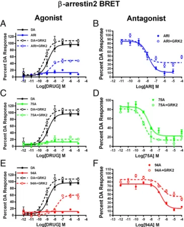
interactions and have little or no agonist activity at D2R-G protein signaling (33). However, we show in Fig. 1 that the agonist versus
antagonist profiles of these ligands at D2R-βarr2 interactions in
HEK293 cells can be modulated by the complement of GRK2 and
βarr2. In a bioluminescence resonance energy transfer
(BRET)-based assay (Fig. 1A,C, andE), compared with DA ARI, 94A and
75A are very weak partial agonists at mediating D2R-βarr2
inter-actions, but increasing GRK2 levels in these cells markedly en-hanced the partial agonist activity of only ARI and 94A. Consistent with pharmacological principles, when tested as antagonists (Fig. 1
B, D, and F), all three ligands fully antagonized DA-mediated
D2R-βarr2 interactions, and high GRK2 levels reduced the
an-tagonist efficacy of only ARI and 94A. The profile of 75A was not significantly changed by GRK2, suggesting that it may have slightly
different properties than ARI or 94A. However, in a D2R–G
protein (GloSensor) assay, ARI and 75A but not 94A behaved as
antagonists (Fig. S1andTable S1), suggesting that only 94A is a
completely selective D2R-βarr2 ligand. These results are consistent
with the established concept thatβarr-dependent GPCR functions
are not only dependent on agonist activation but also, enhanced by phosphorylation of the receptor by GRKs (11, 41) and that the
expression levels ofβarr2 and GRK2 can regulate GPCR signaling
and the pharmacological profile of ligands (42–44).
Previous studies have suggested that the levels of GRKs and
βarrs can vary significantly between tissues and brain areas (45, 46).
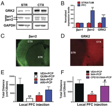
Protein levels ofβarr2 and GRK2 but notβarr1 are higher in the
prefrontal cortex (PFC) compared with the striatum (STR) in mice
(Fig. 2A–D) and humans (Fig. S2AandB) (Ps<0.05). Because
ARI and 94A gain D2R-βarr2 partial agonism on GRK2
over-expression, we hypothesized that these compounds might behave
as agonists in the PFC (highβarr2/GRK2). Previous studies have
shown that local injection of the D2R agonist quinpirole can exert antipsychotic-like effects, because it inhibits locomotion induced by the NMDA receptor antagonist PCP (47). PCP-induced locomo-tion is a pharmacologically induced behavior commonly inhibited by APDs, and the behavioral effects of PCP are thought to be mediated by cortical disinhibition (48). To assess whether PFC
D2R-βarr2 agonism can elicit antipsychotic-like effects, we
mea-sured the effects of injecting quinpirole, ARI, and 94A locally into the PFC of WT mice on PCP-induced locomotion. Consistent with previous findings, local bilateral PFC injection of the unbiased full-agonist quinpirole significantly inhibited PCP-induced locomotion
(Fig. 2E, QUIN) (P<0.01); 94A mimicked this D2R agonist
ac-tion (Fig. 2E, 94A) (P <0.05) and inhibited PCP-induced
loco-motion, whereas the effect of ARI was weaker and did not reach significance. Coadministration of a pharmacological GRK2 inhibitor [compound 101 (cpd101)] prevented the 94A-mediated
inhibition of PCP-induced locomotion (Fig. 2F, cpd101/94A).
These results are consistent with our in vitro data, showing that, in a
D2R-βarr2 interaction BRET assay, the ability of GRK2 to enhance
the agonist effects of both DA and UNC9994A is lost in presence
of the GRK2 inhibitor (cpd101) (Fig. S2C). These observations
Fig. 1. Effects of GRK2 overexpression on the agonist or antagonist profile of D2R ligands in HEK293 cells. (A) ARI, (C) UNC9975A (75A), and (E) UNC9994A (94A) were tested in agonist mode in a D2R-βarr2 interaction BRET assay with endogenous GRK2 (solid lines) or GRK2 overexpression (dashed lines) com-pared with DA (black lines) in HEK293 cells. D2R-βarr2 BRET antagonist assays for (B) ARI, (D) UNC99975A (75A), and (F) UNC9994A (94A) with endogenous GRK2 (solid lines) or overexpressed GRK2 (dashed lines) levels in HEK293 cells. Data are presented as inhibition of the total DA response (mean±SEM).
NEUROSCI
ENCE
PNAS
show that D2R-βarr2 agonism in the PFC is sufficient to inhibit
PCP-induced locomotion, and this effect is dependent onβarr
and GRK2.
Distinct Striatal Vs. Cortical Role of D2R on PCP-Mediated Behavioral Response.Although D2R PFC agonism inhibits PCP-induced lo-comotion, striatal D2R antagonism is thought to be the primary mechanism of action of most APDs (49, 50). To broadly explore this paradoxical role of D2R function behaviorally, we selectively deleted D2Rs in either the PFC by injecting an mCherry-Cre adeno-associated virus (AAV) in the previously described D2R
floxed (D2f/f) mice (51) or the STR by crossing D2f/fmice with
adenosine 2A receptor Cre (A2aCre) mice (Fig. S3), and we tested
the effect on PCP-induced locomotion. We observed that, com-pared with controls, deletion of D2Rs in the PFC resulted in an
enhanced response to acute PCP injection (Fig. 3A) (Ps<0.01),
consistent with D2R agonism playing an inhibitory role on the PCP response (47). However, deletion of D2Rs in the STR attenuated
the PCP response (Fig. 3B) compared with controls (Ps<0.01),
consistent with striatal D2R antagonism inhibiting PCP-mediated responses (49, 50). These pharmacological and behavioral data suggest distinct roles for D2R signaling in the PFC versus the STR in mediating antipsychotic-like effects.
Generation and Characterization of βarr2 Floxed Mice.To further
evaluate the potential opposite role of D2R-βarr2 signaling in the
PFC and STR in antipsychotic-like effects, we generated aβarr2
floxed (βarr2f/f) mouse line for region- and cell-specific
Cre-dependent deletion ofβarr2 (details are inMaterials and Methods
andSI Materials and Methods;Fig. S4AandB);βarr2f/fmice were
(Fig. S4C–E). Deletingβarr2 in all neurons (βarr2f/fCMV-Cre)
recapitulated the decrease in the AMPH locomotor response
originally observed in whole-body βarr2KO mice (27) (Fig. S4
F–H). We have previously shown that the AMPH locomotor response
is mediated at least in part by D2Rs in aβarr2-dependent manner (27,
29), but D2Rs are expressed in multiple regions of the brain, such as
midbrain, STR, and PFC (24, 52–57). To confirm which D2R+
population of neurons is responsible for the AMPH response, we
deletedβarr2 in several distinct neuronal populations;βarr2f/fmice
were crossed with either D2Cre (all D2R+ neurons) or A2aCre
(postsynaptic D2R+ striatal neurons) mice as well as the D1Cre
(D1R+ neurons) or ChaTCre (cholinergic interneurons) mice as
controls. Using immunohistochemistry (IHC) and real-time
quantita-tive PCR techniques, we confirmed selecquantita-tive deletion ofβarr2 in
ap-propriate neuronal populations of all Cre lines crossed with theβarr2f/f
mice (Fig. S5). As shown in Fig. 4,βarr2 in striatal D2R+neurons
seems to play the most prominent role in the AMPH response,
be-cause the locomotor response is reduced only in D2R+or A2aR+
neurons lackingβarr2 (Fig. 4E–H) but not affected by deletion of
βarr2 in either D1R+neurons alone or cholinergic interneurons (Fig.
4C,D, andI). These data suggest that deletingβarr2 in D2R striatal
neurons is sufficient to mimic antipsychotic-like activity. Importantly,
the mice in whichβarr2 is inactivated in select D2R+neuronal
pop-ulations provide unprecedented models to critically examine the in
vivo antipsychotic-like function of our D2R-βarr2–biased compounds.
Region-Specific Responses of Antipsychotics and βarr2-Biased D2R Ligands.AMPH- and PCP-induced hyperlocomotion are the two commonly used pharmacological models to test APD efficacy. Most APDs are D2R partial agonists or antagonists with varying potencies and efficacies (31, 32, 40, 58) and inhibit either the AMPH- or PCP-induced locomotor response in mice (59). The AMPH-induced locomotor response is dependent on striatal DA release, whereas the behavioral effects of PCP are thought to be mediated by cortical disinhibition and activation of the cortico-striatal pathway (48, 50). We tested the ability of representative first, second, and third generation APDs, such as haloperidol,
clozapine, and ARI, respectively, along with theβarr2-biased D2R
ligands 94A and 75A in both of these pharmacological models.
Optimal doses for APDs and the D2R-βarr2–biased ligands were
based on previous studies (30, 33, 35).
For the AMPH-induced locomotor response, AMPH was in-jected at a dose (3 mg/kg) at which there were no significant
Fig. 2. Cortical and striatal expression patterns ofβarr2 and GRK2 as well as D2R PFC agonism. (A) Western blot analysis from WT mice probed with antibodies to GRK2,βarr2, andβarr1 and GAPDH in cortex (CTX) compared with STR and (B) quantification of Western blot band intensities normalized to GAPDH (loading control;Ps<0.05). IHC images of mouse brain sections (cortical-striatal) stained with antibodies to (C)βarr2 and (D) GRK2. Loco-motor responses to (E) bilateral local PFC injection 1μg per side quinpirole (QUIN), UNC9994A (94A), and aripiprazole (ARI) in WT mice followed by systemic PCP injection (6 mg/kg i.p.;n=8–11) or (F) bilateral local PFC in-jection of 1μg per side UNC9994A (94A) with or without cpd101 (0.5μg per side) in WT mice followed by systemic PCP injection (6 mg/kg i.p.;n=8–10). *P< 0.05, compared with VEH+PCP; **P<0.01, compared with VEH+PCP;#P< 0.05, compared with 94A+PCP.
genotype differences between mice (Fig. 4C–H). This higher dose allowed us to achieve better separation of effects when treating with APDs and biased compounds. Consistent with their antagonist
activity, all tested APDs and biased D2R ligands significantly
inhibited AMPH-induced locomotion in all controlβarr2f/fmice
(Fig. 5, black bars) (Ps <0.01). No genotype differences were
observed in mice lacking βarr2 in D1R+neurons for all
com-pounds (Fig. 5AandB) (Ps<0.01). All three APDs and 75A
also inhibited the AMPH response (Ps<0.01) in mice lacking
βarr2 in either all D2R+ (Fig. 5C) or striatal A2aR+neurons
(Fig. 5E). However, in these mice, 94A showed markedly
di-minished antipsychotic-like activity compared with genotype
controls (Ps<0.05), consistent with this compound being a
se-lective βarr2-biased D2R antagonist. Compared with other
APDs, the observations with 94A indicate that D2R-βarr2
an-tagonism is sufficient but not necessary for efficacious antipsy-chotic-like activity in the AMPH pharmacological model, which is consistent with previous observations (60).
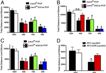
For PCP-induced responses, in mice lacking βarr2 in D1R+
neurons, all drugs significantly inhibited PCP-induced locomotion
compared with vehicle (VEH)-treated controls (Fig. 6A) (Ps <
0.05), and there were no genotype differences. ARI and 75A also
inhibited PCP-induced locomotion in mice lackingβarr2 in D2R+
striatal neurons (Fig. 6B) (Ps<0.05) or all D2R+neurons (Fig. 6C)
(Ps<0.05) compared with genotype and VEH controls. In contrast
to the AMPH response (Fig. 4), however, we observed a loss of
94A activity only in mice lacking βarr2 in all D2R+ neurons
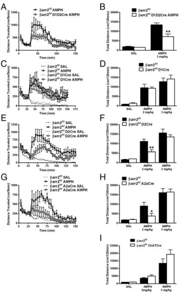
Fig. 4. Deletion of βarr2 in D2R-expressing neurons inhibits the AMPH re-sponse. (A)βarr2f/fmice were crossed with both D1Cre and D2Cre mice to delete
βarr2 in all striatal neurons simultaneously to generateβarr2f/fD1D2Cre and injected with 3 mg/kg AMPH; distance traveled was calculated for 120 min after 30 min of habituation compared with controlβarr2f/fmice. Data were analyzed using a two-way RMANOVA test [genotype×time interaction,F(29, 145)= 1.956,P<0.01] followed by Bonferroni comparisons. (B) Total cumulative dis-tance after injection of saline (SAL) and 3 mg/kg AMPH was calculated forβarr2f/f D1D2Cre andβarr2f/fcontrols. **P<0.01, using a two-way ANOVA (Bonferroni) test (n=7 mice for each genotype). (C)βarr2f/fD1Cre, (E)βarr2f/fD2Cre, or (G)
βarr2f/fA2aCre and their respective Cre-negativeβarr2f/fcontrols were injected with SAL or 2 mg/kg AMPH after 30 min of habituation, and locomotor activity was measured for 120 min. Data were analyzed using a three-way RMANOVA for D1βarr2 [genotype×time interaction,F(29, 116)=0.4045,P=0.9967; genotype× treatment interaction,F(1, 480)=3.463,P=0.0634], A2aβarr2 [genotype×time interaction,F(87, 435)=4.760; genotype×treatment interaction,F(1, 600)=47.15, P<0.001], and D2βarr2 [genotype×time interaction,F(87, 261)=3.324,P<0.001; genotype×treatment interaction,F(1, 360)=66.37,P<0.001] with post hoc Bonferroni tests. (D,F, andH) Total cumulative (120 min) postinjection distance after SAL or 2 or 3 mg/kg AMPH;n=8 mice for each group. *P<0.05, compared withβarr2f/fusing a two-way ANOVA (Bonferroni) test; **P<0.01, compared with
βarr2f/fusing a two-way ANOVA (Bonferroni) test. (I) ChAT-Cre mice were crossed withβarr2f/fmice to generateβarr2f/fChATCre orβarr2f/fcontrols, and total cu-mulative distance after postinjection of SAL or 2 or 3 mg/kg AMPH was calculated; n=8 mice for each group.
Fig. 5. UNC9994A loses its antipsychotic-like activity in response to AMPH in D2R+and A2aR+neuron-specificβarr2KO mice. Control mice (βarr2f/f) and (A)
βarr2f/fD1Cre, (C)βarr2f/fD2Cre, or (E)βarr2f/fA2aCre mice were injected with vehicle (VEH); the antipsychotics haloperidol (HAL; 0.5 mg/kg), ARI (0.5 mg/kg), or clozapine (CLOZ; 2 mg/kg); orβarr2-biased drugs UNC9994A (94A; 2 mg/kg) or UNC9975A (75A; 0.5 mg/kg) followed by 3 mg/kg AMPH injection. Total cumulative distance postinjection of AMPH for 120 min was calculated and shows that all APDs and drugs, except UNC9994A, are able to inhibit the AMPH response in theβarr2f/fD2Cre andβarr2f/fA2aCre mice. **P<0.01, compared with respective VEH control;$P<0.001, compared with respective VEH control;#P<0.05 compare 94A between genotypes using a two-way ANOVA (Bonferroni) test;##P<0.01 compare 94A between genotypes using a two-way ANOVA (Bonferroni) test. Representative graphs of AMPH inhibition by 94A for (B)βarr2f/fD1Cre, (D)βarr2f/fD2Cre, or (F)βarr2f/fA2aCre mice compared with controls (βarr2f/f);n=8 mice for each group. Data were ana-lyzed by two-way RMANOVA [genotype×treatment interaction,F(1, 420)= 2.053,P=0.1526, forβarr2f/fD1Cre; genotype×treatment interaction, F(29, 420)=3.858,P<0.05, forβarr2f/fA2aCre; and genotype×treatment interaction,F(29, 420)=2.285,P<0.01, forβarr2f/fD2Cre] with post hoc Bonferroni tests.
NEUROSCI
ENCE
PNAS
(P<0.05), suggesting a role for βarr2 outside the STR, because A2aCre is essentially STR-selective. Although the D2Cre is
expressed in STR, PFC, and midbrain, a cortical role forβarr2 in
94A-mediated inhibition of PCP-induced locomotion is most likely, because evidence suggests against a role for midbrain DA neuron
βarr2 (61) and the primary site of action for PCP is in the PFC.
However, to further rule out the contribution of midbrain
pre-synaptic βarr2 and confirm a role for corticalβarr2, we deleted
βarr2 in PFC and STR or PFC alone by injecting mCherry-Cre
AAV in the PFC of βarr2f/fA2aCre (PFC+ STRβarr2KO) or
βarr2f/fmice (PFCβarr2KO) (Fig. S6); 94A inhibited PCP-induced
locomotion in PFCβarr2KO mice (Fig. 6D) (P<0.05) but lost its
antipsychotic-like activity whenβarr2 was deleted in both PFC and
STR (Fig. 6D), thus confirming a dual dependence on cortical and
striatalβarr2. These data suggest that cortical and striatalβarr2 are
necessary for the antipsychotic-like effect of 94A. Thus, our be-havioral data further support our initial supposition that distinct mechanisms might regulate the antipsychotic-like effect of
D2R-βarr2 signaling in the PFC (agonism) versus the STR (antagonism).
We next wanted to confirm the neuronal mechanism of these distinct phenomena electrophysiogically in the PFC and STR.
Effect of 94A on Excitability of Cortical D2R+Fast-Spiking Interneurons.
The PFC comprises multiple neuronal cell types, and many of these neurons, in particular GABA interneurons and pyramidal
glutamatergic neurons, express D2Rs (56, 62–64). GABAergic
interneurons are thought to play a critical role in schizophrenia pathophysiology in humans and animal models of schizophrenia.
Postmortem brain analyses of patients (65–71) and behavioral
studies in rodents have implicated GABAergic parvalbumin+
(PV+) fast-spiking interneurons (FSIs) in altered excitation–
inhibition imbalance and cognitive impairment in schizophrenia
adult rodents in a similar fashion as D1R agonists (64), suggesting that this excitatory effect is not mediated by the canonical
in-hibitory Gαi/oactivation but presumably, is through a G
protein-independent pathway. Our data suggest that the higher levels of
βarr2 and GRK2 in the cortex might support this G
protein-independent agonist signaling of D2R ligands (Figs. 1 and 2).
Additionally, the elevatedβarr2 and GRK2 expression is present in
PFC PV+FSI (Fig. S7) and presumably, pyramidal neurons
(non-PV+cells) as well. Given the pharmacological, genetic, and
be-havioral evidence for the importance of FSIs in schizophrenia pathophysiology, we chose to determine the functional impact of
higher cortical levels ofβarr2 and GRK2 in FSIs. We performed
whole-cell, current clamp slice recordings in prefrontal GAD67+
FSI from control (βarr2+/+) and globalβarr2KO mice and assessed
the effects of UNC9994A. FSIs were visually identified in acute slices from Gad1-eGFP adult mice and further identified by their responses to hyperpolarizing and depolarizing current injections
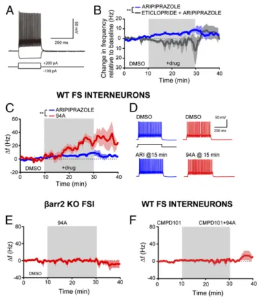
(Fig. 7A). As expected from previous studies with D2R agonists,
like quinpirole (64), ARI and 94A increased prefrontal FSI ex-citability as measured by changes in FSI action potential frequency in response to minimal amounts of depolarizing current injection.
The D2R partial agonist ARI (10μM) elicited a modest increase
in action potential firing (+6.2±1.4 Hz after 20 min of exposure
relative to a 10 min predrug baseline;n=6) (Fig. 7B). This effect
was prevented by the D2R antagonist eticlopride (10 μM) and
even led to a slight decrease in FSI excitability (−2.1±2.2 Hz after
20 min of exposure of 10μM eticlopride and 10μM ARI;n=4)
(Fig. 7B). These data suggest that ARI has modest D2R
agonist-like activity on FSIs in the PFC and are consistent with the partial
agonist activity observed with ARI when GRK2 and βarr2 are
overexpressed in HEK293 cells (Fig. 1B,D, andF). Interestingly,
94A (10μM) elicited a significantly more robust increase in FSI
excitability compared with ARI (+25.8±3.8 Hz after 20 min of
94A;+6.1±1.4 Hz after 20 min of 10μM ARI;n=4 andn=6,
respectively;P<0.01) (Fig. 7CandD). Bath application of
eti-clopride (10μM) also prevented the increase in FSI excitability by
94A and again, led to a slight decrease in FSI excitability [−5.0±
12.6 Hz after 20 min of exposure to eticlopride and 94A (10μM);
n=3]. Additionally, the effects of 94A were dependent onβ
arr2-signaling, because UNC9994A failed to enhance FSI excitability in
βarr2KO mice [+1.3±1.6 Hz after 20 min of 94A (10μM)] (Fig.
7E). Finally, the presence of the membrane-permeable GRK2
inhibitor cpd101 (30μM) prevented the increase in FSI excitability
elicited by 94A (Fig. 7F) (−2.3±3.2 Hz after 20 min of exposure of
cpd101 and 94A;n = 5). These data further highlight that 94A
requires GRK2 in addition toβarr2 to produce its D2R-dependent,
agonist-like effects in cortical FSI.
Effect of 94A on Excitability of Striatal D2R+Medium Spiny Neurons.
Based on the above behavioral and biochemical observations,
becauseβarr2 and GRK2 levels are significantly lower in the STR
than in the PFC, one might expect that the agonist activity of 94A would be reduced in striatal neurons. The D2R agonist quinpirole decreased excitability of striatal medium spiny neurons [MSNs;
−6.8±2.2 Hz after 20 min of quinpirole (10μM);n=8] (Fig. 8B,
black line). These findings are consistent with previous reports showing that D2R agonists reduce the excitability of striatal MSNs (74). Unlike quinpirole, 94A produced a negligible reduction in
striatal MSN excitability [−2.6 ± 1.0 Hz after 20 min of 94A
(10μM);n=8;P=0.0137 compared with quinpirole] (Fig. 8B
and C). Thus, 94A lacks agonist-like activity in striatal MSNs,
showing that D2R-βarr2–biased ligands can have distinct
electro-physiological actions in the PFC and STR.
Discussion
We used neuron-specificβarr2KO mice to characterize the role
ofβarr2 in striatal and cortical circuits and their involvement in
pharmacological models of antipsychotic-like action. Theβ arr2-biased D2R ligand 94A showed a paradoxical pharmacological profile in biochemical, behavioral, and electrophysiological as-says; 94A reversed the hyperlocomotor responses to both AMPH and PCP through both striatal antagonism and cortical agonism. Furthermore, the agonist activity of 94A increased the firing of
PFC D2R-expressing FSI in aβarr2- and GRK2-dependent
man-ner but did not mimic quinpirole’s ability to inhibit firing of striatal
D2R+MSNs. These contrasting electrophysiological, behavioral,
and biochemical effects are consistent with higher PFC expression
ofβarr2 and GRK2 compared with STR. These data suggest that
biased agonism of D2R-βarr2 in the PFC could add a dimension of
cortical benefits in addition to the striatal antagonist profile char-acteristic of clinically efficacious APDs.
The opposite pharmacological action of 94A in the STR and PFC in both behavioral and electrophysiological assays is
con-sistent with the higher expression ofβarr2 and GRK2 in PFC
compared to STR. Although we show that both ARI and 94A
lack basal agonist activity when direct D2R-βarr2 interactions
are assessed by BRET in HEK293 cells, the partial agonist ac-tivity of these compounds can be revealed when using assays such as the TANGO and DiscoverX (33), which markedly amplify the
signal. Similarly, when the D2R-βarr2 BRET assay is performed
in the presence of overexpressed GRK2, which recapitulates PFC expression patterns, both 94A and ARI now show similar
D2R-βarr2 partial agonist activity. However, somewhat
unex-pectedly, 94A is more efficacious than ARI not only at inhibiting PCP-induced locomotion following local PFC injection but also, in
enhancing firing of PV+FSIs of the PFC in a D2R-, GRK2-, and
βarr2-dependent manner. This enhanced PFC antipsychotic-like
effect and excitability of PV+PFC FSIs by 94A can be attributed
to the relative balance between Gαi/o(neuronal inhibition) and
βarr2 (neuronal excitation) agonist activities; 94A has no Gαi/o
agonist activity (33), whereas ARI is a Gαi/opartial agonist. In
contrast to the PFC, the STR has low expression of GRK2 and
βarr2, and we showed in vitro that, with low levels of GRK2
ex-pression, 94A is an antagonist at the D2R-βarr2 pathway.
Fur-thermore, 94A does not mimic the effect of the D2R agonist
quinpirole on striatal D2R+MSN firing and inhibits the striatal
AMPH response consistent with its antagonist-like properties in vivo. Therefore, although behaviorally, 94A has the same effect (i.e., inhibition of the locomotor response), it has opposite effects pharmacologically and on neuronal firing in the PFC versus STR. Consistent with the above observations with 94A, D2R deletion also has opposite effects in PFC versus STR on the PCP response. Although the antipsychotic-like effect of 94A is selectively
de-pendent on the D2R-βarr2 pathway, other APDs do not require
βarr2 for their antipsychotic-like activity, suggesting that targeting
βarr2 is sufficient but not necessary for antipsychotic-like activity
(60). However, our data suggest that targeting the D2R-βarr2
pathway may generate unique antipsychotic-like actions that cur-rent APDs do not achieve.
D2R antagonist and partial agonist APDs are the primary treatment options for schizophrenia, but there are several disease domains, such as negative symptoms and cognitive deficits, that are unaffected by APDs. The prevalent DA hypothesis of schizophrenia posits that this disorder presents with reduced cortical DA tone but enhanced striatal DA release, with these spatially distinct
mani-festations being interdependent (20, 75–77). Brain imaging studies
Fig. 7. UNC9994A hasβarr2- and GRK2-dependent, agonist-like effects in pre-frontal GABAergic FSIs. (A) Sample recording from a prefrontal FSI showing re-sponses to hyperpolarizing or depolarizing current injection. (B) ARI (10μM) increases action potential firing in prefrontal FSIs [+6.2±1.4 Hz (relative to a 10-min predrug baseline) after 20 min of exposure;n=6]. Bath application of eticlopride (10μM) prevented the increase in excitability elicited by ARI (−2.08± 2.2 Hz after 15 min of exposure of eticlopride+ ARI; n= 4). **P< 0.01. (C) UNC9994A (94A; 10μM) elicited a greater increase in FSI excitability (+25.86± 3.8 Hz after 20 min of exposure;n=4) compared with ARI. Data were analyzed using a standard two-way ANOVA test. **P<0.01. (D) Sample responses of FSIs to depolarizing current pulses in various pharmacologic conditions showing an increase in FSI excitability after bath application of 94A or ARI. (E) The 94A-mediated increase in FSI excitability is absent inβarr2KO mice (+1.32±1.68 Hz after 20 min of exposure;n=4). (F) Bath application of the GRK2 inhibitor cpd101 (30μM) prevented the increase in excitability elicited by 94A (−2.3± 3.2 Hz after 20 min of exposure of cpd101+94A;n=5). FS, fast spiking.
Fig. 8. UNC9994A lacks agonist-like effects in the STR. (A) Sample recording from striatal MSNs showing responses to hyperpolarizing or depolarizing current injection. (B) The D2R agonist quinpirole (10μM) decreases action potential firing in striatal MSNs (−6.8±2.2 Hz relative to a 10-min predrug baseline after 20 min of exposure;n=8). UNC9994A (94A; 10μM) elicits a significantly smaller decrease in MSN excitability compared with quinpirole (−2.6±1.0 Hz after 20 min of exposure;n=8). Data were analyzed using a standard two-way ANOVA test. *P =0.0137. (C) Representative traces of striatal MSNs to depolarizing current pulses in control conditions and after quinpirole or 94A application.
NEUROSCI
ENCE
PNAS
release is enhanced in the STR (78) (hyperdopaminergia), whereas a recent study has shown, for the first time, cortical hypo-dopaminergia in schizophrenia patients (79). Partial agonists, such as ARI, were originally developed to counteract these opposite phenomena but have largely been unsuccessful in correcting cortical dysfunction (80, 81). Our behavioral and
electrophysio-logical data show that the ARI-derivedβarr2-biased D2R ligand,
94A, can act as a D2R-βarr2 agonist in the PFC but a D2R-βarr2
antagonist on striatal D2 MSNs, highlighting the feasibility of a pharmacological approach, where a drug could potentially si-multaneously counteract both the cortical hypodopaminergia
with D2R-βarr2 agonism and striatal hyperdopaminergia with
D2R-βarr2 antagonism in schizophrenia.
Our results indicate that 94A acts as an agonist at D2R-βarr2
signaling in GABAergic FSIs. This effect may contribute to the antipsychotic-like action that we observed in PCP-induced loco-motion and most likely, will contribute to resetting excitation in-hibition balance in cortical circuits. However, we cannot exclude the contribution of D2R-expressing pyramidal neurons that may
also have elevated levels ofβarr2 and GRK2. However, DA
re-leased by PFC-projecting mesocortical DA neurons inhibits pyra-midal neurons primarily by activating interneurons (82), suggesting a temporally preceding role for FSIs. To target the exact subtype of cortical neurons for more specific behavioral analyses would require developing novel Cre driver lines involving at least triple-intersectional approaches. Appropriate selectively targeted Cre lines for various D2R neuronal subtypes in the PFC (83, 84) are currently lacking. After appropriate targeting is achieved, signaling
effectors downstream of D2R-βarr2 in the PFC can be elucidated.
Although the downstream cellular targets of D2R-G protein, such as PKA/DARPP32 signaling, are well-established (23, 25, 85),
possible downstream D2R-βarr2 signaling effectors, such as
GSK3β, AMPA, and NMDA receptors, are only now beginning
to be revealed (86, 87). In conclusion, our data provide evidence suggesting that combining the concept of functional selectivity and taking into account region- and cell-specific receptor and transducer expression patterns have the potential to generate more effective therapies and at the same time, reduce side effects.
Materials and Methods
Animals and Drugs.All mouse studies were conducted in accordance with the NIH guidelines for animal care and use and through animal protocols ap-proved by the Duke University Animal Care and Use Committee and Pfizer’s Institutional Animal Care and Use Committee. All mice were housed in a 12-h light–dark cycle at a maximum of five per cage, provided with food and water ad libitum, and tested at 10–20 wk of age. Details of mouse lines used, generation ofβarr2f/fanimals, and drugs and chemicals used are inSI
Ma-terials and Methods.
Locomotor Activity.Activity was measured in an Accuscan Activity Monitor (Accuscan Instruments) and performed as described (30, 88). Briefly, mice were allowed to habituate to the open field for 30 min, injected with various drugs, and returned to the open field. Locomotor activity was measured in 5-min intervals, and data were analyzed for the distance traveled in 5-min increments over 120 min. All APDs and tool compounds were administered i.p. and injected 10 min before AMPH or PCP injections.
IHC. Fifty-micrometer-thick vibratome cut sections of formalin-fixed mouse brains were processed for IHC analyses as described previously (30). To assess neuron-specific deletion ofβarr2, antibodies toβarr2 (generated in rabbit; at 1:300; gift from Jeff Benovic, Thomas Jefferson University, Philadelphia), Cre recombinase (mouse antibody MAB3120; at 1:500; EMD Millipore), and D2 neuron marker enkephalin (ENK; rabbit antibody; AB5026; at 1:500; EMD Millipore) were used. Because both antibodies toβarr2 and ENK were from rabbit, they were used in combination with antibodies to Cre only. Addition-ally, becauseβarr2 levels in the STR are much lower, a TSA Amplification Sys-tem (Perkin-Elmer) was used to enhance detection ofβarr2. Antibodies to PV (1:500; PVG-213; Swant Inc.) and GRK2/3 (1:500; 05–465; EMD Millipore) were used to label PV+interneurons and assess the levels of GRK2, respectively, in
three mice for each group.
Western Blot.Western blot analyses were performed on postmortem human or drug-naïve mice brain tissue as described previously (30). For human brain tissue, all procedures were carried out in compliance with an approved pro-tocol from the University of Mississippi Medical Center Institutional Review Board. Written informed consent was obtained from legally defined next of kin for tissue collection and informant-based retrospective diagnostic inter-views (Table S2). Human or mouse tissue lysates were loaded onto SDS gels followed by transfer onto nitrocellulose membranes. Membrane blots were incubated with primary antibody followed by IR secondary antibody (LICOR), and the blots were developed using an LICOR Odyssey Detection System. Details are inSI Materials and Methods.
Stereotaxic Surgeries and Virus.Deletion ofβarr2 or D2Rs in the PFC was achieved by a viral approach;βarr2f/for D2f/fmice were stereotaxically injected bilaterally with 0.5μL AAV serotype 2/8 (UNC Viral Vector Core).βarr2f/f(PFC
βarr2KO) or A2aCre βarr2f/f(PFC+ STRβarr2KO) mice were injected with mCherry-Cre AAV, whereas D2f/f mice were injected with either GFP or mCherry-Cre AAV at coordinates+2.5 mm anteroposterior (AP),±0.3 mm mediolateral (ML), and−1.8 mm dorsoventral (DV) from bregma to target the prelimbic/infralimbic region of the PFC. Mice were allowed to recover for 3 wk to allow for viral expression of GFP or mCherry-Cre before behavioral testing.
Cannulation and Local PFC Drug Injections.For local PFC injection of drugs, we inserted bilateral guide cannulas (Plastics One) into the PFC of WT mice. The bilateral guide cannulas were inserted at+2.0 mm AP with 1.0-mm spacing (±0.5 mm ML) and−2.0 mm DV and fixed to the skull with dental cement. Mice were allowed to recover for 2 wk, and then, drugs were injected using an automated injection system. Drugs were dissolved in VEH [10% (vol/vol) DMSO and 20% (vol/vol) hydroxypropyl cyclodextrin], and either 1μg (Quinpirole and UNC9994A) or 0.5μg (cpd101–GRK2/3 inhibitor; Hello Bio, Bristol, UK) per 0.5μL were injected per side at a rate of 0.4μL/min. After local injection, mice were placed in the activity monitors for 10 min before systemic (i.p.) injection with PCP (6 mg/kg), and then, locomotor activity was recorded.
cAMP Inhibition Assay.To measure D2R Gαi-mediated cAMP inhibition, a split luciferase-based cAMP biosensor (GloSensor; Promega) in HEK293T cells was used. The assay was performed in a 384-well plate using a Wallac TriLux Microbeta (Perkin-Elmer) Luminescence Counter. Details are inSI Materials and Methods.
βarr BRET Assay.To measure D2R-mediated βarr2 recruitment, a mouse D2Longreceptor fused to C-terminalrenillaluciferase and a Venus-tagged N-terminalβarr2 (a gift from Jonathan Javitch, Columbia University, New York) expressed in HEK293T cells were used in a BRET assay. The assay was performed in a 96-well plate using a Mithras LB940 Multimode Plate Reader (Berthold Technologies). Details are inSI Materials and Methods.
Electrophysiology.
Slice preparation.Three hundred-micrometer-thick coronal slices were cut from 7- to 10-wk-old mice of either sex using a Leica VT1200S Microtome. Gad1-EGFP Tg (Jackson Immunoresearch Laboratories Inc.) and globalβarr2−/−mice were
used in our electrophysiological studies. Acute slices were secured by placing a harp along the midline between the two hemispheres.
Intracellular recording.Whole-cell patch recordings were obtained from visually identified interneurons in layer V of infralimbic or prelimbic cortex or MSNs in the STR using differential contrast video microscopy on an upright microscope (BX51WI; Olympus). Recordings were obtained from FSI or MSNs of Gad1-eGFP adult mice (n=1 per animal), which were further identified by their responses to hyperpolarizing and depolarizing current injections. Recordings were col-lected using a Multiclamp 700A (Molecular Devices). Patch electrodes (tip re-sistance=4–6 MΩ) were filled with the following (in mM): 115 K-Gluconate, 10 HEPES, 20 KCl, 2 MgCl, 2 Mg-ATP, 2 Na-ATP, and 0.3 GTP. Slices were sub-merged in artificial cerebrospinal fluid containing the following: 125 mM NaCl, 25 mM NaHCO3, 3.5 mM KCl, NaH2PO4, 2 mM CaCl2, 1 mM MgCl2, and 10 mM glucose. All recordings were made at 32.5 °C±1 °C. Series resistance was usually 15–20 MΩ, and experiments were discontinued if the series resistance exceeded 30 MΩ.
treatments, or doses were compared using a post hoc Bonferroni’s test. Data were analyzed for normality with equal variance, and only parametric tests were used. Data points were excluded based on previously established criterion and set to±2 SDs from the group mean. Data are presented as mean±SEM.
ACKNOWLEDGMENTS.We thank Xiuqin Zhang and Benjamin Phillips for maintenance of the mouse colony. Antibodies toβarr2 for Western blot anal-yses (A2CT) and βarr2-specific IHC antibody were generous gifts from Dr. Robert Lefkowitz (Duke University) and Dr. Jeff Benovic (Thomas Jefferson University), respectively. Human postmortem brain samples were obtained from Dr. Craig Stockmeier (Postmortem Brain Core, University of Mississippi Medical Center). We also acknowledge the assistance of Dr. James C. Overholser,
Dr. George Jurjus, and Lesa Dieter in psychiatric assessments and Gouri Mahajan in tissue preparation. This work was supported, in part, by NIH Grants 5R37-MH-073853 and 5U-19-MH-082441. Support from the Sidney R. Baer Jr. Foundation (N.M.U.) and the Pall Family Foundation (M.G.C.) for parts of this work is also greatly appreciated. This study was also supported by an award from the Ruth K. Broad Biomedical Research Foundation (T.F.P.) and the National Cancer Institute (NCI) Clinical Oncology Research Career Development Program NCI 5K12-CA100639-10 (to J.C.S.). Some of the behavioral experiments were conducted with equipment and software purchased with a North Carolina Biotechnology Center grant. The Postmortem Brain core is supported by Institutional Develop-ment Award (IDeA) Centers of Biomedical Research Excellence (COBRE) Program of NIH/National Institute of General Medical Sciences Grant P30 GM103328. We acknowledge the support of the Cuyahoga County Medical Examiner’s Office.
1. Hopkins AL, Groom CR (2002) The druggable genome.Nat Rev Drug Discov1(9): 727–730.
2. Allen JA, Roth BL (2011) Strategies to discover unexpected targets for drugs active at G protein-coupled receptors.Annu Rev Pharmacol Toxicol51:117–144.
3. Benovic JL, Strasser RH, Caron MG, Lefkowitz RJ (1986) Beta-adrenergic receptor kinase: Identification of a novel protein kinase that phosphorylates the agonist-occupied form of the receptor.Proc Natl Acad Sci USA83(9):2797–2801. 4. Lohse MJ, Benovic JL, Codina J, Caron MG, Lefkowitz RJ (1990) beta-Arrestin: A
protein that regulates beta-adrenergic receptor function. Science 248(4962): 1547–1550.
5. Ferguson SS, et al. (1996) Role of beta-arrestin in mediating agonist-promoted G protein-coupled receptor internalization.Science271(5247):363–366.
6. Goodman OB, Jr, et al. (1996) Beta-arrestin acts as a clathrin adaptor in endocytosis of the beta2-adrenergic receptor.Nature383(6599):447–450.
7. Laporte SA, et al. (1999) The beta2-adrenergic receptor/betaarrestin complex recruits the clathrin adaptor AP-2 during endocytosis.Proc Natl Acad Sci USA96(7):3712–3717. 8. Attramadal H, et al. (1992) Beta-arrestin2, a novel member of the
arrestin/beta-arrestin gene family.J Biol Chem267(25):17882–17890.
9. Gainetdinov RR, Premont RT, Bohn LM, Lefkowitz RJ, Caron MG (2004) De-sensitization of G protein-coupled receptors and neuronal functions.Annu Rev Neurosci27:107–144.
10. Luttrell LM, et al. (1999) Beta-arrestin-dependent formation of beta2 adrenergic receptor-Src protein kinase complexes.Science283(5402):655–661.
11. Lefkowitz RJ, Shenoy SK (2005) Transduction of receptor signals by beta-arrestins.
Science308(5721):512–517.
12. DeFea KA, et al. (2000) The proliferative and antiapoptotic effects of substance P are facilitated by formation of a beta -arrestin-dependent scaffolding complex.Proc Natl Acad Sci USA97(20):11086–11091.
13. Urban JD, et al. (2007) Functional selectivity and classical concepts of quantitative pharmacology.J Pharmacol Exp Ther320(1):1–13.
14. Violin JD, Lefkowitz RJ (2007) Beta-arrestin-biased ligands at seven-transmembrane receptors.Trends Pharmacol Sci28(8):416–422.
15. Walters RW, et al. (2009) beta-Arrestin1 mediates nicotinic acid-induced flushing, but not its antilipolytic effect, in mice.J Clin Invest119(5):1312–1321.
16. Packard MG, Knowlton BJ (2002) Learning and memory functions of the Basal Gan-glia.Annu Rev Neurosci25:563–593.
17. Saint-Cyr JA, Taylor AE, Nicholson K (1995) Behavior and the basal ganglia.Adv Neurol65:1–28.
18. Zhou QY, Palmiter RD (1995) Dopamine-deficient mice are severely hypoactive, adipsic, and aphagic.Cell83(7):1197–1209.
19. Schultz W (2002) Getting formal with dopamine and reward.Neuron36(2):241–263. 20. Howes OD, Kapur S (2009) The dopamine hypothesis of schizophrenia: Version III–the
final common pathway.Schizophr Bull35(3):549–562.
21. Bernheimer H, Birkmayer W, Hornykiewicz O, Jellinger K, Seitelberger F (1973) Brain dopamine and the syndromes of Parkinson and Huntington. Clinical, morphological and neurochemical correlations.J Neurol Sci20(4):415–455.
22. Pauls DL, Abramovitch A, Rauch SL, Geller DA (2014) Obsessive-compulsive disorder: An integrative genetic and neurobiological perspective.Nat Rev Neurosci15(6): 410–424.
23. Greengard P (2001) The neurobiology of slow synaptic transmission. Science
294(5544):1024–1030.
24. Missale C, Nash SR, Robinson SW, Jaber M, Caron MG (1998) Dopamine receptors: From structure to function.Physiol Rev78(1):189–225.
25. Svenningsson P, et al. (2003) Diverse psychotomimetics act through a common sig-naling pathway.Science302(5649):1412–1415.
26. Bateup HS, et al. (2010) Distinct subclasses of medium spiny neurons differentially regulate striatal motor behaviors.Proc Natl Acad Sci USA107(33):14845–14850. 27. Beaulieu JM, et al. (2005) An Akt/beta-arrestin 2/PP2A signaling complex mediates
dopaminergic neurotransmission and behavior.Cell122(2):261–273.
28. Beaulieu JM, et al. (2008) A beta-arrestin 2 signaling complex mediates lithium action on behavior.Cell132(1):125–136.
29. Beaulieu JM, et al. (2007) Regulation of Akt signaling by D2 and D3 dopamine re-ceptors in vivo.J Neurosci27(4):881–885.
30. Urs NM, Snyder JC, Jacobsen JP, Peterson SM, Caron MG (2012) Deletion of GSK3βin D2R-expressing neurons reveals distinct roles forβ-arrestin signaling in antipsychotic and lithium action.Proc Natl Acad Sci USA109(50):20732–20737.
31. Creese I, Burt DR, Snyder SH (1976) Dopamine receptor binding predicts clinical and pharmacological potencies of antischizophrenic drugs.Science192(4238):481–483.
32. Seeman P, Lee T, Chau-Wong M, Wong K (1976) Antipsychotic drug doses and neuroleptic/dopamine receptors.Nature261(5562):717–719.
33. Allen JA, et al. (2011) Discovery ofβ-arrestin-biased dopamine D2 ligands for probing signal transduction pathways essential for antipsychotic efficacy.Proc Natl Acad Sci USA108(45):18488–18493.
34. Chen X, et al. (2012) Structure-functional selectivity relationship studies ofβ -arrestin-biased dopamine D2receptor agonists.J Med Chem55(16):7141–7153.
35. Park SM, et al. (2016) Effects ofβ-arrestin-biased dopamine D2 receptor ligands on schizophrenia-like behavior in hypoglutamatergic mice.Neuropsychopharmacology
41(3):704–715.
36. Abi-Dargham A, Laruelle M (2005) Mechanisms of action of second generation an-tipsychotic drugs in schizophrenia: Insights from brain imaging studies.Eur Psychiatry
20(1):15–27.
37. Keefe RS, Silva SG, Perkins DO, Lieberman JA (1999) The effects of atypical antipsy-chotic drugs on neurocognitive impairment in schizophrenia: A review and meta-analysis.Schizophr Bull25(2):201–222.
38. King DJ (1998) Drug treatment of the negative symptoms of schizophrenia.Eur Neuropsychopharmacol8(1):33–42.
39. Klewe IV, et al. (2008) Recruitment of beta-arrestin2 to the dopamine D2 receptor: Insights into anti-psychotic and anti-parkinsonian drug receptor signaling.Neuropharmacology
54(8):1215–1222.
40. Masri B, et al. (2008) Antagonism of dopamine D2 receptor/beta-arrestin 2 interaction is a common property of clinically effective antipsychotics.Proc Natl Acad Sci USA
105(36):13656–13661.
41. Krasel C, Bünemann M, Lorenz K, Lohse MJ (2005) Beta-arrestin binding to the beta2-adrenergic receptor requires both receptor phosphorylation and receptor activation.
J Biol Chem280(10):9528–9535.
42. Ménard L, et al. (1997) Synergistic regulation of beta2-adrenergic receptor seques-tration: Intracellular complement of adrenergic receptor kinase and beta-arrestin determine kinetics of internalization.Mol Pharmacol51(5):800–808. 43. Zhang J, et al. (1998) Role for G protein-coupled receptor kinase in agonist-specific
regulation of mu-opioid receptor responsiveness.Proc Natl Acad Sci USA95(12): 7157–7162.
44. Peterson SM, Pack TF, Caron MG (2015) Receptor, ligand and transducer contributions to dopamine D2 receptor functional selectivity.PLoS One10(10):e0141637. 45. Erdtmann-Vourliotis M, Mayer P, Ammon S, Riechert U, Höllt V (2001) Distribution of
G-protein-coupled receptor kinase (GRK) isoforms 2, 3, 5 and 6 mRNA in the rat brain.
Brain Res Mol Brain Res95(1-2):129–137.
46. Ahmed MR, Bychkov E, Gurevich VV, Benovic JL, Gurevich EV (2008) Altered expres-sion and subcellular distribution of GRK subtypes in the dopamine-depleted rat basal ganglia is not normalized by l-DOPA treatment.J Neurochem104(6):1622–1636. 47. Del Arco A, Mora F, Mohammed AH, Fuxe K (2007) Stimulation of D2 receptors in the
prefrontal cortex reduces PCP-induced hyperactivity, acetylcholine release and do-pamine metabolism in the nucleus accumbens.J Neural Transm (Vienna)114(2): 185–193.
48. Suzuki Y, Jodo E, Takeuchi S, Niwa S, Kayama Y (2002) Acute administration of phencyclidine induces tonic activation of medial prefrontal cortex neurons in freely moving rats.Neuroscience114(3):769–779.
49. Fell MJ, et al. (2009) In vitro and in vivo evidence for a lack of interaction with dopamine D2 receptors by the metabotropic glutamate 2/3 receptor agonists 1S,2S,5R,6S-2-aminobicyclo[3.1.0]hexane-2,6-bicaroxylate monohydrate (LY354740) and (-)-2-oxa-4-aminobicyclo[3.1.0] Hexane-4,6-dicarboxylic acid (LY379268).J Pharmacol Exp Ther331(3):1126–1136.
50. White IM, et al. (1995) Phencyclidine-induced increases in striatal neuron firing in behaving rats: Reversal by haloperidol and clozapine.J Neural Transm102(2):99–112. 51. Anzalone A, et al. (2012) Dual control of dopamine synthesis and release by
pre-synaptic and postpre-synaptic dopamine D2 receptors.J Neurosci32(26):9023–9034. 52. De Mei C, Ramos M, Iitaka C, Borrelli E (2009) Getting specialized: Presynaptic and
postsynaptic dopamine D2 receptors.Curr Opin Pharmacol9(1):53–58.
53. Gerfen CR (1992) The neostriatal mosaic: Multiple levels of compartmental organi-zation in the basal ganglia.Annu Rev Neurosci15:285–320.
54. Kreitzer AC (2009) Physiology and pharmacology of striatal neurons.Annu Rev Neurosci32:127–147.
55. Lidow MS, Goldman-Rakic PS, Rakic P, Innis RB (1989) Dopamine D2 receptors in the cerebral cortex: Distribution and pharmacological characterization with [3H]ra-clopride.Proc Natl Acad Sci USA86(16):6412–6416.
56. Santana N, Mengod G, Artigas F (2009) Quantitative analysis of the expression of dopamine D1 and D2 receptors in pyramidal and GABAergic neurons of the rat prefrontal cortex.Cereb Cortex19(4):849–860.
NEUROSCI
ENCE
PNAS
receptors in rat medial prefrontal cortex.J Neurosci13(6):2551–2564.
58. Lieberman JA, et al. (2008) Antipsychotic drugs: Comparison in animal models of efficacy, neurotransmitter regulation, and neuroprotection.Pharmacol Rev60(3): 358–403.
59. Powell SB, Geyer MA (2007) Overview of animal models of schizophrenia.Curr Protoc Neurosci9:9.24.
60. Schmid CL, Streicher JM, Meltzer HY, Bohn LM (2014) Clozapine acts as an agonist at serotonin 2A receptors to counter MK-801-induced behaviors through aβ arrestin2-independent activation of Akt.Neuropsychopharmacology39(8):1902–1913. 61. Bohn LM, et al. (2003) Enhanced rewarding properties of morphine, but not cocaine,
in beta(arrestin)-2 knock-out mice.J Neurosci23(32):10265–10273.
62. Tseng KY, et al. (2008) A neonatal ventral hippocampal lesion causes functional deficits in adult prefrontal cortical interneurons.J Neurosci28(48):12691–12699. 63. Tseng KY, O’Donnell P (2004) Dopamine-glutamate interactions controlling
pre-frontal cortical pyramidal cell excitability involve multiple signaling mechanisms.
J Neurosci24(22):5131–5139.
64. Tseng KY, O’Donnell P (2007) Dopamine modulation of prefrontal cortical inter-neurons changes during adolescence.Cereb Cortex17(5):1235–1240.
65. Akbarian S, et al. (1995) Gene expression for glutamic acid decarboxylase is reduced without loss of neurons in prefrontal cortex of schizophrenics.Arch Gen Psychiatry
52(4):258–266.
66. Thompson M, Weickert CS, Wyatt E, Webster MJ (2009) Decreased glutamic acid decarboxylase(67) mRNA expression in multiple brain areas of patients with schizo-phrenia and mood disorders.J Psychiatr Res43(11):970–977.
67. Volk DW, Austin MC, Pierri JN, Sampson AR, Lewis DA (2000) Decreased glutamic acid decarboxylase67 messenger RNA expression in a subset of prefrontal cortical gamma-aminobutyric acid neurons in subjects with schizophrenia.Arch Gen Psychiatry57(3): 237–245.
68. Fung SJ, et al. (2010) Expression of interneuron markers in the dorsolateral prefrontal cortex of the developing human and in schizophrenia.Am J Psychiatry167(12): 1479–1488.
69. Hashimoto T, et al. (2008) Conserved regional patterns of GABA-related transcript expression in the neocortex of subjects with schizophrenia.Am J Psychiatry165(4): 479–489.
70. Nakazawa K, et al. (2012) GABAergic interneuron origin of schizophrenia patho-physiology.Neuropharmacology62(3):1574–1583.
71. Lewis DA, Curley AA, Glausier JR, Volk DW (2012) Cortical parvalbumin interneurons and cognitive dysfunction in schizophrenia.Trends Neurosci35(1):57–67. 72. Sohal VS, Zhang F, Yizhar O, Deisseroth K (2009) Parvalbumin neurons and gamma
rhythms enhance cortical circuit performance.Nature459(7247):698–702. 73. Cho KK, et al. (2015) Gamma rhythms link prefrontal interneuron dysfunction with
cognitive inflexibility in Dlx5/6(+/-) mice.Neuron85(6):1332–1343.
74. Surmeier DJ, Ding J, Day M, Wang Z, Shen W (2007) D1 and D2 dopamine-receptor modulation of striatal glutamatergic signaling in striatal medium spiny neurons.
Trends Neurosci30(5):228–235.
75. Pycock CJ, Kerwin RW, Carter CJ (1980) Effect of lesion of cortical dopamine terminals on subcortical dopamine receptors in rats.Nature286(5768):74–76.
dependent effects of amphetamine on mesoaccumbens dopamine release and loco-motion.Neuropsychopharmacology29(1):72–80.
77. Weinberger DR (1987) Implications of normal brain development for the pathogen-esis of schizophrenia.Arch Gen Psychiatry44(7):660–669.
78. Laruelle M, Abi-Dargham A (1999) Dopamine as the wind of the psychotic fire: New evidence from brain imaging studies.J Psychopharmacol13(4):358–371.
79. Slifstein M, et al. (2015) Deficits in prefrontal cortical and extrastriatal dopamine release in schizophrenia: A positron emission tomographic functional magnetic res-onance imaging study.JAMA Psychiatry72(4):316–324.
80. Kern RS, et al. (2006) The neurocognitive effects of aripiprazole: An open-label comparison with olanzapine.Psychopharmacology (Berl)187(3):312–320. 81. Rajagopal L, Massey BW, Huang M, Oyamada Y, Meltzer HY (2014) The novel object
recognition test in rodents in relation to cognitive impairment in schizophrenia.Curr Pharm Des20(31):5104–5114.
82. Kabanova A, et al. (2015) Function and developmental origin of a mesocortical in-hibitory circuit.Nat Neurosci18(6):872–882.
83. Madisen L, et al. (2015) Transgenic mice for intersectional targeting of neural sensors and effectors with high specificity and performance.Neuron85(5):942–958. 84. Madisen L, et al. (2010) A robust and high-throughput Cre reporting and
character-ization system for the whole mouse brain.Nat Neurosci13(1):133–140.
85. Bateup HS, et al. (2008) Cell type-specific regulation of DARPP-32 phosphorylation by psychostimulant and antipsychotic drugs.Nat Neurosci11(8):932–939.
86. Du J, et al. (2010) A kinesin signaling complex mediates the ability of GSK-3beta to affect mood-associated behaviors.Proc Natl Acad Sci USA107(25):11573–11578. 87. Li YC, Xi D, Roman J, Huang YQ, Gao WJ (2009) Activation of glycogen synthase
ki-nase-3 beta is required for hyperdopamine and D2 receptor-mediated inhibition of synaptic NMDA receptor function in the rat prefrontal cortex.J Neurosci29(49): 15551–15563.
88. Urs NM, Daigle TL, Caron MG (2011) A dopamine D1 receptor-dependentβ-arrestin signaling complex potentially regulates morphine-induced psychomotor activation but not reward in mice.Neuropsychopharmacology36(3):551–558.
89. Rossi J, et al. (2011) Melanocortin-4 receptors expressed by cholinergic neurons reg-ulate energy balance and glucose homeostasis.Cell Metab13(2):195–204. 90. Heiman M, Kulicke R, Fenster RJ, Greengard P, Heintz N (2014) Cell type-specific
mRNA purification by translating ribosome affinity purification (TRAP).Nat Protoc
9(6):1282–1291.
91. Heiman M, et al. (2014) Molecular adaptations of striatal spiny projection neurons during levodopa-induced dyskinesia.Proc Natl Acad Sci USA111(12):4578–4583. 92. Livak KJ, Schmittgen TD (2001) Analysis of relative gene expression data using
real-time quantitative PCR and the 2(-Delta Delta C(T)) Method.Methods25(4):402–408. 93. Lowe JD, et al. (2015) Role of G protein-coupled receptor kinases 2 and 3 inμ-opioid
receptor desensitization and internalization.Mol Pharmacol88(2):347–356. 94. Zhu H, et al. (2012) Quantitative analysis of focused a-to-I RNA editing sites by
ultra-high-throughput sequencing in psychiatric disorders.PLoS One7(8):e43227. 95. Cobb JA, et al. (2013) Hippocampal volume and total cell numbers in major depressive