Dissecting the human serum antibody
response to secondary dengue virus
infections
Bhumi Patel1, Patti Longo2, Michael J. Miley2, Magelda Montoya3, Eva Harris3, Aravinda M. de Silva1*
1 Department of Microbiology and Immunology, University of North Carolina School of Medicine, Chapel Hill, North Carolina, United States of America, 2 Department of Pharmacology, University of North Carolina School of Medicine, Chapel Hill, North Carolina, United States of America, 3 Division of Infectious Diseases and Vaccinology, School of Public Health, University of California, Berkeley, California, United States of America
Abstract
Dengue viruses (DENVs) are mosquito-borne flaviviruses and the causative agents of den-gue fever and denden-gue hemorrhagic fever. As there are four serotypes of DENV (DENV1-4), people can be infected multiple times, each time with a new serotype. Primary infections stimulate antibodies that mainly neutralize the serotype of infection (type-specific), whereas secondary infections stimulate responses that cross-neutralize 2 or more serotypes. Previ-ous studies have demonstrated that neutralizing antibodies induced by primary infections recognize tertiary and quaternary structure epitopes on the viral envelope (E) protein that are unique to each serotype. The goal of the current study was to determine the properties of neutralizing antibodies induced after secondary infection with a different (heterotypic) DENV serotypes. We evaluated whether polyclonal neutralizing antibody responses after second-ary infections consist of distinct populations of type-specific antibodies to each serotype encountered or a new population of broadly cross-neutralizing antibodies. We observed two types of responses: in some individuals exposed to secondary infections, DENV neutraliza-tion was dominated by cross-reactive antibodies, whereas in other individuals both type-spe-cific and cross-reactive antibodies contributed to neutralization. To better understand the origins of type-specific and cross-reactive neutralizing antibodies, we analyzed sera from individuals with well-documented sequential infections with two DENV serotypes only. These individuals had both type-specific and cross-reactive neutralizing antibodies to the 2 types responsible for infection and only cross-reactive neutralizing antibodies to other sero-types. Collectively, the results demonstrate that the quality of neutralizing (and presumably protective) antibodies are different in individuals depending on the number of previous expo-sures to different DENV serotypes. We propose a model in which low affinity, cross-reactive antibody secreting B-cell clones induced by primary exposure evolve during each secondary infection to secrete higher affinity and more broadly neutralizing antibodies.
a1111111111 a1111111111 a1111111111 a1111111111 a1111111111 OPEN ACCESS
Citation: Patel B, Longo P, Miley MJ, Montoya M,
Harris E, de Silva AM (2017) Dissecting the human serum antibody response to secondary dengue virus infections. PLoS Negl Trop Dis 11(5): e0005554.https://doi.org/10.1371/journal. pntd.0005554
Editor: Amy C Morrison, University of California,
Davis, UNITED STATES
Received: August 19, 2015
Accepted: April 5, 2017
Published: May 15, 2017
Copyright:©2017 Patel et al. This is an open access article distributed under the terms of the
Creative Commons Attribution License, which permits unrestricted use, distribution, and reproduction in any medium, provided the original author and source are credited.
Data Availability Statement: All relevant data are
within the paper and its Supporting Information files.
Funding: The studies were supporting by the
Author summary
The four dengue virus serotypes are emerging mosquito-borne flaviviruses and the causa-tive agents of dengue fever and dengue hemorrhagic fever/dengue shock syndrome. Infected people develop protective immunity to the infecting serotype but remain suscep-tible to secondary infections with new serotypes. Both antibodies and T-cells are responsi-ble for protection against re-infection by the same serotype. The goal of the current study was to analyze the properties of antibodies in people who have been exposed to a single or secondary dengue virus infections. We found that people exposed to a single infection have neutralizing antibodies that mainly bind sites that are unique to the infecting sero-type. In contrast, secondary infections induced more complex mixtures of neutralizing antibodies that target regions that unique to each serotype and regions that are conserved between serotypes. Our results have implications for understanding protective responses after natural infections as well as responses induced by dengue vaccines.
Introduction
Dengue Virus (DENV) is a mosquito-borneflavivirusand the causative agent of dengue fever and dengue hemorrhagic fever (DHF) [1]. Several hundred million people are estimated to acquire DENV infections each year[2]. Dengue infections can be clinically inapparent or lead to symptoms that range from an undifferentiated fever to severe DHF and dengue shock syn-drome (reviewed in [3,4]).
The DENV complex consists of 4 viruses designated as serotypes (DENV1-4)[1]. Primary infection by DENV leads to long-term protection against the serotype of infection (homolo-gous serotype) but not other serotypes (heterolo(homolo-gous serotypes) [5–7]. Subsequent secondary infection with a new serotype results in serotype cross-neutralizing antibodies that correlate with durable protection against 2 or more serotypes [5,7]. Recent studies have defined the properties of human antibodies responsible for serotype-specific neutralization after primary infection [8–12]. In the present study we investigated the properties of serum neutralizing anti-bodies produced after recovery from secondary DENV infections.
The DENV envelope (E) protein, which binds to cellular receptors and mediates viral entry and fusion, is the main target of neutralizing and protective antibodies [13]. The ectodomain of E protein is composed of three domains: I, II and III (EDI, EDII and EDIII)[14]. Each DENV particle has 180 monomers of E that are organized into 90 dimers that cover the entire surface of the virus. The E proteins are arranged with icosahedral symmetry with each asym-metric unit containing portions of three homodimers[15]. After primary DENV infection, people develop a mix of DENV serotype reactive and type-specific antibodies. The cross-reactive antibodies are weakly neutralizing and have been implicated in antibody dependent enhanement of DENVs during secondary infections [8,16–18]. The serotype-specific antibod-ies are strongly neutralizing and, presumably, responsible for long-term protection aganst re-infection with the same serotype. Quaternary epitopes formed after assembly of E molecules into higher order structures required for virion assembly are major targets of type-specific neutralizing antibodies [8–12].
Here we report on the properties of serum neutralizing antibodies in people exposed to 2 or more DENV infections. The studies were designed to test if people exposed to secondary infec-tions have neutralizing antibodies that mainly recognize epitopes that are conserved between serotypes or epitopes that are unique to each serotype previously encountered. Unlike primary DENV infections that result in predominantly serotype-specific polyclonal neutralizing
Sciences Institute) Pediatric Dengue Vaccine Initiative grant VE-1 (PI E. Harris, UCB). The funders had no role in study design, data collection and analysis, decision to publish, or preparation of the manuscript.
Competing interests: Dr. de Silva has consulted
antibody responses, some people exposed to secondary infections had neutralizing antibodies that mainly recognized epitopes conserved between serotypes, while others had antibodies that targeted both type-specific and conserved epitopes.
Materials and methods
Ethics statement
Blood donations were obtained from individuals who had traveled to dengue-endemic regions and experienced a primary DENV infection. These human samples were obtained with informed consent approximately 2 to 10 years after DENV infection. All blood donations were collected in compliance with the Institutional Review Board of the University of North Caro-lina at Chapel Hill. Written informed consent was obtained from all subjects before participa-tion in the study. Blood donaparticipa-tions were obtained from individuals who had traveled to dengue-endemic regions or people living in endemic areas. All blood donations were collected in compliance with the Institutional Review Board of the University of North Carolina at Chapel Hill and the University of California at Berkeley. Written informed consent was obtained from all subjects before participation in the study.
Immune sera
Dengue traveler study. Human sera were collected from individuals who had
experi-enced a DENV infection during travel to a DENV endemic area (Table 1). All donations were collected in compliance with the Institutional Review Board of the University of North Caro-lina at Chapel Hill (Protocol #08–0895). Samples were classified as primary infections if they had neutralizing antibody to only one serotype (1:40 Neut50titers) or a titer that is 4-fold
higher for one DENV serotype compared to low titers to the other serotypes. Samples were classified as secondary infections if they had Neut50titers greater than a 1:40 dilution for two
or more DENV serotypes.
Nicaraguan Pediatric Dengue Cohort Study. The Nicaraguan Pediatric Dengue Cohort
Study is an ongoing community-based prospective study of children 2–14 years old established in 2004 in Managua, Nicaragua [19,20]. Participants are encouraged to present at the first sign
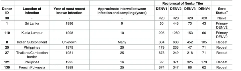
Table 1. Panel of naïve and late convalescent DENV-immune human sera.
Reciprocal of Neut50Titer
Donor ID
Location of infection
Year of most recent known infection
Approximate interval between infection and sampling (years)
DENV1 DENV2 DENV3 DENV4 Sero Status†
30 - - - <20 <20 <20 <20 Naïve
1 Sri Lanka 1996 9 50 443 70 43 Primary
DENV2
110 Kuala Lumpur 1998 10 205 1280 153 96 Primary
DENV2
0 Indian Subcontinent Unknown Many 304 630 452 105 Repeat
25 Philippines 1975 25 179 233 47 71 Repeat
27 Thailand/Cambodian border
1981 25 878 249 218 71 Repeat
121 Philipines 1995 16 92 371 325 179 Repeat
130 French Polynesia 1989 25 674 347 86 62 Repeat
†Primary defined as reciprocal Neut
50titer greater than 40 for one serotype only or a reciprocal Neut50titer that is at least 4-fold greater for one serotype.
Secondary defined as reciprocal Neut50titer greater than 40 for two or more serotypes and the highest titer not 4-fold greater than the 2ndhighest titer.
of illness to the HCSFV, where study physicians provide medical care and screen for signs and symptoms of dengue. Suspected dengue cases, as defined by the traditional WHO dengue case definition (WHO Guidelines 1997) and febrile participants without other apparent origin (undifferentiated febrile illnesses) are analyzed for DENV infection using serological, molecu-lar and virological methods in acute-phase (days 1–5) and convalescent-phase (14–21 days post-onset of fever) blood samples [19,20]. Healthy annual blood samples are also collected for serological analysis. Neut50titers were determined in annual samples using a flow
cytome-try-based assay with reporter viral particles representing the four DENV serotypes in human monocytic U937 cells expressing the DENV attachment factor DC-SIGN[21,22]. The PDCS was approved by the Institutional Review Boards of the University of California, Berkeley, and the Nicaraguan Ministry of Health. Parents or legal guardians of all subjects provided written informed consent, and subjects 6 years of age and older provided assent.
Virus and recombinant envelope protein
WHO reference strains, DENV1 (West Pac 74), DENV2 (S-16803), DENV3 (CH-53489) and DENV4 (TVP-376) were used in the present study. The strains were grown in C6/36 mosquito cells to generate infectious stocks and Vero-81 mammalian cells to generate purified antigen. DENVs from culture media were purified by density gradient and ultracentrifugation as previ-ously described [23]. DENV2 (NGC) purified live virion antigen was purchased from Microbix Biosystems, Inc. (Mississauga, Ontario, Canada). DENV2 envelope protein was produced as a soluble recombinant protein (amino acids 1–397 of S-16803) using the Bac-to-Bac Baculovirus Expression System (Invitrogen Life Technologies) and Sf9 insect cells. The protein was puri-fied from cell culture supernatant using an antibody column coated with 4G2, a serotype-cross reactive mouse monoclonal that targets a conserved region on the envelope protein. Structure of the recombinant protein was verified by ELISA using monoclonal antibodies that bind to well-defined epitopes on all three domains of E protein.
Fluorescent Activated Cell Sorting (FACS)-based DENV neutralization
assay
DENV neutralizing activity of human immune sera was assessed using a flow cytometry-based assay with U937 human monocytic cells stably transfected with DC-SIGN as previously described [24]. In brief, serially diluted human sera were incubated with virus for 45 min at 37˚C followed by the addition of U937 DC-SIGN cells. Cells were incubated with virus for 2 hours at 37˚C, washed with media to remove immune sera and unbound virus and incubated for 24 hours at 37˚C. Cells were fixed, permeabilized and stained with 2H2-Alexa Flour 488, a mouse monoclonal that binds to DENV pre-membrane protein. Infected cells were quantified using a Guava flow-cytometer (Milipore). Stained cells were analyzed using GraphPad Prism version 6.00 (La Jolla California USA,www.graphpad.com) to calculate 50% neutralization titers as previously described [25]. We determined if depletion of specific antibodies resulted in statistically significant (P<0.05) changes in Neut50values by comparing neutralization
curves using the Extra Sum of Squares F test (Non-linear regression: Compare function in GraphPad Prism version 6.00). Each control and antigen depleted serum sample was tested in a single neutralization assay in duplicate using all 4 DENV serotypes.
Detection of DENV binding antibodies
pH 9.6 for 2 hours at room temperature. The plates were blocked with Tris-buffered Saline containing 0.05% Tween 20 with 3% Normal Goat Serum followed by an incubation with a 1:40 dilution of control or DENV depleted human sera for 1 hour at 37˚C. Alkaline phospha-tase conjugated Goat anti-human IgG (Sigma) was added to the plates for 1 hour at 37˚C. The plates were washed and then developed by adding p-nitrophenyl phosphate substrate. The optical density at 405nm was recorded using a spectrophotometer.
Whole virus depletion of DENV-specific antibodies from human immune
sera
Purified DENV was absorbed onto 4.5-μm Polybead polystyrene microspheres (Polysciences, Inc.) at a bead (ul) to ligand (ug) ratio of 5:2. Beads were washed three times with 0.1M Borate buffer (pH 8.5) followed by an overnight incubation with purified DENV and 0.1M Borate buffer (pH 8.5) at room temperature (RT). Control beads were incubated with an equivalent amount of BSA. The control and virus absorbed beads were then blocked with a 10 mg/ml BSA solution for 30 minutes at RT followed by four washes with PBS. DENV-specific antibod-ies were depleted from human sera by incubating virus absorbed beads with human sera diluted 1:10 in PBS for 45 minutes at 37˚C. Typically three cycles were performed to remove dengue-specific antibodies in each serum sample. Successful depletion of DENV-specific anti-bodies was confirmed via an ELISA with purified DENV coated plates.
Recombinant E protein depletion of DENV-specific antibodies from
immune sera
Purified DENV2 rE protein was conjugated to magnetic dynabeads M-270 Epoxy (Invitrogen by Life Technologies) with a bead (mg) to ligand (ug) ratio of 5:1. Beads were washed three times with 0.1M Sodium Phosphate (7.4) followed by an overnight incubation with equal vol-umes of purified rE protein, 0.1M Borate buffer (pH 9.5) and 3M Ammonium sulfate at 37˚C. Control beads were incubated with the equivalent amount of BSA. Control and DENV2 rE conjugated beads were then blocked by incubating with a 10 mg/ml BSA solution followed by four washes with PBS. DENV2 rE-specific antibodies were depleted from human sera by incu-bating rE conjugated beads with human sera diluted 1:10 in PBS for 45 minutes at 37˚C three times with end-over-end mixing. Successful depletion of DENV2 rE-specific antibodies was confirmed via an ELISA with purified DENV2 rE coated plates.
Calculating the proportion of re binding neutralizing antibodies
After depletion of sera using DENV2 rE antigen, the percentage of rE binding DENV2 neutral-izing Abs were calculated using the following formula.
%rE binding DENV2neutralizing Abs
¼ ð1 ðNeut50after DV2rE deletion=ðNeut50after control deletionÞÞ 100:
Results
To determine if DENV serotype cross-reactive or type-specific antibodies were responsible for neutralizing activity in each serum sample, we used polystyrene beads coated with purified dengue virions to deplete different populations of antibodies from serum samples. The anti-bodies in each serum sample were depleted using beads coated with purified DENV2 or an equal mixture of DENV1, 3 and 4. As a control, the serum was also incubated with beads coated with BSA. The DENV2 depletions were expected to remove DENV2 type-specific and DENV serotype cross-reactive antibodies, while retaining any DENV1, 3 or 4 type-specific antibodies in the sample. After depleting with DENV2, any reduction in DENV1, 3 and 4 neu-tralizing activity in the sample was attributed to cross-reactive (heterotypic) antibodies. Simi-larly, when serum samples were depleted with beads containing equal amounts of purified DENV1, 3 and 4, any reduction in DENV2 neutralizing antibody was attributed to cross-reac-tive antibodies.
Depletion of DENV binding antibodies from primary DENV2 immune
sera
Depletion of two primary DENV2-immune sera (DT001 and DT110) using beads coated with DENV2 resulted in a reduction of binding to DENV2 as well as DENV1, 3 and 4, demonstrat-ing removal of cross-reactive and type-specific antibodies in the sample (S1 Fig). Antibody depletion with the homologous (serotype of infection) DENV2 antigen led to a significant (P<0.05; Extra Sum of Squares F test to compare neutralization curves) drop in DENV2 neu-tralization (S1 Fig). Following depletion with DENV1, 3 and 4 coated beads, ELISA confirmed removal of cross-reactive antibodies, while still retaining DENV2 type-specific antibodies in the sample (S1 Fig). After removal of cross-reactive antibodies, the samples retained most of the DENV2 neutralizing activity and the neutralization titers of the control and depleted sam-ples were not significantly different (P>0.05; Extra Sum of Squares F test to compare neutrali-zation curves) (S1 Fig).Fig 1A and 1B). These results are in agreement with previous reports that following primary DENV infections neutralization of the homologous serotype is mainly mediated by type-specific antibodies [8].
Depletion of DENV binding antibodies from secondary infection DENV
immune sera
DT121, another subject with neutralizing antibodies to all 4 serotypes, also had a response that was dominated by cross reactive neutralizing antibodies (Fig 1EandS2 Fig).
DT130, a subject that strongly neutralized DENV1 and 2 but not 3 and 4 (Table 1), had a mixture of type-specific and cross-reactive neutralizing antibodies (Fig 1D). Following removal of DENV2 binding antibodies, we observed a major loss (P<0.05; Extra Sum of Squares F test) of DENV2 neutralization and only a partial loss of DENV1 neutralization (S2 Fig). This result indicates that both type-specific and cross reactive antibodies are responsible for the high DENV1 neutralizing activity in this individual (Fig 1D). Reciprocal depletion with DENV1, 3 and 4 antigens removed all the DENV2 neutralizing activity demonstrating that cross-reactive antibodies were responsible for neutralization (Fig 1DandS2 Fig). Samples DT025 and DT027 also exhibited a similar pattern in which both type-specific and cross-reactive antibodies contributed to DENV neutralization (Fig 1F and 1G, andS2 Fig). Unlike primary DENV infections that stimulate durable serotype-specific neutralizing antibody responses, we conclude that secondary infections result in more complex mixtures of neutral-izing antibodies that recognize serotype-specific and cross-reactive epitopes. The proportions of these two classes of antibodies varied between individuals exposed to secondary infections.
Depletion of DENV binding antibodies from individuals exposed to a
known sequence infection with two different DENV serotypes
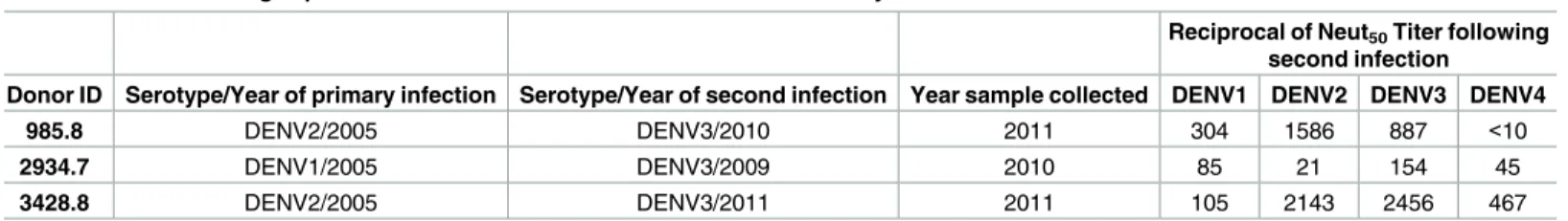
To better understand different patterns of type-specific and cross reactive neutralizing anti-bodies in people exposed to secondary DENV infections, we analyzed serum samples from 3 individuals with well documented histories of two sequential infections with different sero-types of DENV (Table 2). These samples were obtained from a long-term prospective pediatric DENV cohort study in Nicaragua [22]. Two of the subjects had been exposed to a first DENV2 infection followed by a second DENV3 infection (Subjects 985.8 and 3428.8). One subject had been exposed to a DENV1 infection followed by a DENV3 infection (Subject 2934.7). In sera collected several months after the second infection, all three subjects had varying levels of neu-tralizing antibodies to at least 3 different serotypes (Table 2).
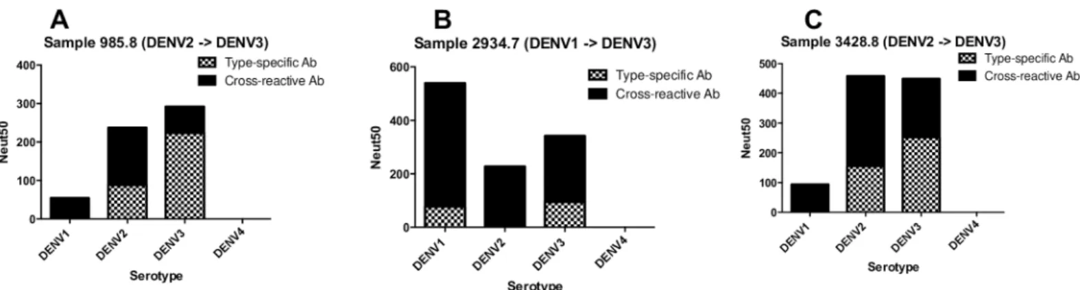
We depleted each post-second infection sample using beads coated with the DENV sero-type of the 1stor 2ndinfection and performed neutralization assays to estimate levels of cross-reactive and type-specific neutralizing antibodies against each DENV serotype. All three sub-jects had a mixture of type-specific and cross-reactive neutralizing antibodies directed to the serotypes of known 1stand 2ndinfection (Fig 2andS3 Fig). For serotypes not “seen” by the individual, neutralization was driven by cross-reactive antibodies only (Fig 2andS3 Fig).
Depletion of DENV recombinant E protein binding antibodies
Our previous studies with primary DENV immune sera revealed that type-specific neutralizing antibodies mostly targeted quaternary epitopes expressed on E protein dimers or higher order structures but not recombinant E protein (rE), which is mainly a monomer in solution[8]. We,
Table 2. Panel of Nicaragua pediatric cohort human immune sera used in the study.
Reciprocal of Neut50Titer following
second infection
Donor ID Serotype/Year of primary infection Serotype/Year of second infection Year sample collected DENV1 DENV2 DENV3 DENV4
985.8 DENV2/2005 DENV3/2010 2011 304 1586 887 <10
2934.7 DENV1/2005 DENV3/2009 2010 85 21 154 45
3428.8 DENV2/2005 DENV3/2011 2011 105 2143 2456 467
next, addressed if neutralizing antibodies induced by secondary DENV infections targeted simple or quaternary epitopes on E protein. When convalescent immune sera from individuals exposed to primary DENV2 infections were depleted of DENV2 rE binding antibodies, we observed no significant loss of DENV2 neutralization (P>0.05; Extra Sum of Squares F test), confirming the importance of quaternary structure neutralizing antibody epitopes after pri-mary infection (Fig 3andS4 Fig). When convalescent immune sera from individuals exposed to secondary DENV infections (Table 1) were depleted of DENV2 rE binding antibodies and tested for DENV neutralization, we observed a significant decrease in DENV2 neutralizing antibody titers (P<0.05; Extra Sum of Squares F test) (Fig 3andS5 Fig). Our results indicate that epitopes displayed on rE protein are a major target of DENV2 neutralizing antibodies after secondary infections but not primary DENV2 infections.
Discussion
Recently several studies have described the properties of neutralizing antibodies generated fol-lowing primary DENV infections and the epitopes targeted by these antibodies [8–10,18,26]. Although primary infections stimulate both DENV serotype-specific and cross reactive bind-ing antibodies, only the type-specific antibodies have been linked to durable neutralizbind-ing and protective responses. These type-specific antibodies target tertiary and quaternary structure E protein epitopes displayed on the surface of the virus [8–10,12].
In the current study we observed that the majority of neutralizing antibodies that develop after secondary DENV infections recognize serotype cross reactive epitopes. The cross-reactive neutralizing antibodies bound to simple epitopes on soluble E protein as well as more complex epitopes displayed on the intact DENV particle. Indeed, serotype cross-reactive and neutraliz-ing human monoclonal antibodies isolated from people exposed to secondary DENV infec-tions bind with high affinity to simple epitopes on domain II as well as quaternary epitopes that span across two E proteins forming a single dimer [16,27–29].
Following secondary infections, some people had a single population of DENV serotype cross-reactive and cross-neutralizing antibodies, whereas others had a mixture of type-specific and cross-reactive neutralizing antibodies. We suspect that subjects like DT000 and 121 who mainly had cross-reactive neutralizing antibodies are likely to have had multiple DENV expo-sures resulting in broad neutralization of all 4 serotypes. Subjects like DT27 and 130, who had
Fig 2. Properties of DENV neutralizing antibodies after sequential infections with two different DENV serotypes. Subjects 985.8 (A) and 3428.8 (C) experienced sequential DENV2!DENV3 infections. Subject 2934.7 (B) experienced sequential DENV1!DENV3 infections. Polystyrene beads coated with DENV serotypes that infected each individual were used to deplete different populations of antibodies in the samples. After confirming depletion of relevant antibodies, the sera were tested for neutralization of DENV1-4 (seeS3 Fig). The graphs depict the levels of DENV serotype-specific and cross-reactive neutralizing antibodies in each subject after the second infection.
a mixture of type-specific and cross-reactive neutralizing antibodies are likely to represent peo-ple who have been exposed to sequential infections with 2 serotypes only. Indeed, when we tested samples from three subjects enrolled in a long-term prospective pediatric cohort study in Nicaragua with know exposure to just two sequential DENV serotype infections, all three subjects had a mixture of type-specific and cross reactive neutralizing antibodies to the sero-types responsible for the first and second infections. Tsai and colleagues recently characterized DENV neutralizing antibodies in volunteers infected with a single serotype monovalent live attenuated DENV vaccine or sequentially infected with two monovalent DENV vaccines rep-resenting different serotypes [30]. They also observed type-specific neutralizing antibodies in volunteers who received the single serotype vaccine and mixture of type-specific and cross reactive neutralizing antibodies after sequential infection with 2 different serotypes.
We propose that low affinity DENV cross reactive memory B-cells derived from primary infections undergo antibody somatic hyper mutation and each subsequent DENV exposure selects and expands rare affinity matured clones with greater neutralization breadth and potency (Fig 4). This model is supported by studies comparing the avidity and neutralization of both monoclonal antibodies and polyclonal sera from people after primary and secondary
Fig 3. People exposed to secondary DENV infections develop neutralizing antibodies that target simple epitopes on recombinant E protein. Primary DENV2 or secondary DENV infection convalescent immune sera depleted of rE protein binding antibodies were assessed for neutralization of DENV2. In people exposed to secondary DENV infections, a greater proportion of DENV2 neutralizing antibodies bound to epitopes displayed on rE protein compared to people exposed to primary DENV2 infections (Mean of 75% vs. 26%;*P<0.018 unpaired Student t Test to compare means). The primary immune sera data are from 2 subjects analyzed in this study (see S4 Fig) and two subjects analyzed in a previous study[8]. All the secondary infection data are from samples analyzed for current study.
DENV infections. Analysis of polyclonal human sera following DENV infections revealed that the avidity of DENV antibodies following secondary infection was higher than that of antibod-ies generated following a primary infection [31]. In agreement with this, studies focusing on group-reactive MAbs derived from primary and secondary DENV infected patients found that the group-reactive MAbs from patients with secondary infection had stronger neutralization potencies and higher binding avidities than those derived from patients with primary infection [29,32,33]. Additional studies have identified a class of broadly neutralizing human antibod-ies produced by plasmablasts in hospitalized cases of secondary DENV infections. Structural analysis of these broadly neutralizing antibodies in complex with rE revealed that these anti-bodies recognize serotype invariant sites at the E dimer interface. Collectively, these studies support the idea that low affinity, weakly neutralizing antibody clones generated followed pri-mary DENV infections give rise to antibodies of increasing breadth and neutralization potency with each subsequent exposure (Fig 4).
In this study we analyzed in-depth convalescent blood samples from 10 individuals exposed to DENV infections. While the small sample size is a weakness, it is challenging to perform antibody depletion studies on larger panels because of the complexity of the studies and the volume of blood required. Another limitation of our study is that the infection history of some of the study subjects was inferred by the neutralizing antibody profile and travel history, there-fore, definite conclusions relating antibody population characteristics to the number of sec-ondary infections cannot be made. However, three subjects with known sequences of two DENV infections support our conclusion that sequential infections with two serotypes result in a mixture of cross-reactive and type-specific neutralizing antibodies to serotypes responsible for infections, while inducing cross-reactive neutralizing antibodies only to the serotypes not
Fig 4. Model of B cell maturation following sequential DENV infections. With each successive DENV infection, the ratio of serotype-specific (TS) and cross-reactive (CR) antibodies that contribute to DENV neutralization changes. During a primary infection (DENV1 in this example), dengue-specific naïve B cells are activated and these cells give rise to both memory B cells (MBCs) and antibody secreting long lived plasma cells (LLPCs). This primary response is dominated MBC and LLPCs clones producing low affinity, weakly neutralizing serotype CR antibodies. The primary response also contains rare MBC and LLPCs producing TS antibodies that strongly neutralize DENV1. Following a secondary infection with a new serotype (DENV3 in this example), the overall DENV-specific B-cell response will be dominated by the activation and expansion of DENV1 and 3 cross-reactive MBCs induced by the primary infection. MBCs producing CR antibodies that bind to the second infecting serotype with high affinity will be preferentially activated. These activated cells will re-enter germinal centers and undergo further rounds of somatic hyper mutation. CR B-cells with high affinity for the second serotype will be selectively expanded to give rise to cross-reactive MBC and LLPCs that strongly cross-neutralize multiple serotypes. In the figure this increase in affinity and neutralization is depicted by an increase in the color gradient (light yellow to bright orange) of CR B-cells. Following a tertiary infection (DENV2 in this example), this process is secondaryed again and results in a population of CR MBCs and LLPCs that dominate the neutralizing antibody response. While the B-cell clones producing TS strongly neutralizing antibodies are also likely to be maintained through each successive round of infection, the TS response will account for only a small fraction of the total neutralizing response.
“seen” by the host. Studies are currently in progress using additional samples from human dengue cohort studies with well-defined infection histories to further test our model about the roles of sequential infections and antibody somatic hypermutations in cross-protective immunity.
Our studies show that the prior DENV immune status of an individual has a profound effect on the quality of neutralizing antibodies that develop after an infection. These findings are relevant to the development of live attenuated dengue vaccines that strive to provide simul-taneous protection against all four serotypes. In DENV naïve individuals who are vaccinated with a single dose of live attenuated DENV vaccines, protection is likely to require type-spe-cific protective antibody responses to each serotype. In the case of tetravalent live DENV vac-cine formulations, these responses may not be balanced towards each serotype [34,35]. On the other hand, DENV-immune individuals receiving a tetravalent vaccine are likely to generate broadly neutralizing and protective responses even if individual components in the vaccine perform poorly. Indeed, a recent live attenuated tetravalent dengue vaccine efficacy trial dem-onstrated higher efficacy in dengue pre-immune individuals compared to DENV naïve indi-viduals [36,37]. Overall, it is clear that a better understanding of the antibody response transition from primary to secondary infection is needed to understand and improve the per-formance of dengue vaccine in the current pipeline.
Supporting information
S1 Fig. Binding and neutralization properties of primary infection DENV2-immune human sera following depletion of DENV-binding antibodies. Polystyrene beads coated
with either DENV2 or a mix of DENV1, 3 and 4 were used to deplete DENV-binding antibod-ies from DENV2 primary immune sera, DT001 (A,B,C,D,E and F) and DT 110 (G,H,I,J,K and L). Following depletion of DENV-binding antibodies, sera was tested for binding (A, D, G and J) and neutralization of DENV1-4 (B, C, E, F, H, I,K and L). Error bars indicate Standard Error of the Mean (SEM).
(DOCX)
S2 Fig. Binding and neutralization properties of repeat infection DENV-immune human sera following depletion of DENV-binding antibodies. Polystyrene beads coated with either
DENV2 or a mix of DENV1, 3 and 4 were used to deplete DENV-binding antibodies from repeat infection DENV-immune sera, DT000, DT130, DT121, DT027 and DT025. Following depletion of DENV-binding antibodies, sera was tested for binding (A, D, G, J, M, P, S, V, Y and BB) and neutralization (B, C, E, F, H, I, K, L, N, O, Q, R, T, U, W, X, Z, AA, CC and DD) of DENV1-4. Error bars indicate Standard Error of the Mean (SEM).
(DOCX)
S3 Fig. Binding and neutralization properties of post-second infection DENV-immune human sera following depletion of DENV-binding antibodies. Subjects 985 (A—D) and
3428 (I—L) experienced DENV2!DENV3 infections. Subject 2934 (E-H) experienced DENV1!DENV3 infections. Polystyrene beads coated with either the DENV serotype of the first or second infection were used to deplete specific populations of DENV-binding antibod-ies from sera collected after the second infection. Following depletion, sera was tested for bind-ing (A, E and I) and neutralization (B, C, D, F, G, H, J, K and L) of DENV1-4.
(DOCX)
S4 Fig. Binding and neutralization properties of primary infection DENV2-immune human sera following depletion of DENV2 rE- binding antibodies. Beads conjugated to
sera were assessed for binding to rE (A, E) and whole virions (B, F) from DENV1-4 as well as neutralization (C, D, G and H) of DENV1-4.
(DOCX)
S5 Fig. Binding and neutralization properties of repeat infection DENV-immune human sera following depletion of DENV2 rE-binding antibodies. Beads conjugated to DENV2 rE
were used to deplete rE-binding antibodies in human immune sera. The depleted sera were assessed for binding to rE (A, E, I, M, Q) and whole virions (B, F, J, N, R) from DENV1-4 as well as neutralization (C, D, G, H, K, L, O, P, S, T) of DENV1-4. Error bars indicate Standard Error of the Mean (SEM).
(DOCX)
Acknowledgments
We thank Eileen McGowan, Rukie de Alwis and Anne Broadwater for technical help including recruitment and collection of samples from travelers exposed to dengue infection. We are extremely grateful to the travelers with suspected dengue exposure who participated in this study. We also thank Leah Katzelnick for assistance in sample selection and helpful discussions and Angel Balmaseda and the past and present members of the study team based at the Centro de Salud So´crates Flores Vivas, the National Virology Laboratory in the Centro Nacional de Diagno´stico y Referencia, and the Sustainable Sciences Institute in Nicaragua as well as the study participants and their families.
Author Contributions
Conceptualization: BP MM EH AMdS.
Data curation: BP MM.
Formal analysis: BP MM EH AMdS.
Funding acquisition: EH AMdS.
Investigation: BP MM EH AMdS.
Methodology: BP PL MJM AMdS.
Project administration: EH AMdS.
Resources: PL MJM MM EH AMdS.
Supervision: EH AMdS.
Visualization: BP EH AMdS.
Writing – original draft: BP AMdS.
Writing – review & editing: BP PL MJM MM EH AMdS.
References
1. Fields BN, Knipe DM, Howley PM, Griffin DE. Fields virology. 4th ed. Philadelphia: Lippincott Williams & Wilkins; 2001. xix, 3087 p. p.
3. Halstead SB. Dengue. London Hackensack, NJ: Imperial College Press; Distributed by World Scien-tific Pub.; 2008. xx, 485 p. p.
4. Simmons CP, Farrar JJ, van Vinh Chau N, Wills B. Dengue. New England Journal of Medicine. 2012; 366(15):1423–32.https://doi.org/10.1056/NEJMra1110265PMID:22494122.
5. Guzman MG, Gubler DJ, Izquierdo A, Martinez E, Halstead SB. Dengue infection. Nat Rev Dis Primers. 2016; 2:16055.https://doi.org/10.1038/nrdp.2016.55PMID:27534439.
6. Imrie A, Meeks J, Gurary A, Suhkbaatar M, Truong TT, Cropp CB, et al. Antibody to Dengue 1 Detected More Than 60 Years after Infection. Viral immunology. 2007; 20(4):672–5.https://doi.org/10.1089/vim. 2007.0050PMID:18158740.
7. Murphy BR, Whitehead SS. Immune response to dengue virus and prospects for a vaccine. Annu Rev Immunol. 2011; 29:587–619.https://doi.org/10.1146/annurev-immunol-031210-101315PMID:21219187. 8. de Alwis R, Smith SA, Olivarez NP, Messer WB, Huynh JP, Wahala WM, et al. Identification of human
neutralizing antibodies that bind to complex epitopes on dengue virions. Proceedings of the National Academy of Sciences. 2012; 109(19):7439–44.
9. Fibriansah G, Tan JL, Smith SA, Alwis AR, Ng T-S, Kostyuchenko VA, et al. A potent anti-dengue human antibody preferentially recognizes the conformation of E protein monomers assembled on the virus surface. EMBO Molecular Medicine. 2014; 6(3):358–71.https://doi.org/10.1002/emmm. 201303404PMID:24421336.
10. Fibriansah G, Tan JL, Smith SA, de Alwis R, Ng TS, Kostyuchenko VA, et al. A highly potent human antibody neutralizes dengue virus serotype 3 by binding across three surface proteins. Nat Commun. 2015; 6:6341. Epub 2015/02/24.https://doi.org/10.1038/ncomms7341PMID:25698059;
11. Smith SA, de Alwis AR, Kose N, Jadi RS, de Silva AM, Crowe JE Jr. Isolation of dengue virus-specific memory B cells with live virus antigen from human subjects following natural infection reveals the pres-ence of diverse novel functional groups of antibody clones. J Virol. 2014; 88(21):12233–41.https://doi. org/10.1128/JVI.00247-14PMID:25100837;
12. Teoh EP, Kukkaro P, Teo EW, Lim APC, Tan TT, Yip A, et al. The Structural Basis for Serotype-Specific Neutralization of Dengue Virus by a Human Antibody. Science Translational Medicine. 2012; 4 (139):139ra83-ra83.
13. Halstead SB. Pathogenesis of Dengue: Challenges to Molecular Biology. Science. 1988; 239 (4839):476. PMID:3277268; 00113967.
14. Roehrig JT. Antigenic Structure of Flavivirus Proteins. Advances in Virus Research. Volume 59: Aca-demic Press; 2003. p. 141–75.
15. Kuhn RJ, Zhang W, Rossmann MG, Pletnev SV, Corver J, Lenches E, et al. Structure of Dengue Virus: Implications for Flavivirus Organization, Maturation, and Fusion. Cell. 2002; 108(5):717–25.http://dx. doi.org/10.1016/S0092-8674(02)00660-8. PMID:11893341
16. Schieffelin JS, Costin JM, Nicholson CO, Orgeron NM, Fontaine KA, Isern S, et al. Neutralizing and non-neutralizing monoclonal antibodies against dengue virus E protein derived from a naturally infected patient. Virology Journal. 2010; 7:28-.https://doi.org/10.1186/1743-422X-7-28PMID:20132551. 17. Lai C-Y, Tsai W-Y, Lin S-R, Kao C-L, Hu H-P, King C-C, et al. Antibodies to Envelope Glycoprotein of
Dengue Virus during the Natural Course of Infection Are Predominantly Cross-Reactive and Recognize Epitopes Containing Highly Conserved Residues at the Fusion Loop of Domain II. Journal of Virology. 2008; 82(13):6631–43.https://doi.org/10.1128/JVI.00316-08PMID:18448542.
18. Beltramello M, Williams KL, Simmons CP, Macagno A, Simonelli L, Quyen NTH, et al. The human immune response to Dengue virus is dominated by highly cross-reactive antibodies endowed with neu-tralizing and enhancing activity. Cell host & microbe. 2010; 8(3):271–83.
19. Balmaseda A, Standish K, Mercado JC, Matute JC, Tellez Y, Saborio S, et al. Trends in patterns of den-gue transmission over 4 years in a pediatric cohort study in Nicaragua. J Infect Dis. 2010; 201(1):5–14. https://doi.org/10.1086/648592PMID:19929380;
20. Kuan G, Gordon A, Aviles W, Ortega O, Hammond SN, Elizondo D, et al. The Nicaraguan pediatric den-gue cohort study: study design, methods, use of information technology, and extension to other infec-tious diseases. Am J Epidemiol. 2009; 170(1):120–9.https://doi.org/10.1093/aje/kwp092PMID: 19435864;
21. Katzelnick LC, Montoya M, Gresh L, Balmaseda A, Harris E. Neutralizing antibody titers against dengue virus correlate with protection from symptomatic infection in a longitudinal cohort. Proc Natl Acad Sci U S A. 2016; 113(3):728–33.https://doi.org/10.1073/pnas.1522136113PMID:26729879;
23. Wahala WMPB, Kraus AA, Haymore LB, Accavitti-Loper MA, de Silva AM. Dengue virus neutralization by human immune sera: Role of envelope protein domain III-reactive antibody. Virology. 2009; 392 (1):103–13.http://dx.doi.org/10.1016/j.virol.2009.06.037. PMID:19631955
24. Kraus AA, Messer W, Haymore LB, de Silva AM. Comparison of Plaque- and Flow Cytometry-Based Methods for Measuring Dengue Virus Neutralization. Journal of Clinical Microbiology. 2007; 45 (11):3777–80.https://doi.org/10.1128/JCM.00827-07PMID:17804661.
25. Padmanabhan R, Vasudevan SG. Dengue Methods and Protocols Preface. Dengue: Methods and Pro-tocols. 2014; 1138:27–39.
26. Corbett KS, Katzelnick L, Tissera H, Amerasinghe A, de Silva AD, de Silva AM. Preexisting Neutralizing Antibody Responses Distinguish Clinically Inapparent and Apparent Dengue Virus Infections in a Sri Lankan Pediatric Cohort. Journal of Infectious Diseases. 2015; 211(4):590–9.https://doi.org/10.1093/ infdis/jiu481PMID:25336728
27. Dejnirattisai W, Wongwiwat W, Supasa S, Zhang X, Dai X, Rouvinsky A, et al. A new class of highly potent, broadly neutralizing antibodies isolated from viremic patients infected with dengue virus. Nat Immunol. 2015; 16(2):170–7. Epub 2014/12/17.https://doi.org/10.1038/ni.3058PMID:25501631. 28. Rouvinski A, Guardado-Calvo P, Barba-Spaeth G, Duquerroy S, Vaney MC, Kikuti CM, et al.
Recogni-tion determinants of broadly neutralizing human antibodies against dengue viruses. Nature. 2015. Epub 2015/01/13.https://doi.org/10.1038/nature14130PMID:25581790.
29. Tsai W-Y, Lai C-Y, Wu Y-C, Lin H-E, Edwards C, Jumnainsong A, et al. High-Avidity and Potently Neu-tralizing Cross-Reactive Human Monoclonal Antibodies Derived from Secondary Dengue Virus Infec-tion. Journal of Virology. 2013; 87(23):12562–75.https://doi.org/10.1128/JVI.00871-13PMID: 24027331.
30. Tsai WY, Durbin A, Tsai JJ, Hsieh SC, Whitehead S, Wang WK. Complexity of Neutralizing Antibodies against Multiple Dengue Virus Serotypes after Heterotypic Immunization and Secondary Infection Revealed by In-Depth Analysis of Cross-Reactive Antibodies. J Virol. 2015; 89(14):7348–62.https:// doi.org/10.1128/JVI.00273-15PMID:25972550;
31. Zompi S, Montoya M, Pohl MO, Balmaseda A, Harris E. Dominant Cross-Reactive B Cell Response during Secondary Acute Dengue Virus Infection in Humans. PLoS Negl Trop Dis. 2012; 6(3):e1568. 1http://dx.doi.org/10.1371/journal.pntd.0001568. 1288106844; PMID:22448292.
32. de Souza VA, Tateno AF, Oliveira RR, Domingues RB, Araujo ES, Kuster GW, et al. Sensitivity and specificity of three ELISA-based assays for discriminating primary from secondary acute dengue virus infection. J Clin Virol. 2007; 39(3):230–3.https://doi.org/10.1016/j.jcv.2007.04.005PMID:17509934. 33. Matheus S, Deparis X, Labeau B, Lelarge J, Morvan J, Dussart P. Discrimination between primary and
secondary dengue virus infection by an immunoglobulin G avidity test using a single acute-phase serum sample. J Clin Microbiol. 2005; 43(6):2793–7.https://doi.org/10.1128/JCM.43.6.2793-2797. 2005PMID:15956399;
34. Sabchareon A, Wallace D, Sirivichayakul C, Limkittikul K, Chanthavanich P, Suvannadabba S, et al. Protective efficacy of the recombinant, live-attenuated, CYD tetravalent dengue vaccine in Thai school-children: a randomised, controlled phase 2b trial. The Lancet. 2012; 380(9853):1559–67.http://dx.doi. org/10.1016/S0140-6736(12)61428-7. 1139218052; PMID:22975340.
35. Guy B, Barban V, Mantel N, Aguirre M, Gulia S, Pontvianne J, et al. Evaluation of Interferences between Dengue Vaccine Serotypes in a Monkey Model. The American Journal of Tropical Medicine and Hygiene. 2009; 80(2):302–11. PMID:19190230
36. Capeding MR, Tran NH, Hadinegoro SR, Ismail HI, Chotpitayasunondh T, Chua MN, et al. Clinical effi-cacy and safety of a novel tetravalent dengue vaccine in healthy children in Asia: a phase 3, rando-mised, observer-masked, placebo-controlled trial. Lancet. 2014; 384(9951):1358–65. Epub 2014/07/ 16.https://doi.org/10.1016/S0140-6736(14)61060-6PMID:25018116.