The
Jour
nal
of
P
hysiology
Neuroscience
Synaptic transmission at the endbulb of Held deteriorates
during age-related hearing loss
Ruili Xie
1and Paul B. Manis
2,31Department of Neurosciences, University of Toledo, Toledo, OH 43614-2598, USA
2Department of Otolaryngology/Head and Neck Surgery, The University of North Carolina at Chapel Hill, Chapel Hill, NC 27599-7545, USA 3Department of Cell Biology and Physiology, The University of North Carolina at Chapel Hill, Chapel Hill, NC 27599-7070, USA
Key points
r
Synaptic transmission at the endbulb of Held was assessed by whole-cell patch clamp recordings from auditory neurons in mature (2–4 months) and aged (20–26 months) mice.r
Synaptic transmission is degraded in aged mice, which may contribute to the decline in neural processing of the central auditory system during age-related hearing loss.r
The changes in synaptic transmission in aged mice can be partially rescued by improving calcium buffering, or decreasing action potential-evoked calcium influx.r
These experiments suggest potential mechanisms, such as regulating intraterminal calcium, that could be manipulated to improve the fidelity of transmission at the aged endbulb of Held.Abstract Age-related hearing loss (ARHL) is associated with changes to the auditory periphery that raise sensory thresholds and alter coding, and is accompanied by alterations in excitatory and inhibitory synaptic transmission, and intrinsic excitability in the circuits of the central auditory system. However, it remains unclear how synaptic transmission changes at the first central auditory synapses during ARHL. Using mature (2–4 months) and old (20–26 months) CBA/CaJ mice, we studied synaptic transmission at the endbulb of Held. Mature and old mice showed no difference in either spontaneous quantal synaptic transmission or low frequency evoked synaptic transmission at the endbulb of Held. However, when challenged with sustained high frequency stimulation, synapses in old mice exhibited increased asynchronous transmitter release and reduced synchronous release. This suggests that the transmission of temporally precise information is degraded at the endbulb during ARHL. Increasing intraterminal calcium buffering with EGTA-AM or decreasing calcium influx with ω-agatoxin IVA decreased the amount of asynchronous release and restored synchronous release in old mice. In addition, recovery from depression following high frequency trains was faster in old mice, but was restored to a normal time course by EGTA-AM treatment. These results suggest that intraterminal calcium in old endbulbs may rise to abnormally high levels during high rates of auditory nerve firing, or that calcium-dependent processes involved in release are altered with age. These observations suggest that ARHL is associated with a decrease in temporal precision of synaptic release at the first central auditory synapse, which may contribute to perceptual deficits in hearing.
(Received 20 April 2016; accepted after revision 7 September 2016; first published online 13 September 2016)
Corresponding author R. Xie, PhD: 186 Block Health Science Building, MS1007, Department of Neurosciences, University of Toledo, 3000 Arlington Avenue, Toledo, OH 43614-2598, USA. Email: [email protected]
Abbreviations ABR, auditory brainstem response; ACSF, artificial cerebrospinal fluid; ARHL, age-related hearing loss; eEPSC, evoked excitatory postsynaptic current;Nmin, lower limit of release sites;Pr, initial release probability; sEPSC,
Introduction
Hearing impairment is a common occurrence amongst the elderly, and occurs as a gradual decline in auditory function referred to as age-related hearing loss (ARHL). It is well known that the auditory periphery undergoes structural changes during ageing, including a loss of hair cells (Bohneet al. 1990), a loss of synaptic connections between the hair cells and the spiral ganglion cells (SGCs) (Hequembourg & Liberman, 2001; Sergeyenko
et al. 2013; Viana et al. 2015), and functional changes such as a decrease in the endocochlear potential (Schulte & Schmiedt, 1992). These peripheral changes lead to decreased sensory input into the central auditory system, and are widely accepted as leading proximal causes of ARHL. However, auditory information is further processed in the CNS, which also has been shown to exhibit its own age-related functional and structural changes. In the elderly, central processing deficits are thought to underlie a compromised ability to process the fine temporal structure of sound (Lorenzi et al. 2006; Anderson et al. 2012), and impair sound detection in noisy environments (Frisina & Frisina, 1997). Although age-related changes in sensory processing have been well documented (Casparyet al.2005; Wang & Manis, 2005; Frisina & Walton, 2006), many of the cellular mechanisms that contribute to ARHL remain unexplored.
The endbulb of Held synapse is the first synaptic contact between the SGCs of the auditory nerve and the bushy neurons of the cochlear nucleus. The bushy cells are specialized to encode the temporal information of sound for further processing at higher levels of the auditory system (Manis et al. 2011), including timing cues in the tens to hundreds of microseconds that are utilized by higher auditory structures for azimuthal sound localization (Joris & Yin, 2007) and pitch identification (Shofner, 2008). The endbulb of Held has been widely used to study aspects of synaptic transmission (Manis
et al.2011), but the majority of these studies have been limited to juvenile animals. Given that the processing of fine temporal structure is compromised during ARHL (Andersonet al. 2012), we hypothesized that the ability of the endbulb of Held synapses to transmit temporal cues decreases during ageing.
Using brain slices from CBA/CaJ mice, we found that at low stimulus rates, endbulb of Held synapses show normal transmitter release in mature and old mice. However, at high stimulus rates, endbulb of Held synapses in old mice have decreased phasic release and elevated asynchronous release. In old mice, the degraded trans-mission appears to be associated with calcium-dependent processes in the endbulb synapse, because either the addition of an exogenous calcium buffer or a decrease in action potential-evoked calcium influx could restore phasic release when challenged at high stimulus rates.
The decreased phasic release is expected to result in poor transmission of information about the fine temporal structure of sounds to the central auditory pathways.
Methods Ethical approval
All experiments were carried out under the guideline of the protocols approved by the Institutional Animal Care and Use Committee at the University of North Carolina at Chapel Hill (UNC-CH) and the University of Toledo (UT).
Animals
CBA/CaJ mice of either sex at ages of 2–4 months (mature; 19 female, 13 male) and 20–26 months (old; 12 female, 3 male) were used. The CBA/CaJ strain was chosen in this study because these mice do not carry any known mutations of hearing loss, show stable hearing through adulthood (Zhenget al.1999), but go on to develop ARHL at old ages during natural ageing (Sergeyenkoet al.2013). This pattern of hearing progression closely mimics the development of ARHL in humans. Mice were housed in animal facilities with enriched environments. The mature mice were maintained on a Harlan diet through their life-time. The old mice spent the first 14 months of their life on a Purina diet, but were then switched to the Harlan diet by the UNC Division of Laboratory Animal Medicine. Experiments performed at UT used five old mice trans-ferred from UNC-CH and eight mature mice that were bred on site at the UT Health Science Campus, where only the Harlan diet was used.
Auditory brainstem response (ABR)
condensation and rarefaction polarity were averaged after artifact rejection, using a custom MATLAB program. The visually determined ABR thresholds and the negative to positive peak amplitudes of the first ABR wave were used to assess hearing status (Fig. 1).
Brain slice preparation
Following ABR measurement, mice were given a supplemental dose of the same ketamine/xylazine cocktail, and decapitated. The brainstem was removed and trans-ferred into artificial cerebrospinal fluid (ACSF) at 34°C, gassed with 5% CO2 and 95% O2. Parasagittal slices (350μm) containing the cochlear nucleus were cut with a Vibratome 1000 (Technical Products, Inc., Hubertus, WI, USA) (Xie & Manis, 2013b,a). Slices were bathed in ACSF at 34°C for 30 min, and transferred to a sub-mersion chamber on a fixed-stage FS2 (UNC-CH) or an Axio Examiner (UT) microscope (Zeiss, Oberkochen, Germany) for recordings, which were also performed at 34°C. The ACSF contained (in mM): 122 NaCl, 3 KCl, 1.25 NaH2PO4, 25 NaHCO3, 20 glucose, 3myo-inositol, 2 sodium pyruvate, 0.4 ascorbic acid, 2.5 CaCl2 and 1.5 MgSO4. Inhibitory synaptic transmission was blocked in all recordings with 2μMstrychnine in ACSF.
Data acquisition
Data acquisition was performed with the program ACQ4 (Campagnola et al. 2014) at UNC-CH, or pClamp10 software at UT. Voltage clamp recordings were made from bushy neurons in the anteroventral cochlear nucleus as previously reported (Xie & Manis, 2013a,b), using a Multiclamp 700B amplifier (Axon Instruments, Union City, CA, USA) with 75% compensation in a 15 kHz
bandwidth; recordings were low-pass filtered at 6 kHz and digitized at 50 kHz. The recording pipette was pulled from borosilicate glass (KG-33; King Precision Glass, Claremont, CA, USA) using a Sutter P-2000 puller (Sutter Instruments, Novato, CA, USA), with a resistance of 3–5 M. Data were acquired from neurons with access resistance less than 15 M. The recording pipette contained (in mM): 130 CsMetSO3, 5 CsCl, 5 EGTA, 10 Hepes, 4 MgATP, 0.3 Tris-GTP, 10 Tris-phosphocreatine, and 3 QX-314, pH adjusted to 7.2 with CsOH. Bushy cells were identified morphologically during the experiment by including Alexa Fluor 488 (Molecular Probes, Carlsbad, CA, USA) in the pipette solution and imaging the cell’s fluorescence at the end of the recording session. Cells were held at −68 mV (corrected with a junction potential of−8 mV). A 75μm diameter concentric stimulating electrode (Fredrick Haer Company, Bowdoinham, ME, USA) was placed in the auditory nerve root to evoke excitatory postsynaptic currents (eEPSCs) in bushy neurons. Stimuli were 100μs pulses delivered through an analog stimulator in constant current mode (Dagan Cornerstone S910 and S940). EGTA-AM (Life Technologies, Carlsbad, CA, USA) was made as a 100 mM stock in DMSO (Sigma-Aldrich, St Louis, MO, USA) and diluted to a final concentration of 100μMin ACSF for each use. To load cells and synapses with EGTA-AM, slices were incubated in ACSF containing EGTA-AM at room temperature for 10 min, and then returned to standard ACSF at 34°C for 15 min before commencing recording. The P-type calcium channel blockerω-agatoxin IVA (Alomone Labs, Jerusalem, Israel) was bath-applied at 25 nM.
Spontaneous EPSCs (sEPSCs) were detected and analysed with a custom program in MATLAB using the scaled template method (Clements & Bekkers, 1997). All
4.5
3.0
1.5
0.0
ABR 1st wave amplitude (
µV)
80 60
40 20
dB SPL Mature Old
1 ms 2 µV 80
20 25 30 35 40 45 50 55 60 65 70 75
dB SPL Mature Old
A B
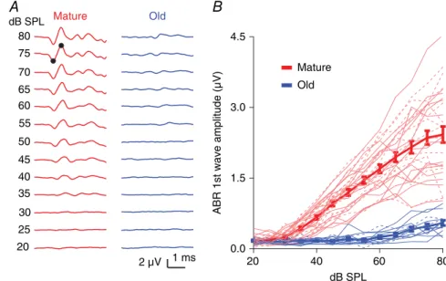
Figure 1. CBA/CaJ mice show ARHL
other analyses used custom programs in Igor Pro (V6.0, WaveMetrics, Inc., Lake Oswego, OR, USA).
Variance-mean analysis
Variance-mean analysis was used to assess the initial release probability (Pr) and to estimate a lower limit of independent release sites (Nmin) (Reid & Clements, 1999; Oleskevich et al. 2000; Wang & Manis, 2005) (Fig. 3). Single eEPSCs were recorded in differentPr conditions obtained by adjusting the calcium concentration (1, 2 and 3 mM) of the external ACSF as previously described (Wang & Manis, 2005). The mean eEPSC amplitude and the variance under eachPrcondition were obtained from stable recordings of 50–100 single eEPSCs with less than 20% drift. Because different calcium concentrations were used in this set of experiments, the acquired data were only used for the variance-mean analysis shown in Fig. 3 and were not included in other analyses. The variance-mean data were fit to a parabola that goes through the origin with the equation:y=Ax+Bx2, whereyis eEPSC vari-ance andxis eEPSC mean amplitude.Pr was calculated as: Pr = x(−B/A)(1+CV2), where the CV (coefficient of variation) was calculated from eEPSCs evoked in the same neuron. A lower limit for the number of release sites at the synapse was then estimated as:Nmin= −1/B.
Statistics
Prism 6.0 (GraphPad Software Inc., La Jolla, CA, USA) was used for all statistical tests except in Fig. 2I. Paired and unpaired Student’sttest (or Mann–Whitney test when the data population failed to pass the normality test), one-way ANOVA as well as two-way ANOVA were used as stated. To test whether paired-pulse ratios (PPRs) were different between ages (Fig. 2I), we used a linear mixed effects model (package lme4 in R, V3.3.1) because, for some cells, not all time intervals had been tested as required for a standard two-way repeated-measures ANOVA. Data are presented as mean and standard deviations (SD), except in Figs 1B, 3F, 4E,GandJ, 6E, F, and 7C,Das specified, where the data are presented as mean±standard error of the mean (SEM) to minimize trace overlap.
Results
ARHL in old CBA/CaJ mice
We evaluated the hearing status of the CBA/CaJ mice by measuring click-evoked ABRs from 20 to 80 dB SPL. Compared to mature mice (2–4 months), old mice (20–26 months) showed an ARHL characterized by a significantly elevated ABR threshold [Fig. 1A; mature:
1 s 50 pA Mature
Old
–200
–150
–100
–50
0
sEPSC amplitude (pA)
Mature Old 20
15
10
5
0
sEPSC frequency (Hz)
Mature Old
0.6
0.4
0.2
0.0
sEPSC decay (ms)
Mature Old A
E
D C
B
–15
–12
–9
–6
–3
0
eEPSC amplitude (nA)
Mature Old
1.5
1.2
0.9
0.6
0.3
0.0
eEPSC decay (ms)
Mature Old
1.4
1.2
1.0
0.8
0.6
Paired pulse ratio (PPR)
2.5 10 50 PPR inter-pulse interval (ms)
Mature Old
2 nA
1 ms Mature Old
20 ms
Mature Old
I H
G F
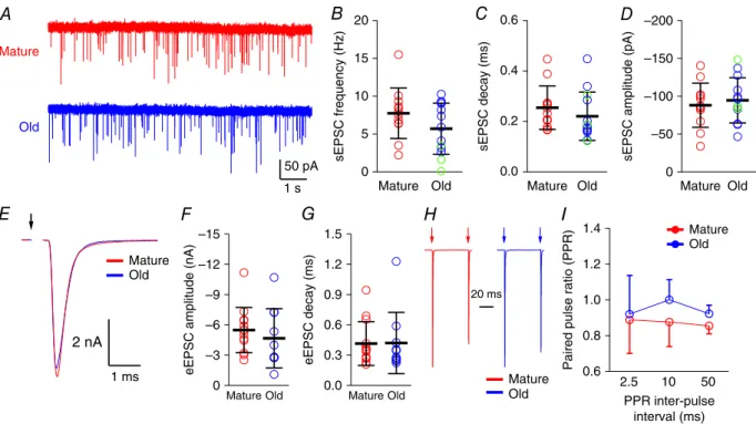
Figure 2. Synaptic transmission at the endbulb of Held in mature and old mice
32 dB SPL (SD 5, n = 29); old: 56 dB SPL (SD 10,
n=13); unpairedttest:t40=10.52,P<0.0001]. Above 40 dB SPL, the amplitude of the first ABR wave was also significantly smaller in old mice (Fig. 1B; two-way ANOVA, age effect: F1, 518 = 660.8, P < 0.0001; sound intensity effect:F12, 518=50.52,P<0.0001; interaction:
F12, 518=28.62,P<0.0001; Sidak’s multiple comparisons
test: P< 0.0001 for wave amplitudes at all levels above 45 dB SPL,P<0.01 at 40 dB SPL). This is consistent with previous reports of ARHL in CBA/CaJ mice (Sergeyenko
et al.2013). Differences in hearing status between male and female mice were not observed in either mature (two-way ANOVA, sex effect:F1,351=2.80,P=0.0951) or old (two-way ANOVA, sex effect:F1, 141=1.17,P=0.281) mice. It has been previously reported that male mice develop ARHL earlier than female mice (Guimaraeset al.
2004; Henry, 2004); that we found no sex difference is probably due to the small number of male mice in the old group.
Synaptic transmission at the endbulb of Held in mature and old mice
We next assessed the quantal transmission at the end-bulb of Held by recording sEPSCs for 100–200 s from each bushy neuron in whole-cell voltage clamp (Fig. 2A). The number of sEPSCs in bushy neurons ranged from 19 to 2949 events, with a median of 970 events in mature mice and 822 events in old mice. There was no significant difference between the two age groups for sEPSC frequency [Fig. 2B; mature: 7.8 Hz (SD 3.3, n= 12); old: 5.7 Hz (SD 3.4,n=13); unpairedttest:t23=1.51,P=0.146], sEPSC decay time constant [Fig. 2C; mature: 0.25 ms (SD 0.09,n=12); old: 0.22 ms (SD 0.10,n= 13); unpaired
ttest:t23=0.93,P=0.362] or sEPSC amplitude [Fig. 2D; mature:−88 pA (SD 29,n =12); old:−95 pA (SD 30,
n = 13); unpaired ttest: t23 = 0.54, P= 0.592]. Thus, the basic aspects of quantal transmission at the endbulb of Held appear to be largely unchanged during ARHL.
5 4 3 2 1 0
eEPSC size (nA)
200 150
100 50
0
eEPSC number
2 mM 1 mM 3 mM
9
6
3
0
eEPSC size (nA)
200 150
100 50
0
eEPSC number
2 mM 1 mM 3 mM
0.4
0.3
0.2
0.1
0.0
V
ariance (nA
2)
V
ariance (nA
2)
8 6
4 2
0
Mean eEPSC (nA)
Pr = 0.48
0.12
0.09
0.06
0.03
0.00
4 3
2 1
0
Mean eEPSC (nA)
Pr = 0.54
[Ca2+] [Ca2+]
A B
C 0.8
0.6
0.4
0.2
Pr
Mature Old
Mature Old
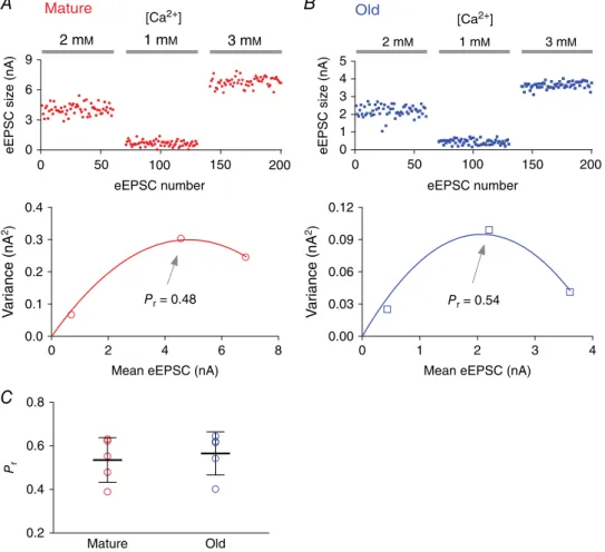
Figure 3. Initial release probability at the endbulb of Held synapses in mature and old mice assessed by variance-mean analysis
A, top: eEPSCs recorded from a bushy neuron of a mature mouse in response to auditory nerve stimulation under different extracellular calcium concentrations. Bottom: variance-mean plot of eEPSCs for the three calcium concentrations. The initial release probability (Pr) at 2 mM[Ca2+] was estimated by fitting the data to a parabola
We next evaluated how ARHL affects evoked release at the endbulb of Held. In these experiments, the stimulus level was gradually increased until reliable eEPSCs were obtained. This level was then used for all eEPSC recordings in a given cell. All 13 bushy neurons in the mature group of mice responded with eEPSCs with an average stimulation amplitude at 270μA (SD 170). In the old mice, stimulation produced eEPSCs in 10 of 13 bushy neurons with an average stimulation amplitude of 440μA (SD 320). The difference in stimulus levels between mature and old mice was not statistically significant (unpairedttest:t21=1.55, P = 0.135). In three neurons from old mice, we were unable to obtain any eEPSCs even after the stimulus was increased to a level (2 mA) that was high enough to cause an electrolytic lesion in the tissue.
For all neurons with evoked responses, neither eEPSC amplitudes nor decay time constants were different between mature and old groups [Fig. 2E, F, amplitude: mature: −5.5 nA (SD 2.2, n = 13); old: −4.7 nA (SD 2.9, n = 10); unpaired t test: t21 = 0.75, P = 0.459; Fig. 2G, decay time constant: mature: 0.42 ms (SD 0.22,
n=13); old: 0.42 ms (SD 0.30,n=10); Mann–Whitney test: P = 0.633]. We also examined the PPR at 2.5, 10 and 50 ms inter-pulse intervals (Fig. 2H). PPRs were not significantly different between mature and old mice (Fig. 2I; linear mixed effects model; PPR as the dependent variable, age and interval as the factors, and subject as a random variable; age effect: t39.5 = 1.405, P= 0.168; interval time effect:t42.6= −0.697,P=0.490; interaction between age and interval:t43.8= −0.070,P=0.945).
To more directly assess changes in release, in a different set of experiment we assessed the initial release probability (Pr) and estimated the minimum number of release sites (Nmin) at the endbulb of Held synapses using variance-mean analysis. Mean release was varied using different extracellular Ca2+concentrations (see Methods). As shown in Fig. 3, the estimatedPrat 2 mM[Ca2+] was not significantly different between mature (0.54, SD 0.10;
n = 5) and old mice (0.57, SD 0.10; n = 5) (unpaired
ttest: t8 = 0.474,P= 0.648). There was no significant difference either in the estimated Nmin between the two age groups (mature: 69 sites, SD 42,n=5; old: 69 sites, SD 49,n=5; unpairedttest:t8=0.0074,P=0.994). The similar estimate ofPrandNminbetween mature and old mice is consistent with the observation that synapses in both age groups show similar quantal (Fig. 2A–D) and single-stimulus evoked synaptic transmission (Fig. 2E,
F), and PPR (Fig. 2H, I). Together these observations suggest that synaptic transmission during the quiescent state remains unchanged during ageing at the endbulb of Held synapse in CBA/CaJ mice. It is worth noting that in a different mouse model of early onset ARHL, the DBA/2J mouse (Johnsonet al.2008),Prwas significantly reduced with concurrently enhanced PPR during hearing loss at the age of45 days (Wang & Manis, 2005). This suggests
that different mechanisms of synaptic transmission are affected by the hearing loss of these two different mouse strains.
Synaptic transmission during high-rate repetitive stimulation
Auditory nerve fibres can fire at sustained rates up to at least 450 Hz for environmentally relevant sound levels in cat (Kiang et al. 1965; Wen et al. 2009) and mouse (Taberner & Liberman, 2005). To examine how release at the endbulbs changes during high rates of afferent firing, we stimulated the nerve with 50-pulse stimulus trains at 100 and 400 Hz. At 100 Hz, every presynaptic stimulus evoked a reliable and precisely timed eEPSC in bushy neurons in both mature (Fig. 4A) and old (Fig. 4B) mice. At 400 Hz, release was maintained in mature mice, although with greater depression than at 100 Hz (Fig. 4C). However, in old mice, phasic release decreased part way through the 400 Hz train (Fig. 4D). The eEPSCs during the last 30 pulses of the train in old mice showed both reduced peak amplitudes and an increase in asynchronous events (Fig. 4D). The asynchronous events continued for tens of milliseconds after the end of the stimulus. To illustrate the kinetics of the synaptic depression, we measured the peak amplitudes of eEPSCs throughout the train, normalized to the first eEPSC (Fig. 4E). Steady state depression was quantified by averaging the normalized amplitude of the last 30 eEPSCs of the train. As shown in Fig. 4F, the steady state depression of eEPSC peak amplitude at 100 Hz showed no significant difference between mature (0.48, SD 0.12, n = 12) and old (0.51, SD 0.15, n = 9) mice (unpairedttest:t19=0.481,P=0.636). At 400 Hz, the eEPSC peak amplitude was significantly more depressed during the steady state in old mice (mature: 0.18, SD 0.07, n= 13; old: 0.10, SD 0.06,n=9; unpaired ttest:
t20 = 2.618, P = 0.0165). To investigate asynchronous release (Fig. 4D), we measured the charge transferred within each response window as the integrated area under each eEPSC, throughout the 50-pulse stimulus trains, and normalized this measure by the charge of the first eEPSC in the train (Fig. 4G). Steady state depression was again quantified by averaging the normalized eEPSC charge of the last 30 eEPSCs of the train. As shown in Fig. 4H, eEPSC charge in young and old mice depressed to similar levels during the trains at both 100 Hz (mature: 0.53, SD 0.11, n=12; old: 0.57, SD 0.12,n= 9; unpairedt-test:
t19 = 0.796, P= 0.436) and 400 Hz (mature: 0.32, SD 0.10, n=13; old: 0.32, SD 0.12,n= 9; unpairedt-test:
20 ms 1 nA
20 ms 1 nA
1nA
20 ms 5 ms
0.5 nA 20 ms 1 nA 5 ms 0.5 nA 50ms 1nA 50ms 1nA 10ms 1nA 50ms 1nA 50ms 1nA 10ms 1nA A I F E D C B 0.5 ms 0.2 nA syn asyn asyn 0.5 ms 0.2 nA syn asyn asyn Mature Old Mature Old 100Hz 400Hz Ctrl 0.8 0.6 0.4 0.2 0.0
Fraction of asynch charge 0 10 20 30 40 50
Pulse number Mature Old 400 Hz 0.8 0.6 0.4 0.2 0.0
Fraction of asynch charge 0 10 20Pulse number30 40 50
Mature
Old 100 Hz
0.6
0.4
0.2
0.0
Fraction of asynch charge
(A
vg first 20 pulses)
Mature Old
0.6
0.4
0.2
0.0
Fraction of asynch charge
(A
vg last 30 pulses)
Mature Old 1.0 0.8 0.6 0.4 0.2 0.0 50 40 30 20 10 0 Pulse number 400Hz 1.0 0.8 0.6 0.4 0.2 0.0
eEPSC size (norm)
50 40 30 20 10 0 Pulse number 100Hz J K 1.0 0.8 0.6 0.4 0.2 0.0
Steady state depression Mature Old
100Hz 0.6 0.4 0.2 0.0 Mature Old 400Hz 1.0 0.8 0.6 0.4 0.2 0.0 50 40 30 20 10 0 Pulse number 400Hz 1.0 0.8 0.6 0.4 0.2 0.0
eEPSC charge (norm)
50 40 30 20 10 0 Pulse number 100Hz G H 1.0 0.8 0.6 0.4 0.2 0.0
SS charge depression
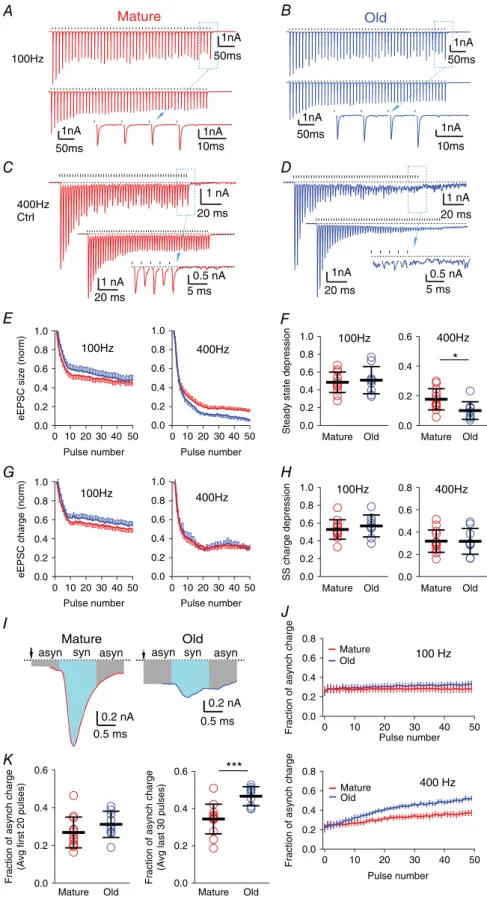
Mature Old 100Hz 0.8 0.6 0.4 0.2 0.0 Mature Old 400Hz *** * Figure 4. Asynchronous release is
increased during high frequency stimulation in old mice
A, eEPSCs of a bushy neuron from a mature mouse in response to a 50-pulse stimulus train at 100 Hz. Top: single trial; bottom: average trace of 10 trials; inset: expanded view of the last eEPSCs from the dashed box in the top trace. Ticks above the traces mark stimulation times; dashed lines indicate the resting current. Plots inB–Dare arranged the same way as inA.B, eEPSCs of a bushy neuron from an old mouse in response to a 50-pulse stimulus train at 100 Hz.CandD, eEPSCs of bushy neurons from mature (C) and old (D) mice in response to 50 pulse-trains at 400 Hz. Notice inDthat eEPSCs show reduced peak amplitude and increased asynchronous release during the late part of the train.
E, eEPSC peak amplitude throughout the stimulus train, normalized to the amplitude of the first eEPSC, at 100 and 400 Hz.
F, summary of the steady state depression (average peak amplitude of the last 30 eEPSCs of the train normalized to the first eEPSC) at 100 and 400 Hz. Each symbol represents an individual neuron.G, eEPSC charge (integrated area under each eEPSC) throughout the stimulus train normalized to the charge of the first eEPSC, at 100 and 400 Hz.H, summary of the steady state (SS) charge depression (average charge of the last 30 eEPSCs of the train normalized to the first eEPSC) at 100 and 400 Hz.I, the last eEPSCs from the 400 Hz train inC
(mature) andD(old). Syn: synchronous eEPSC response (cyan area); asyn: asynchronous eEPSC response (grey area).
J, fraction of asynchronous release charge for each stimulus throughout the 50-pulse train at 100 Hz (top) and 400 Hz (bottom).
K, average fraction of asynchronous charge of the first 20 stimuli (left) and the last 30 stimuli (right) of the 400 Hz trains in all bushy neurons. Data inE,GandJare presented as mean±SEM; data inF,Hand
Kare presented as mean±SD .∗P<0.05,
SD 16,n=9; unpairedt-test:t20=0.689,P=0.499). These results suggest that the total release during the stimulus trains does not change between mature and old mice, and the eEPSC charge is depressed with similar kinetics throughout the trains at both 100 and 400 Hz (Fig. 4G,H). Therefore, the greater depression of eEPSC peak amplitude in old mice at 400 Hz (Fig. 4E,F), which was accompanied by an increase in asynchronous events (Fig. 4D), represents a shift in vesicle release from synchronous to asynchronous modes during high afferent firing rates.
To quantify the shift of vesicle release from synchronous to asynchronous during the stimulus train, we defined synchronous and asynchronous portions of each stimulus response. As shown in Fig. 4I, ‘synchronous release’ was defined as the charge occurring in a 1 ms window surrounding the eEPSC peak (0.4 ms before and 0.6 ms after), and ‘asynchronous release’ was defined as the remainder of the charge between successive stimuli (e.g. over the remaining 9.0 ms within a stimulus cycle for 100 Hz and 1.5 ms for 400 Hz, respectively). The fraction attributed to asynchronous release was consistent after the first few stimuli throughout the 50-pulse train at 100 Hz in both mature and old mice (Fig. 4J). Across the entire 100 Hz train, asynchronous response accounts for 28.1% (SD 10.7%,n=12) of the total response in mature mice and 30.8% (SD 7.9%,n=9) in old mice (unpairedt-test:
t19=0.64,P=0.528). During the first 20 pulses of the 400 Hz train (Fig. 4J,K), asynchronous release accounts for 26.9% (SD 8.2%,n=13) of the charge in mature mice and 31.2% (SD 6.9%,n=9) in old mice (unpairedt-test:
t20=1.30,P=0.208). However, the asynchronous release increased significantly during the last 30 pulses of the train in old mice (old: 46.7%, SD 5.1%,n=9, mature: 34.4%, SD 8.0%,n=13; unpairedt-test:t20=4.06,P=0.0006). Conversely, the magnitude of synchronous release is sub-stantially diminished at old synapses when challenged with sustained high frequency presynaptic firing rates (Fig. 4E, right panel), which is expected to reduce the temporal pre-cision of synaptic transmission at the endbulb synapses.
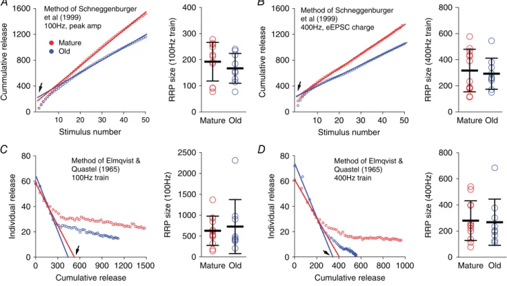
To investigate whether there were different sizes of readily releasable vesicle pools (RRPs) at the endbulb of Held, using eEPSCs to 100 and 400 Hz stimulus trains, we used two different analyses of the responses to stimulus trains. First, eEPSC peak amplitudes in 100 Hz trains were measured to calculate the total quantal content of the RRP (Fig. 5A) using the method described by Schneggenburger
et al.(1999). For each cell, the number of vesicles released for every stimulus was calculated by dividing the eEPSC peak amplitude by the average amplitude of sEPSC events for that neuron. The portion of the cumulative plot with steady state release (from the 11th to the 50th stimulus) was fit to a line, and back-extrapolated to time 0 to obtain the estimated RRP. There was no significant difference in RRP size between mature (193 vesicles, SD 74, n= 11) and old mice (167 vesicles, SD 57, n = 9) (Fig. 5A,
right panel; unpaired t-test: t18 = 0.844, P = 0.410). We then performed the same analysis for EPSCs in the 400 Hz train. However, because 400 Hz trains evoked more asynchronous responses in old mice (Fig. 4D), we measured the total charge associated with each response in the time between stimuli (Fig. 5B). The estimated number of vesicles released at each stimulus was then calculated by dividing the eEPSC charge by the average charge of sEPSC events for that neuron. Again, no significant difference was found in the RRP size between mature (317 vesicles, SD 163, n = 12) and old mice (293 vesicles, SD 119,
n=9) (Fig. 5Bright panel; unpairedt-test:t19=0.367, P=0.718).
Second, we estimated the RRP size from both 100 and 400 Hz trains using the method of Elmqvist & Quastel (1965) (Fig. 5C,D). Individual evoked response amplitudes were plotted against the cumulative release that occurred prior to the given stimulus. Assuming that there is a minimum of new vesicle recruitment during the early part of the stimulus train, RRP size is estimated from thex-axis intercept of a line fit to responses to the first few stimuli (Fig. 5C, D). Again, the estimated RRP size was not significantly different between mature and old mice from either 100 Hz trains (Fig. 5C; mature: 628 vesicles, SD 350,n=12; old: 727 vesicles, SD 645,n=9; Mann–Whitney test:P=0.786) or 400 Hz trains (Fig. 5D; mature: 280 vesicles, SD 152, n= 12; old: 268, SD 177,
n=9; Mann–Whitney test:P=0.633). These estimates, which use different methods with distinct underlying assumptions, suggest that the RRP size of the endbulb of Held synapse is not different between mature and old mice. However, because of the assumptions behind these analyses, this should be viewed as a tentative conclusion (see Discussion).
Synchronous release in old mice can be restored with exogenous calcium buffering or by decreasing calcium influx
One hypothesis for the increase in asynchronous release is that during high rates of stimulation, calcium is not effectively cleared from the presynaptic terminal so that a buildup of presynaptic calcium triggers continued release of synaptic vesicles. The increase in this asynchronous release may also decrease the availability of docked vesicles for action potential evoked-release, leading to a decrease in synchronous release (Hagler & Goda, 2001). We therefore tested whether intraterminal calcium regulation might contribute to asynchronous release with two manipulations.
(Eggermannet al.2012). In mature mice, pre-incubation of the slice in EGTA-AM did not change the single eEPSC peak amplitude (mature: −5.5 nA, SD 2.2, n = 13; mature+EGTA:−4.7 nA, SD 2.6,n=10; unpairedt-test:
t21 =0.8029, P=0.431), or the steady state depression of eEPSC amplitude in 400 Hz trains (Fig. 6A, E) (mature depression: 0.18, SD 0.07,n=13; mature+EGTA depression: 0.20, SD 0.15, n = 6; Mann–Whitney test:
P=0.999). In old mice, EGTA-AM treatment also did not change the single eEPSC peak amplitude (old:−4.7 nA, SD 2.9,n=10; old+EGTA:−3.4 nA, SD 2.5,n=9; unpaired
t-test: t17 =1.049,P= 0.309). However, the EGTA-AM treatment reduced asynchronous release and enhanced synchronous release during the steady state response phase (Fig. 6B vs.4D). Consequently, the steady state depression was significantly reduced (Fig. 6B,E) (old depression: 0.10, SD 0.06,n=9; old+EGTA depression: 0.21, SD 0.07,n=7; unpairedt-test:t14=3.349,P=0.0048).
Second, we bath-applied 25 nM ω-agatoxin IVA to partially block P-type calcium channels, which
should reduce action potential-evoked calcium influx into the endbulb terminal, and thus decrease calcium accumulation during sustained activity, while not completely blocking transmission. Consistent with a smaller calcium influx, the initial release was reduced with decreased single eEPSC peak amplitude (mature:−5.5 nA, SD 2.2,n=13; mature+agatoxin:−0.8 nA, SD 0.7,n=7; unpaired t-test: t18 = 5.279,P< 0.0001; old: −4.7 nA, SD 2.9,n= 10; old+agatoxin:−0.5 nA, SD 0.4,n= 8; unpaired t-test: t16 = 3.932, P = 0.0012). As expected for a decrease in calcium influx, the PPR of the first two eEPSCs of the 400 Hz train was significantly increased in both mature (Fig. 6C; control PPR: 0.90, SD 0.11; agatoxin PPR: 1.23, SD 0.16;n=7; pairedt-test:t6=4.22, P=0.006) and old mice (Fig. 6D; control PPR: 0.94, SD 0.22; agatoxin PPR: 1.25, SD 0.22; n = 8; paired t-test:
t7 =4.55,P=0.003). However,ω-agatoxin IVA did not change the relative amount of synchronous release during the sustained activity in mature mice (Fig. 6C). In old mice,ω-agatoxin IVA reduced asynchronous release and
1600
1200
800
400
0
Cumulative release
50 40 30 20 10
Stimulus number
Method of Schneggenburger et al (1999)
400Hz, eEPSC charge
1600
1200
800
400
0
Cummulative release
50 40 30 20 10
Stimulus number
Method of Schneggenburger et al (1999)
100Hz, peak amp
Mature Old
800
600
400
200
0
RRP
size (400Hz train)
Mature Old 400
300
200
100
0
RRP
size (100Hz train)
Mature Old
A B
C D 80
60
40
20
0
Individual release
1000 800 600 400 200 0
Cumulative release
Method of Elmqvist & Quastel (1965) 400Hz train
80
60
40
20
0
Individual release
1500 1200 900 600 300 0
Cumulative release
Method of Elmqvist & Quastel (1965) 100Hz train
800
600
400
200
0
RRP
size (400Hz)
Mature Old 2500
2000
1500
1000
500
0
RRP
size (100Hz)
Mature Old
Figure 5. Estimates of RRP size in mature and old mice
partially restored synchronous release (Fig. 6D vs.4D). As shown in Fig. 6F, the average fraction of asynchronous release during the first 20 pulses of the 400 Hz train was not different between control, EGTA-AM treated and
ω-agatoxin IVA treated slices in mature mice [mature control: 26.9% (SD 8.2%, n = 13); mature EGTA-AM: 25.0% (SD 10.9%,n=6); matureω-agatoxin IVA: 25.7% (SD 10.4%, n = 7); one-way ANOVA: F2,23 = 0.0893, P=0.915], nor was it different in old mice [old control: 31.2% (SD 6.9%,n=9); old EGTA-AM: 31.6% (SD 9.1%,
n = 7); old ω-agatoxin IVA: 30.1% (SD 9.0%, n = 8); one-way ANOVA: F2,21 = 0.0732, P = 0.930]. During the last 30 pulses, the asynchronous response was also not different in mature mice [mature control: 34.4% (SD 8.0%, n = 13); mature EGTA-AM: 28.2% (SD 11.0%,
n=6); matureω-agatoxin IVA: 31.6% (SD 9.3%,n=7); one-way ANOVA:F2,23=0.997,P=0.384]. However, both treatments significantly decreased asynchronous release during the last 30 stimuli in the train in old mice [old control: 46.7% (SD 5.1%,n=9); old EGTA-AM: 36.3% (SD 11.2%,n=7); oldω-agatoxin IVA: 37.1% (SD 7.6%,
n = 8); one-way ANOVA: F2,21 = 4.342, P = 0.0264; Dunnett’s multiple comparisons test:P <0.05 between control and old EGTA-AM treatment, and between control and ω-agatoxin IVA treatment]. These results support the hypothesis that changes in intraterminal calcium handling in the endbulb of Held underlie the increased asynchronous release in mice with ARHL. It is also possible that there is a change in the calcium sensitivity of other mechanisms in the release process.
20 ms 0.5 nA
20 ms 0.5nA
20 ms 0.5nA
5 ms 0.5nA 20 ms 1 nA
20 ms 1 nA
5 ms 1 nA
20 ms 0.5 nA
5 ms 0.5 nA 20 ms
1 nA
20 ms 1 nA
5 ms 0.2 nA A
E
D C
B Old
400Hz Agatoxin
400Hz EGTA
0.8
0.6
0.4
0.2
0.0
Fraction of asynch charge
50 40 30 20 10 0
Pulse number Mature
Mature+EGTA Mature+Aga Old Old+EGTA Old+Aga
400 Hz Mature
2.0
1.5
1.0
0.5
0.0
eEPSC size (norm)
50 40 30 20 10 0
Pulse number Mature Mature+EGTA Mature+Aga Old Old+EGTA Old+Aga 400 Hz
F
Figure 6. Synchronous synaptic transmission at 400 Hz is rescued by manipulations that decrease intraterminal calcium
AandB, eEPSCs in response to 400 Hz stimulus trains after treatment with EGTA-AM in mature (A) and old (B) mice. Top: single trial; bottom: average trace of 10 trials; inset: expanded view of the last eEPSCs from the dashed box in the top trace. Ticks above the trace mark stimulation times. Plots inCandDare arranged in the same format.CandD, eEPSCs in response to 400 Hz stimulus trains after bath application of 25 nMω-agatoxin IVA in
Recovery from rate-dependent synaptic depression is faster in old mice
The rate of recovery in EPSC amplitude after depression by high-frequency stimulus trains depends on presynaptic calcium levels achieved during the stimulus train. Higher calcium levels produced by high-frequency stimuli have been shown to lead to a faster recovery of the release process (Wang & Kaczmarek, 1998; Wang & Manis, 2008). We assessed the recovery of eEPSC peak amplitude at the endbulb of Held by presenting 50-pulse stimulus trains followed by single test pulses with delays of 20, 70, 200, 500 or 1000 ms (Fig. 7A, B). The EPSC recovery was significantly faster in old mice than in the mature mice after both 100 Hz stimulus trains (Fig. 7C; mature:n=13, old: n = 10; two-way ANOVA, age effect: F1,84 = 8.93, P = 0.0037; post-train delay time effect: F3,84 = 4.50, P = 0.0056; interaction: F3,84 = 0.199, P = 0.897) and 400 Hz stimulus trains (Fig. 7D; mature: n = 13, old: n = 8; two-way ANOVA, age effect: F1,76 = 6.94, P= 0.0102; delay time effect:F3,76 =4.39, P=0.0067; interaction: F3,76 = 0.770, P = 0.515). We also tested EPSC recovery following EGTA-AM pre-incubation. After a 100 Hz stimulus train (Fig. 7C), synapses in slices from mature mice pretreated with EGTA-AM did not differ from controls in their post-train recovery (n=6; two-way ANOVA, EGTA-AM effect: F1,68 = 2.78, P = 0.100; delay time effect:F3,68 = 5.21, P = 0.0027; interaction: F3,68=0.0277,P=0.994). EGTA-AM similarly had little effect in old mice for 100 Hz trains (n = 5; two-way ANOVA, EGTA-AM effect: F1,52 = 2.14, P = 0.150; delay time effect: F3,52 = 1.88, P = 0.144; interaction: F3,52 = 0.00812, P = 0.999). However, following a 400 Hz train (Fig. 7D), EPSC recovery in EGTA-AM treated slices was significantly slower in both mature mice (n=5; two-way ANOVA, EGTA-AM effect:F1,64 =19.4, P< 0.0001; delay time effect:F3,64 =7.64, P=0.0002; interaction: F3,64 = 1.09, P = 0.360) and old mice (n=5; two-way ANOVA, EGTA-AM effect:F1,44 =5.49, P=0.024; delay time effect:F3,44=2.37,P=0.083; inter-action:F3,44=0.225,P=0.879). The faster rate of recovery in old mice, and the return of the recovery time course following EGTA-AM pre-treatment towards that seen in mature mice, is also consistent with the hypothesis that the endbulb of Held synapses in old mice have an elevated intraterminal calcium level relative to mature mice during high rates of afferent firing. However, age-related changes in the expression of calcium binding proteins, or the sensitivity of calcium sensors that participate in synaptic transmission cannot be excluded.
Discussion
We compared synaptic transmission at the endbulb of Held synapses between mature and old CBA/CaJ mice.
No significant difference was found at the endbulbs between two age groups of mice in the basal synaptic conductance amplitude and time course, the size of the RRP or the initial release probability. In old mice with ARHL, synaptic transmission at the endbulb synapse
1.2 1.0
0.8
0.6 0.4
0.2
0.0
EPSC size (normalized)
1600 1200
800 400
0
Time (ms)
Mature Mature+EGTA Old
Old+EGTA 100 Hz
0.1 s
1.2
1.0 0.8
0.6
0.4 0.2
0.0
EPSC size (normalized)
1200 800
400 0
Time (ms) Mature Mature+EGTA Old
Old+EGTA 400 Hz
0.1 s 1 nA
A
D C B
Figure 7. Recovery of eEPSC amplitude after the termination of stimulus trains is faster in old mice
appears to function normally at low firing rates, but was unable to support sustained, time-locked transmission at high firing rates. Instead, synchronous release was decreased at high rates whereas asynchronous release was increased. Furthermore, manipulations that either increase intraterminal calcium buffering or decrease action potential-evoked calcium influx could restore the synchronous release while suppressing asynchronous release in response to high frequency afferent stimulation. These results suggest that endbulb synapses in old mice either have decreased capacity for buffering or clearing calcium in the terminal, increased action potential-triggered calcium influx, or that there is a change in the sensitivity of the calcium-dependent process involved in release.
ARHL and synaptic drive
Determining the proximal causes of the changes in synaptic transmission that we observed in old mice requires consideration of the nature of ARHL. CBA/CaJ mice with ARHL exhibit a loss of synapses between the hair cells and SGCs, and the loss is highly correlated with the amplitude of the first wave of the ABR (Sergeyenko
et al. 2013), suggesting that synaptopathy is involved in the hearing loss. Aged CBA/CaJ mice also exhibit a loss of outer hair cells, which would also be expected to raise thresholds (Ohlemilleret al.2010; Sergeyenkoet al.2013). On the other hand, there is little evidence in this strain for age-related changes in the endocochlear potential or sound transmission through the ossicular chain (Rosowskiet al.
2003; Shaet al. 2008; Ohlemilleret al. 2010). Thus, the primary causes of ARHL in the CBA/CaJ mice appear to be limited to loss of hair cells and synapses onto SGCs. Because SGCs in the adult usually receive input from a single inner hair cell (Spoendlin, 1985; Berglund & Ryugo, 1987), the loss of hair cells and their connectivity with SGCs is expected to abolish both the sensory-evoked and spontaneous firing in affected SGCs. Thus, from the perspective of both spontaneous and acoustically driven action potentials, the subset of endbulb terminals that are disconnected from the periphery will experience an extended period of quiescence, including possible absence of spontaneous activity, with ageing. It is conceivable that there is a homeostatic compensation in the electrical excitability that leads to the generation of spontaneous spikes in the SGCs following hearing loss, but there is no evidence that this occurs. In addition, acoustic thresholds for driving the remaining SGCs will increase with the loss of outer hair cells, which over a lifetime of sound exposure would be expected to decrease the average firing rates and demands on release at the endbulb synapse, although spontaneous activity may be retained. Because the SGCs degenerate at a very slow rate, a large fraction (60% in CBA mice by34 months) is preserved during
ageing (Makaryet al.2011; Sergeyenko et al.2013). For the surviving SGCs, their endbulb synapses also remain and continue to function, although activity-dependent mechanisms within the synapse may exhibit homeostatic adjustments in response to the lower levels of activity.
Because not all SGCs are disconnected from their hair cells during ARHL, there should be a substantial population of auditory nerve fibres and endbulb synapses that continue to have spontaneous activity and some level of acoustically driven activity. If the changes in transmission that we observe are solely dependent on sensory activity, then we might expect that there are two populations of endbulb synapses. However, there is no clear evidence in our data for a segregation of the aged synapses into two groups, one with normal, strong synchronous release and another with more asynchronous release, when tested at 400 Hz (Fig. 4K). This suggests that the factors leading to elevated asynchronous release are generalized across all endbulb synapses, and are at least somewhat independent of the status of the parent SGC.
It is interesting that evoked responses could not be achieved in 23% of the bushy cells from old mice. It is possible that this is an experimental artifact, and that the auditory nerve inputs onto these cells simply were not activated by the stimulating electrode. However, this rarely occurs for cells in slices from mature mice. It is also possible that these bushy cells have lost their auditory nerve inputs from SGCs. The loss of endbulbs would be consistent with the low sEPSC frequencies in these cells (Fig. 2B) when compared to the other bushy cells with evoked responses. Unfortunately, our sample size is too small to clearly distinguish these possibilities, and a morphological assay for the presence of endbulbs on these silent cells would be needed to provide convincing evidence for a loss of inputs.
significantly affect the ability to initiate or sustain action potential initiation during a train.
RRP size of the endbulb of Held in mature and old mice
The calculation of readily release pool size with different methods using eEPSC trains did not show any significant differences between mature and old mice groups (Fig. 5), consistent with the observation of similar spontaneous event amplitudes and rates, and the amplitudes of single evoked synaptic events at both ages (Fig. 2). However, the RRP estimates using these analyses are dependent upon certain assumptions about the mechanisms involved in synaptic transmission (Neher, 2015). For example, the method of Schneggenburgeret al.(1999) used in Fig. 5A
andB assumes that the stimulus train reaches a steady state in which the vesicle release is balanced by recruitment of new vesicles. This assumption may not be met in aged mice, especially at 400 Hz, where it is not clear if new vesicles are reliably replenished to the RRP to balance released vesicles during the trains. By using long stimulus trains (11th to 50th pulses in Fig. 5A,B) we attempted to better reach a steady state of release in old mice, which should improve the reliability of RRP estimates with this method. The method of Elmqvist & Quastel (1965) used in Fig. 5CandDutilizes only early eEPSCs in the train, and assumes that there is little or no new vesicle recruitment during that period. However, at different activity rates, 100 and 400 Hz in this case, different levels of new vesicle recruitment might occur. At 100 Hz, the first four eEPSCs occur over 30 ms whereas the same four eEPSCs are complete in 7.5 ms for the 400 Hz train. We have no independent measure of new vesicle recruitment rates and those rates might differ at different release frequencies. Thus, these RRP estimates are probably rate-specific, and this may explain the differences in the RRP estimate between 100 Hz (Fig. 5C) and 400 Hz (Fig. 5D) trains from the same neurons.
Calcium signalling at the endbulb of Held synapses
Intracellular calcium dysregulation has long been post-ulated to underlie brain ageing and age-related conditions like Alzheimer’s disease (Khachaturian, 1994), and one consequence of changes in calcium handling can be a shift in calcium-dependent processes of exocytosis at synaptic terminals. Synchronous and asynchronous vesicle release are two different modes of exocytosis that often occur together during repetitive presynaptic stimulation (Hagler & Goda, 2001). Synchronous release is tightly regulated by the immediate evaluation of calcium within the nanometre domain surrounding the voltage gated calcium channels upon presynaptic action potential invasion (Eggermannet al.2012). Asynchronous release
is thought to be associated with a buildup of calcium ions in the micrometre domain of the synaptic terminal during repetitive stimulation. These two types of release might utilize different presynaptic calcium sensors (Bacaj
et al.2013), as synchronous release is associated with high calcium cooperativity, whereas asynchronous release is associated with low calcium cooperativity (Sunet al.2007). Synchronous and asynchronous release appear to mobilize vesicles from the same RRP (Hagler & Goda, 2001; Otsu
et al.2004; Sunet al.2007; Yang & Xu-Friedman, 2010), although other evidence suggests that separate pools may be involved (Wen et al. 2013; Kaeser & Regehr, 2014). Regardless, the shift from synchronous release to asynchronous release during high firing rates at the end-bulb of Held in old mice is consistent with the general hypothesis that calcium regulation is impaired during ageing in the CNS. During the post-train transmission, the rapid recovery of eEPSC peak amplitudes in old mice (Fig. 7) with simultaneously diminished asynchronous release suggests a quick switch between asynchronous and synchronous modes. Assuming both forms of release compete for the same RRP, the time course of the asynchronous release reduction presumably resembles the recovery kinetics of synchronous release.
The results from intraterminal calcium manipulations further support the idea that there is a change in calcium handling or calcium sensitivity at the endbulb synapses in aged mice. EGTA-AM treatment largely restored the phasic component of synaptic transmission at the end-bulbs in old mice to a state closer to that of the mature mice (Fig. 6B, F). This result can be interpreted in two ways. First, it is possible that the intraterminal calcium concentration is abnormally elevated when challenged with high-rate stimuli during ageing. During high-rate stimulus trains, calcium builds up in the terminal (Habets & Borst, 2005), and is removed from the presynaptic zones by several mechanisms, including plasma membrane calcium pumps, Na–Ca exchangers, uptake into the endoplasmic reticulum or endoplasmic reticulum-like structures and uptake into mitochondria (Rizzutoet al.
and are highly expressed in fast auditory synapses (Sun
et al. 2007; Xiao et al. 2010). It is conceivable that an age-related upregulation of synaptotagmin-7 and/or downregulation of synaptotagmin-2 at the endbulb of Held synapse could lead to the observed shift in the synaptic transmission from synchronous to asynchronous release in old mice.
Interestingly, partially blocking the P/Q type calcium channels, which are important for release at the end-bulb of Held (Oleskevich & Walmsley, 2002; Lin et al.
2011), also improved synchronized synaptic transmission in old mice (Fig. 6D, F). This is consistent with the idea that elevated intratermial calcium could also come from abnormally increased calcium influx through voltage gated calcium channels. During sustained activity, calcium influx through voltage gated calcium channels at the end-bulb of Held in prehearing mice is facilitated during trains of action potentials (Linet al. 2011), which would lead to enhanced calcium influx during the later phase of the stimulus train. In mature animals, however, calcium influx is likely to be depressive at the endbulb during action potential trains due to calcium channel inactivation, as shown at the calyx of Held in the medial nucleus of the trapezoid body (Forsytheet al.1998). It is possible that a switch from calcium channel inactivation to facilitation, or a decrease in inactivation, at the endbulb of Held during ageing could lead to enhanced calcium influx during the action potential trains in old mice.
In summary, we have shown that the endbulb of Held cannot support synchronous release at high rates in aged mice with ARHL. This deficit may result from the global biological processes of ageing, or from the decrease in activity associated with sensory hearing loss. The results from experimental manipulations of intra-terminal calcium are consistent with the hypothesis that the deficit results from a build up of intraterminal calcium during high frequencies of stimulation, but could also be explained by changes in the calcium sensitivity of proteins involved in the release process. Regardless, these experiments suggest that perceptual deficits associated with ARHL may in part result from an inability of the first central auditory synapses to reliably transmit the temporal information present in sound, as represented by the high firing rates and exceptional temporal precision of spikes in auditory nerve fibres.
References
Anderson S, Parbery-Clark A, White-Schwoch T & Kraus N (2012). Aging affects neural precision of speech encoding.
J Neurosci32, 14156–14164.
Bacaj T, Wu D, Yang X, Morishita W, Zhou P, Xu W, Malenka RC & Sudhof TC (2013). Synaptotagmin-1 and
synaptotagmin-7 trigger synchronous and asynchronous phases of neurotransmitter release.Neuron80, 947–959.
Berglund AM & Ryugo DK (1987). Hair cell innervation by spiral ganglion neurons in the mouse.J Comp Neurol255, 560–570.
Bohne BA, Gruner MM & Harding GW (1990). Morphological correlates of aging in the chinchilla cochlea.Hear Res48, 79–91.
Campagnola L, Kratz MB & Manis PB (2014). ACQ4: an open-source software platform for data acquisition and analysis in neurophysiology research.Front Neuroinform8, 3. Caspary DM, Schatteman TA & Hughes LF (2005). Age-related
changes in the inhibitory response properties of dorsal cochlear nucleus output neurons: role of inhibitory inputs.
J Neurosci25, 10952–10959.
Clements JD & Bekkers JM (1997). Detection of spontaneous synaptic events with an optimally scaled template.Biophys J
73, 220–229.
Eggermann E, Bucurenciu I, Goswami SP & Jonas P (2012). Nanodomain coupling between Ca2+channels and sensors of exocytosis at fast mammalian synapses.Nat Rev Neurosci
13, 7–21.
Elmqvist D & Quastel DM (1965). A quantitative study of end-plate potentials in isolated human muscle.J Physiol178, 505–529.
Forsythe ID, Tsujimoto T, Barnes-Davies M, Cuttle MF & Takahashi T (1998). Inactivation of presynaptic calcium current contributes to synaptic depression at a fast central synapse.Neuron20, 797–807.
Frisina DR & Frisina RD (1997). Speech recognition in noise and presbycusis: relations to possible neural mechanisms.
Hear Res106, 95–104.
Frisina RD & Walton JP (2006). Age-related structural and functional changes in the cochlear nucleus.Hear Res
216–217, 216–223.
Guimaraes P, Zhu X, Cannon T, Kim S & Frisina RD (2004). Sex differences in distortion product otoacoustic emissions as a function of age in CBA mice.Hear Res192, 83–89. Habets RL & Borst JG (2005). Post-tetanic potentiation in the
rat calyx of Held synapse.J Physiol564, 173–187.
Hagler DJ, Jr & Goda Y (2001). Properties of synchronous and asynchronous release during pulse train depression in cultured hippocampal neurons.J Neurophysiol85, 2324–2334.
Henry KR (2004). Males lose hearing earlier in mouse models of late-onset age-related hearing loss; females lose hearing earlier in mouse models of early-onset hearing loss.Hear Res
190, 141–148.
Hequembourg S & Liberman MC (2001). Spiral ligament pathology: a major aspect of age-related cochlear degeneration in C57BL/6 mice.J Assoc Res Otolaryngol2, 118–129.
Johnson KR, Longo-Guess C, Gagnon LH, Yu H & Zheng QY (2008). A locus on distal chromosome 11 (ahl8) and its interaction with Cdh23 ahl underlie the early onset, age-related hearing loss of DBA/2J mice.Genomics92, 219–225.
Joris P & Yin TC (2007). A matter of time: internal delays in binaural processing.Trends Neurosci30, 70–78.
Kaeser PS & Regehr WG (2014). Molecular mechanisms for synchronous, asynchronous, and spontaneous
Khachaturian ZS (1994). Calcium hypothesis of Alzheimer’s disease and brain aging.Ann N Y Acad Sci747, 1–11. Kiang NY, Pfeiffer RR, Warr WB & Backus AS (1965).
Stimulus coding in the cochlear nucleus.Trans Am Otol Soc
53, 35–58.
Lin KH, Oleskevich S & Taschenberger H (2011). Presynaptic Ca2+influx and vesicle exocytosis at the mouse endbulb of Held: a comparison of two auditory nerve terminals.
J Physiol589, 4301–4320.
Lorenzi C, Gilbert G, Carn H, Garnier S & Moore BC (2006). Speech perception problems of the hearing impaired reflect inability to use temporal fine structure.Proc Natl Acad Sci USA103, 18866–18869.
Luo F, Bacaj T & Sudhof TC (2015). Synaptotagmin-7 is essential for Ca2+-triggered delayed asynchronous release but not for Ca2+-dependent vesicle priming in retinal ribbon synapses.J Neurosci35, 11024–11033.
Makary CA, Shin J, Kujawa SG, Liberman MC & Merchant SN (2011). Age-related primary cochlear neuronal degeneration in human temporal bones.J Assoc Res Otolaryngol12, 711–717.
Manis PB, Xie R, Wang Y, Marrs GS & Spirou GA (2011). The endbulbs of Held. InSynaptic Mechanisms in the Auditory
System, ed. Trussell LO, Popper AN & Fay RR, pp. 61–93.
Springer, New York.
Neher E (2015). Merits and limitations of vesicle pool models in view of heterogeneous populations of synaptic vesicles.
Neuron87, 1131–1142.
Ohlemiller KK, Dahl AR & Gagnon PM (2010). Divergent aging characteristics in CBA/J and CBA/CaJ mouse cochleae.
J Assoc Res Otolaryngol11, 605–623.
Oleskevich S, Clements J & Walmsley B (2000). Release probability modulates short-term plasticity at a rat giant terminal.J Physiol524, 513–523.
Oleskevich S & Walmsley B (2002). Synaptic transmission in the auditory brainstem of normal and congenitally deaf mice.J Physiol540, 447–455.
Otsu Y, Shahrezaei V, Li B, Raymond LA, Delaney KR & Murphy TH (2004). Competition between phasic and asynchronous release for recovered synaptic vesicles at developing hippocampal autaptic synapses.J Neurosci24, 420–433.
Reid CA & Clements JD (1999). Postsynaptic expression of long-term potentiation in the rat dentate gyrus
demonstrated by variance-mean analysis.J Physiol518, 121–130.
Rizzuto R, Bernardi P & Pozzan T (2000). Mitochondria as all-round players of the calcium game.J Physiol529, 37–47. Rosowski JJ, Brinsko KM, Tempel BI & Kujawa SG (2003).
The aging of the middle ear in 129S6/SvEvTac and CBA/CaJ mice: measurements of umbo velocity, hearing function, and the incidence of pathology.J Assoc Res Otolaryngol4, 371–383.
Schneggenburger R, Meyer AC & Neher E (1999). Released fraction and total size of a pool of immediately available transmitter quanta at a calyx synapse.Neuron23, 399–409. Schulte BA & Schmiedt RA (1992). Lateral wall Na,K-ATPase and endocochlear potentials decline with age in quiet-reared gerbils.Hear Res61, 35–46.
Sergeyenko Y, Lall K, Liberman MC & Kujawa SG (2013). Age-related cochlear synaptopathy: an early-onset contributor to auditory functional decline.J Neurosci33, 13686–13694.
Sha SH, Kanicki A, Dootz G, Talaska AE, Halsey K, Dolan D, Altschuler R & Schacht J (2008). Age-related auditory pathology in the CBA/J mouse.Hear Res243, 87–94. Shofner WP (2008). Representation of the spectral dominance
region of pitch in the steady-state temporal discharge patterns of cochlear nucleus units.J Acoust Soc Am124, 3038–3052.
Spoendlin H (1985). Anatomy of cochlear innervation.Am J
Otolaryngol6, 453–467.
Sun J, Pang ZP, Qin D, Fahim AT, Adachi R & Sudhof TC (2007). A dual-Ca2+-sensor model for neurotransmitter release in a central synapse.Nature450, 676–682.
Taberner AM & Liberman MC (2005). Response properties of single auditory nerve fibres in the mouse.J Neurophysiol93, 557–569.
Viana LM, O’Malley JT, Burgess BJ, Jones DD, Oliveira CA, Santos F, Merchant SN, Liberman LD & Liberman MC (2015). Cochlear neuropathy in human presbycusis: confocal analysis of hidden hearing loss in post-mortem tissue.Hear Res327, 78–88.
Wang LY & Kaczmarek LK (1998). High-frequency firing helps replenish the readily releasable pool of synaptic vesicles.
Nature394, 384–388.
Wang Y & Manis PB (2005). Synaptic transmission at the cochlear nucleus endbulb synapse during age-related hearing loss in mice.J Neurophysiol94, 1814–1824.
Wang Y & Manis PB (2008). Short-term synaptic depression and recovery at the mature mammalian endbulb of Held synapse in mice.J Neurophysiol100, 1255–1264.
Wen B, Wang GI, Dean I & Delgutte B (2009). Dynamic range adaptation to sound level statistics in the auditory nerve.
J Neurosci29, 13797–13808.
Wen H, Hubbard JM, Rakela B, Linhoff MW, Mandel G & Brehm P (2013). Synchronous and asynchronous modes of synaptic transmission utilize different calcium sources.eLife
2, e01206.
Wen H, Linhoff MW, McGinley MJ, Li GL, Corson GM, Mandel G & Brehm P (2010). Distinct roles for two synaptotagmin isoforms in synchronous and asynchronous transmitter release at zebrafish neuromuscular junction.Proc
Natl Acad Sci USA107, 13906–13911.
Xiao L, Han Y, Runne H, Murray H, Kochubey O, Luthi-Carter R & Schneggenburger R (2010). Developmental expression of synaptotagmin isoforms in single calyx of
Held-generating neurons.Mol Cell Neurosci44, 374–385. Xie R & Manis PB (2013a). Glycinergic synaptic transmission
in the cochlear nucleus of mice with normal hearing and age-related hearing loss.J Neurophysiol110, 1848–1859. Xie R & Manis PB (2013b). Target-specific IPSC kinetics
promote temporal processing in auditory parallel pathways.
J Neurosci33, 1598–1614.
Yang H & Xu-Friedman MA (2010). Developmental
mechanisms for suppressing the effects of delayed release at the endbulb of Held.J Neurosci30, 11466–11475.
Zheng QY, Johnson KR & Erway LC (1999). Assessment of hearing in 80 inbred strains of mice by ABR threshold analyses.Hear Res130, 94–107.
Additional information Competing interests
The authors have no competing financial interests to declare.
Author contributions
R.X. and P.B.M. designed the work. R.X. performed experiments and analysed data. R.X. and P.B.M. wrote the manuscript. Both R.X. and P.B.M. approved the final version of the manuscript and agree to be accountable for all aspects of the work in ensuring
that questions related to the accuracy or integrity of any part of the work are appropriately investigated and resolved. Both R.X. and P.B.M. qualify for authorship, and there are no other qualified authors.
Funding
This work was supported by the US National Institute on Deafness and other Communication Disorders grants R03DC013396 (R.X.), R01DC004551 (P.B.M.), and the Deafness Research Foundation (now Hearing Health Foundation) research grant (R.X.).
Acknowledgements
We thank H. O’Donohue and S. Lin for experimental and organizational support, and H. O’Donohue and M. Kratz for comments on the manuscript.
Authors’ translational perspective