SMALL AND LARGE-SCALE GENE REGULATION IN KAPOSI’S SARCOMA-ASSOCIATED HERPESVIRUS
Isaac Blair Hilton
A dissertation submitted to the faculty of the University of North Carolina at Chapel Hill in partial fulfillment of the requirements for the degree of Doctor of Philosophy in the Curriculum in Genetics and Molecular Biology
Chapel Hill 2013
Approved by:
Dirk P. Dittmer, PhD
Albert S. Baldwin, PhD
Blossom A. Damania, PhD
Ian J. Davis, MD, PhD
iii ABSTRACT
ISAAC BLAIR HILTON: Small and Large-Scale Gene Regulation in Kaposi’s Sarcoma-associated Herpesvirus
(Under the direction of Dirk P. Dittmer, PhD)
Viruses depend upon host cells for genome replication and propagation. Viral genomes must be labile in order to exist inertly within infectious extracellular virions and also dynamically as self-replicating entities within infected host cells. Herpesviruses are large double-stranded DNA viruses which can establish persistent lifelong infections in vertebrates. Kaposi’s Sarcoma-associated Herpesvirus (KSHV) is the most recently discovered human herpesvirus. KSHV infects endothelial and B cells which can lead to three human cancers: Kaposi’s Sarcoma, Multicentric Castleman’s Disease, and Primary Effusion Lymphoma. KSHV alternates between a latent replication phase, during which viral gene expression is restricted and no progeny virions are produced; and a lytic replication phase, which entails robust viral transcription and genome replication as well as host cell lysis and the release of new infectious daughter virions. Both replication patterns are implicated in KSHV pathology, however nearly all infected cells, including those within tumors, display latent infection.
The balance between lytic and latent KSHV infection is sensitive and complex. Viral latency is characterized by the expression of a multifunctional viral protein called the KSHV
iv
transcripts from within the major KSHV latency locus. This selective bidirectional gene expression results in competitive RTA-mediated transactivation, the potency of which varies in proportion to RTA concentration. The resulting transcriptional circuit may have a role in the early establishment of KSHV latency, in attenuating and fine-tuning viral transcription, or both. In a second study presented here I have investigated how nucleosome depletion, or “open chromatin” is dispersed in latent KSHV genomes. This effort integrates our own work with that of others in the KSHV community, and collectively the data indicate that open chromatin in KSHV may be programmed for latency; with select latent regions accessible and most others insulated by CTCF and Cohesin. CTCF-free and CTCF-enriched subsets of latent open chromatin occur proximal to mapped H3-ac/H3K4-me3
modifications on the KSHV genome. Many of these regions also contain identified RTA Recognition Elements (RRE’s) and RNA polymerase II occupancy, including the viral lytic replication origins. This pattern of nucleosome depletion is mostly shared among latent episomes in both B and
v
vi
ACKNOWLEDGEMENTS
I would like foremost to thank my graduate mentor Dr. Dirk Dittmer for his guidance and for the excellent training opportunities I received in his laboratory and under his auspices. I am grateful for his support, understanding, and generosity throughout our time working with one another. I am thankful for his assistance with my first manuscript adapted herein as Chapter II and for his revisions of the manuscript currently in preparation and included as Chapter III. He has taught me the skills needed to effectively perform sophisticated scientific research and to communicate my results. He has allowed me to independently explore my research, while adeptly providing an environment rich with resources, advice, and ideas; and I am deeply grateful. I would also like to thank the members my dissertation committee. Dr. Blossom Damania always proffers remarkable insight, and I am consistently impressed by her scientific acumen in joint lab meetings, at committee meetings, and in passing. Her input has been instrumental in my scientific development and she has been an
encouraging figure throughout my graduate career. I am very thankful for her wisdom, guidance, and support.
vii
gifted scientists to guide my graduate career; all of whom I could respect and from whom I could gain meaningful perspective for which I am thankful.
Dr. Bob Duronio was an excellent director of the Curriculum in Genetics and Molecular Biology; somehow he was able to understand every single student’s projects and needs. He also has a thoughtful question at every student seminar – it’s amazing. Dr. Jeff Sekelsky, who has recently taken the reigns as Curriculum director, is a superb genetics instructor and mentor. His sincere and meaningful advice has been encouraging and motivational. Somehow Bob and Jeff always make time for advising, and the students in the UNC-GMB, including myself, owe a debt of gratitude to these men for their tireless efforts to enable our success. I am also very thankful for Sausyty Hermreck and Cara Marlow. I don’t know how I would have been able to afford groceries or get any administrative tasks performed outside of lab without their assistance. I am proud and grateful to have had the opportunity to interact with the many exceptional members of the Curriculum in Genetics and Molecular Biology, the Microbiology Department, and the Virology community here at UNC-Chapel Hill.
viii
person to get to know. His enthusiasm for science may be unparalleled, and he has been a continual resource for fruitful scientific discussion, motivation, and experimental design, for which I am thankful.
Jeremy Simon was very helpful with the FAIRE-Seq project in Chapter III. His help with the computational aspects of the project were integral to connecting the bench-work to KSHV biology, as was his assistance with troubleshooting sonication and library preparation. I thank him for all of his earnest efforts and also for his friendship. I would like to sincerely thank all of the friends I have made in Chapel Hill over the last six years. Specifically Dr. Kyle Gaulton and Dr. Colin Lickwar, who have been sources of intellectual stimulation and comic relief. Dr. Sara Hanna is a wonderful and exceptionally kind person who I am privileged to know. To list how meaningful all of the friends I have made in graduate school are to me would take a second dissertation. Regardless of where we all end up, many of these relationships will enrich our lives for decades to come. I am happy and lucky to know you all and I am indebted for your friendship.
In addition to intangible contributions such as rich relationships and intellectual guidance, many people including colleagues in the field and the taxpaying populous enabled my efforts. For the work in Chapter II, generous donations of reagents were provided by George Miller, Diane Hayward, and Jae Jung. This work was also supported by Public Health Service grant CA109232 to Dirk P. Dittmer, and I was supported by the virology training grant T32AI007419. For the work in Chapter III, we thank the UNC High-Throughput Sequencing Facility and also funding sources, which include NIH grants CA109232 to Dirk P. Dittmer, CA019014 to Blossom Damania, CA166447 to Ian J. Davis, T32GM067553 to Jeremy M. Simon, and T32AI007419 to me; Isaac B. Hilton.
As a first-generation college student I am constantly mindful of my parent’s appreciation of the opportunities which education can provide. Their hard work every single day and their
ix
x
TABLE OF CONTENTS
LIST OF TABLES ... xiii
LIST OF FIGURES ... xiv
LIST OF ABBREVIATIONS ... xvii
I. INTRODUCTION ... 1
1.1 Kaposi’s Sarcoma-associated Herpesvirus... 2
1.2 KSHV-Associated Malignancies ... 3
Kaposi’s Sarcoma ... 3
Primary Effusion Lymphoma ... 5
Multicentric Castleman’s Disease ... 5
1.3 Latency and Reactivation of KSHV ... 6
KSHV Latent Gene Expression... 6
Transcripts from the KSHV Latency Locus ... 7
Latent Transcription Outside of the Latency Locus ... 8
KSHV Latent Genome Maintenance and Replication ... 9
KSHV Reactivation and Lytic Gene Expression ... 10
RTA Promoter Specification ... 11
The RTA:RBPjκ Interaction ... 12
1.4 Selected KSHV-Encoded Oncogenic Factors ... 13
Latently Expressed Oncogenic Factors ... 14
Variably Expressed Oncogenic Factors ... 18
1.5 Epigenetic Regulation of the KSHV Genome ... 21
Epigenetically Programming the KSHV Episome for Latency. ... 22
Reactivation from Latency in the Epigenetic Context ... 24
1.6 Figures ... 26
II. QUANTITATIVE ANALYSIS OF THE BIDIRECTIONAL VIRAL G-PROTEIN-COUPLED RECEPTOR AND LYTIC LATENCY-ASSOCIATED NUCLEAR ANTIGEN PROMOTER OF KAPOSI’S SARCOMA-ASSOCIATED HERPESVIRUS ... 32
xi
2.2 Introduction ... 33
2.3 Materials and Methods ... 37
Plasmids ... 37
Tissue Culture and Transfection ... 38
Luciferase Data Acquisition ... 39
Data Fitting and Analysis ... 39
Immunoblotting ... 40
2.4 Results ... 41
LANApi and K14p Form a Bidirectional Promoter ... 41
Bidirectional Promoter is Dominated by K14p ... 42
LANApi TATA Element Limits K14p Activity ... 43
Direct DNA Binding by RTA Augments Promoter Efficiency ... 45
Sequence Architecture between TBP Elements Provided Directionality for RBPjκ Transactivation ... 48
RBPjκ binding sites are RTA specific and insensitive to Notch ... 48
2.5 Discussion ... 49
2.6 Figures ... 52
III. THE OPEN CHROMATIN LANDSCAPE OF KAPOSI’S-SARCOMA ASSOCIATED HERPESVIRUS ... 64
3.1 Overview ... 64
3.2 Introduction ... 65
3.3 Materials and Methods ... 67
Cell Culture ... 67
FAIRE-Seq ... 67
Sequence Analysis ... 68
3.4 Results ... 68
Open Chromatin Map of the KSHV Episome ... 68
Latent Episomal Open Chromatin is Adjacent to Activating Histone Markings... 71
Open Chromatin Regions Overlap with CTCF Binding Sites at Lytic, but not Latent Promoters ... 72
Open Chromatin Regions Overlap with RNA PolII Deposition at Active and Poised Promoters ... 74
3.5 Discussion ... 75
xii
IV. CONCLUSIONS AND PERSPECTIVES ... 88
4.1 The Nature of Bidirectional Transactivation via RTA:Paired RPBjκ ... 89
Potential Functions of the LANApi/K14p Bidirectional Promoter ... 89
A Conserved Transcriptional Brake in KSHV ... 92
Mechanistic Nuances of the LANApi/K14p Bidirectional Locus ... 93
4.2 Applications and Extensions of the LANApi/K14p Bidirectional Promoter ... 95
in vivo Mechanistic Consequences ... 95
Manipulation of the Bidirectional RTA:RBPjκ Mechanism: Implications for Therapy ... 96
(Re) Engineering the LANApi/K14p Transcriptional Circuit: Implications for Industry ... 96
4.3 Toward a Functional KSHV Epigenetic Atlas ... 97
KSHV Genome-Wide Epigenetic Regulation... 97
Local Epigenetic Modifications and DNA Methylation in the KSHV Genome... 102
Addressing the Missing Links in a Functional KSHV Epigenetic Atlas ... 105
4.4 Connecting the Dots; Open Chromatin:CTCF:Cohesin:RTA:LANA ... 107
Programmed Nucleosome Depletion and an Open Chromatin Code in the KSHV Genome ... 107
Interpreting the Code: KSHV Transcription ... 110
Transcriptional Utility of CTCF-Free Open Chromatin ... 110
Transcriptional Utility of CTCF-Enriched Open Chromatin ... 112
Interpreting the Code: KSHV Genome Conformation and Replication ... 113
Ghost(s) in the Machine: Potential Variance Associated with Cell Type, Cell Division, and Heterogeneic KSHV Genomes ... 117
The KSHV Episome Comes Full-Circle ... 121
Potential Targeting of Latent KSHV Open Chromatin ... 124
Potential Engineering of Gammaherpesviral Genomes; Substrates for Programmable Genetic Networks ... 126
4.5 Figures... 127
xiii
LIST OF TABLES
xiv
LIST OF FIGURES
1.1 Human Herpesvirus Phylogeny ... 26
1.2 The KSHV Episome ... 27
1.3 KSHV-Associated Malignancies ... 28
14 The KSHV Life Cycle ... 29
1.5 Transcripts from the KSHV Latency Locus ... 30
1.6 The KSHV Lytic Switch Protein: RTA ... 52
2.1 KSHV Genomic Organization of the LANApi and K14p TSS’s ... 53
2.2 WT LANApi and K14p Adhere to First-Order Hill Kinetics ... 54
2.3 LANApi and K14p are a Bidirectional Promoter Activated by RTA ... 55
2.4 The Relative Advantage in Efficiency of WT K14p over WT LANApi is Static ... 56
2.5 RTA Transactivation Based on Fit to Noncooperative Response Model (n = 1) ... 57
2.6 The LANApi-Proximal TATA Box Binding Protein (LANApi TATA) Element Limits K14p Output... 58
2.7 Both RBPjκ Elements are Essential ... 59
2.8 Enhanced DNA Binding by RTA Augments Promoter Activity ... 60
2.9 At Saturating Levels, RTA DNA Binding Becomes Dispensable ... 61
2.10 Directionality of RTA-RBPjκ Transactivation ... 62
2.11 LANApi/K14p Response to Intracellular Notch (ICN) ... 63
2.12 Summary and Putative Model of LANApi/K14p Activation by RTA ... 64
xv
3.2 Regions of Latent Open Chromatin across the KSHV Genome (BC1)... 81
3.3 Regions of Open Chromatin are Conserved in Latent KSHV Infected Endothelial and B cells ... 83
3.4 Regions of Latent KSHV Open Chromatin Occur Near Activated Histone Modifications... 85
3.5 Regions of KSHV Open Chromatin Coincide with CTCF Binding Sites ... 86
3.6 RNA PolII Designates KSHV Latent Promoters ... 87
4.1 Potential Functions of the LANApi/K14p ... 127
4.2 A Conserved Transcriptional Brake in KSHV ... 128
4.3 RTA DNA Binding Increases Efficiency via TSS-Proximal Regulatory Elements ... 129
4.4 Promoter Reprogramming Creates a Refractory Period and Limits kcat in trans ... 130
4.5 Applications and Extensions of the LANApi/K14p Bidirectional Promoter ... 131
4.6 Overview of KSHV Epigenetic Regulation ... 132
4.7 Roadmap to a Functional KSHV Epigenetic Atlas ... 133
4.8 An Open Chromatin Code for Latent KSHV Transcription ... 134
4.9 Overview of CTCF and Cohesin-Mediated Looping in the KSHV Episome ... 135
4.10 An Open Chromatin Code for Latent KSHV Conformation ... 136
4.11 Transcriptional Profile of “Latent” KSHV in BC1 PEL Cells ... 137
xvi
4.13 The Zipper Model of Latent Infection ... 139 4.14 Regions of Conserved Latent Viral Open Chromatin Contain CTCF binding sites and
xvii
ABBREVIATIONS
AIDS Acquired immune deficiency syndrome
bp base pair
CTCF CCCTC binding factor
DNA Deoxyribonucleic acid
EBV Epstein-Barr virus
H3K9/K14-ac Histone H3 lysine 9/14 acetylation H3-ac Histone H3 acetylation
H3K4-me3 Histone H3 lysine 4 trimethylation H3K9-me3 Histone H3 lysine 9 trimethylation H3K27-me3 Histone H3 lysine 27 trimethylation
HIV Human immunodeficiency virus
“K” prefix ORF unique to the KSHV genome
K12p Kaposin promoter
K14p K14/vGPCR promoter
LANA Latency-associated nuclear antigen
LANApc Constitutive LANA promoter
LANApi Inducible LANA promoter
MCD Multicentric Castleman’s Disease miRNA Micro-ribonucleic acid
NELF Negative elongation factor
ORF Open reading frame
OriLyt KSHV lytic replication origin
PEL Primary effusion lymphoma
xviii
PAN Polyadenylated Nuclear RNA
RNA Ribonucleic acid
RTA KSHV Replication and Transcription Activator
KS Kaposi’s sarcoma
KSHV Kaposi’s sarcoma-associated herpesvirus
TR Terminal repeat
TSS Transcription Start Site
1 CHAPTER I INTRODUCTION
Life expectancy in humans continues to rise as does the rate of human population growth (Burger et al., 2012). Along with other factors these realities translate into increasing rates and prevalence of oncogenesis in humans. This issue represents a philosophical, humanitarian, and economic burden; our mechanistic understanding of which is linked to our understanding of our biological origin and evolution as a species. The biomedical scientist is charged with the privilege and responsibility of furthering the collective understanding of our world on basic biological levels and in relation to human health and quality of life.
2 1.1 Kaposi’s Sarcoma-associated Herpesvirus
At present, several oncogenic human viruses have been identified (reviewed in (2012; Butt and Miggin, 2012; Moore and Chang, 2010)). While many other human viral pathogens exhibit tumorigenic properties, their links to human cancers have yet to be verified. Truly oncogenic human viruses can be identified across the spectrum of virology; from retroviruses to persistent DNA viruses. The herpesviridae are a family of large, double-stranded DNA viruses which can persist within
infected host cell nuclei for life (Davison, 2007). All herpesviruses are enveloped, double-stranded DNA viruses with relatively complex genomes. The herpesvirus lineage is biologically prevalent and is capable of infecting a wide range of vertebrate hosts.
To date eight human herpesviruses have been identified (Zamora, 2011). KSHV (also called Human herpesvirus 8; HHV-8) is the most recently discovered human herpesvirus and was identified in 1994 from skin lesions in an HIV-infected patient (Chang et al., 1994). The virus is a member of the gammaherpesvirus subfamily (Moore et al., 1996b) (Figure 1.1). Two known human
gammaherpesviruses exist; Epstein-Barr virus (EBV) and KSHV; and both are etiologically linked with malignancy in infected human hosts.
3
D (Hayward, 1999). The variance observed among KSHV subtypes (~0.4%) is principally due to differences within the coding regions of two viral membrane signaling proteins near the genomic termini called K1 (30% variability) and K15 (predominant; P, and minor; M, alleles) (Poole et al., 1999).
1.2 KSHV-associated Malignancies
The principle target of KSHV in vivo is the B cell, however the virus is also found in endothelial cells and cells surrounding associated skin lesions (Ambroziak et al., 1995; Dupin et al., 1999; Parravicini et al., 2000). KSHV is the etiologic agent in three human malignancies (Figure 1.3). KSHV-mediated oncogenesis manifests in endothelial cells as Kaposi’s sarcoma (KS) (Chang et al., 1994; Neipel and Fleckenstein, 1999). The virus also infects B cells to generate two
lymphoproliferative malignancies; Primary Effusion Lymphoma (PEL) (Cesarman et al., 1995a) and Multicentric Castleman’s Disease (MCD) (Soulier et al., 1995) described below.
Kaposi’s Sarcoma
Kaposi’s sarcoma (KS) is a tumor of endothelial cell origin and is caused by KSHV. KS was first described by Dr. Moritz Kaposi, a Hungarian dermatologist in the late 19th century (Kaposi, 1872). Dr. Kaposi described the skin as cutaneous lesions, which were evident in older men of Mediterranean origin. KS is histologically complex and at present, four forms of the disease exist (Dittmer et al., 2012; Ganem, 2006); the original form described by Dr. Kaposi is termed “Classical KS”. Three other types of KS are now recognized: endemic KS, transplant-associated KS, and AIDS-associated KS.
4
certain parts of Africa the overwhelming prevalence of KSHV has made endemic KS the leading cancer in the male population. Treatment options are often limited, underscoring the need for a more thorough understanding of pathogenesis.
Transplant-associated KS is restricted to patients undergoing organ transplants and receiving immunosuppressant therapy as a preventative measure against transplant rejection. The onset is predicated upon KSHV seroprevalence in either the transplant donor or recipient and as such this disease sub-classification is closely correlated with KSHV infection rates. Hence, in locations where KSHV infection rates are high and transplantations are medically feasible such as Italy, Saudi Arabia, and Turkey, the incidence of transplant-associated KS is concordantly relatively high (Barozzi et al., 2003; Dittmer et al., 2012).
The last sub-classification of KS is AIDS-associated KS, or AIDS-KS. While classical KS is not generally life-threatening (Safai, 1984), KS in the context of HIV infection is often much more aggressive and can affect large areas of the body surface and viscera (Dezube, 1996; Ganem, 2010). Complications from AIDS-KS can give rise to disfiguring lesions and life-threatening issues; such as respiratory failure and gastrointestinal bleeding. Multifocal lesions are often observed in AIDS-KS, suggestive of independent multicentric occurrence rather than single lesion origin. In fact KSHV genomes from different lesions, but from the same AIDS-KS patient, are often from distinct viral subpopulations based on analysis of the viral genomic termini (Duprez et al., 2007).
5 Primary Effusion Lymphoma
Subsequent to the identification of KSHV as the driving factor in KS in 1994, the virus was identified within AIDS-related B cell lymphomas (Cesarman et al., 1995a). One variant of these KSHV-associated lymphomas exhibit an accumulation of fluid in the serous cavities of the body, such as within the pleural and peritoneal cavities, and are known as primary effusion lymphomas; or PEL. PEL is a rare form of AIDS-related non-Hodgkin’s lymphoma and the disease has a very poor prognosis (Cesarman, 2011; Komanduri et al., 1996; Nador et al., 1996; Wen and Damania, 2010). Most PEL presents within serous cavities in the absence of a solid mass, however there are instances of extra-cavity exceptions (Chadburn et al., 2004; Grubb et al., 2006).
Unlike in KS, PEL presents as a clonally expanded neoplastic entity of B cell origin. However despite bearing a B cell genotype, PEL often lack B cell-associated antigens (Cesarman, 2011; Nador et al., 1996). Similar to KS, KSHV is a necessary cofactor for the genesis of PEL. Within infected PEL cell nuclei there are estimated to be 50-100 copies of the double-stranded KSHV episome (Renne et al., 1996a), and in addition to harboring the KSHV genome, the majority of infected PEL cells are also co-infected with multiple copies EBV episomes; complicating both the understanding and treatment of this virally induced malignancy.
Multicentric Castleman’s Disease
A second malignant entity of B cell origin has been confirmed as linked to KSHV infection: a plasmablastic variant of Multicentric Castleman’s Disease, or MCD (Dupin et al., 2000; Hillier et al., 2004; Soulier et al., 1995). KSHV-associated MCD lymphomas are not co-infected with EBV, and are thought to arise from more naïve B cell precursor (Cesarman, 2011; Du et al., 2001). Moreover MCD exhibits a polyclonal origin, unlike PEL (Dupin et al., 1999) and MCD progression may be linked to alterations in key cytokines such as IL-6 (Parravicini et al., 1997). While it is clear that KSHV is linked to MCD, this disease is the least understood of the KSHV-associated
6 1.3 Latency and Reactivation of KSHV
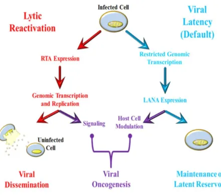
All viruses have obligatorily parasitic lifestyles which require host cell infrastructure for persistence and propagation. Outside of cells, human herpesvirus genomes are enclosed within proteinaceous viral capsids, which are further surrounded by a layer of tegument and finally by a lipid bilayer containing distinct viral glycoproteins, called the envelope (Chakraborty et al., 2012; Trus et al., 2001). The first stages of viral infections involve binding and entry into naïve cell targets, after which viral genomes are rapidly trafficked and accessed to modify host cells for infection and exploitation. A hallmark of herpesvirus infection is the ability to establish a persistent lifelong infection within host cells called latency. KSHV is no exception and after primary infection the virus can persist in latency, a semi-quiescent state, which can be reversibly interrupted by viral reactivation and subsequent reinfection (Figure 1.4). During KSHV latency the Latency-associated Nuclear Antigen (LANA) is expressed and ensures episome persistence (Ballestas et al., 1999). Latent infection is disrupted upon expression of the KSHV lytic-switch protein; the Replication and Transcription Activator (RTA) (Sun et al., 1998). Both replication phases are involved in KSHV-mediated oncogenesis; the autocrine/paracrine signaling and viral spread associated with lytic infection, and the modulation of host cells and maintained viral reservoirs characteristic of viral latency.
KSHV Latent Gene Expression
7
such that the potential for viral recognition by host immune surveillance is minimized (Coscoy and Ganem, 2000, 2001; Ishido et al., 2000). Viral transcripts that are expressed during latency play important roles in modifying the host cell and the virus to ensure the persistence of stable latent infection.
Transcripts from the KSHV Latency Locus
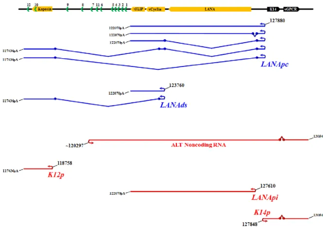
The linearized incoming viral genome is circularized as an episome in the infected host cell nucleus and expression is limited during latency (Dezube et al., 2002; Fakhari and Dittmer, 2002; Jenner et al., 2001; Renne et al., 1996a). The most well characterized transcripts originate from a cluster known as the major KSHV latency locus (Figure 1.5). This locus encodes the viral genes LANA, vCyclin, vFLIP, all 3 isoforms of Kaposin (A-C), as well as 12 stem loops from which the KSHV miRNA’s are derived (Cai et al., 2005; Dittmer et al., 1998; Pfeffer et al., 2005; Samols et al., 2005). LANA, vCyclin, and vFLIP are generated from the major latent promoter (the constitutive LANA promoter; LANApc) via the alternative splicing of co-terminal RNA’s (Bieleski and Talbot, 2001; Dittmer et al., 1998; Jeong et al., 2004; Sarid et al., 1999; Talbot et al., 1999). Splicing events also allow derivation of the viral miRNA’s and Kaposin isoforms from the LANApc (Cai and Cullen, 2006). Although the results are controversial, a second latently active downstream promoter called the LANAds is thought to generate all forms of Kaposin through alternative splicing and to also direct the expression of vCyclin and vFLIP and viral miRNA’s, (Cai and Cullen, 2006; Li et al., 2002; Pearce et al., 2005).
8
encoding LANA/vCyclin/vFLIP or a bicistronic message encoding a viral immunomodulatory glycoprotein (K14/vOX2) and a viral G-protein coupled receptor (vGPCR) (Hilton and Dittmer, 2012; Kirshner et al., 1999). Furthermore a lytically induced antisense transcript of unknown function called “antisense to latency transcripts” (ALT) originates upstream of miR-K7 from the ALTp, and is antisense to several viral miRNA’s, vFLIP, vCyclin, and LANA (Chandriani et al., 2010).
Latent Transcription Outside of the Latency Locus
All of the products derived from true latent promoters at the latency locus are present in latently infected PEL and KS cells (Dittmer, 2003; Fakhari and Dittmer, 2002; Marshall et al., 2007). Certain viral transcripts also originate outside of the latency locus expressed during latent infection, albeit with more variable patterns. For instance vIRF-3 (also called LANA-2) is latently expressed in PEL but not KS, and thus latent expression of this viral ORF is restricted to B cells (Rivas et al., 2001). Latent expression of other viral ORF’s is more controversial. vIRF-1 has classically been considered a lytic phase gene product (Jenner et al., 2001; Sarid et al., 1998), however others have observed latent expression, including within KS tumor cells (Chen et al., 2000; Cunningham et al., 2003; Dittmer, 2003). vIRF-1 may also use two distinct promoter elements, suggesting the possibility of cell type-specific gene expression. Evidence has also surfaced highlighting the fact that vIL-6, often considered to be a lytic gene, exhibits consistently high levels of expression during KSHV latency, but only in certain conditions, particularly MCD (Chandriani and Ganem, 2010; Nicholas et al., 1997).
9
production of previously underappreciated mRNA from the both strands of the latent KSHV episome, including antisense transcription at critical latent loci (Chandriani and Ganem, 2010; Dresang et al., 2011; Xu and Ganem, 2010). In the closely related EBV there are three distinct latency programs and given the phylogenetic and biological similarities between KSHV and EBV the prospect of adaptable patterns of latent KSHV infection has precedent and rationale. The situation is further complicated by the fact that in any given population of KSHV-infected cells (at least in vitro), a small percentage of cells undergo spontaneous lytic reactivation (Renne et al., 1996b), and vIRF-3, vIL-6, K1, and K15 are all induced early during lytic reactivation.
KSHV Latent Genome Maintenance and Replication
During latent infection the KSHV LANA protein tethers the viral genome to host chromatin through interactions between the episomal TR’s and host chromatin (Ballestas et al., 1999; Cotter and Robertson, 1999; Grundhoff and Ganem, 2003). Both the amino (N) and carboxy (C) termini of LANA interact with chromatin (Barbera et al., 2004; Kelley-Clarke et al., 2007). The N-terminus of LANA binds to an acidic pocket between histones H2A and H2B (Barbera et al., 2006;
Chodaparambil et al., 2007) and the LANA C-terminus binds the viral TR’s (reviewed in (Ballestas and Kaye, 2011)). This interaction physically links the viral and host genomes during latency. LANA binds to host chromatin in the presence and absence of the viral genome, however in the presence of KSHV distinct punctate foci are formed at the sites of KSHV episomes, whereas without the viral genome LANA binding is diffuse on host chromatin (Ballestas et al., 1999).
10
latent episome requires the replication of host cell machinery (Hu et al., 2009; Purushothaman et al., 2012; Stedman et al., 2004), however the field remains bereft of a full understanding of how latent episomal replication occurs.
KSHV Reactivation and Lytic Gene Expression
While viral latency is relatively stable and is the default replicative phase for KSHV, it is not indefinite. The physiologic stimuli which interrupt KSHV latency are varied and are still being elucidated. Activation of toll-like receptors, cellular cytokines and transcription factors, and treatment with chemicals which activate the protein kinase C pathway or which inhibit histone deacetylase (HDAC) activity; such as 12-O-tetradecanoyl-phorbol-13-acetate (TPA) or sodium butyrate respectively, can interrupt latency and reactivate KSHV (Chang et al., 2000; Gregory et al., 2009; Lan et al., 2006; Mercader et al., 2000; Renne et al., 1996b; Yu et al., 2007; Yu et al., 1999). While these inputs are diverse, all reactivation stimuli are ultimately coordinated through a single viral gene encoded by KSHV ORF50 called the Replication and Transcription Activator; RTA (Sun et al., 1998).
RTA expression from the KSHV genome (or ectopic expression) initiates an ordered cascade of viral gene expression and genome replication. This ultimately results in host cell lysis and the production of new infectious virions. During reactivation nearly the entire KSHV genome is robustly transcribed in a stage-specific manner, with transcripts temporally designated as: (i) immediate-early (IE) – based upon their expression occurring in the absence of de novoprotein synthesis. (ii)
11
pattern may be more complex, with large portions of both strands of the KSHV genome abundantly transcribed in both latency and reactivation (Chandriani and Ganem, 2010; Dresang et al., 2011; Xu and Ganem, 2010). Moreover RTA-independent mechanisms of KSHV lytic gene induction have been characterized (Chang et al., 2005a; Toth et al., 2012) and variance in viral gene expression across host cell cycle may also occur (Kang and Lieberman, 2009). Emerging data challenge the idea of such a clear transcriptional demarcation between two static viral lifestyles, and instead seem to suggest a more variable (and likely more adaptable), expression pattern.
RTA Promoter Specification
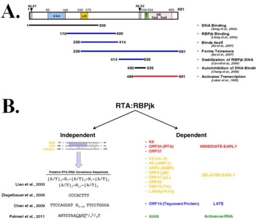
The RTA protein contains 691 amino acids (aa), two nuclear localization signals (NLS), an N-terminal DNA binding and dimerization domain, and a C-terminal transactivation domain, as shown in Figure 1.6a (Lukac et al., 1998). RTA is a DNA binding protein and physically interacts with a number of cellular and viral proteins (including itself) to activate transcription of cellular and viral genes; including activation of its own promoter (Bu et al., 2007; Carroll et al., 2006; Carroll et al., 2007; Chang et al., 2008; Deng et al., 2000; Gould et al., 2009; Gwack et al., 2002; Gwack et al., 2003b; He et al., 2010; Izumiya et al., 2003; Liang et al., 2002; Lukac et al., 2001; Lukac et al., 1999; Palmeri et al., 2007; Sakakibara et al., 2001; Song et al., 2002; Wang et al., 2003; Yada et al., 2006; Yang et al., 2008; Yu et al., 2005). Purified RTA rapidly forms multimers of decamers and tetramers by size-exclusion chromatography, however the transcriptionally active state of RTA appears to occur only in a tetrameric form (Bu et al., 2007).
12
RTA-response element (RRE) (Chang et al., 2002). A similar RRE is bound by RTA at lytic replication origins (OriLyt’s) (AuCoin et al., 2004; Wang et al., 2004d). Although RTA recognizes RRE’s at many loci, a conserved consensus sequence for binding has not yet been established (Chang et al., 2002; Chen et al., 2009; Liao et al., 2003; Palmeri et al., 2011; Song et al., 2001; Ziegelbauer et al., 2006). Recent data suggests that RTA may recognize a 14bp consensus sequence containing the core nucleotides of Cytosine-Adenine-N-Thymine (where N is any base); termed “CANT” repeats (Palmeri et al., 2011). CANT repeat elements may co-occur with other cis recognition elements at RTA-responsive promoters, however only one instance of CANT repeat utility has been demonstrated (Guito and Lukac, 2012; Palmeri et al., 2011).
The RTA:RBPjκ Interaction
While RTA interacts with several proteins to activate transcription; the interaction with RBPjκ (also called CSL) is perhaps the most well established and ubiquitous (Liang et al., 2002). RBPjκ is the downstream effector on the Notch signaling pathway (reviewed in (Kovall and Blacklow, 2010)). The RBPjκ molecule canonically acts as a transcriptional repressor and binds to the sequence motif 5’-GTGGGAA-3 (Tun et al., 1994); although there is a significant degree of motif redundancy, including within the KSHV genome (Persson and Wilson, 2010). Moreover the
13
The RTA:RBPjκ interaction is transcriptionally robust; even artificial promoters containing RBPjκ recognition sites can be transcriptionally activated by RTA (Liang et al., 2002). RBPjκ binding to DNA is likely more dynamic than initially characterized, and it is thought that the RTA tetramer is able to “stabilize” RBPjκ binding to initiate a transactivation response at RTA-responsive promoters (Carroll et al., 2006). The mechanisms underlying this stabilization are not fully known, but recent evidence suggests that the presence of CANT repeats or AT repeats could coordinate this interaction (Liao et al., 2003; Palmeri et al., 2011). Both gammaherpesviruses rely on RBPjκ for the effective coordination of gene expression (Hayward, 2004). Given the widespread decoration of the KSHV genome with RBPjκ binding sites, it is not surprising that intracellular Notch, (the canonical activator of RBPjκ) is able to transactivate some RBPjκ-dependent viral genes, and has even been shown to reactivate latent KSHV (Lan et al., 2006). However many RPBjκ-dependent KSHV genes do not respond to Notch signals, indicating that at the majority of responsive loci, the RTA:RBPjκ interaction is exclusively required (Chang et al., 2005a; Hilton and Dittmer, 2012). The RTA:RBPjκ interaction is further complicated by the fact that LANA has also been shown to interact with RBPjκ with functional consequences (Jin et al., 2012; Lan et al., 2005a; Lan et al., 2005b). The interactions between RBPjκ and the major viral latent (LANA) and lytic (RTA) transcriptional regulators imply that RBPjκ is a central player in mediating the balance between latency and lytic reactivation.
1.4 Selected KSHV-Encoded Oncogenic Factors
14
replication are involved in the malignant pathology of the virus and outlined below are selected products generated by the virus which have characterized roles in viral oncogenesis.
Latently Expressed Oncogenic Factors
LANA
The KSHV Latency-associated nuclear antigen (LANA) is a virally encoded protein with a multitude of diverse functions in infected cells (reviewed in (Ballestas and Kaye, 2011)). LANA is constitutively expressed from the major latency locus of the viral genome in all infected cells
(Dittmer et al., 1998; Gao et al., 1996; Kedes et al., 1997; Rainbow et al., 1997). The LANA protein is responsible for maintaining the KSHV genome and for systemic coordination of latency within infected cells.
LANA is critical for persistence of the KSHV genome though infected cellular divisions (Ballestas et al., 1999). LANA mediates this feat by tethering the KSHV episome to host
chromosomes through a bridging interaction between the viral TR’s and host nucleosomes (Ballestas and Kaye, 2001; Barbera et al., 2006; Cotter and Robertson, 1999; Grundhoff and Ganem, 2003; Skalsky et al., 2007a). This arrangement ensures that the viral genome is maintained during cellular mitotic division(s). In addition LANA directs episomal DNA replication at the latent viral replication origin during latency by binding to specific sequence elements within the viral TR’s (Ballestas and Kaye, 2001; Hu et al., 2002; Stedman et al., 2004; Verma et al., 2006). LANA also has
transcriptional and protein:protein interactions which modulate infected host cells to ensure continued cellular proliferation and growth and the stable preservation of viral latency.
15
protein also interacts with DNA methyltransferases, chromatin remodelers, and cellular transcription factors which contribute to its transcriptional modulatory functions (An et al., 2004; Bubman et al., 2007; Cai et al., 2006; Chen et al., 2012b; Kusano and Eizuru, 2010; Lan et al., 2005a; Lim et al., 2001; Lim et al., 2000; Liu et al., 2007; Roupelieva et al., 2010; Sakakibara et al., 2004; Shamay et al., 2006; Stuber et al., 2007; Verma et al., 2004). Furthermore, LANA interferes with cellular tumor suppressors functions, such as p53 and retinoblastoma protein, and several other pathways important to cell proliferation and oncogenesis (Ballestas and Kaye, 2011; Chen et al., 2010; Fakhari et al., 2006; Friborg et al., 1999; Fujimuro and Hayward, 2003; Fujimuro et al., 2003; Katano et al., 2001; Radkov et al., 2000). The transcriptional capacity and the interactions with other important proteins in infected cells make the KSHV LANA protein a nexus of viral genome persistence and cellular proliferation and oncogenesis.
vCyclin
vCyclin is a virally encoded homologue of cellular cyclin. In KSHV pirated homologues of cellular genes are denoted by a preceding “v” (see Figure 1.2). The protein is constitutively
expressed from the viral latency locus along with LANA (Bieleski and Talbot, 2001; Dittmer et al., 1998; Pearce et al., 2005; Talbot et al., 1999). vCyclin is a functional cyclin that can stimulate retinoblastoma protein (Rb) to overcome cell-cycle arrest (Godden-Kent et al., 1997). Unlike cellular cyclin D, vCyclin is capable of degrading the cyclin-dependent kinase (CDK) p27Kip when
16 vFLIP
Viral FADD-like interleukin-1-β-converting enzyme (FLICE) inhibitory protein (called vFLIP) is the KSHV homologue of cellular FLIP (cFLIP). vFLIP is also latently expressed co-terminally with LANA and/or vCyclin (Bieleski et al., 2004; Bieleski and Talbot, 2001; Dittmer et al., 1998; Grundhoff and Ganem, 2001; Pearce et al., 2005; Talbot et al., 1999). vFLIP inhibits apoptosis induced by cellular death receptors which may provide a growth advantage to infected cells (Djerbi et al., 1999; Low et al., 2001; Thome et al., 1997). vFLIP can also induce the expression of
anti-apoptotic genes via activation of NF-κB; a mechanism which protects cells against Fas-induced death (Chaudhary et al., 1999; Field et al., 2003; Guasparri et al., 2006; Matta and Chaudhary, 2004). vFLIP is essential for tumor cell survival in in vitro systems and can also prevent autophagy in B cells (Guasparri et al., 2004; Lee et al., 2009b). Additionally, transgenic mice bearing the vFLIP ORF develop B cell malignancies (Ballon et al., 2011; Chugh et al., 2005).
KSHV miRNA’s
KSHV also constitutively expresses several miRNA’s derived from 12 stem loops within the latency locus, some of which are also induced during lytic phase replication (Cai and Cullen, 2006; Cai et al., 2005; Gottwein, 2012; Pearce et al., 2005). At present the complete functionality of the KSHV miRNA’s is unknown and our understanding thereof is continually expanding. Emerging evidence demonstrates that KSHV miRNA’s are important in the context of oncogenesis and that they can modulate apoptosis, cell growth, cell cycle, and transcription in infected cells (Gottwein, 2012; Ramalingam et al., 2012). The KSHV miRNA’s show some capacity to reprogram infected cells both transcriptionally and even epigenetically (Gottwein et al., 2011; Haecker et al., 2012; Hansen et al., 2010; Lu et al., 2010), and miRNA expression levels may even correlate with KSHV-associated disease progression (O'Hara et al., 2009; O'Hara et al., 2008).
17
overcome apoptosis in infected cells. KSHV miRNA expression also enhances angiogenic and proliferative phenotypes (Boss et al., 2011; Gottwein et al., 2007; Liu et al., 2012; Samols et al., 2007; Skalsky et al., 2007b). Although we do not fully understand the breadth of the KSHV miRNA repertoire, the known functions of these virally encoded factors suggest involvement in KSHV-associated malignancy.
Kaposin
18 Variably Expressed Oncogenic Factors
K1
Genes which are not found in other organisms but are instead unique to the KSHV genome are designated by the prefix “K”. The K1 protein of KSHV is a transmembrane signaling protein encoded by the first (“leftmost”) ORF of the viral genome. The K1 ORF is one of the most polymorphic viral ORF’s (Hayward, 1999; Lagunoff and Ganem, 1997; Poole et al., 1999). K1 expression is induced during the viral lytic cycle, however low levels of latent expression have been observed (Chandriani and Ganem, 2010; Lagunoff and Ganem, 1997; Wang et al., 2006a). K1 has transforming potential and can immortalize endothelial cells in culture and can enhance angiogenesis and tumor vasculature in vivo (Lee et al., 1998b; Wang et al., 2006a). Cell signaling pathways relating to oncogenesis, angiogenesis, and proliferation are also perturbed via the K1 protein (Lagunoff et al., 1999; Lee et al., 1998a; Tomlinson and Damania, 2004; Wang et al., 2004a). Furthermore in transgenic mice the K1 protein can produce sarcoma-like tumors and lymphomas (Prakash et al., 2002).
K15
19 Pietrek et al., 2010).
vIRF-1
KSHV encodes 4 genes with homology to cellular interferon regulatory factors (IRF’s) known as vIRF’s. These proteins have roles in modulating host interferon system and innate immunity (reviewed in (Lee et al., 2009a)). In addition to innate immune perturbation, vIRF-1 and vIRF-3 have reproducibly demonstrated oncogenic properties (reviewed in (Jacobs and Damania, 2011)). vIRF-1 is considered a lytic gene, although latent expression has been detected in culture and in primary KS lesions (Dittmer, 2003; Jenner et al., 2001; Moore et al., 1996a; Sarid et al., 1998). Moreover, the vIRF-1 ORF is thought to contain two different start sites which could be under differential temporal regulation (Chen et al., 2000; Cunningham et al., 2003). vIRF-1 is capable of interfering with the tumor suppressor p53 and blocking p53-mediated apoptotic and transcriptional activities (Nakamura et al., 2001; Seo et al., 2001). vIRF-1 has also exhibited transforming potential in vivo (Gao et al., 1997), at least in part mediated through interactions with c-Myc (Jayachandra et al., 1999).
vIRF-3
20 vIL-6
Human interleukin-6 (hIL-6) is an important cytokine involved in a wide array of cellular activities including cell grown and differentiation (reviewed in (Kishimoto, 2010)). KSHV encodes a homologue of hIL-6, called vIL-6 (reviewed in (Sakakibara and Tosato, 2011)). vIL-6 is expressed at low levels during viral latency and is up-regulated during lytic replication (Chandriani and Ganem, 2010; Moore et al., 1996a; Nicholas et al., 1997). The vIL-6 transcript can initiate at two different start sites and is responsive to RTA (Deng et al., 2002). RBPjκ binding is thought to mediate the RTA response and unlike most other viral genes also allows vIL-6 to respond to intracellular Notch (Chang et al., 2005a). Although vIL-6 expression has been differentially detected in KS, PEL, and MCD tumors; the role of vIL-6 in KSHV-associated malignancy has been established through various experimental means (Parravicini et al., 2000; Staskus et al., 1999).
T-cell immunodeficient mice injected with NIH3T3 cells transduced with vIL-6 demonstrated the potency of vIL-6 in augmenting angiogenesis, tumor growth, and plasmocytosis (Aoki et al., 1999). vIL-6 activates pathways such as MAPK, JAK/STAT, etc. similarly to hIL-6 (Molden et al., 1997; Osborne et al., 1999), which in addition to other effects can lead to increased VEGF expression and signaling effects (Liu et al., 2001). vIL-6 is not limited by the physiologic checkpoints that regulate endogenous hIL-6 activity, and hence this viral mimic has enhanced hIL-6-reponsive signaling capacity relative to hIL-6 (Molden et al., 1997).
vGPCR
21
angiogenesis and proliferation including VEGF, p38, MAPK, Akt, mTOR, PI3 kinase, and small GTPases (Bais et al., 2003; Martin et al., 2011; Montaner, 2007; Montaner et al., 2001; Rosenkilde et al., 1999; Shepard et al., 2001; Sodhi et al., 2000). vGPCR can also upregulate transcription factors including NF-κB, HIF-α, CREB, and AP-1 which subsequently induce cytokines and
proliferative/angiogenic factors (Cannon et al., 2003; Cannon and Cesarman, 2004).
Further, vGPCR is capable of direct immortalization and transformation of certain cell lines and can also generate tumors reminiscent of KS lesions in nude mice (Bais et al., 1998; Bais et al., 2003; Yang et al., 2000). vGPCR is expressed early in lytic infection as part of a bicistronic transcript which also encodes K14 (vOX2) (Kirshner et al., 1999), however expression may occur outside of viral reactivation during cell cycling (Kang and Lieberman, 2009) or in a subset of infected cells in culture or in tumors (Cesarman et al., 1995a) . Both vGPCR and vOX messages are
expressed from the K14 promoter (K14p). The K14p is unusual as it is a member of the RTA-responsive bidirectional LANApi/K14p promoter which can also synthesize latent messages (Hilton and Dittmer, 2012; Matsumura et al., 2005). Notably, vGPCR expression can lead to both autocrine and paracrine signaling effects (Martin et al., 2011; Montaner et al., 2004), and hence vGPCR expression can affect infected cells and nearby cells which may or may not be infected.
1.5 Epigenetic Regulation of the KSHV Genome
22
intrinsically interwoven with the understanding of chromatin biology in general. Moreover, such endeavors may prove invaluable in the pursuit of antiviral strategies.
Chromatin modifications during viral infections often occur in both the host and invading viral genomes. Large DNA viruses, especially those which establish long-live persistent infections are not excluded from epigenetic regulation. This is exemplified in KSHV, and the virus has evolved intricate tactics to utilize host epigenetic machinery to navigate existence within human cell nuclei, which are outlined below and are discussed in greater detail in Chapter IV.
Epigenetically Programming the KSHV Episome for Latency
In the incoming virion the linear KSHV genome is devoid of DNA methylation and nucleosomes (Bechtel et al., 2005; Gunther and Grundhoff, 2010). Shortly after de novo infection, the viral genome circularizes by joining of the TR’s. Early in the stages of infection, before latency is effectively established, little is known about how the viral genome is epigenetically programmed for latency. Despite the relatively high GC content across the KSHV genome, DNA methylation of the viral genome is not an early event, but in culture models takes several days. The most noticeable effects of DNA methylation upon the viral life cycle are observed at the RTA promoter region, with a notably paucity of methylated viral DNA at the latency locus (Chen et al., 2001; Gunther and
Grundhoff, 2010). DNA methylation occurs in KSHV during latency, but it is not required for the establishment of latency, and has only extensively (and inconsistently) been observed, at late time points in viral infection (Chen et al., 2001; Gunther and Grundhoff, 2010).
23
Activating marks are also present at many immediate-early (IE) and delayed-early (DE) lytic cycle genes. IE/DE genes also contain so-called “bivalent” chromatin structures (Gunther and Grundhoff, 2010; Toth et al., 2010); histone tails modified with H3-ac or H3K4-me3 in addition trimethylated H3K27 (H3K27-me3). ORF’s of the viral genome encoding late-lytic phase (L) products are generally marked by the sole presence of heterochromatic H3K27-me3 or trimethylation H3K9 (H3K9-me3). These observations have led the field to theorize that the modification of histone tails during latency generates poising at key lytic IE/DE genes and repression at L genes.
Histone-modifying enzymes are also bound to the viral genome including; (i) a Polycomb group repressive complex 2 (PRC2) member called EZH2, and (ii) a histone H3K9-me3 demethylase called JMJD2A (Chang et al., 2011; Toth et al., 2010). The EZH2 methyltransferase is co-localized with H3K27-me3 on the KSHV genome is thought to maintain the bivalency or repression at lytic loci. JMJD2A binding to the viral genome is anti-correlated with H3K9-me3 and instead binds at activated histone modifications where it serves to prevent H3K9 methylation. Other reports suggest wide scale genomic binding of the heterochromatic organizing protein Kruppel-associated box domain-associated protein-1 (KAP-1) (Chang et al., 2009).
24
locus, manipulate KSHV lytic transcription, control episomal maintenance, and coordinate long range inter-episomal linkages (Chen et al., 2012a; Kang et al., 2013; Kang et al., 2011; Stedman et al., 2008).
CTCF is emerging as a master regulator of stable latency in both human gammaherpesviruses (Kang et al., 2011; Knipe et al., 2013; Tempera et al., 2011). CTCF is a zinc finger DNA binding protein that can insulate chromatin, mediate interactions between distal regulatory elements, structurally configure DNA into looped structures, and regulate transcription (Donohoe et al., 2009; Merkenschlager, 2010; Phillips and Corces, 2009; Rubio et al., 2008; Wendt et al., 2008). In addition CTCF affects nucleosome positioning and the modification of histone tails (Barski et al., 2007; Fu et al., 2008). CTCF binding is widespread in the latent KSHV episome and a major peak exists within the KSHV latency locus. Here CTCF/Cohesins are thought to regulate a balance between latent and lytic transcription through local PolII programming and physical connections to other episomal (Chen et al., 2012a; Kang et al., 2013; Kang and Lieberman, 2009; Kang et al., 2011; Stedman et al., 2008). CTCF/Cohesin may be involved in silencing lytic viral gene expression and in coordinating long-range interactions between the viral genome during latency. Several questions still remain to be addressed regarding CTCF/Cohesin-mediated epigenetic regulation of KSHV latency, including how long-range interactions are specified and how CTCF programming unfolds at specific loci.
Reactivation from Latency in the Epigenetic Context
25
“bivalent” chromatin but is repressed by PRC2 and histone deacetylases (Toth et al., 2010). Upon reactivation the KSHV lytic gene PAN chaperones H3K27-me3 histone demethylases and H3K4-me3 methyltransferases to the RTA promoter (Rossetto and Pari, 2012). RTA then recruits histone
acetyltransferases and chromatin remodelers to lytic promoters; including its own (Gwack et al., 2003a).
26
Figure 1.1: Human Herpesvirus Phylogeny. The eight known human herpesviruses are shown based upon amino acid similarity in respective major capsid proteins. The α-herpesviruses; varicella-zoster virus (VZV) and herpes simplex viruses 1 and 2 (HSV-1/2), are shown in orange. The β-herpesviruses; human
27
28
29
Figure 1.4: The KSHV Life Cycle. After KSHV infection the default viral replication program is latency, during which the viral genome exhibits very restricted gene expression. A handful of genes escape repression during latency including the KSHV Latency Associated Nuclear Antigen (LANA). LANA maintains the viral reservoir by assisting in viral genome replication and maintenance and also modulates the host cell
30
31
Figure 1.6: The KSHV Lytic Switch Protein: RTA A. The RTA protein is 691 amino acids (aa) in length and contains two putative nuclear-localization signals (NLS1/2). The protein is shown along with the
32 CHAPTER II
QUANTITATIVE ANALYSIS OF THE BIDIRECTIONAL VIRAL G-PROTEIN-COUPLED RECEPTOR AND LYTIC LATENCY-ASSOCIATED NUCLEAR ANTIGEN PROMOTER
OF KAPOSI’S SARCOMA-ASSOCIATED HERPESVIRUS1
2.1 Overview
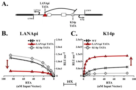
Kaposi’s sarcoma-associated herpesvirus (KSHV) establishes sustained latent persistence in susceptible cells. This is dependent on the latency-associated nuclear antigen (LANA). Understanding how LANA transcription is regulated thus aids our fundamental understanding of KSHV biology. Two hundred ninety-four base pairs are sufficient to regulate LANA transcription in response to the viral RTA protein and RBPjκ. The same region controls K14/viral G-protein-coupled receptor (vGPCR) transcription in the opposite direction. We used a quantitative analysis in conjunction with specific nucleotide substitutions and defined gain-of-function and loss-of-function RTA mutants to dissect this region. We used a bidirectional reporter driving red and green luciferase to study the LANApi and K14p promoters simultaneously. This established that LANApi/K14p functions as a canonical bidirectional promoter. Both were TATA dependent. K14p was favored by 50-fold in this context. Eliminating the distal LANApi TATA box increased maximal output and lowered the induction threshold (T) of K14p even further. Two RBPjκ binding sites were independently required; however, at high concentrations of RTA, direct interactions with an RTA-responsive element (RRE) could complement the loss of one RBPjκ binding site. Intracellular Notch (ICN) was no longer able to activate RBPjκ in the viral context.
1This chapter has been adapted from: Hilton, I.B., and Dittmer, D.P. (2012). Quantitative analysis of the
33
This suggests a model whereby KSHV alters ICN-RBPjκ gene regulation. When the architecture of this pair of head-to-head RBPjκ binding sites is changed, the sites now respond exclusively to the viral transactivator RTA and no longer to the host mediator ICN.
2.2 Introduction
Kaposi's sarcoma-associated herpesvirus (KSHV) is a human oncogenic gammaherpesvirus. The KSHV genome is 137,000 bp long and encodes more than 70 open reading frames (ORFs). KSHV is associated with Kaposi's sarcoma (KS), primary effusion lymphoma (PEL), and multicentric Castleman's disease (MCD). Viral transcription is tightly regulated and can be divided into two well-defined states (Dittmer, 2003; Jenner et al., 2001). (i) During the lytic phase, the genome replicates and every viral promoter is active. (ii) During latency, the viral genome persists within the nucleus as a circular plasmid (episome) and is subject to the same regulation as human chromosomes (Gunther and Grundhoff, 2010; Pantry and Medveczky, 2009; Toth et al., 2010). As a result, this
minichromosome is transcriptionally silent, with the exception of some key genes: the KSHV latency locus and a few genes that respond to cell type-specific and environmental stimuli. The KSHV latency locus encodes vital viral genes, which drive latent episome persistence: for instance, the latency-associated nuclear antigen (LANA) gene, as well as all viral microRNAs (Cai and Cullen, 2006; Dittmer et al., 1998; Ganem, 2010; Kang and Lieberman, 2009; Pearce et al., 2005; Talbot et al., 1999; Wen and Damania, 2010). Latent genes are central to KSHV tumorigenesis, since abrogation of LANA protein expression by small interfering RNA (siRNA) results in a loss of the KSHV plasmid and induction of apoptosis (Godfrey et al., 2005). Conversely, LANA expression can drive B cell hyperplasia in vivo (Fakhari et al., 2006; Sin et al., 2010).
34
Cullen, 2006; Dittmer et al., 1998; Li et al., 2002; Pearce et al., 2005; Sarid et al., 1998; Talbot et al., 1999). The LANA promoter ensures the coordinated expression of this KSHV latent gene cluster, including all viral microRNA. Elucidating the molecular details of this regulation can be expected to contribute significantly to our understanding of KSHV persistence and the AIDS-defining
malignancies, KS and PEL. A contiguous ∼1,200-bp fragment contains all cis regulatory elements to ensure constitutive LANA promoter (LANApc) activity (Dittmer et al., 1998; Jeong et al., 2001; Jeong et al., 2002; Jeong et al., 2004). The LANA promoter is never methylated and is free of repressive histone marks (Chen et al., 2001; Gunther and Grundhoff, 2010; Toth et al., 2010). Thus, this locus provides the opportunity to investigate general principles of promoter structure and function in a defined genomic context; however, the situation is more complicated.
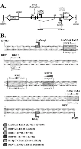
During latency, LANApc, a largely constitutive promoter, drives transcription of the LANA mRNA (Fig. 2.1A). It initiates transcription at position 127880. It is B cell-specific in transgenic mice (Jeong et al., 2002) and constitutively active in a large number of tissue culture cell lines. During reactivation, additional promoters are used. These are termed the LANApi, which is a promoter that can drive LANA transcription in response to the viral immediate-early transactivator RTA, and K14p, which is a promoter that drives a large K14/viral G-protein-coupled receptor (vGPCR) mRNA. Transcripts initiating from these two promoters have thus far been detected only in lytically reactivating cells. Nested within the LANApc untranslated region (UTR) is the bidirectional
35
gammaherpesvirus, Epstein-Barr virus (EBV), also exploits this mode of regulation (Jimenez-Ramirez et al., 2006).
A number of studies have looked at either K14p or LANApi by itself (Lan et al., 2005b; Liang and Ganem, 2004; Matsumura et al., 2005; Staudt and Dittmer, 2006) but thus far not at both in the bidirectional context. The LANApi TSS is positioned 313 bp upstream of the LANA protein translation initiation site (Matsumura et al., 2005; Staudt and Dittmer, 2006), and the K14p TSS initiates transcription 35 bp upstream of the K14 translation initiation site on the opposite strand (Chiou et al., 2002; Kirshner et al., 1999; Nador et al., 2001). The LANApi TSS utilizes a canonical TATA element (Matsumura et al., 2005; Staudt and Dittmer, 2006). A K14p TATA element has been predicted but not yet confirmed by functional studies. In sum, features of the LANApi/K14p pair resemble the architectural (spacing and strand identity) and functional (coexpression and
coregulation) features of bidirectional promoters.
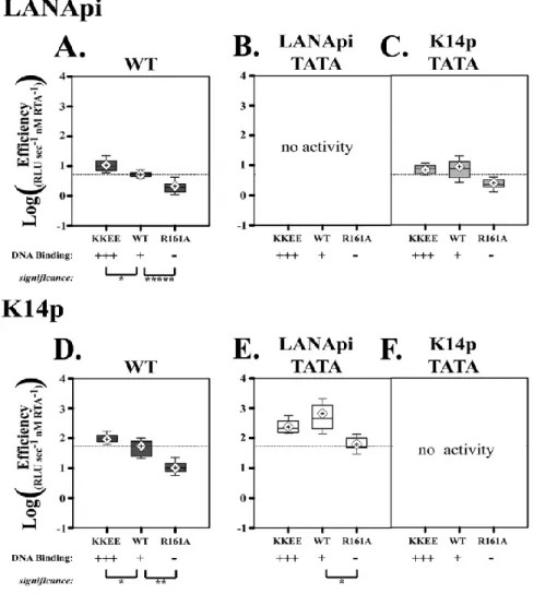
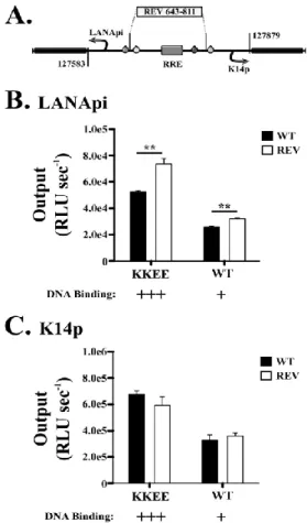
The LANApi and K14p TSSs are induced by the KSHV RTA transactivator. In fact, K14p is the most highly induced TSS in the entire KSHV genome (Damania et al., 2004). The RTA-mediated transactivation of LANApi and K14p displays a strict reliance upon two binding sites (Fig. 2.1B) for the human transcriptional adaptor RBPjκ (Liang and Ganem, 2004; Matsumura et al., 2005). The RTA-RBPjκ complex binds to these sites and activates the TSS. RTA functions as a multimer (Bu et al., 2007; Palmeri et al., 2011). The situation is more complex, however, since RTA can also bind DNA directly through a loosely defined RTA-responsive element (RRE) (Chen et al., 2009; Song et al., 2003; Song et al., 2002) and thus can activate other viral promoters independent of RBPjκ. Further, RBPjκ can be activated by its host partner intracellular Notch (ICN) independent of any viral proteins; reviewed in -(Miele, 2011). The LANApi/K14p region contains two RBPjκ elements, as well as a centrally located RRE, which could function to regulate K14p, LANApi, or both.
36
DNA binding domain. Transactivators (trans inputs) and their cognate sequence elements (cis inputs) coalesce to regulate mRNA production in the form of a transcriptional regulatory circuit (Kim et al., 2009). These regulatory circuits can be described in quantitative terms using the Hill function (see equation (1)) (Kim and O'Shea, 2008; Kim et al., 2009; Kuhlman et al., 2007; Rosenfeld et al., 2005). This equation relates promoter output to transactivator concentration. Traditional, enzymatic studies use purified proteins and enzyme activity relates to the enzyme protein concentration. In our
transfection studies, we did not know the intracellular RTA concentration, but we established that within the boundaries of our transfection series, doubling the input of a transfected expression plasmid resulted in a linear increase in the RTA protein. Hence, we can use this equation to characterize promoter behavior.
Three parameters describe the Hill function: the maximal output, measured herein as RLU s−1max; T, the induction threshold; and n, the Hill coefficient, which is a measure of cooperativity. By determining these parameters, we can make inferences about regulation and the biochemical
mechanism of action.
This mathematical framework is well known and has also been applied to study promoter activity in transfected cells (Kim and O'Shea, 2008; Kim et al., 2009; Kuhlman et al., 2007; Rosenfeld et al., 2005). For instance, the induction threshold T determines at which activator concentration the promoter is 50% active. A promoter with a lower T will be more active at lower transactivator concentrations. The Hill coefficient, n, indicates the cooperativity of the response. A Hill coefficient of >1 indicates a high degree of cooperativity and a step-like, “all-or-nothing” response curve; a Hill coefficient of ≤1 indicates a more gradual response. The maximal output provides a measure of promoter strength. A weak promoter will produce fewer transcripts
37
output is as follows: at the limit, the promoter initiates as many new transcripts per time unit as possible. Adding more specific transactivator no longer increases this rate, which is determined by how fast the general transcription factor complex can assemble and “reset” at the TATA element.
Using this framework, we investigated the LANApi/K14p response to RTA and report two new findings. (i) Our studies revealed a competitive relationship between the two TSSs, i.e., we found that one function of the LANApi TSS is to dampen the K14p response to RTA. (ii) Since
LANApi/K14p contains two RBPjκ sites, we expected RTA-dependent transactivation to be highly cooperative. This was not the case. We found an unconventional utilization of the head-to-head RBPjκ element pair reminiscent of sequence-paired site (SPS) Notch signaling (Arnett et al., 2010; Cave et al., 2005; Ong et al., 2006) but evolved to respond solely to the viral transactivator and no longer to ICN.
2.3 Materials and Methods
Plasmids
38
and -2029_3-1), RBP A (pDD2024, -2028, and -2031), RBP B (pDD2022, -2023, and -2030), and K14pTBP mutants (pDD2013, -2015, and -2016), respectively, were generated using the GeneTailor site-directed mutagenesis system (Invitrogen) and the GeneAmp high-fidelity PCR system (Applied Biosystems). The bidirectional mutant variants, RRE (pDD2038) and 611-843 REV (pDD2034), were designed as synthetic oligonucleotides (Blue Heron Biotechnology Inc.) with flanking HindIII sites for subcloning into the HindIII self-ligated bidirectional empty vector (pDD2045). Regulatory elements were identified using the software program Alibaba 2.1 and a literature review. The wild-type (WT) ORF50 expression vector was a kind gift from J. Choe (Gwack et al., 2001). 4X-RBPjκ Luc (4X-CBF Luc) was generously provided by S. D. Hayward (Hsieh et al., 1996). The ORF50 mutant expression vectors (ORF50 KK/EE and ORF50 R161A) were generous gifts from G. Miller (Chang and Miller, 2004; Chang et al., 2005b). The Flag-tagged human intracellular Notch- and tagged RTA (Nakamura et al., 2003)-encoding constructs were kindly provided by J. Jung. Myc-tagged ORF50 KK/EE (pDD2032) and ORF50 R161A (pDD2033) were generated using the same methods as in reference -(Nakamura et al., 2003).
Tissue Culture and Transfection
SLK cells (Herndier et al., 1994) were cultured in Dulbecco's modified Eagle's medium (DMEM) supplemented with 5% fetal bovine serum, penicillin (0.05 μg/ml), and streptomycin (5 U/ml) (Invitrogen Inc.) at 37°C under 5% CO2. SLK cells were seeded at a density of 1.0 × 10⁴ cells per well in 96-well plates (Sarstedt). The next day, transfection mixes were prepared using the RoboGo liquid handling system (Aviso) (or by hand in the case of Fig. 8 only) and then mixed with incomplete medium (DMEM without serum or antibiotic) and Superfect (Qiagen) as per the
39
purposes. We therefore relied on extensive biological replicates. Transfections utilized the MWG RoboGo liquid handling system and were performed in triplicate, at least three different times. Those transfections that were performed by hand were performed in duplicate, at least two different times.
Luciferase Data Acquisition
Cells were lysed with 100 μl 1X cell Culture lysis reagent (Promega), undergoing gentle orbital rotation for 10 min at room temperature. Lysate was then mixed with the Chromaglo luciferase assay system (Promega) substrate or with luciferase assay system (Promega) substrate as per the manufacturer's instructions. Luciferase activity was measured using a FLUOstar Optima 96-well luminometer (BMG Labtech). Red and green signal outputs were separated as per the manufacturer's instructions using a 590-nm long-pass and 510/60-nm filter (Chroma Corp.), respectively. Luciferase activity was measured from each well for 10 s at 1-s intervals, with the final values derived by the luminometer software as the average of all interval readings (n = 10), such that the output therein was expressed as relative light units observed per second (RLU s−1obs). Filter correction was achieved using the Chroma-Luc technology calculator (Promega).
Data Fitting and Analysis
40
for each individual trial (i.e., n ≥ 9 data points per titration curve). Global values were subsequently calculated by fitting the averaged outputs across all runs from each condition to generate a global fit, with standard error derived from the variance among individual runs. Initial analyses revealed adherence to first-order Hill kinetics (see Fig. 2.2); as such, we fixed the Hill coefficient to n = 1 for subsequent calculations (see equation (2)) (Chow et al., 2011; Kim and O'Shea, 2008; Ong et al., 2010). The apparent kcat and efficiency T were calculated as described above (see equations (3) and (4)). The initial concentration (Et) used to calculate the apparent kcat was defined as the concentration (in nM) of measurable input reporter construct (held constant throughout each titration curve). This definition expresses the observed output as a function of total detectable molecular quantities, and other definitions (such as potential binding sites, etc.) arbitrarily dilute this relationship. Due to the differences in molecular weight, the single reporter alone, bidirectional reporter, and single reporter in trans (1:1) thus had corresponding values of Et of 138 nM, 99 nM, and 69 nM, respectively.
Immunoblotting