The role of the default mode network in
contextual control
Verity Holly Lim Smith
Trinity Hall
MRC Cognition and Brain Sciences Unit
This dissertation is submitted for the degree of Doctor of Philosophy at the University of
Cambridge
2
Preface
I declare that this dissertation is the result of my own work and includes nothing which is the outcome of work done in collaboration. Where reference is made to the work of others, the extent to which that work has been used is indicated. I further state that no substantial part of my dissertation has already been submitted, or, is being concurrently submitted for any such degree, diploma or other qualification at the University of Cambridge or any other University or similar institution.
Data from the studies outlined in Chapter 4 and Chapter 5 have been published in a peer-reviewed journals. The references for these publications are as follows:
Smith V, Mitchell DJ, Duncan J. 2018. Role of the default mode network in cognitive transitions. Cerebral Cortex. 28, 3685-96.
Smith V, Mitchell DJ, Duncan J. 2019. The effect of rule retrieval on activity in the default mode network. NeuroImage. 116088.
The study outlined in Chapter 3 was done in collaboration with M Roca and C Pinasco, who designed the Hotel and Situations tasks, and C Pinasco and J Achterberg who collected the data with myself.
This dissertation does not exceed 60 000 words in length.
3
Acknowledgements
Thank you to all those who made this thesis possible:My supervisor, John Duncan, thank you for all your guidance throughout the last four years and for encouraging me to follow my research interests. My co-supervisor, Danny Mitchell, thank you for your endless patience in teaching me all I know about fMRI analysis. It’s been a pleasure to discuss this project with both of you and learn from your insights and critiques.
Thank you to the MRC Cognition and Brain Science Unit Imaging Community and Admin Team, none of these projects would have been possible without your expertise.
Thank you to the Medical Research Council for funding this PhD.
4
The role of the default mode network in
contextual control
Verity Holly Lim Smith
While extensive theories outline the importance of meaningful context in guiding goal directed behaviour, little evidence has emerged about the underlying cognitive mechanisms involved. This thesis aims to addresses this gap in the literature by integrating two commonly disparate topics in neuroscience: cognitive control and the default mode network.
Chapter 2 considers why current studies of contextual control do not implicate DMN regions by comparing context-dependent decision making using rich, meaningful scenes, in comparison to arbitrary letter stimuli. DMN regions of the posterior cingulate cortex, parahippocampus and posterior inferior parietal cortex are found to show increased activity during decision making in the lifelike context only.
Chapter 3 asks whether regions beyond the ‘task-positive’ multiple demand network are necessary for adequate performance in more lifelike naturalistic tasks. This neuropsychology experiment used behavioural data accumulated from brain lesioned patients across a series of naturalistic tasks and a standard IQ task. Naturalistic tasks were found to capture control processes beyond IQ and multiple demand network function, most likely depending on many processes and brain regions.
Chapter 4 aims to understand to what extent the DMN contributes to non-spatial executive tasks. Replicating (Crittenden et al. 2015), DMN regions were found to represent the broader task domain and respond with greater activation to larger task switches and task restarts. A role for the DMN in transitions between distinct cognitive tasks is suggested.
Chapter 5 assesses an alternative explanation for the switch effects of the previous chapter. The fMRI experiment presented in this chapter asks whether the activation of the DMN at cognitive transitions reflects changes in task rule retrieval difficulty instead of degree of task switch. To this end, this study directly manipulated the rule retrieval demands. Contrary to the retrieval account, increased
5
Contents
Preface ... 2
Acknowledgements ... 3
Verity Holly Lim Smith ... 4
Chapter 1 ... 9
Introduction ... 9
OUTLINE ... 9
THE DEFAULT MODE NETWORK ... 10
Social Cognition ... 12
Episodic Memory ... 15
Episodic Simulation ... 17
Navigation ... 18
Broad Function of the DMN ... 21
CONTEXT-DEPENDENT DECISION MAKING ... 24
THE MULITPLE DEMAND NETWORK ... 26
DMN AND MD IN ‘EXTERNAL’ TASK-RELATED COGNITION ... 30
THESIS OVERVIEW ... 38
Chapter 2 ... 41
Context-dependent decision making in naturalistic contexts ... 41
INTRODUCTION ... 41 METHODS ... 42 Participants... 42 Task... 43 Training ... 46 Data acquisition ... 47
6 Preprocessing ... 47 Regions of Interest ... 47 Univariate Analysis ... 50 Multivariate Analysis ... 50 RESULTS ... 53 Behaviour ... 53 fMRI: Univariate ... 54 fMRI: Multivariate ... 60 DISCUSSION ... 65 Chapter 3 ... 68
Naturalistic task performance in brain damaged patients ... 68
INTRODUCTION ... 68 METHODS ... 70 Patients ... 70 Testing ... 70 Tasks ... 70 Neuroradiological Assessment ... 76 Regions of Interest ... 76 RESULTS ... 78 Situations Task ... 79
Differences between patients and controls ... 79
Effects of lesion volume ... 81
Between task correlations ... 82
Residual patient impairment ... 83
DISCUSSION ... 84
Chapter 4 ... 86
The role of the default mode network in cognitive transitions ... 86
INTRODUCTION ... 86
7 Participants... 87 Task... 87 Training ... 90 Data acquisition ... 90 Preprocessing ... 90 Regions of Interest ... 91 Univariate analyses ... 92 Multivariate analyses ... 93
Finite Impulse Response Model ... 93
RESULTS ... 94
Behavioural switch costs ... 94
Increased DMN activity on rest trials ... 94
Increased DMN activity for large task switches ... 95
Large increases in DMN activity for task restarts ... 96
Component ROIs within each subnetwork ... 97
MD activity across trial types ... 100
Increased activity at task restart is distinct from prolonged rest activity ... 101
Individual voxels in DMN and MD regions show sensitivity to both rest and between-domain task switches ... 102
DMN and MD activity patterns distinguish task domains ... 103
DISCUSSION ... 104
Chapter 5 ... 108
The effect of rule retrieval on activity in the default mode network ... 108
INTRODUCTION ... 108 METHODS ... 109 Participants... 109 Task... 110 Training ... 112 Data acquisition ... 112
8 Preprocessing ... 112 Regions of Interest ... 112 Analysis ... 113 RESULTS ... 114 Behavioural Performance ... 114 ROI analysis ... 115
Whole brain analysis... 122
DISCUSSION ... 123
Chapter 6 ... 127
Discussion ... 127
The DMN is not ‘task-negative’ ... 128
The DMN and perception ... 129
What constitutes a ‘broad context’? ... 130
DMN Subnetworks... 132
The DMN and non-human animals ... 135
MD and DMN ... 136
Conclusions ... 138
9
Chapter 1
Introduction
OUTLINE
Spatial, semantic and social context is considerably helpful to everyday cognition. Context can help us to understand ambiguous sentences, to work out the meaning of a new word in the presence of learned words, and to understand a person’s actions or feelings. The behavioural benefit of contextual information has been studied for many decades, particularly in memory recall and language comprehension (Zwaan and Radvansky 1998). Bransford and Johnson (1972) found that participants showed better understanding and more detailed recall of written passages when first read with a title encompassing the broad context of the passage compared to when read with no title available. Godden and Baddeley (1975) found that memory recall for word lists was more accurate for divers when the recollection phase was in the same context (underwater or on land) as the memory encoding phase where the word lists were first presented. Spivey et al. (2002) found that participants were able to use the visual context to parse syntactically ambiguous sentences such as ‘Put the apple on the towel in the box’ (see also Spivey and Tanenhaus 1998; Mirman 2008). The researchers found that if the participants could see an apple already on a towel, this visual context could override participants preferred sentence parsing (that the towel was in the box) in favour of moving the apple on top of a towel to an empty box. These studies therefore suggest that
representations of our surroundings can influence our understanding of ongoing events.
Many theories also suggest contextual knowledge is important in guiding goal-directed behaviour. These theories suggest that contextual information, built up through repeated experiences in similar contexts, can be used to guide future behaviours by representing situational constraints and
simulating common courses of events (Bar 2007, 2009; Zacks et al. 2007; Ranganath and Ritchey 2012). Levels of contextual representation can include very generalised (semantic) information (e.g. you must be quiet in libraries), to more specific knowledge of a particular place (e.g. in the study room of this library you can talk quietly) or episodic knowledge of a specific event (e.g. at this library open day you can talk).
10
Despite extensive theories outlining the importance of meaningful context in guiding goal directed behaviour, little evidence has emerged about the underlying cognitive mechanisms involved. This thesis aims to addresses this gap in the literature by integrating two commonly disparate topics in neuroscience: cognitive control and the default mode network. Cognitive control refers to the executive process influencing cognition in accordance with ongoing goals and is commonly associated with a network of lateral frontal and parietal regions (Duncan and Owen 2000; Duncan 2010, 2013). The default mode network (DMN) is a network of functionally connected brain regions most commonly associated with scene, episode and situational context representation in internally generated thoughts (Buckner and Carroll 2007; Hassabis and Maguire 2007; Buckner et al. 2008). Despite being often found to be negatively related to task control (Fox et al. 2005a; Kelly et al. 2008) its role in contextual representation marks its potential importance in contextual control.
THE DEFAULT MODE NETWORK
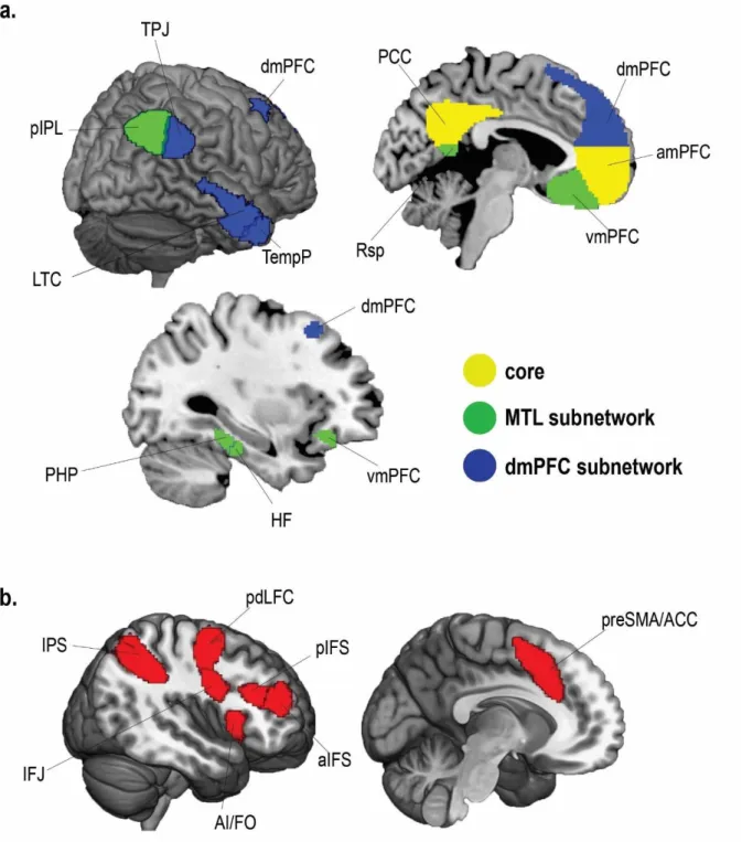
The DMN is one of the most well established brain networks, with its emergence consistently replicated through functional connectivity (Greicius et al. 2003, 2009; Fransson and Marrelec 2008; Spreng et al. 2013; Liang et al. 2016), structural connectivity (Greicius et al. 2009; Khalsa et al. 2014; Yin et al. 2016) and univariate activity (Raichle et al. 2001; Andrews-Hanna et al. 2010; see Spreng et al. 2009 for a meta-analysis). It is most commonly found to include posterior medial cortex (posterior cingulate cortex, precuneus and retrosplenial cortex), anterior medial prefrontal cortex, posterior inferior parietal lobe, parahippocampus, hippocampus, temporal parietal junction and middle lateral temporal lobes. More recent findings have also suggested the DMN may be split into core regions and two sub-networks (Andrews-Hanna et al. 2010; Yeo et al. 2011). Andrews-Hanna et al. (2010) used hierarchical clustering analysis on task-based and resting-state functional connectivity fMRI data, revealing a highly connected core network of posterior cingulate cortex (PCC) and anterior medial prefrontal cortex (amPFC) as well as a Medial Temporal subsystem and a Dorsal Medial subsystem (see Figure 1.1a). The Medial Temporal subsystem including parahippocampus (PHP), hippocampal formation (HF), retrosplenial cortex (Rsp), posterior inferior parietal lobe (pIPL) and ventromedial prefrontal cortex (vmPFC). The Dorsal Medial subsystem included regions of the dorsomedial prefrontal cortex (dmPFC), temporo-parietal junction (TPJ), lateral temporal cortex (LTC), and temporal pole (TempP). Strong functional connectivity between subnetwork regions and core DMN has been proposed to allow efficient transmission of information across subnetworks via core DMN hubs. Similar DMN subdivisions were further uncovered in a large-scale brain parcellation by (Yeo et al. 2011). Using resting state functional connectivity MRI data in a sample of 1000
11
included the DMN. In a further finer-grained analysis, a 17 network solution was uncovered which further divided the DMN into 3 sub-networks. As shown in Figure 1.1b, this approach revealed similar subnetworks to Andrews-Hanna et al. (2010) with a few differences within each subnetwork. Firstly, the vmPFC was not classified within the DMN network but a limbic network instead. Secondly, Yeo et al. (2011) found that the DM subsystem also included some lateral prefrontal regions and was left lateralized, while the core network also included the angular gyrus and right anterior temporal lobe.
Figure 1.1. Proposed DMN Subnetworks from a. Andrews-Hanna et al. (2010) and b. Yeo et al. (2011). Figure adapted from Andrews-Hanna et al. (2014). PCC = posterior cingulate cortex, aMPFC = anterior medial prefrontal cortex, Rsp = retrosplenial cortex, PHC = parahippocampal cortex, HF+ =
hippocampal formation, vMPFC = ventromedial prefrontal cortex, pIPL = posterior inferior parietal lobe, TempP = temporal pole, LTC = lateral temporal cortex, TPJ = temporo-parietal junction, dMPFC = dorsomedial prefrontal cortex.
Early research looking at DMN activations characterised the DMN as ‘task-negative’, finding
reductions in activity during many externally-focused tasks in comparison to rest or easier versions of the same task (Shulman et al. 1997; McKiernan et al. 2003, 2006). DMN activity has instead been associated with off-task thinking or mind wandering during external task performance (McKiernan et al. 2006; Mason et al. 2007; Christoff et al. 2009). For example, using an event sampling technique, Christoff et al. (2009) found stronger DMN activation immediately before thought probes where participants indicated off-task thoughts compared to before thought probes indicating on-task cognition. These inter-trial increases in DMN activity during external task performance have also been linked to attention lapses and negative effects on performance (Weissman et al. 2006). In line with these findings of task-related deactivations and associations between DMN activity and poor performance, the DMN has often been found to be negatively correlated with regions associated with on-task executive function (Fox et al. 2005a; Kelly et al. 2008; Uddin et al. 2009; Newton et al. 2011). As such, early research suggested the DMN was a ‘task-negative’ network, preferentially active during a default state of rest.
12
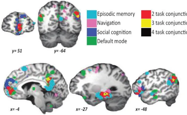
Since these initial findings, the DMN has been implicated in a number of different tasks, indicating its role in representing scenes, episodes and contexts. In an influential meta-analysis using the
activation likelihood estimation approach, Spreng et al. (2009) found extensive overlap in voxels active in episodic (autobiographical) memory retrieval, social cognition (measured by Theory of Mind tasks), navigation and the DMN, as presented in Figure 1.2. These findings were replicated by Spreng and Grady (2010) who also found common neural activation during cognitive processes involved in social cognition, episodic memory retrieval and future episodic simulation within the same 16 participants.
Figure 1.2. Results of Spreng et al. (2009) activation likelihood estimation (ALE) meta-analysis for each term (episodic memory, navigation, social cognition and default mode) and their conjunction. Figure adapted from Spreng et al. (2009). MNI coordinates presented for slices.
Social Cognition
Social cognition, or mentalizing, refers to the ability to infer someone else’s thoughts, feelings and emotions. This ability is often tested using false belief (Theory of Mind) tasks. In these tasks (see Figure 1.3) participants are presented with scenarios where the subject of a story has a belief which is at odds with the current reality. Participants are asked to infer the subject’s false belief in order to predict their actions. Neuroimaging studies have revealed DMN involvement in a number of social cognition tasks including false beliefs, moral judgements, person perception and self-knowledge tasks (Fletcher et al. 1995; Greene et al. 2001; Frith and Frith 2003; Amodio and Frith 2006; Gilbert et
13
al. 2007). In one of the first imaging studies to research the neural basis of social cognition, Fletcher et al. (1995) presented participants with stories in which one of the characters acts in line with their false beliefs. In comparison to reading unlinked sentences, medial prefrontal, posterior medial and the superior temporal sulcus showed increased activations when reading the stories. Further studies with more stringent baselines have also implicated these regions as being particularly active when participants were asked to make judgements about other people’s beliefs (see Frith and Frith 2003 for a review).
14
Figure 1.3. The Sally-Anne false belief Task. Image from Felisberti and King (2017).
Within the DMN, social cognition has been particularly related to lateral temporal, temporo-parietal junction and dorsal medial prefrontal regions. Using automated meta-analytic software
(NeuroSynth), Andrews-Hanna et al. (2014) found meta-analyses maps for social cognition terms such as ‘mentalizing’, ‘theory of mind’ and ‘social’ were particularly strongly related to the Dorsal Medial subsystem of the DMN. Similarly Kumaran and Maguire (2005) found dmPFC and core DMN
15
regions were strongly related to social cognitive processes. In a novel task the researchers asked participants to work out how to get a case of wine from Friend A to Friend B by exchanging the wine through as many other friends as possible. In the social condition, participants could only exchange wine between their friends who knew each other. In the spatial condition the case of wine had to be passed closer and closer to the goal location (Friend B’s house) via other friend’s houses. Despite no significant behavioural differences between the two conditions, the researchers found increased activity in core DMN, dmPFC, vmPFC, superior temporal sulcus and the temporal poles when directly comparing the social condition to the spatial condition.
Episodic Memory
As was made famous by amnesic patient HM, the medial temporal lobe is known to play a
fundamental role in episodic memory encoding and retrieval (Scoville and Milner 1957; Penfield and Milner 1958; Corkin 2002). HM had a bilateral medial temporal lobectomy, in order to cure his epilepsy, incurring lesions to the hippocampus, parahippocampus, amygdala and entorhinal cortex bilaterally. His surgery left him with a striking impairment in forming new memories (anterograde amnesia) and, to a lesser extent, recalling pre-surgery events (retrograde amnesia). HM showed severe anterograde memory deficits regardless of the domain of to-be remembered stimuli (words, sounds, pictures, personal events, and public events) and the type of memory test used (free recall, cued recall, multiple choice recognition). HM’s retrograde amnesia, on the other hand, appeared to be temporally graded in that HM’s memory impairment affected events from the three years prior to surgery but most of his memories before this period in time were intact. Despite such prolific
memory impairments, HM’s general intelligence and language were unaffected, if not improved by reduced epileptic symptoms (Scoville and Milner 1957). Intriguingly performance on short term memory tasks were also intact (Sidman et al. 1968; Corkin 1982) and HM also showed good
visuomotor skill learning. Over repeated sessions HM was asked to trace a star while seeing only the mirror image of his drawing hand. Despite no memory or feelings of familiarity for any of the testing events, HM was able to show marked improvement on a mirror drawing task as well as other procedural learning tasks where an explicit knowledge of learnt events is unnecessary. Given these findings, researchers have suggested that these medial temporal regions are particularly important in forming new declarative memories and consolidating them into long term memory (Squire and Zola 1996).
Due to the severity of memory impairments in patient HM, most lesion and animal electrophysiology research in memory initially focused on medial temporal lobe regions. With the development of new neuroimaging methods, researchers have been able to further establish regions beyond the medial temporal lobe that are related to episodic memory. These studies tend to implicate DMN regions
16
within and beyond the medial temporal lobe in episodic memory retrieval (Lepage et al. 2000; Diana et al. 2007; Schacter et al. 2007; Vilberg and Rugg 2012).
Lepage et al. (2000) developed the idea of an episodic retrieval mode (REMO), a state where one is focused on holding a personal past event in mind and using external information to cue memory for specific details of the event. The researchers suggest that neural correlates of REMO should show increased activity for attempted retrieval regardless of retrieval success and as such, tested for a conjunction between brain regions active during successful item retrieval (‘old’ judgements) and unsuccessful item retrieval (‘new’ judgements) compared to a control task block in which there was no requirement for memory retrieval. The researchers identified activity in anterior medial frontal regions as well as the anterior cingulate cortex and frontal operculum related to attempted retrieval.
Other researchers have investigated the neural correlates of episodic memory by testing for regions associated with accurate memory for contextual details surrounding a cued event. In Hayama et al.'s (2012) source memory study participants were given two sets of 30 words to study. Each word started with a unique three-letter word stem. In the study phase words were presented to the left or right of the screen and participants were asked to make judgements about the degree of
abstractness of the words. At the test phase 90 three-letter word stems (60 studied, 30 new) were presented to the centre of the screen. Participants were asked to complete the word stem with the previously studies word (if possible) and, if the word had been previously studied, retrieve the position of the studied word. Hayama et al. (2012) found DMN regions of the anterior medial prefrontal cortex, parahippocampus, posterior medial cortex and posterior inferior parietal lobe were active for both successful (vs. unsuccessful) word recall and successful (vs. unsuccessful) word position retrieval, implicating the DMN regions in successful memory for episodic details of an event.
Tulving (1985) outlined three key properties of episodic memory: a subjective sense of time (a feeling of mental time travel), connection to the self (self-relevance), and autonoetic consciousness (the cognitive ability to mentally project oneself to an imagined time and place). Particularly supporting these ideas, DMN regions have been found to show increased activity with increased self-relevance and vividness of the remembered episode, and subjective reliving of an event (Andrews-Hanna et al. 2010; St. Jacques et al. 2011; Richter et al. 2016). D’Argembeau et al. (2005) further found
dorsomedial prefrontal, ventromedial prefrontal, middle temporal gyrus and temporal pole regions were particularly active whilst engaging in self-referential reflections. In accordance with these ideas, Buckner and Carroll (2007) have argued that self-projection, or imagining events from a first person perspective, may be the main function of the DMN.
17
Episodic Simulation
In line with the understanding of episodic memory retrieval as mental time travel to past events, much research has been carried out to test for neural overlap between episodic memory recall and episodic simulation of future events, which can be thought of as mental time travel to the future. As predicted, DMN regions have been found to be implicated in simulating both past episodes and future events as well as imaging events in the present (Addis et al. 2007, 2009; Szpunar et al. 2009; Andrews-Hanna et al. 2010). Addis et al. (2009) tested for neural overlap as well as differences in neural activation between recalling autobiographical past events and imagining autobiographical past and future events using spatiotemporal partial least squares analysis. On each trial Addis et al. (2009) presented participants with three cues corresponding to location, person and object details for three different past events, personal to each participant. With these cues participants were asked to either recall the cued details from each event or reimagine each of the cued details in a new event situated in the past or future. Confirming the hypothesis that autobiographical retrieval and
construction involve overlapping cognitive processes, conjunction analyses showed neural overlap between these three conditions, relative to a sentence construction control, in DMN regions of posterior medial cortex, anterior medial prefrontal cortex, hippocampus, parahippocampus, middle temporal gyrus and posterior inferior parietal lobe. However, recollection was more associated with the hippocampus, parahippocampus and visual cortex, perhaps due to more rich visual imagery. In comparison, imagined events were more related to medial prefrontal cortex, anterior hippocampus, posterior medial cortex and inferior frontal gyrus, perhaps reflecting construction and integration processes.
Andrews-Hanna et al. (2010) further tested whether DMN regions were activated for imagining either self-relevant or non-self-relevant events in the present as well as the future by asking participants to make decisions concerning different scenarios varying in temporal situation and self-relevance. All DMN regions showed increased sensitivity for autobiographical decisions compared to non-self-relevant conditions. Unlike Addis et al. (2009), who found no differences in activity between events imagined at different points in time, Andrews-Hanna et al. (2010) found MTL subnetwork regions were preferentially active during future self judgements whereas dmPFC subnetwork regions were preferentially active during present self judgements. Extending these findings, the researchers found that several self-reported ratings explained the variance in activity in core and MTL
subnetworks, suggesting some underlying contents of cognition that these regions are most involved in. Core DMN activity was particularly related to ratings of personal significance, self-introspection and evoked emotion, whereas the MTL DMN subnetwork was associated with episodic memory, event imagination and scene content ratings.
18
Hassabis et al. (2007a, 2007b), however, suggest that DMN regions are still necessary for detailed imagination of events that are not personally relevant or positioned in time. Hassabis et al. (2007a) asked 5 bilateral hippocampal lesioned patients with amnesia and 10 matched controls to imagine a new event based on a short verbal cue. Participants were then asked to describe the contents of their imagination and judge the degree of vividness. Their descriptions were also scored by researchers on the basis of information content, spatial coherence and overall quality. Although there was no significant difference between patient and controls on subjective judgements of vividness, patients scored lower in informational content, spatial coherence and quality. The researchers suggest that the hippocampus might be particularly important for setting up the spatial context in which further details of a scene can be bound. To check whether other regions beyond the hippocampus were also related to construction of imagined experiences, the researchers scanned healthy participants whilst undertaking a similar version of this task. In this fMRI version, participants were asked to imagine or remember scenes and objects. Hassabis et al. (2007b) found that, in comparison to object construction, during construction of imagined and recalled scenes there was increased activity in the hippocampus, parahippocampus, retrosplenial cortex, posterior cingulate cortex, posterior parietal cortex and anterior medial prefrontal cortex. However, in comparison to the recalled events, imagined events showed less activity in the core DMN regions.
In accordance with Hassabis et al. (2007a), Szpunar et al. (2009) suggest the key component driving DMN activation during event simulation is not self-relevance but context familiarity. The researchers asked 27 participants to imagine autobiographical events in a cued spatial context that had
happened in the past or could happen in the future. In 24 of the trials participants were asked to imagine future events in an unfamiliar context (e.g. a hot air balloon) and in 48 trials participants were asked to either imagine future events or recall past events in familiar contexts. Whole brain analyses found increased activity in posterior medial cortex, parahippocampus, anterior medial prefrontal cortex and temporo-parietal junction for familiar remembering and imagining in familiar contexts compared to unfamiliar contexts.
Navigation
While amnesiac patient HM was most famous for having difficulties with episodic memory formation and retrieval, he was also found to show impairments in spatial navigation of new environments first experienced post-surgery (Scoville and Milner 1957; see also Spiers et al. 2001). Findings from animal lesion studies have continued to implicate posterior DMN regions in navigation. Lesions to the hippocampus, parahippocampus and retrosplenial cortex have been found to impair performance during radial arm maze and water maze tasks where the subject is required to navigate from memory
19
towards a rewarded spatial location or hidden platform (Logue et al. 1997; Vann and Aggleton 2002; Bohbot et al. 2006; Pothuizen et al. 2008).
The finding that these regions play a major role in spatial navigation has been extended by
electrophysiology recordings in animals. O’Keefe and Dostrovsky (1971) first discovered the presence of place cells in the hippocampus of rats and since their initial finding, place cells have been
identified in monkeys and humans (Ono et al. 1991; Ekstrom et al. 2003). Place cell populations characteristically fire at unique locations within an environment regardless of the orientation or trajectory of the animal, allowing for representation of one’s allocentric spatial location in an environment. Furthermore, place fields have been found to be differently configured for distinct environments and remain stable in familiar environments for several weeks, suggesting the hippocampus can generate long-term memories unique to specific spatial contexts (Lever et al. 2002).
Location-sensitive cells have also been identified in another DMN region in the medial temporal lobe: the parahippocampus. In comparison to hippocampal place cells, place cells in the parahippocampus have much broader place fields and are more sensitive to changes in external environment (Burwell and Hafeman 2003). Results from imaging studies also suggest that the parahippocampus is
particularly sensitive to the external visual properties of an environment. The parahippocampus has been found to show increased activity in response to pictures of scenes, places and landmarks compared to other visual stimuli (Epstein and Kanwisher 1998; Epstein et al. 1999; Janzen and Van Turennout 2004; Mullally and Maguire 2011). As such, a region within the parahippocampus has been termed the “Parahippocampal Place Area” (Epstein and Kanwisher 1998). In line with the findings from neuroimaging, patients with lesions to the parahippocampus have been found to show poor recognition of familiar rooms or buildings, termed landmark agnosia, despite maintaining the ability to draw accurate allocentric spatial maps (Landis et al. 1986; Takahashi and Kawamura 2002).
The retrosplenial cortex has also been found to contain neurons which encode information important for navigation. Chen et al. (1994) reported cells in the retrosplenial cortex of rats which showed selective preference for certain head orientations even in the absence of visual cues or when rats were in the dark. In line with an emerging role for the retrosplenial cortex in representing information about one’s orientation, damage to the retrosplenial cortex in humans is associated with topographical amnesia. Topographical amnesia is a condition which is associated with a poor ability to orient oneself by visual landmarks despite preserved scene perception (Aguirre and D’Esposito 1999; Epstein 2008).
20
According to the cognitive map theory (O’Keefe and Nadel 1978), and the model presented in Byrne et al. (2007), the hippocampus is important in representing an allocentric, spatially coherent world, given visual inputs from the parahippocampus and orientation information from the retrosplenial cortex. These allocentric maps are thought to be translated into a first person perspective with the aid of heading information from head direction cells in the retrosplenial cortex. The resulting egocentric scene is proposed to be represented in the posterior cingulate cortex. This translation of stored allocentric spatial information into an egocentric image has been suggested to be important for simulating an upcoming journey during navigation but also constructing a spatial environment during episodic simulation (Bird and Burgess 2008; Ranganath and Ritchey 2012).
In line with findings from electrophysiology, imaging studies also find activity in posterior DMN regions related to spatial navigation (Spiers and Maguire 2007; Howard et al. 2014; Javadi et al. 2017, 2018). Howard et al. (2014) asked participants to navigate around London’s Soho district. Increased activation in many DMN regions was found during the active navigation condition compared to the passive control condition which kept the same visual background but did not require participants to navigate from memory. Regions of the posterior medial cortex and medial temporal lobe also
showed strong activity at decision points (when participants had to choose which road to turn down). Interestingly, the posterior hippocampus appeared to code path distance to goal, showing greater activity during travel periods further from goal than closer to the goal. Furthermore, in a similar paradigm, Javadi et al. (2017) found that after entering a new street the posterior hippocampus showed increased activity to increases in the number of upcoming routes available. The researchers suggest that the posterior hippocampus might be simulating all upcoming routes and therefore showing increased activation when the number of route available increased.
Further imaging studies suggest other regions of the DMN are also related to spatial navigation. Balaguer et al. (2016) found the ventromedial prefrontal cortex and hippocampus coded distance to goal with these regions showing increased activity the closer to goal (although this ramping of DMN activity at the end of a trial may not be specific to spatial navigation, or to the DMN, see Farooqui and Manly 2018). Patai et al. (2019) scanned participants during virtual navigation through newly experienced university campuses and frequently visited university campuses with which participants had a minimum of 2 years’ experience. The researchers found that navigation through the newly learnt campus was associated with a map-like representation of space and related to hippocampal encoding of distance to goal. In contrast, navigation though the well-known campus was found to be more associated with egocentric experiences and in this case, the retrosplenial cortex was found to code distance to goal. These findings may link well to the model presented by Byrne et al. (2007) suggesting medial posterior regions are important for translating spatial information into an
21
egocentric perspective whereas the hippocampus is more important for establishing an allocentric map of space.
While the Spreng et al. (2009) meta-analysis showed great overlap between regions related to episodic memory, social cognition and navigation, it should be noted that most studies of spatial navigation do not implicate the Dorsal Medial subnetwork of the DMN. Indeed, when contrasting activity during spatial and social tasks, Kumaran and Maguire (2005) found the Rsp, PHP, and pIPL were more active during the spatial version compared to the social version of the wine task
(described above) marking the Medial Temporal DMN subnetwork as especially important in spatial navigation. These regions were also more active during a spatial control task, asking participants to assess the building type of different friends’ houses, compared to a social task, where participants answered whether various friends wore glasses. These findings suggest that the MTL subnetwork of the DMN plays a role in spatial processing that is more general than just spatial navigation.
Broad Function of the DMN
How the DMN contributes to all these disparate cognitive processes is a subject of ongoing discussion. Consistent among all these tasks is the requirement for the construction of egocentric scenes or episodes from internal sources of information. As a result, the role of the DMN has been reconceptualised from a ‘task-negative’ network to having a main role in projection of oneself into an imagined scene (Buckner and Carroll 2007) or construction of a spatial context with which to bind social or episodic details (Hassabis and Maguire 2007). Some studies have attempted to differentiate between these theories, finding conflicting results. Hassabis et al. (2007a) found DMN activity related to imagination for scenes that are not self-relevant whereas Alzheimer’s patient DB showed specific impairment in thinking about personal futures compared to imagining futures that were not self-relevant. Despite these differences, one commonality is the focus on internally constructed
simulations, distinct from that of the current surroundings (Buckner and Carroll 2007; Buckner et al. 2008; Andrews-Hanna 2012).
This focus on internal scene representations has led researchers to overlook the simple possibility that the DMN also represents the current external context for context-guided cognition. With new multivariate methods, more recent studies have found a role for the DMN in representing the content of both externally perceived, recalled and imagined scenes and events (Baldassano et al. 2016, 2017; Chen et al. 2017; Robin et al. 2018). In Chen et al. (2017) participants watched a 50 minute episode of Sherlock and then immediately recalled the episode from memory in an MRI scanner. Average voxel activity for 50 independently labelled scenes was computed for each individual during the movie watching and movie recall phases. Using a spatial searchlight, the
22
researchers found that patterns of activity in posterior medial cortex, medial prefrontal cortex and parahippocampal cortex were more similar during watching and recall of the same scene compared to different scenes. Along with sensory regions, these regions also showed highly similar scene-specific patterns of activity between subjects during movie watching, indicating a role for DMN regions in perception of the current scene which is stable across participants. Furthermore, Baldassano et al. (2017) found activity patterns in the same data set were more stable across a longer time scale in several brain regions, including posterior DMN, compared to sensory regions, during both the movie watching and recall phases (see Figure 1.4). Medial prefrontal DMN regions were not included in the searchlight analysis. By defining reductions in cross-temporal correlations as neurally defined event boundaries, the researchers went on to compare neurally defined event boundaries from movie watching data with data when hearing an audio-description of the same movie and perceived event boundaries as judged by human observers. Compared to sensory regions, neural event boundaries from movie watching data in DMN regions of interest, the angular gyrus and posterior medial cortex, were found to better match with boundaries defined by human annotations and were consistent across movie perception modalities (watching and audio-narration). These findings led Baldassano et al. (2017) to suggest that the angular gyrus and posterior medial cortex represent a stable, gist-like representation of a scene (see also Simony et al. 2016).
Figure 1.4. Figure from Baldassano et al. (2017). Results from a neural event segmentation
searchlight from movie watching data in cortical regions with high between-subject correlation (Chen et al. 2017). The optimal number of events found, as tested using a Hidden Markov Model, were
23
found to vary throughout the cortex. Early sensory regions optimally divide the movie into greater numbers of short events (purple regions) and higher multimodal cortex divide the movie into fewer numbers of long events (yellow regions). Events here are defined as periods of similarity in cross-temporal correlation. Example cross-cross-temporal pattern correlations during movie watching for long timescale events and short timescale events are presented from posterior medial cortex (top) and early visual cortex (bottom), respectively.
In keeping with these findings, Ranganath and Ritchey (2012) suggest that the DMN, particularly the parahippocampus, hippocampus and retrosplenial cortex, are important for the construction and representation of situation models. Situation models have been described as a mental
representation of the current situational context (or schema) along with its social, semantic and temporal associative relationships. For example, the situation model for the event of studying with a friend at a library would hold information about the place of study (along with further locational information about libraries), the person you are with (along with further information about them and their connection to you), what you are doing and perhaps the temporal order of events leading up to the requirement to study. An example situational model, as described in Ranganath and Ritchey (2012) is presented in Figure 1.5. The researchers suggest that the parahippocampus is particularly important for identifying the current spatial context while the retrosplenial cortex integrates external information with stored associative information. The hippocampus has been proposed to integrate situational context with more specific details encoded in the perirhinal cortex and connected anterior temporal regions while the situation model itself is suggested to be represented in other regions of the DMN. The representation of this associative structure of the current context has been thought to be important for context-guided cognition and action by representing situational constraints and simulating common courses of events (see also Bar 2007, 2009; Zacks et al. 2007). According to these theories, the DMN should therefore be fundamental for context-dependent decision making.
24
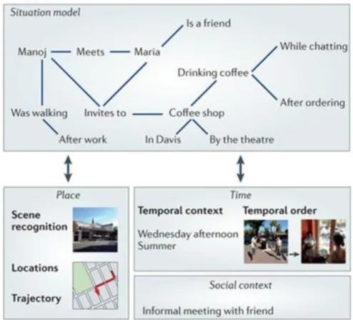
Figure 1.5. Figure from Ranganath and Ritchey (2012) displaying the associative spatial, temporal and social information proposed to be represented in the posterior medial (PM) system, closely
overlapping with posterior DMN regions, in the event of having coffee with a friend after meeting them in the street. The situation model represents associative links including the sequence of events and spatial trajectory, semantic information about Maria (a friend), spatial information about the coffee shop (by the theatre in UC Davis) and the overall social context (an informal meeting).
CONTEXT-DEPENDENT DECISION MAKING
Despite the relation between DMN and context representation, the role of the DMN in context-dependent decision making is far from well established. Studies aiming to understand the neural basis of context-dependent decision making have mainly used arbitrary cues as substitutes for life-like contextual control. In one such task, Koechlin et al. (2003) compared brain activity between 3 different levels of cognitive control with increasing levels of rule abstraction. The experimental design is presented in Figure 1.6. In the Sensory Control condition, button presses were cued by stimulus colour (i.e. colour-response trials). In the Contextual Control condition, participants were asked to make either vowel/consonant or uppercase/lowercase judgements based on the colour of
25
the presented letter (i.e. feature-response trials). In the Episodic Control condition, colour-response mappings were not fixed but were cued at the start of each block so participants were required to keep in mind temporally abstract information (i.e. the start of block cue-colour-response). The experimenters also designed control blocks for each experimental condition also presented in Figure 1.6. Comparing experimental block to control block activity, Koechlin et al. (2003) implicated the dorsolateral prefrontal cortex, not DMN regions, in contextual control, with the more anterior inferior frontal sulcus implicated in episodic cognitive control and more posterior regions of the premotor cortex associated with sensory control. Furthermore, using effective functional
connectivity methods the researchers suggested a direction of information transfer from the anterior frontal regions to posterior regions.
Since this original study, similar cognitive control gradients have also been found in a number of different studies manipulating different types of task complexity (Badre and D’Esposito 2007, 2009; Race et al. 2009; Badre and Nee 2018). Although the specific regions identified in contextual control vary across these experiments (Badre and Nee 2018), a lack of DMN contextual control related activity is common across them all. In fact, the lateral prefrontal regions identified in these cognitive control tasks fall within another well-established brain network; the multiple demand (MD) network (Duncan 2010, 2013).
Importantly for this thesis, Badre and Nee (2018) now suggest a difference between ‘contextual control’, associated with the dorsolateral prefrontal cortex in the MD network, and ‘schematic control’. Schematic control refers to generalised knowledge gained after repeated experiences and is suggested to be related to the anterior prefrontal cortex and ventromedial prefrontal cortex in the DMN. The extent to which the DMN and MD network contribute to cognitive control processes in naturalistic contexts which include both schematic and more context-specific information is, as yet, an unanswered question in the field.
26
Figure 1.6. Hierarchical cognitive control experimental design, adapted from Koechlin and
Summerfield (2007). In Sensory Control trials (ai.) respond using different button presses (R1 and R2) depending on the colour of stimuli with participants asked to make no response (NG=no go) to white squares. In Contextual Control trials (bi.) the colour of the letters represents the task (T1 and T2, vowel/consonant judgement and upper/lowercase judgement) with white letters reflecting a no-go (NG) trial. Episodic Control trials (ci.) were similar to the Sensory Control task where button presses and no-go trials (R1, R2 and NG) were cued by colour of stimulus but the colour-response mappings were not fixed but dependent on a cue prior to the block of episodic control trials. ii. Control tasks used for each executive control condition.
27
The MD network is a set of highly connected frontoparietal regions including parts of the inferior frontal sulcus, dorsal prefrontal cortex, inferior frontal junction, anterior insula, presupplementary motor area and intraparietal sulcus (often accompanied by activity in lateral occipital cortex). Some researchers suggest that the MD system can be further subdivided into a frontoparietal network (FPN) containing lateral frontal and parietal MD regions, and the cinglular-opercular, or salience network (SN) containing the anterior insula and presupplementary motor area and sometimes anterior frontal cortex (Dosenbach et al. 2006, 2007, 2008; Seeley et al. 2007; Crittenden et al. 2016). A map of canonical MD regions is presented in Figure 1.7. While resting-state functional connectivity analyses have typically found these regions to be highly temporally correlated (Seeley et al. 2007; Vincent et al. 2008), this network is most typically defined from univariate task activations. These frontoparietal regions have been consistently linked to task demand in a diverse range of executive tasks including working memory, response inhibition, executive planning, memory span and fluid intelligence (Duncan and Owen 2000; Duncan et al. 2000; Duncan 2006; Bishop et al. 2008; Fedorenko et al. 2013; Assem et al. under review). In Fedorenko et al. (2013) 40 participants were scanned whilst performing a selection of seven different tasks testing language, arithmetics, response inhibition and spatial and verbal working memory. The tasks were presented in a blocked design with participants tested on hard and easier versions of each task in order to assess activity related to cognitive demand. One of the tasks performed by all participants was used to localise MD voxels in each subject. Activation for the hard vs. easy contrasts in all other tasks was found to overlap with voxels sensitive to the localiser. Uniquely, only voxels within MD regions, and not neighbouring language selective regions, showed consistent sensitivity to the difficulty contrast.
28
Figure 1.7. MD network regions from group-level hard vs. easy contrasts across all seven tasks in Fedorenko et al. (2013). The map presented was made symmetrical across hemispheres by averaging across hemispheres and thresholding the map at t = 1.5.
Further evidence from the neuropsychology literature demonstrates the necessity of MD regions for good cognitive function. Woolgar et al. (2010, 2018) tested 80 patients with focal brain lesions on a fluid intelligence task, the Cattell Culture Fair (Cattell 1971). This fluid intelligence measure contained four timed tasks including series completion, finding the odd-one-out, matrix judgements and
topological relations. Cattell performance has been found to be predictive of task performance on a number of executive tasks (Roca et al. 2010, 2011; Duncan et al. 2012) and is thought to be a robust measure of executive function. Woolgar et al. (2010, 2018) found that patient MD lesion volume, but not total lesion volume or lesion volume in language specific regions, was negatively related to fluid intelligence scores.
The properties of MD regions are perhaps most clearly understood in studies of single unit activity in monkeys. These studies find a large percentage of neurons in lateral prefrontal cortex and
intraparietal sulcus that selectively and flexibly code for a wide range of task-relevant information including target stimulus identity and location, response, reward and task rules (Rao et al. 1997; Duncan et al. 2000; Freedman et al. 2001; Freedman and Assad 2006; Roy et al. 2010). For example, in Roy et al. (2010) monkeys were trained to make orthogonal categorisation decisions on a single stimulus set depending on the colour of cue preceding the trial (see Figure 1.8). Separate populations of prefrontal neurons were found to show category selectivity with stronger categorical responses measured when that dimension was relevant in the current trial. Further research has also shown that task selective coding in prefrontal cortex dynamically changes throughout a trial (Rao et al. 1997; Kusunoki et al. 2009; Stokes et al. 2013).
29
Figure 1.8. a. The stimulus set presented in Roy et al. (2010), grouped according to each category
decision. The stimulus set was comprised of morphed images of cats and dogs of two different types. Each of the corner stimuli represent prototype cats and dogs while stimuli in between represent mixtures. Category scheme A was cat/dog, category scheme B was type 1/2. b. Responses of an example prefrontal neuron showing category sensitivity. The neuron specifically distinguishes between cats and dogs (showing greater activation for cats) but only when that is the relevant category decision.
30
Adaptive coding of task-relevant information has also been found in human MD regions in fMRI studies. Woolgar et al. (2011a) found that patterns of activity in MD regions could be used to discriminate between all task-relevant features in a simple stimulus location-response task including stimulus position, stimulus-response rule and response. Furthermore, Woolgar et al. (2011b) went on to establish that the degree of task-relevant feature coding was related to the level of task demand. At low levels of perceptual difficulty in identifying object location, patterns of activity in MD regions did not discriminate object location. However, at odds with visual discriminability, in trials of high perceptual difficulty, MD regions did discriminate object location. These findings suggest that the greater univariate activity in the MD system during more demanding tasks may reflect adaptive coding of task features most challenging for task completion.
Given the importance of MD regions across such a diverse range of task demands, Duncan (2010, 2013) suggests that the core function of the MD system is to control complex behaviour in line with current task goals. This function is thought to be achieved through decomposition of abstract goals into achievable sub-goals (Christoff et al. 2001; Bhandari and Duncan 2014), selective coding of information relevant to the current sub-goal (Duncan 2001), and influencing selection bias in other brain systems to process task-relevant information (Desimone and Duncan 1995; Stokes et al. 2009).
DMN AND MD IN ‘EXTERNAL’ TASK-RELATED COGNITION
As discussed above, the MD system has been found to be important for task control and the DMN has been implicated in representing context. Given these findings it seems plausible that both networks would work together during real-world contextual control. Historically, however, the DMN and MD network have been found to be negatively related.
As described previously, in univariate analyses of demanding external tasks, MD regions show increases in activity with increased task demand (Fedorenko et al. 2013) whereas DMN regions show the opposite pattern of task related deactivations (McKiernan et al. 2003, 2006). Functional
connectivity studies have also been used to assess the relationship between these two networks. Using resting state functional connectivity Fox et al. (2005a) found that ‘task-negative’ regions (anterior medial prefrontal, posterior cingulate and posterior inferior parietal lobe) and ‘task-positive’ regions (dorsolateral prefrontal, inferior parietal sulcus and middle temporal region) as defined using peak coordinates from previously published data, were negatively correlated. Kelly et al. (2008) further established that the strong negative correlation between these ‘task-negative’ and
31
‘task-positive’ regions in each subject during the Eriksen flanker task was associated with low intra-subject variability in task reaction times. Given that intra-intra-subject variability has also been found to be negatively related to general intelligence (Jensen 1992), the researchers suggest that a competitive relationship between MD and DMN regions is important for good task performance. In accordance with this proposal, Newton et al. (2011) found that the degree of negative correlation between ‘task-positive’ and ‘task-negative’ regions, which closely reflect MD and DMN regions respectively,
strengthened with increased task load. Cole et al. (2012) further highlighted the importance of these negative correlations between DMN and task-positive regions in task performance. The researchers tested the connectivity strength, during an N-back task, between networks defined through meta-analyses of network names (e.g. “Default Mode Network”) with the lateral prefrontal cortex whose activity was found to predict N-back performance. The researchers found that the degree of negative correlation between DMN and the lateral prefrontal cortex was predictive of participant IQ as tested by the Cattell Culture Fair (“Institute for Personality and Ability Testing” 1973) and Raven’s
progressive matrices (Raven 1981). In comparison, the strength of positive connectivity between cognitive control regions and lateral prefrontal cortex and the absolute strength of sensory-motor network connectivity with lateral prefrontal cortex was predictive of IQ (see Figure 1.9).
Figure 1.9. Regions of interest and correlation results from Cole et al. (2012). a. The network regions of interest as found through meta-analyses by using network names as search terms. Red = cognitive control network, yellow = sensory-motor network, blue = default mode network. b. Significant correlations between connectivity strength of networks with lateral prefrontal cortex vs. fluid intelligence score. Figure from Cole et al. (2012).
The above functional connectivity studies defined seed regions as ‘task-negative’ and ‘task-positive’ based on univariate activity from the same task or using pre-defined negative’ and ‘task-positive’ regions of interest. More recent studies using whole brain network connectivity analyses to define DMN and MD on the basis of connectivity alone, also confirm the negative relationship between DMN and MD regions, suggesting this result is not just a consequence of how these regions of interest have been defined. Cole et al. (2014) compared functional connectivity between brain
32
networks from rest and task-related activity in two data sets. The first data set, from Cole et al. (2010) consisted of a 10 minute resting state scan followed by 10 short task runs. The task, presented in Figure 1.10a, asked 15 participants to make relational judgements about item properties by particular button presses. The relation, property and button to press would change for each trial creating 64 different ‘tasks’. A second dataset from the Human Connectome Project (Van Essen et al. 2013) was also used as a comparison. This fMRI data was collected in 118 participants over two days. Each scanning session started with 28 minutes of rest followed by 30 minutes of task. FMRI data for 7 different tasks were collected over the two days; the particular tasks used are presented in Figure 1.10b. Cole et al. (2014) calculated functional connectivity matrices for the task-based and resting-state data for each of the datasets from time course correlations across 264 brain regions as defined by Power et al. (2011). The researchers then used a standard community detection approach to cluster the 264 regions into brain networks from their functional connectivity profiles. The resulting parcellation showed good correspondence to the resting state networks found by Power et al. (2011) identifying the same networks including the DMN and FPN. When comparing connectivity between these networks during rest and task, Cole et al. (2014) found that in both data sets, connectivity between DMN regions and FPN regions decreased during tasks compared to rest (see Figure 1.10c), suggesting the negative relationship between DMN and MD regions is particularly strong during tasks. Whole brain network analyses therefore mirror the findings of initial ‘task-negative’ connectivity experiments suggesting that DMN and MD are negatively related during task performance in a way that affects performance.
33
Figure 1.10 Tasks and connectivity results from Cole et al. (2014). a. Experimental design from Cole et al. (2010). The task asked participants to use 64 different combinations of logic x sensory x motor rules to make relational judgements based on properties of pairs of items. b. The 7 tasks from the Human Connectome Project scans (Van Essen et al. 2013). c. Differences in connectivity strength between task functional connectivity and resting state functional connectivity for 264 brain regions as organized by brain network. Cole et al. (2010) dataset on the left and Human Connectome Project dataset on the right. The black box signifies the increasingly negative correlation between DMN and FPN for task-state functional connectivity compared to resting-state functional connectivity. Figure adapted from Cole et al. (2014).
34
Given that several studies report DMN deactivation related to improved task performance (Daselaar et al. 2004; Shulman et al. 2007; Anticevic et al. 2010) and increased DMN activity predictive of attention lapses (Weissman et al. 2006), it has been suggested that the cognitive control regions are responsible for suppressing DMN activity during task performance (Fox et al. 2005a; Sridharan et al. 2008). Effective functional connectivity methods have been used to assess the direction of these negative correlations in order to test this prediction. Some papers have found a modulatory
influence of MD regions on DMN regions (Sridharan et al. 2008; Spreng et al. 2013; Liang et al. 2016). For example, Sridharan et al. (2008) used Granger causality methods to test the direction of influence of proposed MD subnetworks SN and FPN with the DMN during an auditory event segmentation task, a visual oddball task and periods of rest. The researchers found a causal influence of SN regions, presupplementary motor area and particularly anterior insula, on DMN and FPN. The degree of influence to DMN and FPN changed depending upon the task with SN regions showing greater connectivity with DMN regions during rest and with FPN regions during task. However, other papers come to the opposite conclusion (Uddin et al. 2009). Uddin et al. (2009) performed seed-based resting state functional connectivity from two spherical core DMN seeds based on peak coordinates from a previous study (De Luca et al. 2006). From these two posterior cingulate and anterior medial prefrontal seeds the researchers found thresholded maps of regions positively (amPFC-, PCC-) and negatively (amPFC+, PCC+) correlated to the seed regions. While there were some differences, the PCC+ and amPFC+ connectivity maps were generally associated with DMN regions including
extensive medial prefrontal cortex, precuneus, temporo-parietal junction, posterior inferior parietal lobe, middle temporal cortex and parahippocampus. PCC and amPFC were negatively associated with MD regions including pre-supplementary motor area, anterior insula, dorsal lateral prefrontal cortex, intraparietal sulcus and extrastriate visual cortex. To test for causal relationships between DMN regions and the regions negatively correlated to DMN, the researchers performed Granger causality analysis on the average time course of the positive and negative connectivity maps generated by each seed region. Uddin et al. (2009) found that changes in signal in amPFC+ and PCC+ could
significantly predict activity in amPFC- and PCC- at a later point in time. In comparison to the findings from Sridharan et al. (2008), therefore, Uddin et al. (2009) suggest DMN activity modulates activity in task-positive regions during rest.
Despite prevalent findings of a competitive relationship between the DMN and MD network, more recent research has suggested that these networks are not exclusively in opposition. During a visuospatial planning task and an autobiographical planning task, Spreng et al. (2010) assessed functional connectivity and univariate activation patterns in independently defined DMN and FPN regions. The Tower of London task was used as the visuospatial planning task. In the fMRI version of
35
this task, participants were presented with a starting configuration and a goal configuration of three coloured discs on three sticks of different heights (see Figure 1.11a). By only moving one disk at a time participants had to judge the fewest number of moves required to transition the disks from the starting positions to the goal positions. The autobiographical planning task used a similar visual layout (see Figure 1.11b). Participants were given an autobiographical goal in the bottom of the screen and shown 3 steps and obstacles associated with achieving that goal on the top half of the screen in the three disks. Participants were asked to plan a way of achieving the desired goal using the three steps in a way specific to the participant. Upon completion of their personalised plan, participants were asked to press a button and indicate how detailed their plan was. As a control condition, participants were also asked to count the number of vowels from the 6 discs on the screen (see Figure 1.11c). Between-network connectivity results showed that DMN regions were
significantly positively correlated with the FPN during the goal-directed autobiographical planning task. These results were also reflected in the univariate analyses, with both the DMN and FPN showing increased activation during autobiographical planning task relative to control. During the visuospatial planning task FPN and DMN were not significantly correlated. FPN regions showed univariate increases in activity and DMN regions showed univariate decreases relative to the control task. These results suggest that, depending on the task, DMN activity can couple with ‘task-positive’ MD regions to support goal-directed cognition. Similarly, Gao et al. (2013) found that in comparison to rest, correlations between PCC seed regions and MD regions, pre-supplementary motor area and anterior insula, during the Navon global/local letter task (Navon 1977) became positive, with the degree of between network connectivity reflecting accuracy.
36
Figure 1.11. Task stimuli from Spreng et al. (2010). a. Visuo-spatial planning task – participants were asked the minimum number of moves it would take to change the starting configuration of disks to the goal configuration by moving one disk at a time. b. Autobiographical planning task – participants were asked to imagine a detailed plan to achieve the goal using the three points. c. Control task – participants were asked to count the vowels. Figure adapted from Spreng et al. (2010).
Other research assessing univariate activations also suggests the competitive relationship between ‘task-positive’ MD regions and ‘task-negative’ DMN is not as clear cut as has been previously suggested. Increased activity in DMN regions has been linked to external task performance and MD activations have even been associated with off-task cognition. Intriguingly, Christoff et al. (2009) implicated the MD network as well as the DMN in off-task mind wandering. In the event sampling study described above, Christoff et al. (2009) found regions of the MD network (as well as DMN
37
regions) that were more active immediately before probes where participants reported off-task thoughts compared to when participants reported on-task cognition. The researchers suggest that MD regions are not only important for maintaining focus during task control but also important for maintaining focus on self-generated, task-unrelated thoughts.
DMN has been linked to goal-related activity in a number of demanding cognitive control tasks. Spreng et al. (2014) found increases in DMN activity during a 2-back working memory task using famous faces rather than anonymous faces, mirroring a performance benefit for famous face matching. This finding also held after trial activity was split by reaction time. DMN activity was still greater during slower famous face matches than faster anonymous face matches, suggesting that the DMN activity during famous face matching represents more than just a difficulty effect. The
researchers suggest that the DMN contributes to task performance when required to retrieve information from long term knowledge. Furthermore, in a series of studies, Smallwood et al. found increased DMN activation when participants were required to make decisions about stimuli
presented in the previous trial compared to when making perceptual decisions about current stimuli (Smallwood et al. 2013; Konishi et al. 2015; Murphy et al. 2018). These researchers suggest that DMN activations during externally-directed tasks are due to retrieval of learnt information which is not available in the current environment. The content of retrieved information, they suggest, does not have to be rich and semantic in content in order to require DMN for retrieval (Murphy et al. 2018, 2019).
However, the role of the DMN in externally-focused tasks does not appear to be limited to memory retrieval. In a cued attention task, Hahn et al. (2007) asked participants to indicate the presence of a target as quickly as possible by button press. The target could be in one of four locations on the screen. Prior to the target’s appearance, a cue was presented to the middle of the screen giving information about the upcoming target location. The cue varied in specificity from ‘specific location’ to ‘uncued’ by pointing to 1, 2, 3 or all 4 of the 4 target locations. Activity in core DMN and left pIPL was negatively related to reaction time for uncued trials when participants did not have prior information about the target location. These findings hint at a role for the DMN during task performance when attention is focused more broadly in the absence of top-down endogenous control.
Esterman et al. (2012) suggest a more complicated relationship between DMN univariate activity and task performance. From participant performance during a sustained attention task, the researchers identified two attentional states from reaction time variability data. The ‘in the zone’ state was characterized by periods of relatively stable reaction time and the ‘out the zone’ state was