DOI:10.19026/rjaset.14.3982
ISSN: 2040-7459; e-ISSN: 2040-7467 © 2017 Maxwell Scientific Publication Corp.
Submitted: May 20, 2015 Accepted: June 19, 2015 Published: January 15, 2017
Corresponding Author: Tian-Swee, Tan, Department of Biotechnology and Medical Engineering, Faculty of Biosciences and Medical Engineering, UniversitiTeknologi Malaysia, Malaysia
This work is licensed under a Creative Commons Attribution 4.0 International License (URL: http://creativecommons.org/licenses/by/4.0/).
Research Article
Preprocessing Digital Retinal Images for Vessel Segmentation
1Tian-Swee, Tan, 1Nurul Emaan Ameen, 2Wan Hazabah Wan Hitam, 3Yan-Chai Hum and
4Chong-Keat Teoh
1Department of Biotechnology and Medical Engineering, Faculty of Biosciences and Medical
Engineering, UniversitiTeknologi Malaysia,
2Department of Ophthalmology, School of Medical Sciences, UniversitiSains Malaysia,
3National R&D Center in ICT, MIMOS Berhad,
4Department of Computer Science, Faculty of Computing, UniversitiTeknologi Malaysia, Malaysia
Abstract: The information contained in the retinal vasculature is used to diagnose the onset of retinal diseases such
as diabetic retinopathy. However, due to non-uniform illumination and variations in imaging modalities, the contrast between the retinal blood vessels network and the background is very low, encumbering the analysis and the diagnosis processes. This prompts the need for preprocessing digital fundus images to remove noise and improve contrast thus increasing the segmentation accuracy of the retinal vasculature. In this study, we address issues of non-uniform illumination and low contrast by developing a framework that implements shade correction, image enhancement and prepares the digital fundus images for the next stage.
Keywords: Binary mask generation, contrast enhancement, morphological operations, retinal fundus images
INTRODUCTION
The growing applications of computer vision in the medical sciences has enabled the medical community to automate and optimize screening and diagnostic processes (Dehghani et al., 2012). Modern ophthalmology, in particular, has benefitted greatly from the automation and standardization of an otherwise labor-intensive, subjective screening and diagnostic processes which, until recently, depended heavily on visually oriented signs (Murugan and Reeba, 2012).
The retina gives insight into the microcirculation on the human body as the retinal microvasculature is the only part of the central nervous system that can be imaged directly and non-invasively (Thakur and Juneja, 2014). In recent years, much progress has been made towards the development of semi-automated diagnostic systems with respect to conditions such as diabetic retinopathy, Retinopathy of Prematurity (ROP) and macular degeneration (Ponnaiah and Baboo, 2013).
Retinal vasculature analysis, the segmentation and inspection of retinal blood vessel features such as diameter, illumination and tortuosity and optic disk morphology serve as crucial structural indicators for assessing the presence and stage of the afore-mentioned
diseases. The manual grading of low-contrast, blood vessels in retinal fundus images is time-consuming and resource-intensive. Inter-grader variability and the qualitative and objective nature of manual inspection may also lead to inefficiencies (Youssif et al., 2006). Hence, the need for software tools that can conduct retinal vasculature analysis.
Preprocessing of digital fundus images is the primary stage to the development of these algorithms. It simplifies the consequent steps such as optic disc localization, vasculature network segmentation and classifying abnormal structures. Our goal, in this study, is to increase the contrast of the retinal vessel tree against the background region. This study is part of an effort to use information extracted from the segmented retinal vessel tree to develop computer-assisted diagnostic tools for diseases such as diabetic retinopathy.
LITERATURE REVIEW
Inter-image variability is due to differences in cameras, illumination, acquisition angle and retinal pigmentation (Narasimhan et al., 2012; Prentašić et al., 2013). Youssif et al. (2007) published a study that compares the performances of various preprocessing methods which can be categorized into mask generation, illumination equalization, color normalization and contrast enhancement.
Contrast enhancement methods mainly focus in the image spatial domain (Shimahara et al., 2004; Sun et al., 2005). Histogram equalization is commonly used to enhance the image contrast. However, it tends to over-enhance the image. Contrast Limited Adaptive Histogram Equalization (CLAHE), a modification of the original histogram equalization method, has also been extensively used for the enhancement of retinal vasculature. Nevertheless, it has been found to create artifacts in the enhanced image and the selection of contrast gain limit is image-dependent (Hani and Nugroho, 2009). Both HE and CLAHE employ a neighborhood based approach on the pixels, such that the noisy background also contributes to overall enhancement, implying that these two techniques are image dependent. Another contrast enhancement technique, utilizes the Mahalanobis Distance approach which takes into account the fact that the background may be considerably different in intensity than the foreground (Rahim et al., 2014).
Fundus image illumination normalization methods are usually carried out in the frequency domain (Yi and Zhang, 2011). Sumathy and Poornachandra (2012) used background equalization and vessel central light removal to normalize images. Saravanan et al. (2013) applied background subtraction after converting the fundus images to green channel and subtracted by median filtered gray scale image. A brightness correction approach in the HSV colorspace is introduced by Hatanaka et al. (2008). This method, however, does not achieve desired results when performed on fundus images with a high number of exudates.
METHODOLOGY
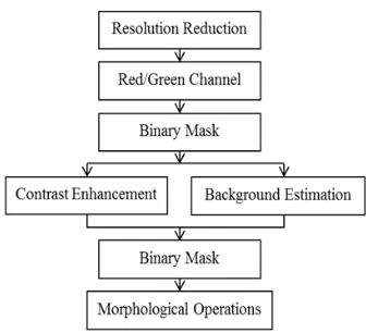
The proposed preprocessing method is summarized in Fig. 1. It consists of a sequence of steps namely, resizing for decreased computation time, intensity homogenization, shade correction and morphological processing for enhancing features. Each of these steps is detailed in the following sections.
Image acquisition: To implement the pre-processing
approach described in the following steps, digital fundus images of dimensions 2000x3008 were captured using a KOWA VX-10, non-mydriatic retinal camera with a 50º field-of-view, at Hospital Universiti Sains
Fig. 1: Overview of the pre-processing approach
Fig. 2: Fundus Image of a normal eye as provided by Hospital Universiti Sains Malaysia
Malaysia (HUSM) situated in Kelantan, Malaysia. Figure 2 shows one such retinal image.
Resolution reduction: This step was implemented for
the high-resolution fundus images collected at Hospital Universiti Sains Malaysia. The dimensionality of the data is reduced using bi-cubic interpolation reduces the dimensionality of the data which reduces processing time, while keeping the information loss to a minimum. The output pixel value, calculated by bi-cubic interpolations, is the weighted average of pixels in the nearest 4×4 neighborhood. This approach produces sharper results than other non-adaptive interpolation algorithms (Manikantan et al., 2012).
Grayscale conversion: Experiments conducted by
other researchers demonstrate that the red channel of the fundus photo is often oversaturated in the central papilla region, whereas the blue channel often has noise and the green channel has the highest degree of contrast between the background and the vasculature network (Meier et al., 2007). To maximize the benefits of both the green channel and the red channel, a hybrid red/green channel is formed using Eq. (1) as proposed by Hashim et al. (2013):
. 1 (1)
Res. J. App. Sci. Eng. Technol., 14(1): 1-6, 2017
= The intensity component = A constant
and = The red and green channels respectively
Binary mask generation: The purpose of generating a
binary mask is to increase computation speed by excluding pixels belong to the background from further processing (Hashim et al., 2013). With respect to fundus images it labels the pixels of the circular retinal fundus as Region-of-Interest (ROI), whereas the black pixels of the background are excluded (Gagnon et al., 2001; Goatman et al., 2003; Youssif et al., 2007). In this study, the binary mask was automatically generated by thresholding the red color band, followed by morphological operators-opening, closing by a disc-shaped structuring element.
CLAHE: The Contrast Limited Adaptive Histogram
Equalization (CLAHE) technique is a modification to the original histogram equalization approach. It has been used successfully to improve the contrast of medical images (Shimahara et al., 2004). It operates by partitioning the image into regions and applies the histogram equalization to each one. This, in turn, distributes the intensity values over the entre gray value range, making hidden features more visible (Garg et al., 2011). The amount of contrast enhancement is directly proportional to the gradient of the Cumulative Distribution Function (CDF). CLAHE limits contrast enhancement by limiting the gradient of the CDF by pre-determining the height of the histograms for that particular bin (Garg et al., 2011; Lal and Chandra, 2014).
Median filtering: Fundus images also display
variations in background intensity due to non-uniform illumination. With the purpose of removing these differences, the shade correction method proposed by authors (Saleh and Eswaran, 2012) is applied. A background estimate is obtained by filtering the input with a median filter kernel. Median filtering, is the where the neighboring pixels of a central pixel are ranked according to intensity and the median value becomes the new value for said pixel. These filters do a credible job of rejecting extreme intensity values and impulse noise. The difference between the input image and the background estimate is then calculated for each pixel. The shade-corrected image is finally obtained by transforming this difference into integers covering the entire range of possible gray values from 0-255 (Suero et al., 2013).
Shade correction: The background of the image as
represented by the median filtered version of the image
is subtracted from the CLAHE image to remove non-uniform illumination.
Morphological operations: Finally, consecutive
morphological operations-opening, closing and erosion where performed to obtain preliminary vessel segmentation. The morphological operations are all carried out using the same structuring element. As stated in Phyo and Khaing (2014), Radha and Lakshman (2013) and Tripathi et al. (2013) and by equation (2) below, the opening of A by B is the erosion of A by B, followed by dilation of the resultant image by B:
(2)
Equation (3) displays the closing of A by B as the dilation of A by B, followed by erosion of the resultant image by B:
(3)
RESULTS AND DISCUSSION
An overview of the resultant images at every stage of the pre-processing approach is illustrated in Fig. 3.
As can be seen the hybrid Red/Green channel has embraced the favorable properties of both parent channels. The red channel contributed to increasing the brightness of the fundus, whereas the green channel enhanced the contrast. The method to generate the binary mask proved to be appropriate for the purposes of this study. Enhancing the contrast of the foreground and subsequently subtracting the large median filter approximation of the background produced a shade-corrected image. The drawback to this method is that the size of the median filter must be user-defined for different resolutions of retinal images. Although this type of shade correction is adequate for our purposes, it shows a tendency towards artifacts. During the course of this study, it is hoped that a better and more robust representation of the background can be obtained. Finally, the shade corrected image underwent consecutive morphological operations (opening, closing and erosion) to obtain preliminary vessel segmentation results.
Figure 4 displays the histograms corresponding to various stages of the pre-processing approach.
As can be observed, the red channel and the green channel are skewed in different directions. By creating the hybrid, the histogram has the range of the red channel and the intensity of the green channel. Performing contrast limited adaptive histogram equalization spread the intensities across the entire
o
Fig. 3: Resu
Fig. 4: The
ultant image at ev
changes in the sp
very stage of the
pread of the ima
e preprocessing a
age histogram at approach
Res. J. App. Sci. Eng. Technol., 14(1): 1-6, 2017
range of the gray values. The final step maps the intensity values in the CLAHE image to new values such that 1% of the data is saturated at low and high intensities. While the CLAHE was successful in increasing contrast, the oversaturation it caused in the central parts of the fundus is undesirable. Implementing variations of adaptive histogram equalization approaches is the next step in improving results.
CONCLUSION
The aim of this study is to address the problem of low contrast and non-uniform illumination in digital fundus images. To do this, we propose an approach that performs CLAHE on a hybrid red/green channel to enhance the contrast as well as correcting illumination variations by subtracting a median filtered background representation from the enhanced image. Finally, morphological operations were conducted on the resultant image to obtain preliminary vessel segmentation results. We intend to improve our approach by using a more robust representation of the background. For validation purposes, we intend to apply our algorithm to publically available databases such as DRIVE, STARE and ARIA. We also intend to do a performance analysis using a standard evaluation metric against previous works. The findings reported in this study are part of a long-term project in which the goal is to segment and extract the retinal vasculature from fundus images and employ this information to design a reliable and robust diagnostic tool for diabetic retinopathy.
ACKNOWLEDGMENT
This research is collaborated effort between Universiti Teknologi Malaysia (UTM) and Hospital Universiti Sains Malaysia (HUSM). The authors would like to express their gratitude for the Research University Grant, vot no: 10H93 provided by the Research Management Centre of Universiti Teknologi Malaysia, Johor Bahru, Malaysia.
REFERENCES
Dehghani, A., H.A. Moghaddam and M.S. Moin, 2012. Optic disc localization in retinal images using histogram matching. EURASIP J. Image Video Process., 19: 1-11.
Gagnon, L., M. Lalonde, M. Beaulieu and M.C. Boucher, 2001. Procedure to detect anatomical structures in optical fundus images. Proseeding of Conference on Medical Imaging 2001: Image Processing, pp: 1218-1225.
Garg, R., B. Mittal and S. Garg, 2011. Histogram equalization techniques for image enhancement. IJECT, 2(1): 107-111.
Goatman, K.A., A.D. Whitwam, A. Manivannan, J.A. Olson and P.F. Sharp, 2003. Colour normalisation of retinal images. Proceeding of Medical Image Understanding and Analysis, pp: 49-52.
Hani, A.F.M. and H.A. Nugroho, 2009. Model-based retinal vasculature enhancement in digital fundus image using independent component analysis. Proceeding of IEEE Symposium on Industrial Electronics and Applications (ISIEA, 2009), 1: 160-164.
Hashim, F.A., N.M. Salem and A.F. Seddik, 2013. Preprocessing of color retinal fundus images. Proceeding of Japan-Egypt International Conference on Electronics, Communications and Computers (JEC-ECC), pp: 190-193.
Hatanaka, Y., T. Nakagawa, Y. Hayashi, T. Hara and H. Fujita, 2008. Improvement of automated detection method of hemorrhages in fundus images. Proceeding of the Annual International Conference of the IEEE Engineering in Medicine and Biology Society, pp: 5429-5432.
Lal, S. and M. Chandra, 2014. Efficient algorithm for contrast enhancement of natural images. Int. Arab J. Inform. Technol., 11(1): 95-102.
Manikantan, K., M.S. Shet, M. Patel and S. Ramachandran, 2012. DWT-based illumination normalization and feature extraction for enhanced face recognition. Int. J. Eng. Technol., 1(4): 483-504.
Meier, J., R. Bock, G. Michelson, L.G. Nyúl and J. Hornegger, 2007. Effects of preprocessing eye fundus images on appearance based glaucoma classification. Proceeding of the 12th International Conference on Computer Analysis of Images and Patterns (CAIP’07), pp: 165-172.
Murugan, R. and K. Reeba, 2012. An automatic screening method to detect optic disc in the retina. Int. J. Adv. Inform. Technol., 2(4): 23
Narasimhan, K., V.C. Neha and K. Vjayarekha, 2012. A review of automated diabetic retinopathy diagnosis from fundus image. J. Theore. Appl. Inform. Technol., 39(2): 225-238.
Phyo, O. and A. Khaing, 2014. Automatic detection of optic disc and blood vessels from retinal images using image processing techniques. Int. J. Res. Eng. Technol., 3(3): 300-307.
Ponnaiah, G.F.M. and S.S. Baboo, 2013. Automatic optic disc detection and removal of false exudates for improving retinopathy classification accuracy. Int. J. Sci. Res. Publications (IJSRP), 3(3): 1-7. Prentašić, P., S. Lončarić, Z. Vatavuk, G. Benčić, M.
Radha, R. and B. Lakshman, 2013. Retinal image analysis using morphological process and clustering. Signal Image Process. Int. J., 4(6): 55-69.
Rahim, H.A., A.S. Ibrahim, W.M.D.W. Zaki and A. Hussain, 2014. Methods to enhance digital fundus image for diabetic retinopathy detection. Proceeding of the IEEE 10th International Colloquium on Signal Processing and Its Applications (CSPA, 2014), pp: 221-224.
Saleh, M.D. and C. Eswaran, 2012. An automated blood vessel extraction algorithm in fundus images. Proceeding of the IEEE International Conference on Bioinformatics and Biomedicine, pp: 1-5. Saravanan, V., B. Venkatalakshmi and V. Rajendran,
2013. Automated red lesion detection in diabetic retinopathy. Proceeding of the IEEE Conference on Information and Communication Technologies (ICT, 2013), pp: 236-239.
Shimahara, T., T. Okatani and K. Deguchi, 2004. Contrast enhancement of fundus images using regional histograms for medical diagnosis. Proceeding of the SICE 2004 Annual Conference, 1: 650-653.
Suero, A., D. Marin, M.E. Gegundez-Arias and J.M. Bravo, 2013. Locating the optic disc in retinal images using morphological techniques. Proceeding of the IWWBBIO 2013, pp: 593-600. Sumathy, B. and S. Poornachandra, 2012. Retinal blood
vessel segmentation using morphological structuring element and entropy thresholding. Proceeding of 3rd International Conference on Computing Communication and Networking Technologies (ICCCNT, 2012), pp: 1-5.
Sun, C.C., S.J. Ruan, M.C. Shie and T.W. Pai, 2005. Dynamic contrast enhancement based on histogram specification. IEEE T. Consum. Electron., 51(4): 1300-1305.
Thakur, N. and M. Juneja, 2014. Analysis of various techniques used for optic disc and optic cup segmentation for glaucoma diagnosis. Int. J. Adv. Res. Comput. Sci. Software Eng., 4(12): 933-936. Tripathi, S., K.K. Singh, B.K. Singh and A. Mehrotra,
2013. Automatic detection of exudates in retinal fundus images using differential morphological profile. Int. J. Eng. Technol., 5(3): 2024-2029. Yi, Y. and D. Zhang, 2011. Observation model based
retinal fundus image normalization and enhancement. Proceeding of the 4th International Congress on Image and Signal Processing (CISP, 2011), 2: 719-723.
Youssif, A.A.A., A.Z. Ghalwash and A.S. Ghoneim, 2006. Comparative study of contrast enhancement and Illumination equalization methods for retinal vasculature segmentation. Proceeding of 3rd Cairo International Biomedical Engineering Conference, pp: 5.