_____________________________________________________________________________________________________
*Corresponding author: E-mail: [email protected];
(Past name: British Journal of Medicine and Medical Research, Past ISSN: 2231-0614, NLM ID: 101570965)
Age Estimation Using Digital IOPA- A Short Study
BR. Sathvikalakshmi
1*, CH. Uma Reddy
1,
R. Kirthika
1, L. Chandrashekar
1and R. Sudarshan
11
Department of Oral Medicine and Radiology, Best Dental Science College, Madurai - 625104, Tamil Nadu, India.
Authors’ contributions This work was carried out in collaboration between all authors. Author BS designed the study, performed the statistical analysis, wrote the protocol and wrote the first draft of the manuscript. Authors CUR and RS managed the analyses of the study. Author RS managed the literature searches. All authors read and approved the final manuscript.
Article Information
DOI: 10.9734/JAMMR/2017/36843
Editor(s):
(1)Emad Tawfik Mahmoud Daif, Professor, Oral & Maxillofacial Surgery, Cairo University, Egypt.
Reviewers:
(1)Rodrigo Lorenzi Poluha, State University of Maringá, Brazil. (2)Andrea Scribante, University of Pavia, Italy. Complete Peer review History:http://www.sciencedomain.org/review-history/22106
Received 19th September 2017 Accepted 12th November 2017 Published 30th November 2017
ABSTRACT
Aims and Objectives: The aim of the study is estimating the age of a person from his teeth particularly using the pulp to tooth area ratio. This ratio will be measured from the digital intraoral radiographic images taken for different patients with a sole purpose of estimation the age of the. Each patient’s maxillary central incisors will be evaluated and their pulp to tooth area ratio is taken.
Materials and Methods: The study comprises of 60 patients divided into two subsets of 10 test subjects and 50 study subjects. The RVG images of maxillary central incisors were collected and the pulp to tooth area ratio was measured using Image‑Pro Plus ІІ software (Media, Cybernetics, USA). Intraobserver variation was evaluated with paired t-test.
Results: The AR was a significant (p-value <0.001) predictor of age with a mean absolute error (MAE) of 4.908 years using the formula, Age = 89.778-379.020 (AR). This equation when applied on the test subset revealed no statistically significant difference (p = 0. 232) between estimated and chronological ages with no gender differences.
Conclusion: Considering the possible errors ±10 years were added to this method of age estimation provides a fairly accurate and reliable method.
Keywords: Pulp tooth ratio; Kvaals; dental age.
1. INTRODUCTION
Age is one of the essential factors, which play an important role in every aspect of life [1]. Person identification is an important aspect of forensic medicine and dentistry [1]. Age, gender, race, and so on is used for identification of a person. Chronological age, as recorded by registration of birth date, is referred throughout an individual’s life. Age is an important factor in clinical practice; research and court of law. Major dental clues once neglected are increasingly used to solve the crime [1]. Age is estimated on the basis of chronological age and bone age, dental age, mental age, and others [1]. Dental age is considered to be vital as tooth development shows less variability than other developmental features and also low variability in relation to chronological age [1]. Hence, dental age is considered to be vital in establishing the age of an individual. Different morphological stages of mineralization correlate with the different developmental stages [1]. RVG estimation serves quick and easy method for the study style and also fearless for the patient giving them comfort and convenience. Furthermore unlike the extra oral radiological techniques they provide a high precision image with under lower radiation doses for accurate calculation of the tooth pulp ratios.
1.1 Aims and Objectives
This study aimed at estimating the age of an individual from maxillary central incisors using the pulp to tooth area ratio (AR) measured from the digital intraoral radiographic images or radiovisuographic images (RVGs).
2. MATERIALS AND METHODS
The study comprised of 60 patients divided into two subsets of 10 test subjects and 50 study subjects. The RVG images of maxillary central incisors were acquired and the pulp to tooth area
ratio was measured using the software for RVG using Kvaals method. Intraobserver variation was evaluated with paired t-test. This was a comparative study. RVG of maxillary central incisors using paralleling technique were obtained of 60 subjects in the age group 15 to 50 years, relatively well distributed across the different age groups. Radiographs of the teeth (either left or right side) were made using the paralleling technique. The subjects were randomly divided into two subsets, the study subset (n = 50) and the test subset (n = 10) (Table 1). The study subset was used to derive a specific regression formula to calculate the age from the pulp to tooth AR and the test subset was used to test the accuracy of this formula.
Grossly destructed teeth, teeth with carious lesions or periapical lesions, teeth with any developmental anomalies that affect the structure of the tooth, malaligned or rotated teeth, and teeth with any prosthetic fittings were excluded from the study.
Table 1. Gender distribution of the study sample
Gender Male Female Total
Sample 34 16 50
Test subset 5 5 10
2.1 Age Estimation
The AR value was then used to estimate the age from an independently generated linear regression formula. The error was calculated from the estimated age and the chronological age. The absolute value of the errors, the ‘mean absolute error’ or Mean absolute error (MAE) was derived. The regression equation was applied on the test subset to test its accuracy in age prediction. The pulp area and tooth area of 25 randomly selected RVG images were measured again after 1 weeks to test for intraobserver variation.
Table 2.Mean estimated age and mean average error of the study and test subsets
Groups Total no. of patients
Mean chronological age
Mean estimated age
Mean absolute error
Study subset
50 33.584 35.432 4.908
2.2 Statistical Analysis
SPSS (Statistical Package for Social Science) V10 was used to do the statistical analysis. Regression analysis was done on the AR and a formula was derived to estimate the age. In addition to the MAE, the number/percentage of estimates with errors <10 years were also tabulated. Independent samples t-test was performed between chronological age and estimated age in the test subset. Paired t-test was done to evaluate the intra observer variation. A p-value < 0.05 was considered to be statistically significant.
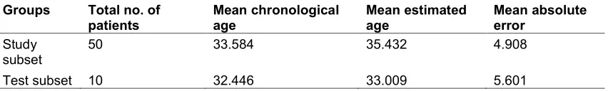
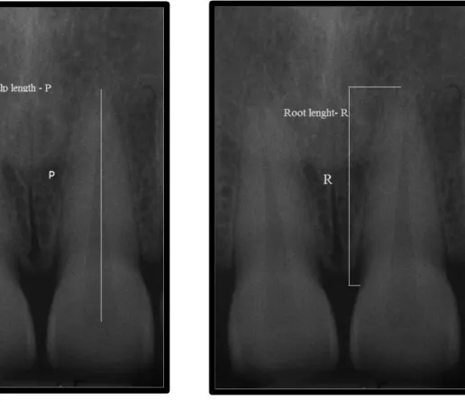
The ratios calculated were:
• P = Pulp length/root length • R = Pulp length/tooth length.
Ratios of the pulp/root width at three different levels:
• At the ECJ (A)
• At the midpoint between ECJ and mid root level (B)
• At the mid root level (C)
Pulp-to-tooth ratio method by Kvaal
• In this method, pulp-tooth ratio is calculated for six mandibular and maxillary teeth, such as maxillary central and lateral
incisors; maxillary second premolars; mandibular lateral incisor; mandibular canine; and the first premolar [2].
• The age is derived by using these pulp to tooth ratios in the formula for age determination given by Kvaal et al. [2].
Age = 129.8 – (316.4 × m) (6.8 × [W-L])
M = Mean value of all the ratios excluding T (T = tooth length)
W = Mean value of width ratios from level B and C
L = Mean value of length ratios P and R W − L = Differences between W and L
M = P + R + A + B + C/5 W = B + C/2
L = P + R/ 2
3. RESULTS
The mean age of patients included in the study is 35.432 years with a standard deviation of 1.635.
There was no significant difference between the actual age and calculated age in males (p = 0.552) and females (p = 0.701).
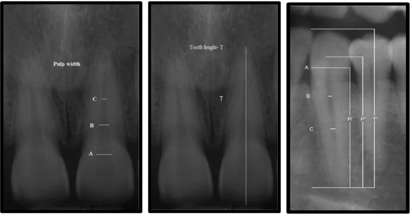
Fig. 3. Pulp width ratio Fig. 4. Total length Fig. 5. Pulp length
4. DISCUSSION
Investigators use numerous tools for forensic age estimation among which there are destructive and non-destructive techniques [3]. Non-destructive techniques provide the advantage that it can be used in living subjects as well as collected forensic specimen [3,2]. Radiographic and computerized tomography-based methods for age estimation are non-destructive tools for the forensic investigator 3. In the last decade, adult age estimation using radiographs have proved to be less precise [4]. The technique of using pulp to tooth area ratio for adult age estimation has been able to provide better accuracy than other methods. Most researchers employed the radiographs of anterior teeth, while few authors, utilized posterior teeth. Babshet et al observed that premolars were more reliable than using canines and so we have used maxillary central incisors in our study [5]. The pulp/tooth AR progressively decreased as age increased. The technological advantage that RVG offers over IOPA could be the reason why we obtained more accurate age estimations [6]. From radiographs commonly used in dentistry many evaluations and predictors have been proposed. Panoramic radiograph could assess skeletal maturation with analysis of wisdom teeth [7] or canines [8]. Moreover tele radiography could help clinicians in the evaluation of subject growth [9] and in the interception of the
radiograph showed in the present report could help clinicians with a simplified method with a lower radiation dose.
In the present study, there was no significant difference between the actual and calculated age in both males (p = 0.552) and females (p = 0.701), which was in concordance with other age estimation studies done on Indian population [11]. The paired t-test revealed that [t-value is – 0.643 and p-value is 0.40 (non-significant)] there was a significant intraobserver reliability in terms of the measurements. Repeated measurements did not affect the age estimation. The recent reports advocate constructing population specific equations to enhance age prediction. Bosmans et al. [12] Meinl et al. [13] and Singaraju et al. [14,2] have developed different regression formulae for this technique. The high correlation between the AR and the chronological age prompted us to develop a linear regression formula, specific for South Indian population.
5. CONCLUSION
pulp to tooth area ratio or pulp height to crown height ratios [2].
CONSENT
As per international standard or university standard, patient’s consent has been collected and preserved by the authors.
ETHICAL APPROVAL
It is not applicable.
COMPETING INTERESTS
Authors have declared that no competing interests exist.
REFERENCES
1. Priyadharshini, et al. Dental age estimation a method review. International Journal of Advanced Health Sciences. 2015;1:2. 2. Joseph CC, Reddy BHS, Cherian NM,
Kannan SK, George G, Jose S. Intraoral digital radiography for adult age estimation: A reliable technique. J Indian Acad Oral Med Radiol. 2013;25(4):287-290.
3. Kvaals, et al. A non destructive dental method for age estimation. Journal of Forensic Odontostomatology. 1994;12(1): 6-11.
4. Piyush, et al. Age estimation using dental radiographs. Journal of Forensic Dental Sciences. 2013;5(2):118–122.
5. Babshet M, Acharya A, Venkatesh NG. Age estimation in Indians from pulp/tooth area ratio of mandibular canines. Forensic Science International. 2010;197:125.e1-125.e4.
6. Shruthi K. Patil, et al.Estimation of age by Kvaal's technique in sample Indian population to establish the need for local Indian-based formulae. Journal of Forensic Dental Sciences. 2014;6(3):166– 170.
7. Mehta N, et al. Evaluation of skeletal maturation using mandibular third molar development in Indian adolescents. J Forensic Dent Sci. 2016;8(2):112.
8. Trakinienė G, et al. Evaluation of skeletal maturity using maxillary canine, mandibular second and third molar calcification stages. Eur J Orthod. 2016; 38(4):398-403.
9. Torres FC, et al. Evaluation of the cervical vertebrae maturation index in lateral cephalograms taken in different head positions. Braz Dent J. 2013;24(5): 462-6.
10. Scribante A, et al. Sella turcica bridging and dental anomalies: Is there an association? Int J Paediatr Dent; 2017. DOI: 10.1111/ipd.12301
11. Saxena S. Age estimation of Indian adults from orthopantomographs. Braz Oral Res. 2011;25:225-229.
12. Bosmans N, Ann P, Aly M, Willems G. The application of Kvaal’s dental age calculation technique on panoramic dental radiographs. Forensic Sci Int. 2005; 153(2-3):208-212.
13. Meinl A, Tangl S, Pernicka E, Fenes C, Watzek G. On the applicability of secondary dentin formation to radiological age estimation in young adults. Journal of Forensic Sciences. 2007;52: 438-441.
14. Singaraju S, Sharada P. Age estimation using pulp/tooth area ratio: A digital image analysis. J Forensic Dent Sci. 2009;1: 37-41.
_________________________________________________________________________________
© 2017 Sathvikalakshmi et al.; This is an Open Access article distributed under the terms of the Creative Commons Attribution License (http://creativecommons.org/licenses/by/4.0), which permits unrestricted use, distribution, and reproduction in any medium, provided the original work is properly cited.
Peer-review history: