RESIDENT & FELLOW SECTION
Clinical Reasoning: Rapid progression of
reversible cognitive impairment in an 80-year-old
man
Walid Bouthour, MD, Lukas Sveikata, MD, Maria Isabel Vargas, MD, Johannes Alexander Lobrinus, MD, and Emmanuel Carrera, MD
Neurology
®
2018;91:1109-1113. doi:10.1212/WNL.0000000000006647Correspondence Dr. Carrera emmanuel.carrera@ hcuge.ch
Section 1
An 80-year-old right-handed man was accompanied by his wife to the neurology clinic for difficulties in performing activities of daily living. He was reported to struggle using cutlery and remembering how to dial a phone number. His wife had noticed changes in his speech, which had become incomprehensible. Symptom onset was insidious, with progressive worsening over a couple of weeks. The patient himself had no complaint. His medical history included an episode of transient vertical diplopia 2 months prior to admission, diagnosed as a TIA in the midbrain. Brain MRI at the time of TIA showed no acute lesion but moderate vascular white matter disease (WMD), related to arterial hypertension and type II diabetes, and 11 lobar microbleeds (bilateral temporal, parietal, and frontal).
Neurologic examination at the time of the current admission revealed right homonymous hemianopia, production of jargon and impaired comprehension suggestive of Wernicke aphasia, ideational apraxia, executive dysfunction, and anosognosia. The rest of the physical examination was normal. Episodes of paroxysmal atrialfibrillation were detected during the patient’s hospital stay.
Questions for consideration:
1. Can you relate the clinical picture to a single lesion? 2. What is your differential diagnosis at this stage?
Section 2
Both Wernicke aphasia and ideational apraxia suggest a dys-function in the left posterior hemisphere; more specifically in temporoparietal associative cortices for the Wernicke aphasia and in the supramarginal gyrus for ideational apraxia. Right homonymous hemianopia indicates involvement of the left visual pathways. Although executive functions are widely distributed throughout the brain, executive dysfunction is classically associated with pathologies of prefrontal brain regions. Anosognosia is the result of a lesion in the right temporoparietal lobe. The symptoms and signs suggest widespread bihemispheric dysfunction predominantly affect-ing the posterior part of the left hemisphere.
In our patient, the rapidly progressive evolution of cognitive decline points towards several etiologies including vascular, infectious, and inflammatory processes.1 Vascular causes should be considered because of the history of chronic arterial hypertension, possible TIA, and WMD. However, the absence of an acute or stepwise evolution does not favor a vascular etiology. Among infectious causes, herpes simplex virus en-cephalitis was ruled out because of the subacute evolution of the disease and the absence of fever or seizures. Neurosyphilis
and HIV dementia have a more indolent evolution, and the patient had no sexual risk factors. There was no tick bite or erythema migrans history to support Lyme disease. Voltage-gated potassium channel (VGKC), NMDA receptor, and other antibody-related autoimmune encephalitis are fre-quently associated with myoclonus or seizures. Paraneoplastic autoimmune encephalitis may also cause rapidly progressive cognitive and neuropsychiatric symptoms. Other in-flammatory causes include cerebral vasculitis, which could present with subacute cognitive impairment. Inflammatory cerebral amyloid angiopathy is a rare and newly recognized entity that may cause subacute dementia in predominantly older patients. Creutzfeldt-Jacob disease can present with rapid cognitive decline, sometimes associated with visualfield defects, myoclonus, or seizures. In the absence of preexisting cognitive decline reported by the family, Alzheimer disease and frontotemporal disease were considered less likely. As for tumor-related pathologies, lymphoma typically presents with progressive cognitive decline, and brain metastases can cause rapidly progressive cognitive symptoms.
Question for consideration:
1. Which ancillary tests would you perform to make the diagnosis?
Section 3
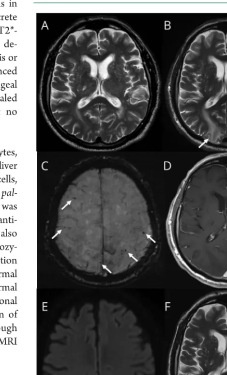
Brain MRI, FSE T2-weighted sequence, showed bilateral and asymmetric cortical and subcortical hyperintense lesions in the left parietal, temporal, and occipital lobes with a discrete mass effect on adjacent structures (figure 1). Axial T2*-weighted images showed the same lobar microbleeds as de-scribed previously. There was no superficial hemosiderosis or signs of past intracerebral hemorrhage. Gadolinium-enhanced T1-weighted sequence showed no parenchymal or meningeal enhancement. Magnetic resonance spectroscopy revealed a decrease of the N-acetylaspartate/choline ratio, but no characteristic pattern of brain tumor.
Laboratory investigations, including blood electrolytes, C-reactive protein, erythrocyte sedimentation rate, liver function, and blood cell count (particularly white blood cells, 5.9 g/L, normal 4–11), were normal. HIV, Treponema pal-lidum hemagglutination, and Lyme disease screening was negative. VGKC and NMDA antibodies, antinuclear anti-bodies, and anti-double-stranded DNA antibodies were also negative. Characterization ofAPOEgene showed a heterozy-gous APOE with e4/e3 alleles. Finally, CSF examination revealed increased protein concentration (1.45 g/L, normal <0.45), normal cell count (<1 M/L, normal 0–5), normal glucose level (4.0 mmol/L, normal 2.8–4.0), no oligoclonal bands, and no abnormal cells. The subacute evolution of the clinical picture and apraxia should prompt a thorough assessment for Creutzfeldt-Jacob disease. However, the MRI did not support this hypothesis.
Questions for consideration:
1. What is your diagnosis at this stage? 2. How would you confirm the diagnosis?
GO TO SECTION 4
Figure 1Brain MRI slices before admission, upon admis-sion, and during follow-up
Section 4
The clinical picture, imaging, CSF, and blood workup are suggestive of inflammatory cerebral amyloid angiopathy (CAA-I). The patient also carried theAPOEe4 allele, which increases the risks ofβ-amyloid peptide deposition and CAA-related hemorrhage compared to carriers of the commone3 alleles.
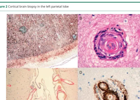
To confirm the diagnosis, we performed a brain biopsy in the left posterior parietal lobe including cortical and subcortical brain tissue. Cerebral biopsy showed transmural inflammatory infiltrates and amyloid deposition within vessels, and peri-vascular inflammatory infiltrates with reactive astrogliosis and
microglial activation (figure 2). This provides evidence for 2 variants of CAA-I. The transmural inflammatory infiltrate is a destructive vasculitic process that characterizes Aβ-related angiitis (ABRA), and perivascular inflammatory infiltrates are characteristic of CAA-related inflammation (CAA-ri).2
Both subtypes can coexist in patients with CAA-I3and respond to immunosuppressive treatment, but it is not established whether such pathologic subtypes translate into separate entities that require different therapeutic management.2
Questions for consideration:
1. Which treatment would you start?
2. How would you relate the history of transient symptoms (vertical diplopia) to the diagnosis of CAA-I?
GO TO SECTION 5
Figure 2Cortical brain biopsy in the left parietal lobe
Section 5
We initiated immunosuppressive treatment, consisting of oral prednisone, 1 mg/kg a day, tapered over 3 months, with a good initial clinical and radiologic evolution. However, 1 month after the end of immunosuppression (i.e., 4 months after diagnosis of CAA-I), the patient experienced rapid worsening of cognitive functions. MRI at the time showed the recurrence of the same initial hyperintensity at the same location, and an acute, asymptomatic diffusion-weighted imaging (DWI) punctiform ischemia in the right subcortical parietal lobe. We reinitiated oral prednisone and introduced oral azathioprine with a favor-able clinical evolution. Follow-up MRI did not show recurrence of the inflammatoryfindings of CAA, but 6 new lobar micro-bleeds were reported over the 36-month follow-up.
Regarding the episode of regressive vertical diplopia presented by our patient 2 months prior to admission, several hypotheses may exist. Transient focal neurological episodes or“amyloid spells,” defined as short, recurrent, stereotyped, spreading neurologic symptoms, are frequently reported in patients with CAA and are often related to superficial hemosiderosis or subarachnoid hemorrhage.3A TIA is also possible given the age, cardiovascular risk factors, and detection of episodes of atrial fibrillation. Interestingly, small asymptomatic DWI ischemic lesions have been described recently in patients with CAA over the course of the disease.3,4
Discussion
The diagnosis of CAA-I was established based on (1) cerebral MRI showing rapid onset leukoencephalopathy and edema; (2) CSF analysis displaying mildly elevated protein concen-tration, but no pleiocytosis; and (3) neuropathology dem-onstrating perivascular inflammation associated with amyloid deposits in cortical vessels and transmural and perivascular inflammation. Before confirmation with cerebral biopsy, our patient met the diagnostic criteria of probable CAA-I: (1) age >40 years; (2) clinical symptoms, such as headaches or cog-nitive disturbances, not directly attributable to intracerebral hemorrhage; (3) asymmetric WMD patterns that extend to the immediately subcortical area; and (4) presence of cortico–subcortical hemorrhagic lesions.5
The inflammatory variants of CAA have gained a great amount of interest lately because of their increased incidence in the elderly population. Unlike classical CAA, inflammatory variants do not usually present with intracerebral hemorrhage, and the symptoms may be reversed by immunomodulatory therapy.4However, CAA without inflammation can also ex-hibit infiltrative white matter processes.6 In order to avoid undue immunosuppressive treatment and the side effects thereof, we carried out a brain biopsy to look for vascular inflammation that characterizes CAA-I.
An interesting point raised by this case is the speed of the clinical and radiologic course. The fact that lobar microbleeds were present before the onset of CAA-I supports the pre-existence of CAA. The subsequent cognitive symptoms were more likely linked to the CAA-I, as supported by the clinical and radiologic features that progressed rapidly over 2 months. Based on repetitive MRIs and cerebral biopsyfindings, our case illustrates that the course of CAA-I may be subacute and clearly distinct from the more progressive evolution of the noninflammatory form of CAA.6
A second interesting point is the management of transient neurologic symptoms and the ischemic lesion on DWI at-tributed to CAA.7,8 Our patient had CAA, but also cardio-vascular disease risks factors (diabetes, hypertension) and episodes of atrial fibrillation. The presence of ≥5 cerebral microbleeds carries a major risk of intracerebral hemorrhage with anticoagulant use (odds ratio 5.50 or 2.8%/y).9 There-fore, we opted for a percutaneous left atrial appendage clo-sure, although it is not known if the bleeding risk is increased to the same extent in the inflammatory form of CAA.
Author contributions
Research project conception: E. Carrera. Research project execution: W. Bouthour, L. Sveikata. Writing of thefirst draft: W. Bouthour, L. Sveikata. Specialized material (radiology, pathology): M.I. Vargas, J.A. Lobrinus. Review and critique: E. Carrera, M.I. Vargas, J.A. Lobrinus.
Study funding
No targeted funding reported.
Disclosure
The authors report no disclosures relevant to the manuscript. Go to Neurology.org/N for full disclosures.
References
1. Paterson RW, Takada LT, Geschwind MD. Diagnosis and treatment of rapidly pro-gressive dementias. Neurol Clin Pract 2012;2:187–200.
2. Salvarani C, Hunder GG, Morris JM, Brown RD, Christianson T, Giannini C. Aβ -related angiitis: comparison with CAA without inflammation and primary CNS vas-culitis. Neurology 2013;81:1596–1603.
3. Charidimou A, Peeters A, Fox Z, et al. Spectrum of transient focal neurological episodes in cerebral amyloid angiopathy: multicentre magnetic resonance imaging cohort study and meta-analysis. Stroke 2012;43:2324–2330.
4. Corovic A, Kelly S, Markus HS. Cerebral amyloid angiopathy associated with
in-flammation: a systematic review of clinical and imaging features and outcome. Int J Stroke 2017;73:1–11.
5. Auriel E, Charidimou A, Gurol ME, et al. Validation of clinicoradiological criteria for the diagnosis of cerebral amyloid angiopathy–related inflammation. JAMA Neurol 2015;73:197–202.
6. Salvarani C, Morris JM, Giannini C, Brown RD Jr, Christianson T, Hunder GG. Imagingfindings of cerebral amyloid angiopathy, Aβ-related angiitis (ABRA), and cerebral amyloid angiopathy–related inflammation. Medicine 2016;95: e3613–e3617.
7. Kimberly WT, Gilson A, Rost NS, et al. Silent ischemic infarcts are associated with hemorrhage burden in cerebral amyloid angiopathy. Neurology 2009;72: 1230–1235.
8. van Veluw SJ, Lauer A, Charidimou A, et al. Evolution of DWI lesions in cerebral amyloid angiopathy. Neurology 2017;89:2136–2142.
DOI 10.1212/WNL.0000000000006647
2018;91;1109-1113
Neurology
Walid Bouthour, Lukas Sveikata, Maria Isabel Vargas, et al.
80-year-old man
Clinical Reasoning: Rapid progression of reversible cognitive impairment in an
This information is current as of December 10, 2018
Services
Updated Information &
http://n.neurology.org/content/91/24/1109.full
including high resolution figures, can be found at:
References
http://n.neurology.org/content/91/24/1109.full#ref-list-1
This article cites 9 articles, 6 of which you can access for free at:
Subspecialty Collections
roke
http://n.neurology.org/cgi/collection/other_cerebrovascular_disease__st
Other cerebrovascular disease/ Stroke
http://n.neurology.org/cgi/collection/mri
MRI
http://n.neurology.org/cgi/collection/cognitive_aging
Cognitive aging following collection(s):
This article, along with others on similar topics, appears in the
Permissions & Licensing
http://www.neurology.org/about/about_the_journal#permissions
its entirety can be found online at:
Information about reproducing this article in parts (figures,tables) or in
Reprints
http://n.neurology.org/subscribers/advertise
Information about ordering reprints can be found online:
rights reserved. Print ISSN: 0028-3878. Online ISSN: 1526-632X.
1951, it is now a weekly with 48 issues per year. Copyright © 2018 American Academy of Neurology. All ® is the official journal of the American Academy of Neurology. Published continuously since