ISSN(Online): 2319-8753 ISSN (Print): 2347-6710
International Journal of Innovative Research in Science,
Engineering and Technology
(A High Impact Factor & UGC Approved Journal) Website: www.ijirset.com
Vol. 6, Issue 9, September 2017
Accurate Detection of Abnormal Tissues of
Brain MR Images Using Hybrid Clustering
and Deep Learning Based Classifier Methods
of Image Segmentation
Shivang M. Patel1, Jyotindra N. Dharwa2
Research Scholar, Faculty of Computer Applications, Ganpat University, Kherva, Mehsana, Gujarat, India1
Associate Professor, A. M. Patel Institute of Computer Studies, Ganpat University, Kherva, Mehsana, Gujarat, India2
ABSTRACT: Image analysis by segmentation techniques has wide range of utilities. The research on image analysis includes industrial inspection, object tracking from multiple sequential images, traffic control systems, geographical classification from satellite images, geomorphological mapping at land surfaces, crop analysis in agriculture, treatment planning in medical science and many others. Segmentation of an image is very significant in medical field by improving treatment possibilities like early diagnosis of abnormalities in brain. Manual segmentation for detection of abnormal tissues from large amount of brain MR images are difficult and time-consuming task. In this research paper, brain MR images are taken due to its quality parameters for the purpose of detecting abnormalities compared to other techniques like X-Ray, CT Scan etc. This paper reviews the trade-off between various approaches for analysing the abnormalities from brain Magnetic Resonance Images using different methods of image segmentation. In addition, the proposed study has given a roadmap to improve accuracy and precision to detect the region of interest. From multiple methods of segmentation, segmentation using hybrid clustering approach including K-means and Fuzzy C-means followed by deep learning based classifier method improves the level of precision to detect abnormal tissues from brain MR images. The result by implementing the proposed approach in MATLAB environment leads to plan precise treatment for the medical practitioners.
KEYWORDS: Image Segmentation, Brain MRI, Hybrid Clustering, K-means, Fuzzy C-means, Deep Learning Classifier.
I. INTRODUCTION
ISSN(Online): 2319-8753 ISSN (Print): 2347-6710
International Journal of Innovative Research in Science,
Engineering and Technology
(A High Impact Factor & UGC Approved Journal) Website: www.ijirset.com
Vol. 6, Issue 9, September 2017
This paper presents the current status of various methods of segmentations in medical imaging applications. Current segmentation approaches are reviewed with advantages and disadvantages of various methods. It focuses mainly on comparison of various classification and clustering techniques of image segmentation on brain MRI images in terms of their accuracy and performance for detection of abnormalities based on various criteria and parameters. This research paperanalyses few methods of segmentation on different brain MRI image modalities like T1 and T2 weighted, FLAIR images of various views such as sagittal, axial etc. and compare results for accuracy. It also proposes a hybrid clustering approach followed by deep learning based classifier that improves the level of precision to detect abnormal tissues from brain MR Images. In this hybrid approach, K-means clustering is better for minimal computation time and Fuzzy C-means clustering gives notable accuracy. K-means technique is simple but suffers from incomplete detection of high damaged tissues. Fuzzy C-means technique recalls more information of original source image. The main idea behind hybrid approach is to reduce the number of iterations for initializing the correct cluster centre. Performance is assessed by comparing gathered results with ground truth normal images in terms of accuracy as well as processing time.
II. RELATEDWORK
Segmentation process splits an image into its integral regions till the interested/targeted areas have been detected. Mainly segmentation algorithms depend on similarity or discontinuity in intensity values. Similarity property deals with identification of edges while discontinuity detects regions based on a set of previously defined criteria like region growing, thresholding, region splitting and merging etc. [1][26][27]Different image segmentation approaches are applied in various extents in different fields. Early diagnosis of abnormalities in brain tissues improves treatment planning for the patient by medical practitioners. Let us discuss about medical image segmentation and related methods from the literature survey.The medical image segmentation methods available in the literature can be divided into major following categories. [1][2] Clustering, Thresholding, Classifiers, Region growing, Neural Network, Markov Random Field etc.Manual segmentation involves delineation of the boundaries of tissues manually. It is a tedious task. It requires additional care to mark regions carefully, otherwise it will generate jaggy images and may be result in poor segmentation.In semi-automatic segmentation, human interaction is least as possible. Since it involves both computer and humans’ expertise so the result depends on both as well as subject to variations in between.However, fully automated segmentation is also not without problems to retrieve specific region. In such methods, there is no intervention of human and segmented tissues. Though it involves the human intelligence and is developed with soft computing techniques, which is a difficult task. [26]
Among various methods of image segmentation, classifier and clustering based approaches have received an excessive pact of attention in medical imaging research community. Several classifier and clustering strategies have been used by researchers, each of which has its own special characteristics, merits and demerits. Recently, automatic segmentation through deep learning based technique became more effective to achieve desired results. Image classification techniques are mainly divided in two categories: supervised and unsupervised image classification techniques. [10] In supervised approach, first we need to select some pixels having same characteristics from each class to train pixels so later they can be used for identification of issues having similar properties. Some clustering techniques like log based clustering, relevance feedback, hierarchical clustering, k-means clustering, fuzzy based clustering etc. are used according to requirements. [11] In unsupervised approach, it is not required to train pixels. In cases like absence of sufficient basic information, this approach is useful. Various classifier techniques are available like minimum distance, Bayesian, maximum likelihood, convolutional neural network, decision tree etc. [8]
ISSN(Online): 2319-8753 ISSN (Print): 2347-6710
International Journal of Innovative Research in Science,
Engineering and Technology
(A High Impact Factor & UGC Approved Journal) Website: www.ijirset.com
Vol. 6, Issue 9, September 2017
from brain MR Images using different segmentation approaches. Here, few of them are applied to solve different problems by different researchers and related discussion are described briefly.
A fully automated segmentation which uses SVM classification, splits normal and abnormal tissues before they can be classified into: grey matter cell, cerebrospinal fluid, white matter cell, and tumor area. This approach is more robust and spend less time in whole process. [11]Another approach is Bayesian which is considered as supervised as well as parametric. Here, the mean is calculated using training dataset. [12] Threshold based approach uses binary image. In this method, regions are converted into various grey values. Here, intensity values are separated by anticipated classes. [13] Otsu method ensures high intensity level difference between foreground and background images. Then it converts into binary form as needless region. [14] In [15], automated segmentation method was introduced to find tumor region by more accuracy and less diagnosis time using Multi-Layer Perception Network and contrast level. Statistic features like mean, median, variance and correlation were also used. This approach uses neural network for classification. In [16], applications of intelligent computing techniques were discussed for image classification. This study focuses on AI approach to solve different issues related to various diagnostic. Different methods like SVM neural network, fuzzy C means, mammogram, hybrid approach etc. used for feature extraction are discussed in that research study.Segmentation methods for brain tissue analysis, can be separated in two categories: generative and discriminative model. Generative models depend on related prior knowledge about the presence of healthy as well as damaged tissues. These methods depend on structural models of 3D brain MR image tissues. [17][18][19] Techniques such as SVM [20][21], decision forests [22][23] and random forests [24][25] also proved accurate to get targeted regions.
III. SAMPLE SELECTION PROCEDURE,SAMPLE SIZE AND TOOLS USED TO STUDY
This research study considers 16 normal and 56 abnormal standard size (128 X 128 pixels or 512 X 512 pixels) brain MR images like T1, T2, FLAIR (Fluid Attenuation Inversion Recovery) etc. obtained from various imaging centres and few samples from authenticated online sources like brainweb and other databases. Different views of same images are used for better results. Experiments by different techniques are applied on multiple same images, so comparison will become easier and accurate.This research uses MATLAB tool (R2017a trial version 9.2.0.538062) for performing various operations on brain MR images. For more specific processing according to various parameters on images, it is necessary to use special plug-in that is used for clinical data analysis. Here, for pre-processing stage, ITK-SNAP (Insight Segmentation and Registration Toolkit, version 3.6.0) tool is used to open individual images as well as sequences/series stored in different file formats. Medical images obtained from hospital or imaging centres are in .dcm file format and they can be observed here by DICOM (Digital Imaging and Communications in Machine) viewer tool.
IV. PROPOSED FRAMEWORK
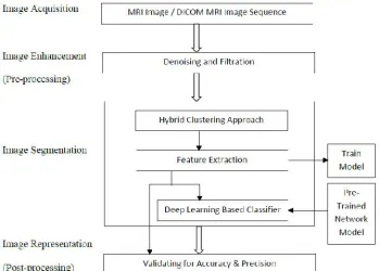
Here this analytical study uses the facts/information already available and analyse these for identification and detection of abnormal tissues in brain MR images. This research paper uses statistical data obtained by implementing proposed hybrid approach and evaluate the accuracy of the results. After applying hybrid clustering and before post-processing, obtained images are entered for further processing using deep learning based classifier. One can also use pre-trained network model of brain MRI images for experiment and create N number of hidden layers as per the diagnostic requirement. Generally, at initial stages, two hidden layers should be considered. Each layer filters data and give to next subsequent layer for further processing. In this process, the input image or sequences are compared with pre-trained labelled dataset to get more specific results. The resulted outcome will give more statistics about the images that will be more helpful in preliminary diagnoses.The entire procedure is presented by proposed below framework.
1.Pre-processing
ISSN(Online): 2319-8753 ISSN (Print): 2347-6710
International Journal of Innovative Research in Science,
Engineering and Technology
(A High Impact Factor & UGC Approved Journal) Website: www.ijirset.com
Vol. 6, Issue 9, September 2017
the image sharpness of observed MR images. As per experimental requirement, the process of removing background like remove skull, scalp, eyes or any other non-interested area is also initiated using brain surface extractor algorithm. It will decrease the processing time.
Fig. 1 Proposed framework to detect abnormal tissues from brain MR images
2.Clustering
After removing the noise, feed that image to proposed technique.
ـ Initialize number of clusters, maximum iterations and termination parameters. Initial number of clusters are not chosen at random so it saves time and effort by reducing number of iterations.
ـ Calculate cluster centre by initial mean = (1:k)*m/(k+1) where k=no. of clusters & m=maximum no. of image count + 1
ـ Find minimum distance between point and cluster centre to allot each point to nearest cluster centre. ـ Then recalculate the cluster centres and repeat this step until some convergence criteria is met.
ـ Some points are scattered and they are far away from any cluster centre. For such points calculate new values of cluster, distance and mean values.
ـ There is no inference between points exists in clusters by reclustering process.
ـ Record output as clustering image, execution time and number of iterations as well as degree of membership by putting each point together which belong to closest cluster.
3.Feature Extraction
After applying the proposed approach, the resulting image is continued to process for feature extraction to get interest area. We can use Thresholding and Contouring function to get region of interest with proper boundary tracking. Then image is entered into binarization process to observe accurate results.
4.An intermediate stage before validating image - Deep Learning based Classifier
ISSN(Online): 2319-8753 ISSN (Print): 2347-6710
International Journal of Innovative Research in Science,
Engineering and Technology
(A High Impact Factor & UGC Approved Journal) Website: www.ijirset.com
Vol. 6, Issue 9, September 2017
deep learning without a huge data set and long computation and training time. Using MATLAB and ‘nntool’ – Neural Network Toolbox, one can train the required convolutional neural network from scratch or use a pretrained model to perform transfer learning. [Source: mathworkhelp][29]
V. PERFORMANCE MEASURES
Two performance measures are used for measuring performance: i) find sensitivity, specificity and accuracy ii) find dice similarity index. For this research study, we have used clustering and classification approach with various parameters. Classification results are not without any error. It may be possible that experimental study may fail to identify the abnormal tissues or may identify the abnormal tissues which are not actually present. This fact may be described by finding the values of true and false positive and true and false negative. [32][33]
Table 1: Condition of classification results and description Condition Description
TP Classification result is positive i.e. presence of abnormality (abnormal tissues are correctly detected) TN Classification result is negative i.e. absence of abnormality (normal tissues are not correctly detected) FP Classification result is positive in absence of abnormality (normal tissues are incorrectly detected as
abnormal tissues)
FN Classification result is negative in presence of abnormality (abnormal tissues are not detected) We can find the performance of applied classifier using sensitivity, specificity and accuracy measures. [35]
Table 2: Performance Matrix
Sensitivity = TP / (TP + FN) * 100 %…….………...eq. (1) Specificity = TN / (TN + FP) * 100 %….………...eq. (2) Accuracy = (TP + TN) / (TP + TN + FP + FN) * 100 %..…eq. (3)
Based on above criteria, we can compare various algorithms by finding FPR, TPR and precisions. Better performance is indicated if the values of accuracy and sensitivity are higher and the value of specificity is lower.
Other measures for testing accuracy and precision of acquired result are Dice and Jaccard similarity index. For each abnormal tissue region, P1 represents the segmented area by the proposed method, and T1 is the actual area in the ground truth. Then, dice and Jaccard index functions [30][31] are written as,
Dice Index (P, T) = (P1.T1) / ( (P1+T1) / 2 ) ………eq. (4) Jaccard Index (P, T) = (P1.T1) / [P1+T1-(P1.T1)] ………eq. (5)
VI. EXPERIMENTALSTUDY
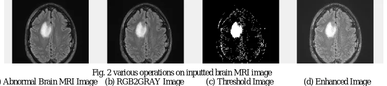
For this experimental study, 16 normal and 56 abnormal brain MR images are considered. Brain MR Image with abnormal tissues of 512 x 512 pixel dimension is taken as input. The next step is preprocessing. In which, operations like removal of noise, skull etc. is done. Then filters like median filter, morphological filter are applied for edge preservation. Following figures demonstratethese steps:
The next stage is to perform various morphological operations on obtained images from previous stage. Performance Measure Normal Abnormal
Normal TN FP
Abnormal FN TP
Fig. 2 various operations on inputted brain MRI image
ISSN(Online): 2319-8753 ISSN (Print): 2347-6710
International Journal of Innovative Research in Science,
Engineering and Technology
(A High Impact Factor & UGC Approved Journal) Website: www.ijirset.com
Vol. 6, Issue 9, September 2017
As we have discussed in proposed framework, after applying hybrid clustering approach the next step is to input acquired images and results to train network model using ‘nntool’ available in MATLAB environment. Here, this is optional as one can use already pretrained network of same domain for further analysis. Following figures show the wizard for how to input data images and phases of processes like training, validating and testing.
VII. RESULTANALYSIS
Following results in forms of various tables and charts depict and prove thatthe proposed approach for segmentation is more precise compared to other methods.
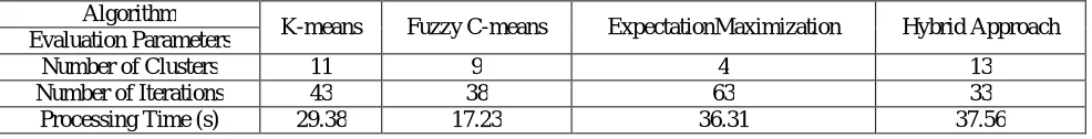
Table 3: Various Parametric Statistics for different segmentation approaches for Image Dataset-I Algorithm
K-means Fuzzy C-means ExpectationMaximization Hybrid Approach Evaluation Parameters
Number of Clusters 11 9 4 13
Number of Iterations 43 38 63 33
Processing Time (s) 29.38 17.23 36.31 37.56
Chart 1: Execution Time vs. various Segmentation methods Fig. 3 Various statistics of inputted abnormal brain MRI image
(a) Detection of abnormal tissues after morphological operations (b) Findings of segmented image after hybrid clustering
ISSN(Online): 2319-8753 ISSN (Print): 2347-6710
International Journal of Innovative Research in Science,
Engineering and Technology
(A High Impact Factor & UGC Approved Journal) Website: www.ijirset.com
Vol. 6, Issue 9, September 2017
Table 4: Comparison of accuracies (Confusion Matrix) by different approaches for Image Dataset-I
Evaluation Parameters
Segmentation Approach
Clustering Approach Classification Approach Hybrid Approach
K-means
Fuzzy C-means
Expectation
Maximization k-NN
Decision
Tree SVM Proposed
TP 36 41 24 39 41 45 47
TN 10 12 14 12 13 16 18
FP 17 14 23 16 14 7 5
FN 9 5 11 5 4 4 2
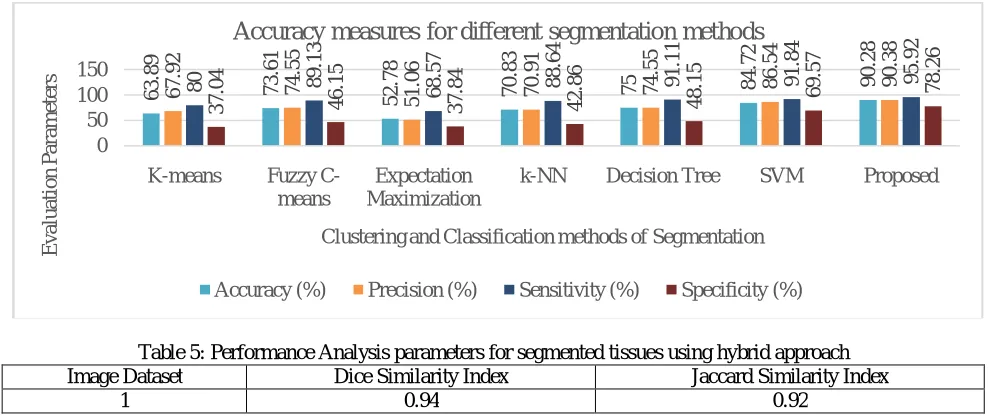
Accuracy (%) 63.89 73.61 52.78 70.83 75.00 84.72 90.28
Precision (%) 67.92 74.55 51.06 70.91 74.55 86.54 90.38
Sensitivity (%) 80.00 89.13 68.57 88.64 91.11 91.84 95.92
Specificity (%) 37.04 46.15 37.84 42.86 48.15 69.57 78.26
Chart 2: Accuracy of proposed framework vs. existing systems
Table 5: Performance Analysis parameters for segmented tissues using hybrid approach
Image Dataset Dice Similarity Index Jaccard Similarity Index
1 0.94 0.92
11 43 9 38 4 17
63 36
29.38 17.23 36.31 39.56
0 50 100
K-means Fuzzy C-means Expectation
Maximization Hybrid Approach E v a lua ti o n P a ra m et er s Segmentation algorithms
Evaluation Parameters for various Segmentation algorithms
Number of Clusters Number of Iterations Processing Time (s)
63. 89 73. 61 52. 78 70. 83 75 84. 72 90. 28 67. 92 74. 55 51. 06 70. 91 74. 55 86. 54 90. 38 80 89. 13 68. 57 88. 64 91. 11 91. 84 95. 92 37. 04 46. 15 37. 84 42. 86 48. 15 69. 57 78. 26 0 50 100 150
K-means Fuzzy
C-means
Expectation Maximization
k-NN Decision Tree SVM Proposed
E v a lua ti o n P a ra m et er s
Clustering and Classification methods of Segmentation
Accuracy measures for different segmentation methods
ISSN(Online): 2319-8753 ISSN (Print): 2347-6710
International Journal of Innovative Research in Science,
Engineering and Technology
(A High Impact Factor & UGC Approved Journal) Website: www.ijirset.com
Vol. 6, Issue 9, September 2017
VIII. CONCLUSION
From this research study, we conclude that the proposed framework for detecting abnormal tissues from brain MRI images is more effective and efficient compare to other similar methods of clustering and classification of segmentation.Proposed segmentation method has achieved Dice coefficient of 94%, Jaccard coefficient of 92%, Accuracy of 90.28% and Precision of 90.38%.The proposed approach performs feature extraction and classification better than other similar segmentation methods. Moreover, the application of the proposed approach can be extended to a varying range of tumor and other abnormalities. It can also be applied on larger dataset of MR images having different aspects to get more realistic and more clinically bounded outcomes.
REFERENCES
[1] Pal NR, Pal SK; “A review on image segmentation techniques, pattern recognition”, 1993; 26(9):1277-1294 http://www.sciencedirect.com/science/ article/pii/003132039390135J
[2] Pham DL, Xu C, Prince JL; “A survey of current methods in medical image segmentation”, Annual Review of Biomedical Engineering, 2000; 2:315338 http://www.tecn.upf.es/~afrangi/ibi/Phan2000.pdf
[3] RajeshwarDass, Priyanka, Swapna Devi; “Image Segmentation Techniques”, International Journal of Electronics & Communication Technology, ISSN 2230-7109 (online), ISSN 2230-9543 (print) vol. 3, Issue I, Jan-March 2012
[4] SpringerLink Multimodality State-of-the-Art medical image segmentation and registration methodologies vol. II ISBN: 978-1-4419-8203-2 (Eds.) A.S. El-Baz; R. Acharya U; A.F. Laine; J.S. Suri
[5] S. Thilagamani and N. Shanthi; “A Survey on Image Segmentation Through Clustering”, International Journal of Research and Reviews in Information Sciences vol. 1, No. 1, March 2011
[6] Ruchi D. Deshmukh and Chaya Jadhav, “Study of Different Brain Tumor MRI Image Segmentation Techniques”, International Journal of Computer Science Engineering and Technology (IJCSET), ISSN: 2231-0711, Vol 4, Issue 4, 133-136, April 2014
[7] J.R. Parker, “Algorithms for Image Processing and Computer Vision”, Second Edition, Wiley Publishing, Inc. ISBN: 978-0-470-64385-3 [8] Sunayana G Domadia, Dr. Tanish Zaveri; “Comparative Analysis of Unsupervised and Supervised Image Classification Techniques”, National
Conference on Recent Trends in Engineering & Technology, 13-14 May 2011
[9] SpringerLink Multimodality State-of-the-Art medical image segmentation and registration methodologies vol. II ISBN: 978-1-4419-8203-2 (Eds.) A.S. El-Baz; R. Acharya U; A.F. Laine; J.S. Suri
[10] S. Thilagamani and N. Shanthi; “A Survey on Image Segmentation Through Clustering”, International Journal of Research and Reviews in Information Sciences vol. 1, No. 1, March 2011
[11] P. John, “Brain Tumour Classification Using Wavelet and Texture Based Neural Network” International Journal of Scientific & Engineering Research”, Vol. 3, Issue 10, pp. 1-7,2012.
[12] S. Jain, “Brain Cancer Classification Using GLCM Based Feature Extraction in Artificial Neural Network” International Journal of Computer Science & Engineering Technology Vol. 4, Issue 7, pp. 966-970, 2013.
[13] R. C. Gonzalez and R. E. Woods, Digital Image Processing, Prentice Hall, New jersy, 2002.
[14] N.Zulpe and V.Pawar, “GLCM Textural Features for Brain Tumor Classification”, International Journal of Computer Science Issues, Vol. 9, Issue 3, pp. 354-359, 2012.
[15] Aqhsa Q. Syed1, K. Narayanan, “Detection of Tumour in MRI Images Using Artificial Neural Networks”, International Journal of Advanced Research in Electrical, Electronics and Instrumentation Engineering, Vol. 3, Issue 9, September 2014.
[16] S.N. Deepa and B. Aruna Devi,” A survey on artificial intelligence approaches for medical image classification”, Indian Journal of Science and Technology, Vol. 4 No. 11 (Nov 2011) ISSN: 0974- 6846
[17] Farahani, K., Menze, B., Reyes, M., 2013-14. Multimodal Brain Tumor Segmentation (BRATS 2013-14).
[18] Bauer, S., Wiest, R., Nolte, L., Reyes, M., 2013. A survey of mri-based medical image analysis for brain tumor studies. Phys. Med. Biol. 58, 97–129.
[19] Doyle, S., Vasseur, F., Dojat, M., Forbes, F., 2013. Fully automatic brain tumor segmentation from multiple mr sequences using hidden markov fields and variationalem. Proc. BRATS-MICCAI.
[20] Bauer, S., Nolte, L.-P., Reyes, M., 2011. Fully automatic segmentation of brain tumor images using support vector machine classification in combination with hierarchical conditional random field regularization. In: MICCAI, Vol. 6893, pp. 354–361.
[21] Bauer, S., Wiest, R., Nolte, L., Reyes, M., 2013. A survey of mri-based medical image analysis for brain tumor studies. Phys. Med. Biol. 58, 97–129.
[22] Zikic, D., Glocker, B., Konukoglu, E., Criminisi, A., Demiralp, C., Shotton, J., Thomas, O., Das, T., Jena, R., Price, S., 2012. Decision forests for tissue-specific segmentation of high-grade gliomas in multi-channel mr. In: Medical Image Computing and Computer-Assisted Intervention–MICCAI 2012. Springer, pp. 369–376.
[23] Zikic, D., Ioannou, Y., Brown, M., Criminisi, A., 2014. Segmentation of brain tumor tissues with convolutional neural networks. Proc. BRATS-MICCAI
[24] Gotz, M., Weber, C., Blocher, J., Stieltjes, B., Meinzer, H.-P., Maier-Hein, K., 2014. Extremely randomized trees based brain tumor segmentation. In: in Proceedings of BRATS Challenge- MICCAI.
ISSN(Online): 2319-8753 ISSN (Print): 2347-6710
International Journal of Innovative Research in Science,
Engineering and Technology
(A High Impact Factor & UGC Approved Journal) Website: www.ijirset.com
Vol. 6, Issue 9, September 2017
[26] Ali, Cem, Melike, “Review of MRI-based brain tumor image segmentation using deep learning methods”, Procedia Computer Science 102 (2016) 317-324, ScienceDirect, Elsevier
[27] Ruchi D. Deshmukh and Chaya Jadhav, “Study of Different Brain Tumor MRI Image Segmentation Techniques”, International Journal of Computer Science Engineering and Technology (IJCSET), ISSN: 2231-0711, Vol 4, Issue 4, 133-136, April 2014
[28] TranosZuva, Oludayo O. Olugbara, Sunday O. Ojo and Seleman M. Ngwira, “Image Segmentation, Available Techniques, Developments and Open Issues”, Canadian Journal on Image Processing and Computer Vision Vol. 2, No. 3, March 2011
[29] Deng, L.; Yu, D. (2014). "Deep Learning: Methods and Applications". Foundations and Trends in Signal Processing. 7 (3–4): 1– 199. doi:10.1561/2000000039
[30] Sorensen, T. (1948). "A method of establishing groups of equal amplitude in plant sociology based on similarity of species and its application to analyses of the vegetation on Danish commons". Kongelige Danske VidenskabernesSelskab. 5 (4): 1–34.
[31] Dice, Lee R. (1945). "Measures of the Amount of Ecologic Association Between Species". Ecology. 26 (3): 297– 302. JSTOR 1932409. doi:10.2307/1932409.
[32] Vipul Singh, “Digital Image Processing with MATLAB and LabVIEW, Elsevier 2013 [33] S. Jayraman, S. Esakkirajan, T. Veerakumal DIP, TMH, 2009
[34] Monica Subashini.M et al. “Brain MR Image Segmentation for Tumor Detection using Artificial Neural Networks”, International Journal of Engineering and Technology (IJET): ISSN: 0975-4024 Vol 5 No 2 Apr-May 2013