Western University Western University
Scholarship@Western
Scholarship@Western
Electronic Thesis and Dissertation Repository
8-6-2014 12:00 AM
Evaluating Human Performance for Image-Guided Surgical Tasks
Evaluating Human Performance for Image-Guided Surgical Tasks
Matthew Kenneth Kramers
The University of Western Ontario
Supervisor Dr. Roy Eagleson
The University of Western Ontario Joint Supervisor Dr. Aaron Fenster
The University of Western Ontario
Graduate Program in Biomedical Engineering
A thesis submitted in partial fulfillment of the requirements for the degree in Master of Engineering Science
© Matthew Kenneth Kramers 2014
Follow this and additional works at: https://ir.lib.uwo.ca/etd
Part of the Bioimaging and Biomedical Optics Commons, Biomedical Commons, Biomedical Devices and Instrumentation Commons, Diagnosis Commons, Medical Education Commons, and the Surgical Procedures, Operative Commons
Recommended Citation Recommended Citation
Kramers, Matthew Kenneth, "Evaluating Human Performance for Image-Guided Surgical Tasks" (2014). Electronic Thesis and Dissertation Repository. 2342.
https://ir.lib.uwo.ca/etd/2342
This Dissertation/Thesis is brought to you for free and open access by Scholarship@Western. It has been accepted for inclusion in Electronic Thesis and Dissertation Repository by an authorized administrator of
EVALUATING HUMAN PERFORMANCE FOR IMAGE-GUIDED SURGICAL TASKS
(Thesis Format: Integrated Article)
by
Matthew Kramers
Graduate Program in Biomedical Engineering
A thesis submitted in partial fulfillment of the requirements for the degree of
Master of Engineering Sciences
The School of Graduate and Postdoctoral Studies The University of Western Ontario
London, Ontario, Canada
Abstract
The following work focuses on the objective evaluation of human performance for two different interventional tasks; targeted prostate biopsy tasks using a tracked biopsy device, and external ventricular drain placement tasks using a mobile-based augmented reality device for visualization and guidance. In both tasks, a human performance methodology was utilized which respects the trade-off between speed and accuracy for users conducting a series of targeting tasks using each device. This work outlines the development and application of performance evaluation methods using these devices, as well as details regarding the implementation of the mobile AR application. It was determined that the Fitts’ Law methodology can be applied for evaluation of tasks performed in each surgical scenario, and was sensitive to differentiate performance across a range which spanned experienced and novice users. This methodology is valuable for future development of training modules for these and other medical devices, and can provide details about the underlying characteristics of the devices, and how they can be optimized with respect to human performance.
Keywords
Co-Authorship Statement
The following thesis is presented in an integrated article format containing chapters based on the following works that are either published or in
preparation for submission:
Chapter 2: Kramers, M., de Ribaupierre, S., Fenster, A. Eagleson, R.:
Evaluating Human Performance for Needle Guidance Tasks Using a Prostate Biopsy Device. (In preparation for submission)
My contributions towards this chapter include the development of the
modified targeted biopsy user interface that enabled the collection of human performance data from the device originally developed in Aaron Fenster’s lab. Aaron Fenster provided me with access to the device used in this study, as well as the assistance via support staff for troubleshooting when problems were encountered. With the guidance of Sandrine de Ribaupierre, Roy Eagleson, and Aaron Fenster, I was able to create and conduct a user
experiment in order to collect the necessary data for evaluation. I performed all of the evaluation of performance including the statistical analysis and result validation.
Chapter 3: Kramers, M., Armstrong, R., Bakhshmand, S.M., Fenster, A., de Ribaupierre, S., Eagleson, R.: A Mobile Augmented Reality Application for Image Guidance of Neurosurgical Interventions. American Journal of Biomedical Engineering 2013, 3(6): 169-174
My contributions towards this chapter include the development of the augmented reality application on the mobile platform. Ryan Armstrong provided the details and implementation of the image processing components of the study, allowing for the anatomical surface models to be constructed from CT images and used in the application. Saeed Bakhshmand provided guidance and inspiration for the implementation of the application by
Sandrine de Ribaupierre provided the original concept and motivation of the mobile application to potentially assist neurosurgeons. Roy Eagleson
provided motivation and guidance with implementation details and contributed feedback during the design process. Aaron Fenster also contributed motivation and guidance for this work.
Chapter 4: Kramers, M, Armstrong R., Bakhshmand, S.M., Fenster A., de Ribaupierre S., Eagleson R.: Evaluation of a mobile augmented reality application for image guidance of neurosurgical interventions. Studies in Health Technology and Informatics. 2014; 196:204-8.
My contributions towards this work include the development and
implementation of the mobile application. Ryan Armstrong contributed the image processing related components necessary to carry out this work. Saeed Bahkshman provided assistance with constructing and running the
experimental setup and data collection. Aaron Fenster provided support and guidance throughout this work. Sandrine de Ribaupierre provided the
Acknowledgments
I would like to thank my supervisors whom both provided the direction and continuous support for this work: Dr. Roy Eagleson who was always available to provide his valuable insight and clever ideas for development and
implementation during my final undergraduate project all the way up to the end of my Master’s degree, and Dr. Aaron Fenster for his unwavering support and direction throughout my time as a summer research assistant and
graduate student. I would like to thank Dr. Sandrine de Ribaupierre for her invaluable clinical and neurosurgical knowledge and expertise and for providing valuable motivation to ensure my work was practical for use in a clinical environment.
I would like to thank all members of both Dr. Eagleson’s lab and Dr. Fenster’s lab for welcoming me as a colleague and providing me with invaluable experience and knowledge, both academically and practically. I thank Lori Gardi for contributing her time and extensive knowledge of software development as well as her experience with medical device development. I thank Dr. David Tessier for his technical support and research lab experience. I thank Jeremey Cepek for providing me with his engineering experience and wisdom as a fellow graduate student. I would also like to thank Kevin Barker for his extensive knowledge and skills in manufacturing, and for always being willing to help out. I would also like to thank Ryan Armstrong for creating great discussions about software, which was very valuable to my work as a graduate student. I would like to recognize several other members of the lab and other colleagues for their valuable discussions, support, and contributions to my work: Jing Jin, Saeed
Edirisinghe, Dr. Derek Cool, Kimberly Booth, Janette Wallace, Debbie Lillie, Christine Ellwood, as well as any other members that helped me during my graduate studies.
I would like to thank my examination committee for their invaluable time spent reviewing and evaluating my work: Dr. Terry Peters, Dr. Aaron Ward, and Dr. Luiz Capretz – I really appreciate your help.
Finally, I would like to thank the many people outside of the lab that contributed support and friendship throughout my time as a graduate student. My loving girlfriend Ashley D’Andrea who always provides support and keeps me positive. My parents, Ken and Lynda, who have forever
supported me and have shaped me into the person I am today. My brother Jordan who has always served as a role model for achieving success and happiness. And of course my friends with whom I’ve shared countless good times: Jacob Hoefnagels, Josh Douwes, Kyle Rosso, Christian Moulton, Tyler Savoie, Scott Haig, Mike Byers, Justin Beland, Rachel Farkas, Peter Martin, Kathryn Manning, and everyone else – you know who you are.
I would also like to acknowledge the sources of funding including the NSERC CREATE Computer-Assisted Medical Interventions Scholarship, and
Table of Contents
Abstract ... ii
Co-Authorship Statement ... iii
Acknowledgments ... v
Table of Contents ... vii
List of Tables ... x
List of Figures ... xi
List of Appendices ... xiii
1. Introduction... 1
1.1 Prostate Cancer: Disease and Diagnosis Techniques ... 1
1.1.1 Diagnosis ... 2
1.2 External Ventricular Drain Insertion † ... 9
1.3 Augmented Reality ... 10
1.3.1 Tracking Techniques ... 11
1.3.2 Display Platforms and Devices ... 13
1.3.3 Visualization Techniques ... 15
1.3.4 Augmented Reality in Medicine and Surgery ... 17
1.4 Human Performance: Theory, Application ... 19
1.4.1 Fitts’ Law ... 19
1.4.2 Application ... 22
1.4.3 Additional Performance Evaluation Methods ... 23
References ... 24
2. Evaluating Human Performance for Needle Guidance Tasks Using a Prostate Biopsy Device† ... 33
2.1 Introduction ... 33
2.2 Modeling Image-Guided Human Performance ... 35
2.3 Methods ... 37
2.3.1 Prostate Biopsy Device ... 37
2.3.3 Participants and Experimental Procedure ... 40
2.3.4 Hypothesis ... 41
2.4 Results ... 42
2.5 Discussion ... 45
2.6 Conclusion ... 47
References ... 48
3. A Mobile Augmented Reality Application for Image Guidance of Neurosurgical Interventions† ... 50
3.1 Related Work ... 50
3.2 Methods ... 52
3.2.1 Vuforia and Augmented Reality Implementation ... 52
3.2.2 Segmentation and Registration of Patient Data ... 55
3.2.3 User Controlled Registration Correction ... 59
3.2.4 Evaluation of System Accuracy ... 60
3.3 Results ... 61
3.4 Discussion ... 61
3.5 Conclusion ... 62
3.6 Acknowledgment ... 62
References ... 62
4. Evaluation of a Mobile Augmented Reality Application for Image Guidance of Neurosurgical Interventions† ... 65
4.1 Introduction ... 65
4.2 Methods ... 65
4.3 Evaluation ... 68
4.4 Results ... 69
4.5 Conclusion ... 70
References ... 71
5. Conclusion ... 73
Appendix A: Additional Experiments... 1
Appendix C: Supplementary Data ... 8
Appendix D: Supplementary Images ... 10
Appendix E: Permissions ... 12
List of Tables
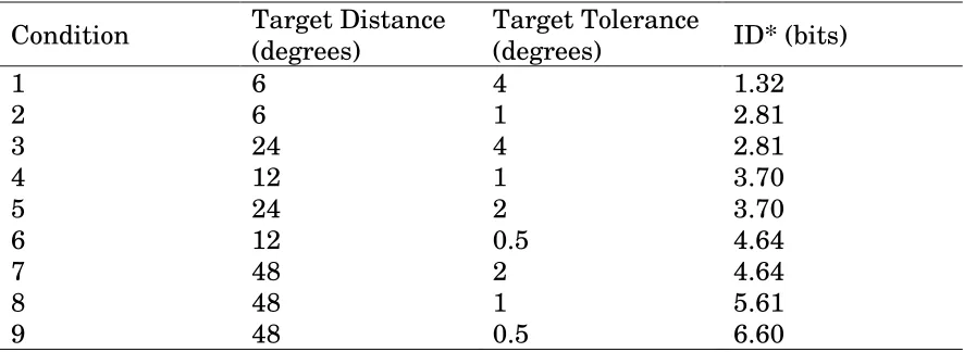
Table 2-1: Target Distance and Tolerance Configuration Parameters ... 39
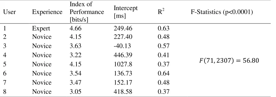
Table 2-2: Performance results for all users without adjustments to accuracy ... 43
Table 2-3: Performance results for all users with accuracy adjustments applied ... 44
Table 2-4: Sum of variance vs. target distance ... 47
Table 3-1: Targeting error measured as the Euclidean distance between targeted corner location and actual corner location ... 61
Table 4-1: Average Intra-User Deviation from Target [mm] ... 69
Table 4-2: Average Inter-User Deviation from Target [mm] ... 70
List of Figures
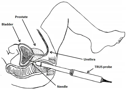
Figure 2-1: Biopsy procedure schematic with TRUS probe inserted into the
patient’s rectum along with needle aligned in parallel to the ultrasound
image plane ... 34
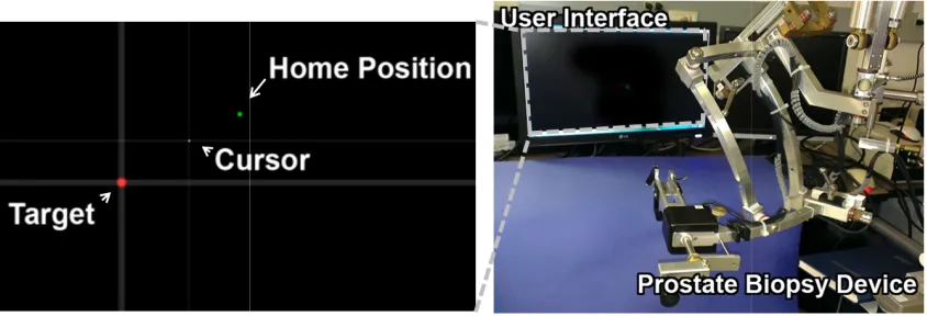
Figure 2-2: User Interface for visualization of biopsy needle and targets and
Prostate Biopsy Device for performing targeting tasks ... 38
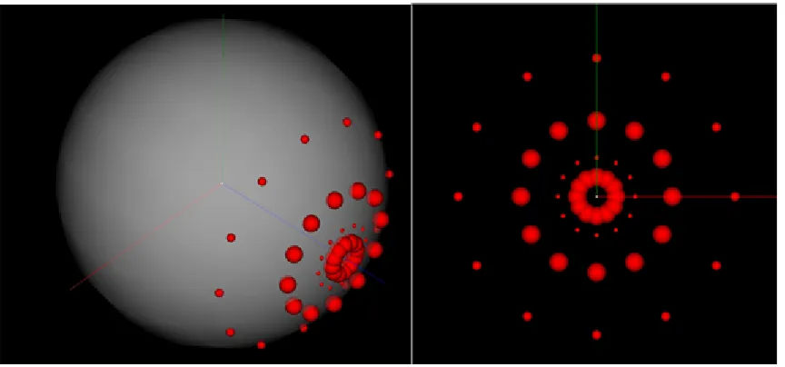
Figure 2-3: Isometric view of all target configurations and approach angles
distributed on a sphere (left) and from users’ viewpoint along the z-axis ... 40
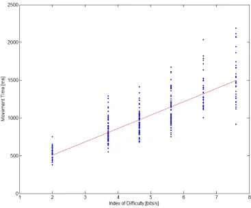
Figure 2-4: Fitts’ Law profile and regression line for a single user for a total
of 216 task configurations ... 43
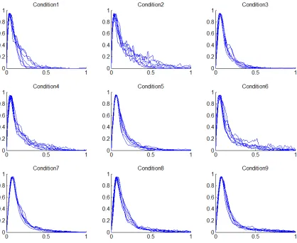
Figure 2-5: Tangential velocity profiles for all users averaged and normalized
for each target condition. ... 45
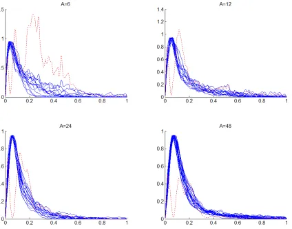
Figure 2-6: Tangential velocity profiles sorted by target distance (solid) and
variance of each trajectory for all users (dotted) ... 47
Figure 3-1: Tracking marker fixed to safety glasses for patient head pose
estimation and registration of anatomy to scene. ... 54
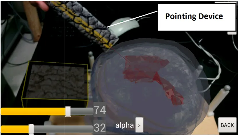
Figure 3-2: The tracked pointing device provides visual feedback for planning
entry point locations and trajectories. The device also allows for additional
interaction between the user and the AR scene. ... 55
Figure 3-3: Anatomy segmented from preoperative images aligns with the
physical tracker and is positioned using the nasion as a positional landmark.
Figure 3-4: Users have access to multiple sliders and buttons to manually
adjust the virtual models to achieve appropriate alignment of anatomy, as
well as visual settings that aid in guidance. ... 59
Figure 4-1: Glasses with attached tracking marker ... 66
List of Appendices
Appendix A: Additional Experiments ... 1
Appendix B: Software Design and Architecture ... 4
Appendix C: Supplementary Data ... 8
Appendix D: Supplementary Images ... 10
Chapter 1
1.
Introduction
This work focuses on the objective evaluation of human performance for two specific surgical tasks. The first task involves a device that is used for
performing targeted TRUS-guided biopsy of the prostate. Due to high prevalence of prostate cancer in men, the targeted biopsy procedure is
becoming an increasingly desirable method of diagnosing cancer in men. For this reason, we proposed a method for the objective evaluation of users using a particular targeted biopsy device. The results obtained in this work can be used towards further optimization of the device towards more efficient user performance. The second surgical task focuses on the neurosurgical procedure of placing an external ventricular drain. This procedure is often performed free-hand and by novice surgeons, and is therefore susceptible to human targeting errors and consequential complications to the patient. The following work highlights the development of a mobile-based augmented reality device that enables users to visualize and perceive the target location within the lateral ventricles of the brain by providing the view of pre-operative 3D scans overlaid in Augmented and Virtual Reality presentation to overlay in the context of the view of the patient’s head. In addition to a description of this development, human performance data was also collected for users operating the device and performing target and trajectory estimation tasks. The
following section outlines relevant literature in prostate cancer diagnosis, the external ventricular drain insertions procedure, augmented reality with a focus on surgical applications, as well as human performance evaluation
1.1
Prostate Cancer: Disease and Diagnosis Techniques
year 2013 [1]. This equates to an approximate probability of 14.3% of men, or 1 in 7, to develop prostate cancer in his lifetime. It is estimated that a total of 3,900 men will die as a result of prostate cancer in the year 2013 [2]. In
addition to premature death as a result of this disease, there are significant impacts upon the health-related quality of life (HRQOL). In a study
comparing the impact on HRQOL for men before and after prostate cancer diagnosis with a control group, Reeve et al. found that there was a significant decline in both the physical and mental health of a patient within the first 6 months of diagnosis compared to the control group [3]. In addition, certain social aspects of the patient’s life were noted to also suffer as a result of diagnosis. Although this particular study was limited to patients above the age of 65 years, it is reasonable to assume that prostate cancer diagnosis has a significant impact on younger men [4]. In addition, many of the therapies that exist to treat prostate cancer have an increased likelihood of increasing the chance for complications that can result in urinary, bowel and sexual health issues [5]. This due to the close proximity of the patient’s rectum, bladder, urethra, and neurovascular bundles with the prostate. One
particular instance of such treatment is the radical prostatectomy procedure which is often detrimental to urinary function [6].
1.1.1
Diagnosis
There are four methods that are most commonly used to diagnose prostate cancer: prostate specific antigen (PSA) test, digital rectal examination (DRE), imaging, and prostate biopsy. The following section describes each of the stated diagnostic methods for detecting and characterizing prostate cancer in men, with a focus on targeted biopsy.
1.1.1.1
Prostate Specific Antigen Test
correlation between the level of PSA in a patient’s blood and the stage of the cancer. In addition, a relationship between the PSA levels and the estimated volume of the tumour was also determined and reported by [8]. The increase in PSA levels detected in the bloodstream has been attributed to the
disturbance of the prostate glandular structure due to the presence of cancer [9]. Although this discovery was a significant step towards more effective prostate cancer diagnosis, there is much debate over the value of such screening methods, since it often results in overdiagnosis, and consequently overtreatment of the disease, which may be undesirable for men with slowly progressing cancer.
1.1.1.2
Digital Rectal Examination
The digital rectal examination (DRE) is a method for cancer detection where a physician uses a finger to palpate the prostate through the patient’s
rectum. This diagnostic examination is sensitive to cancer contained in the peripheral zone (PZ) of the prostate, as this area is located adjacent to the rectal wall. The physician attempts to detect hardened areas of the prostate tissue which may indicate the presence of cancer, often indicated by
1.1.1.3
Prostate Imaging
The following section provides a brief overview of the most commonly used and state-of-the-art prostate cancer imaging modalities. Although there are many modalities used in relation to prostate cancer, including computed tomography (CT), positron emission tomography (PET), and others, this section is limited to ultrasound (US) and magnetic resonance (MR) imaging techniques. The former modalities (PET, CT, etc.) are typically used in the evaluation of metastases and/or lymph node analysis, and therefore will not be discussed in the following section.
1.1.1.3.1
Ultrasound
Ultrasound provides many advantages over MR imaging for prostate cancer imaging. Ultrasound provides real-time image sequences, allowing it to be useful intra-operatively during procedures such as prostate biopsy or brachytherapy [10, 11]. Furthermore, ultrasound has been used to obtain prostate volume information, which is necessary for determining PSA density [10]. In addition, the low cost of ultrasound devices allow for greater access in many hospitals over MR imaging, which is both expensive and physically large.
Although ultrasound provides some clear advantages, it is not without its trade-offs. For instance, ultrasound is typically only sensitive to large, higher grade tumour volumes as noted by [12]. In addition, ultrasound is not
typically sensitive to tumors located in the transition zone of the prostate, which can contain around 20% of prostate cancer [13].
1.1.1.3.2
Magnetic Resonance Imaging
to be possible from the improvement in MR performance with combined modern multiparametric MRI (mpMRI) techniques, such as T1- and T2-weighted images, dynamic contrast, diffusion weighting, and proton spectroscopy [14, 15]. Despite this fact, there is much debate in the professional community over the accuracy and clinical usefulness of such techniques stemming from the variability in quality and methodology of reported studies in the field [16]. To address these issues, Dickinson et al conducted a meeting with 16 European prostate cancer experts to determine a set of recommendations towards a standardized method for interpreting and reporting of prostate mpMRI for prostate cancer detection and
localization [17]. Despite the variability present in the literature, the use of MRI in the clinical setting is certainly promising, especially for determining optimal targets for targeted biopsy in men whom are suspected to have cancer [18].
1.1.1.4
Biopsy
A biopsy of the prostate involves the insertion of specialized needles into the prostate to obtain small tissue samples of the prostate tissue for pathological analysis. The collected samples, also called biopsy cores, are then prepared for further examination under a microscope by a pathologist. A pathologist observes the appearance and structure of the glands and the cells that compose them, and is able to determine if the tissue is cancerous as well as grade the aggressiveness of the identified cancer. The Gleason grading system is used to rate the cancer in all obtained biopsy cores. Although this grading system has become a significant tool for the estimation of the
1.1.1.4.1
Transrectal Ultrasound (TRUS)-Guided Biopsy
The transrectal ultrasound-guided biopsy (TRUS-GB) procedure is currently the most commonly used method for obtaining biopsy cores for the purpose of diagnosing prostate cancer in men. In this approach, the patient is positioned in a lateral decubitus position (lying down on the side) and an ultrasound probe is inserted through the patient’s rectum allowing the physician to visualize the prostate and biopsy needles via ultrasound through the rectal wall. The needles are directed through a needle guide that is attached to the ultrasound probe, such that they can be visualized on an external display. The physician directs these needles to regions within the prostate that are known to have a high incidence of developing prostate cancer and proceeds to collect multiple biopsy cores. Approximately 75% of prostate cancers are found in the peripheral zone (PZ) and as a result, the initial round of biopsies is typically directed at this region. Recently the standard number of biopsy cores collected during a procedure increased from 6 (sextant) to 10-12. This increase is attributed to the result demonstrated in [21] that reveals that in up to 1 in 3 cases the sextant biopsy configuration will underestimate the reported Gleason grade. Although the increased number of biopsy cores collected increases the likelihood of detecting cancer, it also increases the potential symptoms occurring after biopsy procedure, including urinary retention, sepsis and dysuria [22]. Furthermore, the anterior, midline and apex regions of the prostate are difficult to perform a biopsy using the TRUS-GB approach due to their physical locations with respect to surrounding anatomy [23]. The TRUS-GB procedure has potential for delivering an accurate diagnosis of prostate cancer, and recent developments in imaging have gained the targeted biopsy method attention among physicians.
analysis. The repeated biopsy not only increases the patient’s discomfort and the potential for undesirable side-effects, but it has been shown that the repeat biopsy is no more sensitive to detecting cancer than the initial biopsy [24]. Although the physician attempts to obtain biopsy cores from areas that were not sampled in the initial or previous biopsy, there is no guarantee that regions are not resampled. Both groups in [24, 25]showed that the detection rate of a repeated biopsy after an negative first biopsy was in the range of 10-20%, indicating that repeat biopsies have poor sensitivity for cancer
detection in certain patients.
1.1.1.4.2
Targeted Biopsy
To accommodate some of the sensitivity issues with TRUS-GB procedures, there has been growing interest in incorporating magnetic resonance images (MRI) to provide specific targets for biopsy, as well as using such images for detection, localization and grading of the cancer [26]. By incorporating the location of suspicious areas into the biopsy procedure, and ensuring that those areas are sampled, the probability of detecting cancer potentially increases and the number of subsequent biopsies decreases, resulting in a decrease of the accompanying side-effects of the biopsy procedure. By
consequence, the accuracy of the Gleason score obtained from the biopsy can be therefore increased by using targeted biopsy [23]. This becomes important when determining therapeutic treatment options, since each treatment method has different associated side effects [21].
The targeted biopsy method has been evaluated with respect to diagnostic outcomes and clinical utility. Two studies determined that that targets
for detecting cancer using standard systematic biopsy, and a 48% probability for detecting cancer using the target biopsy approach [23]. It was also
determined that 30% of targeted biopsy cores contained cancer, while only 7% did while using the non-targeted approach. Therefore it can be concluded that less biopsy cores are required to detect cancer using the targeted biopsy
method.
1.1.1.4.3
Targeted Biopsy Devices
As a result of the increasing interest in targeted biopsy of the prostate, several groups have developed systems that promote improved diagnosis of cancer through the inclusion of techniques such as tracked US and in-bore MR guided devices. Bax et al. [24] developed a passive mechanical device that utilized existing 2D ultrasound equipment used for biopsy to allow physicians to perform biopsies at predetermine locations with high accuracy. This
1.2
External Ventricular Drain Insertion
†Placing an external ventricular drain (EVD) is a fundamental neurosurgical procedure performed to treat acute hydrocephalus – a condition characterized by an accumulation of cerebrospinal fluid within the ventricular system, either due to obstruction or by a problem of reabsorption [31, 32]. The procedure consists of drilling a burr-hole in the skull, followed by a bling placement of an external ventricular drain using external landmarks for guidance. This procedure allows drainage of cerebrospinal fluid to relieve intracranial pressure. While most neurosurgical interventions are usually performed in an operating room (OR) while the patient is under sedation [33], this is rarely the case for this procedure. Since the insertion of an EVD is usually performed on critically ill patients (either for acute hydrocephalus or after a trauma), the predominant difficulty involves transportation of the patient [34] mostly due to life-support equipment [35]. To accommodate such a scenario, manual operation of mobile drills for burr-hole trephine can be performed within the Intensive Care Unit or in the Emergency room. While advantageous in avoiding the difficulties in relocation to the OR, this
technique precludes the use of certain immobile equipment present within the OR.
While external ventricular drain placements are among the most common and basic of neurosurgical procedures, they are generally performed free-hand, relying on surface landmarks on the patient as well as the spatial reasoning of the operating surgeon to determine optimal trajectory of tools within the complex workspace. While it might be relatively easy to target large ventricles placed in a normal anatomical position, most patients will
†.Kramers, M., Armstrong, R., Bakhshmand, S.M., Fenster, A., de Ribaupierre, S., Eagleson, R., A Mobile Augmented Reality Application for Image Guidance of Neurosurgical
have small ventricles with some anatomical variation, possibly displaced by lesions occurring after the trauma. As a result of navigational error due to free-hand placement, a number of complications can occur, including malposition, non-function, infection and haemorrhage [36]. While
neurosurgeons generally consider the manual procedure to be safe, a number of studies have identified the technique as suboptimal [37-39]. In addition, this procedure is often performed by junior residents who are on-call. Indeed, many of these complications are a result navigational error, often requiring repositioning of the EVD into the ventricular system. In addition to the complications resulting from such misplacements, the procedure time is increased and additional, un-necessary tissue damage occurs.
1.3
Augmented Reality
Augmented reality (AR) is a form of virtual reality that combines the
physical, real-world environment with computer generated elements, such as video and graphics. In a definition provided by Azuma et al. [40], AR is
described as system that can perform the following tasks:
- Combines real and virtual imagery
- Be capable of interaction in real-time
- Be able to perform 3D registration of virtual objects within the real-world scene
The following section discusses different devices and methods for providing users with an immersive AR experience, followed by an overview of several tracking techniques employed in AR. Finally, several existing mobile AR devices aimed towards assisting surgeons will be reviewed.
1.3.1
Tracking Techniques
In a review performed by Zhou et al., it was determined that research directed towards tracking technology in AR was the most popular topic for participants of several AR focused conferences over the last decade [41]. The following section outlines the three main categories of tracking technologies, and their application and feasibility in a surgical environment.
1.3.1.1
Sensor-Based Tracking Techniques
components when magnetic tracking was used [43]. Since many elements of the surgical environment may contain these materials, this type of tracking may be difficult to accommodate. In the case of mechanically tracked AR system, such as the first head-mounted display (HMD) AR device by Sutherland et al. [44], user interaction may be restricted due to the fixed nature of the device. As a result, this may lead to limited mobility of the surgeon, which is disadvantageous.
1.3.1.2
Vision-Based Tracking Techniques
The use of image processing based methods to extract camera pose information was noted as being the most active area within AR tracking research [41]. This type of tracking can be divided into two categories: feature based and model based [45]. Feature based tracking uses the minimization of the distance between features extracted from a 2-D image and projected 3-D object features to determine the camera pose [46]. Some particular feature-based tracking methods use methods such as Harris feature detection along with the random sample consensus (RANSAC) algorithm to match the detected features with a known configuration [47]. In recent work by Pilet, the tracking non-rigid (non-planar) surface markers were achieved using advanced featuring detection and matching techniques In addition, there is a growing trend towards using marker-less tracking by detecting natural features within the scene, such as lines and edges [48-51]. Methods that employ marker-less tracking offer many advantages, despite the associated computational cost. One particular method of natural feature detection, present by Park et al. demonstrated the tracking robustness of initially registering the AR device with known features, and then constantly
researchers have further explored natural feature tracking methods by incorporating gradient and texture information observed in the scene [53].
1.3.1.3
Hybrid Tracking Techniques
There are some specific instances where features of each of the above methods of tracking are desirable, and therefore some researchers have explored the application of a combination of sensor- and vision-based techniques to achieve AR tracking. For example, vision-based tracking has low jitter and no drift but requires extensive computation and therefore can be slower than some of the sensor-based approaches. In addition, quick movements of the camera may result in a disruption in the AR scene since the vision-based algorithms are not robust to such drastic changes in its input. Inertial tracking can act to compliment vision-based techniques, by providing motion prediction information to the tracking algorithms and therefore strengthen the overall accuracy of the system [54, 55]. It has been reported that the hybrid tracking approach is the most favorable method of providing tracking information to the AR scene [56].
1.3.2
Display Platforms and Devices
With respect to augmented reality in surgery, there are three primary categories of display methods that have been adopted: Video see-through displays, optical see-through displays and projective displays [57]. Each of these methods uses one of following display device types to present virtual objects to the user: Head worn displays, hand-held displays and spatial displays. Each of these device types and display methods is discussed in the following section.
1.3.2.1
Video See-Through Displays
the most inexpensive solution for providing users with AR, and in turn often tends to be the easiest to implement and most readily available. One
particular advantage of video see-through displays over alternative
technologies is that the brightness and contrast of the virtual objects can be adjusted such that the objects blend into the scene more naturally [58]. One drawback of this type of display is that it is limited by the screen’s resolution, which for head-mounted displays can be low. In addition, some
implementations provide users with a limited field of view, although this can be addressed by simply increasing the size of the display, if possible.
Depending on the position of the display relative to the user, especially in head-mounted setups, video see-through displays may cause disorientation for the user caused by the eye-offset, which requires the user to adjust their perception of the scene [59]. Another disadvantage of head-mounted video see-through displays is that, depending on the ocular configuration, it may cause the user to experience eye strain and fatigue, which may constrain the effectiveness of AR [60].
1.3.2.2
Optical See-Through Displays
The optical see-through approach involves combining computer graphics with the actual view of the real world. In this approach, a special mirror is
constructed that allows virtual objects to be projected upon the user’s field of view, and may be worn by surgeon [61] or can be fixed upon a patient's bed between the surgeon and the patient [62]. Optical see-through head-mounted displays provide users with a lesser sense of immersion within the
difficulties portraying occlusion, although some researchers have found ways around this [63]. Despite these disadvantages, the optical see-through
method permit more control of the surgeon in an emergency situation, as they have direct view of the patient and operating site and have a higher chance of recognizing misalignment between the scene and the augmented elements [64].
1.3.2.3
Projective Displays
The final primary display technology involves the use of projectors to project virtual objects onto target surfaces. This method is advantageous since it does not require any additional eye wear for the user. In addition, projection alleviates the need for the user to focus their eyes, since the virtual objects do not appear directly in front of their eyes, like in head mounted approaches. This method does, however, require additional input devices to allow for user interaction and registration within the augmented scene. In addition,
projectors require recalibration for every instance that the target moves, which may be difficult to achieve in dynamic settings. Fortunately, there are methods for automated projector calibration that have the potential to
alleviate this issue [65].
1.3.3
Visualization Techniques
Visualization systems are essential to image guided medical interventions. Generally, such systems are used to incorporate preoperative information into the surgery. The data used is usually derived from volumetric medical images, such as CT and MR scans, although other preoperative information can also be used, such as surgeon-defined navigational paths and
annotations. The overall goal of such a system is to provide the operating surgeon with additional information to better perform a task. In many
when the view of target anatomical features is obstructed. With an
augmented reality environment, such internal information can be overlaid onto the patient from the perspective of the operating surgeon, providing context for the task that is otherwise unavailable. While clinicians are familiar with slice-based, two-dimensional views of the imaging data, the spatial reasoning required in mentally reconstructing the scene using these techniques increases cognitive load, which may slow the task and inject error [66]. For these reasons, Traub et al. proposed a hybrid navigation interface that combines the both slice-based approach with the in-situ augmented visualization [67]. By subjecting different surgeons with varying experience levels to each of the different combinations of slice-based and volume
rendering visualization strategies, a comparison of task completion time and accuracy was made. It was found that overall performance was largely
determined by the individual surgeon’s experience with each visualization method. However, it was shown that the hybrid visualization approach improved the surgeon’s accuracy when compared to in-situ visualization methods, and also improved speed when compared to standard, slice-based visualization. Therefore, it is important to consider the method of
visualization within the augmented reality scene and measure surgeon performance to find the optimal means of visualization.
Additionally, specifying the required transformations to fine-tune the algorithm may be a complex task with a steep learning curve.
Isosurface rendering is technically easier to achieve, but requires
significantly more effort within the pre-operative workflow. Features of interest must be segmented from the medical volumes and then represented geometrically. Geometric representations predominantly involve surface meshes composed of a number of vertices in space that define triangles to cover the surface of the structure. While the intensity information is lost from the original image, this approach allows for accurate surface representation without a heavy computational load – ideal for platforms with limited computational capability.
1.3.4
Augmented Reality in Medicine and Surgery
software interfaces, localization and tracking devices, integration of real-time data, and most importantly, judgment and clinical experience, contribute to the variability and probability for error and must be carefully addressed when designing a surgical AR system [70].
Several groups have recently been working towards bringing augmented reality into the field of medicine and image-guided surgery. One particular group explored the application of AR to improve upon the traditional methods of viewing medical images, by developing an interactive display device that uses a Time-Of-Flight camera to provide AR tracking via surface matching algorithms [71]. By aiming the device at a patient, medical images were overlaid such that the user was able to interpret the images with additional spatial context relative to the patient. In work completed by Müller et al., an augmented reality mobile device was evaluated for use with assisting
has an excellent potential to assist both trainee and experienced surgeons to perform tasks more efficiently.
1.4
Human Performance: Theory, Application
The objective evaluation of human performance is important for enabling researchers to quantify the ability of a user to perform motor control tasks while using a particular device. Since many, if not all, modern surgical
procedures require a surgeon to perform a series of tasks using human motor control, the application of objective evaluation methods is essential in order to understand and identify the underlying characteristics of a device that enables a user to perform optimally. Although there have been many
proposed methods of human performance evaluation, our work focuses on the application and various extension of the Fitts’ Law methodology, which involves the use of a human performance model that enables the
quantification of human pointing task performance. The following section discusses the Fitts’ Law methodology and the various extensions that have been developed to allow for the evaluation of tasks of different nature. Several different applications of this particular model are discussed, with a focus on surgical task performance evaluation. In addition, other performance evaluation methods are discussed.
1.4.1
Fitts’ Law
In 1954, researcher Paul Fitts demonstrated that human performance for simple motor control tasks could be modeled using a theory adapted from the information science field [81]. This model identifies a relationship between speed and accuracy that can evaluated by subjecting a user to a series of targeting tasks, each with varying difficulty. In order to quantify task
(1)
where ID represents index of difficulty in units of bits/response, W represents the tolerance range of the target (target width) and D represents the distance or amplitude of the target from the user’s starting position to the centre of the target [81]. This representation was derived in a manner analogous to Shannon’s theorem (for the information capacity of a communication channel) by Fitts with the assumption that the logarithm of the signal-to-noise (SNR) ratio corresponds to the resolution of a signal into quanta that have bands of tolerance [81-83]. Many researchers have developed and applied different variations for the formulation of index of difficulty. Welford’s variation has been adopted by many researchers as it often results in more optimal results.
0.5 (2)
By comparing this with Shannon’s theorem, the SNR is equivalent to the ratio of target amplitude to target width in Fitts’ original equation, and since Fitts’ original experiments had D:W ratios as low as 1:1, it is noted that it may be more favorable to use the direct analogy to Shannon’s theorem as follows:
1 (3)
note is that these logarithmic expressions all provide relative measures which allow subsequent measures to be compared objectively.
Upon selecting the task difficulty model, it must be ensured that an adequate range of task difficulties are selected in order to fully evaluate a user’s
performance for a range of conditions. MacKenzie et al. suggest that a range of 2-8 bits shall be selected in each Fitts’ Law related experiment, and that the user preforms each task difficulty condition around 15-25 times so that a performance tendency can be observed[85]. During each task, the movement time of each task is recorded such that the following linear trade-off of speed and accuracy can be determined as follows:
· (4)
where MT represents observed movement time, ID represents index of difficulty as discussed above, and C1 and C2 represent experimentally
determined parameters calculated by a linear regression of the collected data. It can be noted that by rearranging the linear equation in (4), the value of 1 2⁄ can be interpreted as the index of performance, or IP,measured in bits per second (bps).
To further improve the correlation between speed and accuracy, [82] suggest that adjustments for accuracy can be applied to the observed task completion times in order to fully represent a user’s “actual” performance. By analyzing the collected end-point data, an effective target width, denoted We, can be determined as:
4.133 (5)
where σ represents the standard deviation of measured end-point positions
calculating the mean movement distance between a user’s starting and end-point position independently for each user under each condition. Using both adjusted values to calculate an accuracy adjusted index of difficulty as follows:
1 (6)
It should be additionally noted, that the above models for index of difficulty were originally developed for discrete translational tasks, however the model has been shown to be provide similar results for rotational tasks, where instead of translational units of D and W, angular units can be substituted [86].
1.4.2
Application
The predictive abilities of the Fitts’ Law methodology are often used in graphical user interface (GUI) design, where it is useful to predict the
has also been used to explore the interaction of users with an augmented reality mobile application for selecting buildings in an urban area, as shown by Rohs et al. [90]. In addition, Fitts’ Law has been used in the field of laparoscopic surgery, cardiovascular surgery, as well as neurosurgery to assess the surgical skill of surgeons performing tasks with respect to varying magnification, task amplitude, and approach angle [91-94].
1.4.3
Additional Performance Evaluation Methods
Although Fitts’ Law is capable of accurately describing the trade-off between speed and accuracy for human motor control tasks, other methods of
evaluating human performance in surgery have been developed and applied in human performance evaluation research. For instance, there has been a great deal of interest in the objective evaluation of laparoscopic surgical skill since it is relatively new field of surgery and requires a high degree of skill compared to traditional open-adnominal surgery [95, 96]. This is due to reduced instrument maneuverability, impaired vision of the surgical field, and decreased tactile sensation [97]. In the past, techniques such as the Halstedian Technique had expert surgeons evaluate novice surgeons based on witnessed performance, however it has been noted that this method is prone to variation in how performance is rated due to subjectivity [98]. In contrast, objective evaluation techniques were introduced by Martin et al. that compared the difference in global ratings forms, operation-specific checklists and pass/fail judgments for the evaluation of surgeons working both on live animals, as well as simulators. This study determined that global ratings forms were capable of discriminating between levels of residents, while the checklists and pass/fail judgments were not [99]. Alternative methods were proposed through the development Imperial College Surgical Assessment Device, which uses video recording and motion analysis software to further assess performance in the following areas: compact spatial
orientation, and ambidexterity [100]. Furthermore, techniques involving the application of algorithms such as Hidden Markov models have been applied to hand movements in laparoscopic surgery in order to compare the general behavior of a novice with an expert surgeon model [101, 102]. Although these findings indicate potentially viable methods for evaluating human
performance, the Fitts’ Law methodology remains as one of most widely used methods for evaluating motor task performance from the standpoint of speed and accuracy, which are arguably the two task metrics that form a basis for all motor control tasks. Since the following work focuses on the use and evaluation of novel devices for performing interventional tasks via discrete translational and rotational movements, we primarily utilized the Fitts’ Law methodology for our evaluation.
References
1. R. Siegel, D. Naishadham and A. Jemal, "Cancer statistics, 2013," CA: Cancer J. Clin. 63, 11-30 (2013).
2. Canadian Cancer Society’s Advisory Committee on Cancer Statistics, "Canadian Cancer Statistics 2013", Toronto, ON: Canadian Cancer Society; 2013
3. B. B. Reeve, A. M. Stover, R. E. Jensen, R. C. Chen, K. L. Taylor, S. B. Clauser, S. P. Collins and A. L. Potosky, "Impact of diagnosis and treatment of clinically localized prostate cancer on health‐related quality of life for older Americans," Cancer 118, 5679-5687 (2012).
4. A. L. Potosky, J. Legler, P. C. Albertsen, J. L. Stanford, F. D. Gilliland, A. S.
Hamilton, J. W. Eley, R. A. Stephenson and L. C. Harlan, "Health outcomes after prostatectomy or radiotherapy for prostate cancer: results from the Prostate Cancer Outcomes Study," Journal of the National Cancer Institute 92, 1582-1592 (2000).
5. D. T. Eton and S. J. Lepore, "Prostate cancer and health‐related quality of life: a review of the literature," Psycho‐Oncology 11, 307-326 (2002).
7. T. A. Stamey, N. Yang, A. R. Hay, J. E. McNeal, F. S. Freiha and E. Redwine,
"Prostate-specific antigen as a serum marker for adenocarcinoma of the prostate," New England Journal of Medicine 317, 909-916 (1987).
8. A. R. Rao, H. G. Motiwala and O. Karim, "The discovery of prostate‐specific antigen," BJU international 101, 5-10 (2008).
9. J. I. Izawa, L. Klotz, D. R. Siemens, W. Kassouf, A. So, J. Jordan, M. Chetner and A. E. Iansavichene, "Prostate cancer screening: Canadian guidelines 2011," Canadian Urological Association Journal 5, 235 (2011).
10. H. Hricak, P. L. Choyke, S. C. Eberhardt, S. A. Leibel and P. T. Scardino, "Imaging Prostate Cancer: A Multidisciplinary Perspective 1," Radiology 243, 28-53 (2007).
11. S. Ghai and A. Toi, "Role of transrectal ultrasonography in prostate cancer," Radiologic Clinics of North America 50, 1061-1073 (2012).
12. E. K. Outwater and J. L. Montilla-Soler, "Imaging of Prostate Carcinoma," Cancer Control 20, 161-176 (2013).
13. R. Clements, "Ultrasonography of prostate cancer," European radiology 11, 2119-2125 (2001).
14. A. P. Kirkham, M. Emberton and C. Allen, "How good is MRI at detecting and characterising cancer within the prostate?," European urology 50, 1163-1175 (2006).
15. A. Villers, L. Lemaitre, J. Haffner and P. Puech, "Current status of MRI for the diagnosis, staging and prognosis of prostate cancer: implications for focal therapy and active surveillance," Current opinion in urology 19, 274-282 (2009).
16. G. J. Kelloff, P. Choyke and D. S. Coffey, "Challenges in clinical prostate cancer: role of imaging," AJR. American journal of roentgenology 192, 1455 (2009).
17. L. Dickinson, H. U. Ahmed, C. Allen, J. O. Barentsz, B. Carey, J. J. Futterer, S. W. Heijmink, P. J. Hoskin, A. Kirkham and A. R. Padhani, "Magnetic resonance imaging for the detection, localisation, and characterisation of prostate cancer: recommendations from a European consensus meeting," European urology 59, 477-494 (2011).
18. Y. Mazaheri, A. Shukla-Dave, A. Muellner and H. Hricak, "MR imaging of the prostate in clinical practice," Magnetic Resonance Materials in Physics, Biology and Medicine 21, 379-392 (2008).
20. G. L. Andriole, "Pathology: the lottery of conventional prostate biopsy," Nature Reviews Urology 6, 188-189 (2009).
21. C. R. King, J. E. McNeal, H. Gill and J. C. Presti Jr, "Extended prostate biopsy scheme improves reliability of Gleason grading: implications for radiotherapy patients," International Journal of Radiation Oncology* Biology* Physics 59, 386-391 (2004).
22. U. Patel and D. Rickards, Handbook of transrectal ultrasound and biopsy of the prostate. (Informa Health Care, 2002).
23. C. M. Moore, N. L. Robertson, N. Arsanious, T. Middleton, A. Villers, L. Klotz, S. S. Taneja and M. Emberton, "Image-guided prostate biopsy using magnetic
resonance imaging–derived targets: a systematic review," European urology 63, 125-140 (2013).
24. J. Bax, D. Cool, L. Gardi, K. Knight, D. Smith, J. Montreuil, S. Sherebrin, C.
Romagnoli and A. Fenster, "Mechanically assisted 3D ultrasound guided prostate biopsy system," Medical physics 35, 5397-5410 (2008).
25. D. Keetch, W. Catalona and D. Smith, "Serial prostatic biopsies in men with persistently elevated serum prostate specific antigen values," The Journal of urology 151, 1571-1574 (1994).
26. J. V. Hegde, R. V. Mulkern, L. P. Panych, F. M. Fennessy, A. Fedorov, S. E. Maier and C. Tempany, "Multiparametric MRI of prostate cancer: An update on state‐of‐the‐art techniques and their performance in detecting and localizing prostate cancer," Journal of Magnetic Resonance Imaging 37, 1035-1054 (2013).
27. J. Haffner, L. Lemaitre, P. Puech, G. P. Haber, X. Leroy, J. S. Jones and A. Villers, "Role of magnetic resonance imaging before initial biopsy: comparison of magnetic resonance imaging‐targeted and systematic biopsy for significant prostate cancer detection," BJU international 108, E171-E178 (2011).
28. B. K. Park, J. W. Park, S. Y. Park, C. K. Kim, H. M. Lee, S. S. Jeon, S. I. Seo, B. C. Jeong and H. Y. Choi, "Prospective evaluation of 3-T MRI performed before initial transrectal ultrasound–guided prostate biopsy in patients with high prostate-specific antigen and no previous biopsy," American Journal of Roentgenology 197, W876-W881 (2011).
29. B. A. Hadaschik, T. H. Kuru, C. Tulea, P. Rieker, I. V. Popeneciu, T. Simpfendörfer, J. Huber, P. Zogal, D. Teber and S. Pahernik, "A novel stereotactic prostate biopsy system integrating pre-interventional magnetic resonance imaging and live ultrasound fusion," The Journal of urology 186, 2214-2220 (2011).
office based magnetic resonance ultrasound fusion device," The Journal of urology 189, 86-92 (2013).
31. J. A. Kusske, P. T. Turner, G. A. Ojemann and A. B. Harris, "Ventriculostomy for the treatment of acute hydrocephalus following subarachnoid hemorrhage," Journal of neurosurgery 38, 591-595 (1973).
32. S. A. Mayer and F. Rincon, "Treatment of intracerebral haemorrhage," The Lancet Neurology 4, 662-672 (2005).
33. P. Schödel, M. Proescholdt, O.-W. Ullrich, A. Brawanski and K.-M. Schebesch, "An outcome analysis of two different procedures of burr-hole trephine and external ventricular drainage in acute hydrocephalus," Journal of Clinical Neuroscience 19, 267-270 (2012).
34. P. Ferdinande, "Recommendations for intra-hospital transport of the severely head injured patient," Intensive care medicine 25, 1441-1443 (1999).
35. L. P. Voigt, S. M. Pastores, N. D. Raoof, H. T. Thaler and N. A. Halpern, "Review of a large clinical series: intrahospital transport of critically ill patients: outcomes, timing, and patterns," Journal of intensive care medicine 24, 108-115 (2009).
36. A. Saladino, J. B. White, E. F. Wijdicks and G. Lanzino, "Malplacement of ventricular catheters by neurosurgeons: a single institution experience," Neurocritical care 10, 248-252 (2009).
37. D. R. Huyette, B. J. Turnbow, C. Kaufman, D. F. Vaslow, B. B. Whiting and M. Y. Oh, "Accuracy of the freehand pass technique for ventriculostomy catheter placement: retrospective assessment using computed tomography scans," (2008).
38. U. K. Kakarla, S. W. Chang, N. Theodore, R. F. Spetzler and L. J. Kim, "Safety and accuracy of bedside external ventricular drain placement," Neurosurgery 63, ONS162-ONS167 (2008).
39. C.-C. Hsia, Y.-H. CHen, H.-Y. Wu and M.-Y. Liu, "The misplacement of external ventricular drain by freehand method in emergent neurosurgery," Acta Neurol. Belg 111, 22-28 (2011).
40. R. T. Azuma, "A survey of augmented reality," Presence 6, 355-385 (1997).
41. F. Zhou, H. B.-L. Duh and M. Billinghurst, Proceedings of the 7th IEEE/ACM International Symposium on Mixed and Augmented Reality, 2008.
43. A. Milne, D. Chess, J. Johnson and G. King, "Accuracy of an electromagnetic tracking device: a study of the optimal operating range and metal interference," Journal of biomechanics 29, 791-793 (1996).
44. I. E. Sutherland, Proceedings of the December 9-11, 1968, fall joint computer conference, part I, 1968.
45. M. Pressigout and E. Marchand, Proceedings of the 5th IEEE and ACM International Symposium on Mixed and Augmented Reality, 2006.
46. H. Wuest, F. Vial and D. Strieker, Mixed and Augmented Reality, 2005. Proceedings. Fourth IEEE and ACM International Symposium on, 2005.
47. G. Bishop and S. Julier, "Tracking: how hard can it be?," IEEE Computer Graphics and Applications 22, 0022-0023 (2002).
48. A. I. Comport, E. Marchand, M. Pressigout and F. Chaumette, "Real-time markerless tracking for augmented reality: the virtual visual servoing framework,"
Visualization and Computer Graphics, IEEE Transactions on 12, 615-628 (2006).
49. K. W. Chia, A. D. Cheok and S. J. Prince, Mixed and Augmented Reality, 2002. ISMAR 2002. Proceedings. International Symposium on, 2002.
50. A. I. Comport, É. Marchand and F. Chaumette, Proceedings of the 2nd IEEE/ACM International Symposium on Mixed and Augmented Reality, 2003.
51. V. Ferrari, T. Tuytelaars and L. Van Gool, Augmented Reality, 2001. Proceedings. IEEE and ACM International Symposium on, 2001.
52. J. Park, S. You and U. Neumann, Proc. Int. Workshop on Augmented Reality, 1998.
53. L. Vacchetti, V. Lepetit and P. Fua, Mixed and Augmented Reality, 2004. ISMAR 2004. Third IEEE and ACM International Symposium on, 2004.
54. P. Lang, A. Kusej, A. Pinz and G. Brasseur, Instrumentation and Measurement Technology Conference, 2002. IMTC/2002. Proceedings of the 19th IEEE, 2002.
55. A. Pinz, M. Brandner, H. Ganster, A. Kusej, P. Lang and M. Ribo, "Hybrid tracking for augmented reality," ÖGAI Journal 21, 17-24 (2002).
56. T. Höllerer and S. Feiner, "Mobile augmented reality," Telegeoinformatics: Location-Based Computing and Services. Taylor and Francis Books Ltd., London, UK 21 (2004).
58. Y. Liu, X. Qin, S. Xu, E. Nakamae and Q. Peng, "Light source estimation of outdoor scenes for mixed reality," The Visual Computer 25, 637-646 (2009).
59. F. A. Biocca and J. P. Rolland, "Virtual eyes can rearrange your body: Adaptation to visual displacement in see-through, head-mounted displays," Presence:
Teleoperators and Virtual Environments 7, 262-277 (1998).
60. S. R. Ellis, F. Breant, B. Manges, R. Jacoby and B. D. Adelstein, Virtual Reality Annual International Symposium, 1997., IEEE 1997, 1997.
61. T. Sielhorst, M. Feuerstein and N. Navab, "Advanced medical displays: A literature review of augmented reality," Display Technology, Journal of 4, 451-467 (2008).
62. R. R. Shamir, M. Horn, T. Blum, J.-H. Mehrkens, Y. Shoshan, L. Joskowicz and N. Navab, Biomedical Imaging: From Nano to Macro, 2011 IEEE International Symposium on, 2011.
63. K. Kiyokawa, M. Billinghurst, B. Campbell and E. Woods, Proceedings of the 2nd IEEE/ACM International Symposium on Mixed and Augmented Reality, 2003.
64. J. H. Shuhaiber, "Augmented reality in surgery," Archives of surgery 139, 170-174 (2004).
65. R. Raskar, J. Van Baar, P. Beardsley, T. Willwacher, S. Rao and C. Forlines, ACM SIGGRAPH 2006 Courses, 2006.
66. M. Hegarty, M. Keehner, C. Cohen, D. R. Montello and Y. Lippa, "The role of spatial cognition in medicine: Applications for selecting and training professionals," Applied spatial cognition, 285-315 (2007).
67. J. Traub, P. Stefan, S. M. Heining, T. Sielhorst, C. Riquarts, E. Euler and N. Navab, "Hybrid navigation interface for orthopedic and trauma surgery," in Medical Image Computing and Computer-Assisted Intervention–MICCAI 2006, (Springer, 2006), pp. 373-380.
68. Q. Zhang, T. M. Peters and R. Eagleson, "Medical Image Volumetric Visualization: Algorithms, Pipelines, and Surgical Applications," in Medical Image Processing, (Springer, 2011), pp. 291-317.
69. N. Navab, J. Traub, T. Sielhorst, M. Feuerstein and C. Bichlmeier, "Action-and workflow-driven augmented reality for computer-aided medical procedures," Computer Graphics and Applications, IEEE 27, 10-14 (2007).
70. S.-L. Tang, C.-K. Kwoh, M.-Y. Teo, N. W. Sing and K.-V. Ling, "Augmented reality systems for medical applications," Engineering in Medicine and Biology
71. L. Maier-Hein, A. M. Franz, M. Fangerau, M. Schmidt, A. Seitel, S. Mersmann, T. Kilgus, A. Groch, K. Yung and T. R. dos Santos, "Towards mobile augmented reality for on-patient visualization of medical images," in Bildverarbeitung für die Medizin 2011, (Springer, 2011), pp. 389-393.
72. M. Müller, M.-C. Rassweiler, J. Klein, A. Seitel, M. Gondan, M. Baumhauer, D. Teber, J. J. Rassweiler, H.-P. Meinzer and L. Maier-Hein, "Mobile augmented reality for computer-assisted percutaneous nephrolithotomy," International journal of computer assisted radiology and surgery 8, 663-675 (2013).
73. Y. Masutani, T. Dohi, F. Yamane, H. Iseki and K. Takakura, "Augmented reality visualization system for intravascular neurosurgery," Computer aided surgery 3, 239-247 (1998).
74. C. R. Maurer Jr, F. Sauer, B. Hu, B. Bascle, B. Geiger, F. Wenzel, F. Recchi, T. Rohlfing, C. R. Brown and R. J. Bakos, Medical Imaging 2001, 2001.
75. H. Fuchs, M. A. Livingston, R. Raskar, K. Keller, J. R. Crawford, P. Rademacher, S. H. Drake and A. A. Meyer, Augmented reality visualization for laparoscopic surgery. (Springer, 1998).
76. J. Marescaux, F. Rubino, M. Arenas, D. Mutter and L. Soler, "Augmented-reality– assisted laparoscopic adrenalectomy," Jama 292, 2211-2215 (2004).
77. P. Lamata, W. Ali, A. Cano, J. Cornella, J. Declerck, O. J. Elle, A. Freudenthal, H. Furtado, D. Kalkofen and E. Naerum, "Augmented reality for minimally invasive surgery: overview and some recent advances," Augmented Reality, 73-98 (2010).
78. L.-M. Su, B. P. Vagvolgyi, R. Agarwal, C. E. Reiley, R. H. Taylor and G. D. Hager, "Augmented reality during robot-assisted laparoscopic partial nephrectomy: toward real-time 3D-CT to stereoscopic video registration," Urology 73, 896-900 (2009).
79. F. K. Wacker, S. Vogt, A. Khamene, J. A. Jesberger, S. G. Nour, D. R. Elgort, F. Sauer, J. L. Duerk and J. S. Lewin, "An Augmented Reality System for MR Image–guided Needle Biopsy: Initial Results in a Swine Model 1," Radiology 238, 497-504 (2006).
80. S. Nicolau, A. Garcia, X. Pennec, L. Soler and N. Ayache, "An augmented reality system to guide radio‐frequency tumour ablation," Computer animation and virtual worlds 16, 1-10 (2005).
81. P. M. Fitts, "The information capacity of the human motor system in controlling the amplitude of movement," Journal of experimental psychology 47, 381 (1954).
83. I. S. MacKenzie, "A note on the information-theoretic basis for Fitts’ law," Journal of Motor Behavior 21, 323-330 (1989).
84. E. R. Hoffmann, "Which version/variation of Fitts’ law? A critique of information-theory models," Journal of motor behavior 45, 205-215 (2013).
85. R. W. Soukoreff and I. S. MacKenzie, "Towards a standard for pointing device evaluation, perspectives on 27 years of Fitts’ law research in HCI," International Journal of Human-Computer Studies 61, 751-789 (2004).
86. G. V. Kondraske, Engineering in Medicine and Biology Society, 1994. Engineering Advances: New Opportunities for Biomedical Engineers. Proceedings of the 16th Annual International Conference of the IEEE, 1994.
87. R. William Soukoreff and I. Scott Mackenzie, "Theoretical upper and lower bounds on typing speed using a stylus and a soft keyboard," Behaviour & Information Technology 14, 370-379 (1995).
88. S. K. Card, T. P. Moran and A. Newell, "The keystroke-level model for user performance time with interactive systems," Communications of the ACM 23, 396-410 (1980).
89. X. Bi, Y. Li and S. Zhai, Proceedings of the SIGCHI Conference on Human Factors in Computing Systems, 2013.
90. M. Rohs, A. Oulasvirta and T. Suomalainen, Proceedings of the SIGCHI Conference on Human Factors in Computing Systems, 2011.
91. C. J. Lin and H.-J. Chen, "The Investigation of Laparoscopic Instrument Movement Control and Learning Effect," BioMed Research International 2013, 16 (2013).
92. K. Abhari, J. S. Baxter, E. S. Chen, A. R. Khan, C. Wedlake, T. Peters, R. Eagleson and S. de Ribaupierre, "The role of augmented reality in training the planning of brain tumor resection," in Augmented Reality Environments for Medical Imaging and Computer-Assisted Interventions, (Springer, 2013), pp. 241-248.
93. C. A. Linte, J. White, R. Eagleson, G. M. Guiraudon and T. M. Peters, "Virtual and augmented medical imaging environments: Enabling technology for minimally invasive cardiac interventional guidance," Biomedical Engineering, IEEE Reviews in 3, 25-47 (2010).
95. A. Karniel and G. F. Inbar, "A model for learning human reaching movements," Biological cybernetics 77, 173-183 (1997).
96. A. Gallagher, N. McClure, J. McGuigan, I. Crothers and J. Browning, "Virtual reality training in laparoscopic surgery: a preliminary assessment of minimally invasive surgical trainer virtual reality (MIST VR)," Endoscopy 31, 310-313 (1999).
97. M. T. Gettman, G. V. Kondraske, O. Traxer, K. Ogan, C. Napper, D. B. Jones, M. S. Pearle and J. A. Cadeddu, "Assessment of basic human performance resources predicts operative performance of laparoscopic surgery," Journal of the American College of Surgeons 197, 489-496 (2003).
98. R. W. Barnes, N. P. Lang and M. F. Whiteside, "Halstedian technique revisited. Innovations in teaching surgical skills," Annals of surgery 210, 118 (1989).
99. J. Martin, G. Regehr, R. Reznick, H. MacRae, J. Murnaghan, C. Hutchison and M. Brown, "Objective structured assessment of technical skill (OSATS) for surgical residents," British Journal of Surgery 84, 273-278 (1997).
100. N. Stylopoulos and K. G. Vosburgh, "Assessing technical skill in surgery and endoscopy: a set of metrics and an algorithm (C-PASS) to assess skills in surgical and endoscopic procedures," Surgical innovation 14, 113-121 (2007).
101. L. Rabiner, "A tutorial on hidden Markov models and selected applications in speech recognition," Proceedings of the IEEE 77, 257-286 (1989).
Chapter 2
2.
Evaluating Human Performance for Needle Guidance
Tasks Using a Prostate Biopsy Device
†2.1
Introduction
Prostate cancer is among the leading causes of death in men in North
America, with an estimated 238,590 new cases in the United States in 2013 [1]. In many instances, early diagnosis of this disease can allow for effective treatment and management of the disease [2]. A number of techniques exist to diagnose prostate cancer including prostate specific antigen (PSA) test, digital rectal exam (DRE), prostate biopsy, and imaging. A biopsy of the prostate is often performed under transrectal ultrasound (TRUS) guidance, and allows a clinician to guide a specialized needle into the prostate (see Figure 2-1) to collect samples of prostate tissue (biopsy cores) for analysis by a pathologist to detect the presence and grade of cancer at each of the
biopsied regions. In a typical biopsy procedure, an average of 10 biopsy cores are acquired from the patient’s prostate [3]. This method is prone to
generating a false-negative result upon the patient’s initial biopsy procedure, leading to mischaracterization of the disease and requiring the patient to undergo additional repeat biopsy procedures [4]. Recently, a device was developed by [5] to provide navigation for performing targeted prostate biopsy based on imaging to ensure that physicians could collect tissue
samples at precise 3D positions within the prostate. Using a tracked passive mechanical arm along with a 3D ultrasound (US) imaging system, the device enables biopsy core centre targeting accuracy of approximately 3.87 ± 1.81 mm [5].
Figure 2-1: Biopsy procedure schematic with TRUS probe inserted into the patient’s rectum along with needle aligned in parallel to the ultrasound
image plane
accuracy, a unifying evaluation of performance must be established for objective evaluation of both the user and the device.
We propose a method for the objective evaluation of human performance for user’s performing targeting tasks with the prostate biopsy device. Although there have been a number of groups focusing on evaluation of human
performance in laparoscopic surgery [7-9], there has been limited focus on the objective evaluation of human performance for needle guidance tasks. It is therefore important to apply and validate a standardized method for evaluating human performance for such procedures.
2.2
Modeling Image-Guided Human Performance
We base our evaluation on the Fitts’ Law human movement model [10], which models the relationship between speed and accuracy for human pointing tasks. Fitts’ Law has been used in an assortment of graphical user interface evaluation and optimization studies [11, 12], as it allows designers to evaluate how well users are able to perform pointing tasks within a graphically driven environment. The basis of this paradigm is that a target can be parameterized by its size as well as its distance from a user’s starting position. Using these values, an index of difficulty for a given targeting task can be calculated using the following Shannon formulation [13]:
1 (1)
where ID denotes index of difficulty, D denotes translational distance
between user starting position and target centre, and W denotes target width. The index of difficulty is then used in the following linear equation to relate to task completion time: