International Journal of Nanomedicine
FGF1-gold nanoparticle conjugates targeting
FGFR efficiently decrease cell viability
upon NIR irradiation
Anna Szlachcic1 Katarzyna Pala1
Malgorzata Zakrzewska1 Piotr Jakimowicz1 Antoni Wiedlocha2 Jacek Otlewski1
1Department of Protein Engineering, Faculty of Biotechnology, University of Wroclaw, Poland; 2Centre for Cancer Biomedicine, Department of Biochemistry, Institute for Cancer Research, The Norwegian Radium Hospital, Oslo University Hospital, Oslo, Norway
Correspondence: Jacek Otlewski Department of Protein Engineering, Faculty of Biotechnology, University of Wroclaw, Tamka 2, 50-137 Wroclaw, Poland Tel +48 71 375 2824 Fax +48 71 375 2608 Email [email protected]
Abstract: Fibroblast growth factor receptors (FGFRs) are overexpressed in a wide variety of tumors, such as breast, bladder, and prostate cancer, and therefore they are attractive targets for different types of anticancer therapies. In this study, we designed, constructed, and char-acterized FGFR-targeted gold nanoconjugates suitable for infrared-induced thermal ablation (localized heating leading to cancer cell death) based on gold nanoparticles (AuNPs). We showed that a recombinant ligand of all FGFRs, human fibroblast growth factor 1 (FGF1), can be used as an agent targeting covalently bound AuNPs to cancer cells overexpressing FGFRs. To assure thermal stability, protease resistance, and prolonged half-life of the targeting protein, we employed highly stable FGF1 variant that retains the biological activities of the wild type FGF1. Novel FGF1 variant, AuNP conjugates are specifically internalized only by the cells expressing FGFRs, and they significantly reduce their viability after irradiation with near-infrared light (down to 40% of control cell viability), whereas the proliferation potential of cells lacking FGFRs is not affected. These results demonstrate the feasibility of FGF1-coated AuNPs for targeted cancer therapy.
Keywords: FGF1-conjugates, gold nanoparticles, FGFR-targeted cancer therapy, photothermal therapy
Introduction
Targeted cancer therapies are nowadays extensively developed as very promising strategies for cancer treatment. Specific delivery of the therapeutic agent to the tumor site provides a benefit of higher efficacy of cancer cell elimination, while being less
harmful to the healthy tissues.1 One of the molecular targets that can be used to
distin-guish between the cancerous cells and the surrounding normal cells are transmembrane proteins belonging to the family of fibroblast growth factor receptors (FGFRs), which contains four closely related members (FGFR1–4).
Overexpression of FGFRs has been reported in a wide variety of tumors, such as breast, prostate, bladder, and gastric cancer, and has been associated with tumor
progression and poor patient prognosis.2 Depending on the localization and cancer
type, different types of FGFRs are overexpressed. For example, elevated levels of FGFR1, FGFR2, and FGFR4 are found in breast cancers, the amplification of FGFR2 is observed in gastric cancers, the overexpression of FGFR1 and FGFR3 is associated with papillary thyroid carcinoma, and FGFR4 overexpression occurs in prostate
can-cers.3,4 Moreover, a low level of FGFR expression is found on the surface of noncancer
cells in the tumor vicinity. Therefore, FGFRs represent a potential therapeutic target,
raising their currently significant interest.5 Until now, small molecule tyrosine kinase
Dove
press
O R I G I N A L R E S E A R C H
open access to scientific and medical research
Open Access Full Text Article
International Journal of Nanomedicine downloaded from https://www.dovepress.com/ by 118.70.13.36 on 23-Aug-2020
For personal use only.
Number of times this article has been viewed
This article was published in the following Dove Press journal: International Journal of Nanomedicine
inhibitors and specific antibodies have been developed and
tested to target FGFRs in various cancer types.6
In this study, we employed a natural ligand of FGFR, fibroblast growth factor 1 (FGF1) as a targeting agent specifi-cally aimed at cancer cells. FGF1 is a member of a large fam-ily of growth factors involved in diverse cellular processes such as cell proliferation, migration, angiogenesis, as well as
embryonic and fetal development.7 FGF1 interacts with all
four FGFRs, and upon receptor binding and activation, it is effectively internalized into the cell. This feature makes this growth factor a particularly useful targeting agent.
One of the biophysical features of FGF1 that limits its potential pharmacological use is its poor stability and rela-tively short half-life in vivo. Its denaturation temperature
is close to 40°C, the physiological temperature at which
almost half of the protein molecules exist in an unfolded state. To overcome stability limitations of the wild type (wt) FGF1 we took advantage of highly stable triple mutants previously engineered by us with a denaturation temperature
of 61.5°C.8
Targeted therapy can be supported with recent advances in nanobiotechnology. One of the recently revisited and actively developed techniques is infrared-driven thermal ablation of tumor cells. Metal nanoparticles of defined size can absorb near-infrared (NIR) light, leading to a
tempera-ture increase to over 40°C.9 Such an elevated temperature is
sufficient for the direct killing of tumor cells or for
sensitiz-ing them to chemo- or radiotherapy.10,11 Among extensively
studied metal nanoparticles, gold nanoparticles (AuNPs) are considered to be well suited for cancer therapy and for diagnostic applications due to their structural, optical, and chemical properties such as their stability in physiological conditions, infrared radiation response, and low
immuno-genicity in vivo.12 In order to achieve tumor-specific and
localized thermal effects, nanoparticles can be coated with various targeting molecules that increase their selective cel-lular binding and internalization through receptor-mediated
endocytosis.13
Here, we present an AuNP-based system employing a modified FGF1 stable variant as a targeting agent to reach and destroy tumor cells. Our study clearly shows that the engineered growth factor retains its biological activity and stability profile, and upon conjugation with AuNPs is effectively internalized by cells expressing FGFR1 but not by FGFR-deficient cells. Moreover, irradiation with NIR light resulted in significantly decreased viability of FGFR-expressing cells treated with FGF1-conjugated nanoparticles, as compared to treatment with uncoated AuNPs.
Materials and methods
Construction, expression, and purification
of FGF1 variant
The truncated form of human FGF1 (residues 21–154) cloned in the pET-3c vector was used. Point mutations Gln40Pro, Ser47Ile, His93Gly, Cys117Ser, and N- terminal Cys-Gly-Gly-Gly insertion were introduced with QuikChange™ site-directed mutagenesis kit (Stratagene Cloning Systems, La Jolla, CA) using mutagenic primers (Genomed SA, Warsaw, Poland). Hereafter, the FGF1 variant used in this study will
be abbreviated as FGF1V. Recombinant FGF1V FGF1v was
expressed at 37°C in Escherichia coli BL21(DE3)pLysS
strain and purified on a heparin-Sepharose CL-6B. Purity and identity of a resulting protein was confirmed by sodium dodecyl sulfate polyacrylamide gel electrophoresis (SDS-PAGE) and mass spectrometry (MS).
Conformational and stability
measurements
In order to evaluate the impact of the introduced mutations on the FGF1 structure, circular dichroism (CD) and fluo-rescence spectra were collected at a protein concentration
of 5 × 10⁻⁶ M in 25 mM sodium phosphate buffer, pH 7.3,
using a JASCO J-715 spectropolarimeter (JASCO, Inc, Easton, MD) and JASCO FP-750 spectrofluorimeter (JASCO, Inc), respectively. CD measurements were performed in the 210–260 nm range and fluorescence spectra were recorded in the 300–400 nm range upon excitation at 280 nm.
To determine the stability of FGF1V, a thermal
denatur-ation curve was acquired based on the changes in the fluores-cence emission intensity at 353 nm of the tryptophan residue (Trp107) excited at 280 nm. Measurements were performed in the presence of 0.7 M GdmCl in 25 mM sodium phosphate
buffer, pH 7.3, and at a protein concentration of 2 × 10⁻⁶ M.
Thermodynamic data were analyzed using PeakFit software (Systat Software, Inc, San Jose, CA), assuming a two-state reversible equilibrium transition.
Gold colloid synthesis
Among several procedures of AuNPs synthesis,14,15 the
protocol based on the reduction of chloroauric acid by
sodium citrate and tannic acid was applied.16,17 Briefly,
a 20 mL solution containing 4 mL 1% (w/v) trisodium citrate (Sigma-Aldrich, St Louis, MO) and 0.08 mL 1% tannic acid (Sigma-Aldrich) was simultaneously added to the solu-tion (80 mL), which contained 1 mL 1% (w/v) chloroauric
acid (Sigma-Aldrich). The mixture was heated to 60°C for
Dovepress
Szlachcic et al
International Journal of Nanomedicine downloaded from https://www.dovepress.com/ by 118.70.13.36 on 23-Aug-2020
20 minutes, with magnetic stirring until it became purple. Then it was cooled down to room temperature. The dispersion of nanoparticles (colloid) was verified by measurement of ultraviolet-visible spectroscopy (UV-Vis) spectrum using a Cary 300 spectrophotometer (Varian Medical Systems, Palo Alto, CA). The size was determined by the dynamic light scattering (DLS) Zetasizer version 6.00 beta 2 (Malvern Instruments Ltd, Worcestershire, UK).
Preparation of FGF1
V-AuNP conjugates
The FGF1V-coated AuNPs were prepared by ligand exchange
between reduced protein with exposed N-terminal cysteine residue and citrate ions under the protection of nonionic
sur-factant, n-decyl-β-D-maltoside. Freshly synthesized AuNP
colloids in the aqueous phase were stabilized with citrate counter ions. The addition of nonionic detergent replaced the citrate counter ion, producing the stable protected nanoparticles. Fourteen different detergents of four types were tested to choose the optimal one for this procedure.
The detergents were added to the ∼10 nM solution of gold
colloid to reach a final concentration of a two times critical micelle concentration, gently mixed and left for 1 hour at
room temperature. FGF1V was reduced prior to conjugation
with 0.5 mM TCEP for 30 minutes at room temperature, and the excess of the reducing agent was removed using desalting column (Zeba, Pierce, Thermo Fischer Scientific, Rockford, IL). The reduced protein was added dropwise to the gold colloid solution, mixed gently and incubated under
mild shaking overnight at 4°C.
Protein was added to a final concentration of 150 µM
to provide a 15-fold molar excess over AuNPs. To remove,
the unbound FGF1V colloids were centrifuged five times at
10,000 g for 30 minutes at 4°C. Supernatants were discarded
and nanoparticles were resuspended in sterile phosphate buffer (Life Technologies, Carlsbad, CA). The dispersion
of FGF1V-AuNPs was verified by measurement of UV-Vis
spectrum using Cary 300 (Varian Medical Systems, Inc) spectrophotometer. The size was determined by the DLS Zetasizer Ver 6.00 beta 2 (Malvern Instruments). The
FGF1V-gold conjugates solution was stored at 4°C. The
con-centration of FGF1V conjugated to the AuNPs was estimated
using modified Bradford assay (Bio-Rad Protein Assay,
Bio-Rad Laboratories, Hercules, CA), with the FGF1V-based
calibration.
Mass spectrometry
The presence of the FGF1V bound to the surface of
nanoparticles was confirmed by matrix-assisted laser
desorption/ ionization time-of-flight MS on Applied
Biosystems 4800 (Life Technologies). Colloid-bound FGF1V
was either reduced with 0.5 M dithiothreitol (DTT) or to 0.1 M tris(2-carboxyethyl)phosphine (TCEP) for 1 hour at
37°C, or trypsin digested with subsequent identification of
tryptic peptides. The supernatant from the last wash of the conjugates’ preparation was also subjected to MS analysis
in order to confirm the absence of unbound FGF1V in the
colloid solution.
Cell culture
BJ cells were grown in Quantum 333 medium (PAA Labora-tories GmbH, Pasching, Austria) and mouse fibroblast (NIH 3T3) cells were cultured in Quantum 333 medium contain-ing 2% bovine serum (Life Technologies). U2OS or U2OS stably transfected with FGFR1 (U2OS R1) cells were grown in Dulbecco’s modified Eagle’s medium (Life Technologies) containing 10% fetal calf serum (PAA Laboratories GmbH). Cells were seeded into tissue culture plates the day preceding the start of the experiment. U2OS R1 cells were a kind gift from Dr Ellen M Haugsten from The Norwegian Radium Hospital.
Proliferation assays
The serum-starved NIH 3T3 cells grown on the 96-well plates
were treated with FGF1 wt, FGF1V, FGF1V-AuNP conjugates,
and AuNPs in the presence of heparin (10 U/mL). After incu-bation for 48 hours, 5 mg/mL MTT solution (3-(4,5-dimeth-ylthiazol-2-yl)-2,5-diphenyltetrazolium bromide) was added to each well and the cells were incubated for 4 additional hours. Then equal volume of lysing buffer (13.5% sodium dodecyl sulphate (SDS), 45% dimethylformamide (DMF)) was added, and after 24 hours the absorbance of dissolved formazan crystals (570 nm) was read on a Spectra Max 340 enzyme-linked immunosorbent assay plate reader (Molecular Devices LLC, Sunnyvale, CA). Alternatively, AlamarBlue
(Life Technologies) was added to cells treated with FGF1V,
FGF1V-AuNPs, or AuNPs, and after 4 hours the fluorescence
of the reduced form of the dye was measured on a Spectra Max Gemini XS fluorescence plate reader (Molecular
Devices LLC). FGF1 wt and FGF1V were added to the cells
at concentrations of 1, 10, and 100 ng/mL. For all cellular experiments, we used the same system of concentration
notation; 1*, 10*, and 100* ng/mL FGF1V-AuNPs denotes
the concentration of FGF1V bound to AuNPs, while 1**,
10**, and 100** ng/mL AuNPs denotes the concentration of AuNPs equal to the concentration of AuNPs in 1*, 10*,
and 100* ng/mL FGF1V-AuNPs.
Dovepress FGF1-gold nanoparticle conjugates target FGFR
International Journal of Nanomedicine downloaded from https://www.dovepress.com/ by 118.70.13.36 on 23-Aug-2020
Activation of signaling cascades
Serum-starved NIH 3T3 were stimulated for 15 minutes with
FGF1V, FGF1V-AuNPs, or AuNPs in the presence of heparin
(10 units/mL). Cells were then washed with phosphate buff-ered saline, lysed with SDS sample buffer, and sonicated. Total cell lysates were subjected to SDS-PAGE separa-tion and Western blot analysis. Membranes were probed with antiphospho-p44/42 MAP kinase antibody (Thr202/ Tyr204) (p-Erk1/2) and anti-p44/42 MAP kinase antibody (Erk1/2) (Cell Signaling Technology, Inc, Danvers, MA), antiphospho-FGFR (Tyr653/Tyr654) (p-FGFR) (Santa Cruz Biotechnology, Inc, Santa Cruz, CA; Cell Signaling Technol-ogy, Inc), antiphospho-Akt antibody (Ser473) (p-Akt) (Cell
Signaling Technology, Inc), antiphospho-PLCγ antibody
(Tyr783) (p-PLCγ) (Santa Cruz Biotechnology, Inc), and
visualized with horseradish peroxidase-conjugated second-ary antibodies (Jackson ImmunoResearch Laboratories, Inc, West Grove, PA) and chemiluminescent substrate (Pierce, Thermo Fisher Scientific).
Confocal microscopy
Primary amines on the surface of the FGF1V were
fluores-cently labeled with fluorescein isothiocyanate (FITC) dye (Sigma-Aldrich) prior to conjugation with nanoparticles. The excess of free dye was removed with a disposable spin
column. FGF1V-FITC was then conjugated to AuNPs using
a detergent exchange approach described above.
Serum-starved U2OS and U2OS R1 cells cultured on glass
coverslips were incubated with 100 ng/mL labeled FGF1V
and a respective amount of FGF1V-gold conjugates in the
presence of heparin (10 U/mL) for 30 minutes at 37°C, which
were then washed twice with phosphate buffered saline fol-lowed by fixation with a formalin solution (Sigma-Aldrich).
The incubation time of the growth factor or FGF1V-AuNPs
with the cells was chosen based on previously published
data on FGF1 internalization at 37°C.18,19 Finally, the fixed
cells were incubated with Hoechst dye to visualize the
nuclei. The coverslips were mounted with Mowiol®
(Sigma-Aldrich) and examined with a Zeiss LSM510 Meta confo-cal microscope (Carl Zeiss, Jena, Germany) at the 488 nm
excitation to visualize FITC-labeled FGF1V, and in the
reflec-tance mode under 633 nm excitation to visualize AuNPs.
NIR induced hyperthermia
U2OS, U2OS R1, and BJ cells cultured on 96-well plates
were treated with FGF1V, uncoated gold colloids, and
FGF1V-AuNPs in the presence of heparin (10 U/mL) for
1 hour at 37°C. Then the cells were washed with medium to
remove any unbound nanoparticles and they were exposed to NIR light generated by 808 nm diode laser (1.2 A, OPTEL Opole, Poland) for 20 minutes. A spot of approxi-mately 4 mm in diameter was exposed to the irradiation with an 808 nm diode laser in each treated well. The laser was put in the same distance from the bottom of the well, and therefore it was illuminating the same area in each well with the same relative energy in all photothermal experiments. Cell viability has been assessed 12 hours after treatment with the use of MTT assay. Each experiment was repeated four times.
Results
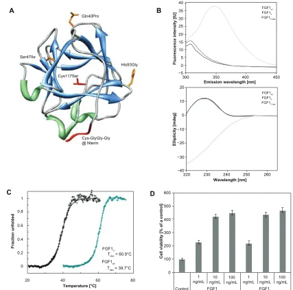
Design, expression, and characterization
of FGF1 mutant
We converted a poorly stable FGF1 and turned it into a molecule of high stability, increased resistance to degradation, and suitable for precise and site specific conjugation to
AuNPs. We took advantage of the FGF1V that we formerly
designed and characterized, containing three mutations (Gln40Pro, Ser47Ile, His93Gly), which provide a dramatic
increase of 21°C in the denaturation temperature and protect
the growth factor against proteolysis.8 Next, to ensure precise
cysteine-mediated linkage to AuNP, a flexible Cys-Gly-Gly-Gly linker was introduced at the protein N terminus. In addition, the exposed Cys117 in the FGF1 molecule was substituted with serine, so that the engineered N-terminal cysteine contained the single thiol group available for the conjugation with a nanoparticle surface (Figure 1A).
Ortega et al20 showed that Cys117Ser mutation did not affect
protein stability and significantly increased the protein half-life in plasma, which is an additional benefit for therapeutic or diagnostic applications.
FGF1V was expressed in E. coli and purified by
affin-ity chromatography using heparin-Sepharose resin. Its CD and fluorescence spectra were virtually identical to the wt
protein, indicating that the engineered FGF1V was native
and properly folded (Figure 1B). Thermal denaturation was monitored by the measurement of the single tryptophan residue fluorescence at 353 nm, which was completely quenched in the folded state and significantly recovered upon protein denaturation. We assumed a two-state revers-ible thermal transition, as we have previously shown, for
a large set of FGF1 mutants.8,21 Based on the denaturation
curve, thermodynamic parameters were calculated using
the Santoro–Bolen equation (Figure 1C).22 The T
den value
of FGF1V is 60.9°C, which is 21.5°C higher than that of wt
FGF1, and closely approximates the Tden of the previously
Dovepress
Szlachcic et al
International Journal of Nanomedicine downloaded from https://www.dovepress.com/ by 118.70.13.36 on 23-Aug-2020
described triple variant (Gln40Pro, Ser47Ile, His93Gly).8 In
conclusion, the addition of the cysteine-containing linker at the N terminus together with Cys117Ser substitution did not lead to any substantial changes in protein stability.
To verify whether the introduced modifications did not affect the growth factor’s biological activities, especially receptor binding and activation, cell proliferation assay in the NIH 3T3 cells was performed (in the presence of heparin).
FGF1V engineered for nanoparticle coating showed the same
proliferation potential as the wt FGF1, which in the presence of heparin is very similar to the triple FGF1 stable mutant (Gln40Pro, Ser47Ile, His93Gly) activity (Figure 1D).
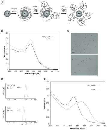
AuNP preparation and characterization
AuNPs are widely used in nanobiotechnology and are
considered to be representative nanomaterial.23 Several
methods of their synthesis have been developed, which dif-fer in the capping agent, product size, shape, and tendency
600
220 230 240 250 260 300
40
FGF1wt
FGF1V
FGF1V den
FGF1wt
FGF1V
FGF1V den
35 30 25 20 15 10 5
−5 0
350
Emission wavelength [nm]
Wavelength [nm]
400 450
−40 −30 −20 −10 0 10 20 Gln40Pro
A B
C D
His93Gly Ser47IIe
Cys117Ser
Cys-GlyGly-Gly @ Nterm
500
400
300
200
100
0 1
0.8
0.6
0.4
0.2
0
20 40 60
Temperature [°C]
Fraction unfolded
Cell viability [% of a control]
Ellipticity [mdeg]
Fluorescence intensity [IU]
80 FGF1V
FGF1wt Tden = 60.9°C
Tden = 39.7°C
Control 1
ng/mL ng/mL10 ng/mL100 ng/mL1 ng/mL10 FGF1V FGF1wt
100 ng/mL
Figure 1 FGF1 variant dedicated for specific conjugation with gold nanoparticles. (A) The spatial structure of FGF1 with the introduced mutations (based on PDB: 2Q9X).
Side chains of mutated residues involved in stability effects are represented in orange; mutations designed to facilitate thiol-mediated conjugation are marked in red.
(B) Fluorescence (upper panel) and circular dichromism (lower panel) spectra of native wild-type FGF1, native FGF1V, and denaturated FGF1V at a protein concentration
of 2 × 10⁻⁶ M in 25 mM sodium phosphate buffer, pH 7.3. (C) Normalized thermal denaturation curves of FGF1 wild-type (in black) and engineered FGF1
V variant (in blue).
Changes in ellipticity were monitored at 228 nm wavelength, in the presence of 0.7 M GdmCl in 25 mM sodium phosphate buffer, pH 7.3, and at a protein concentration of 2 × 10⁻⁶ M. (D) Proliferative activity of FGF1
V and wild-type FGF1 measured by the MTT assay.
Notes: Starved NIH 3T3 cells were incubated with the growth factor for 48 hours in the presence of 10 U/mL heparin. Equal volume of PBS was added to the control samples.
Results shown are mean values from four experiments. Bars indicate +/− standard deviation.
Abbreviations: FGF1, fibroblast growth factor 1; PDB, Protein Data Bank; FGF1V, fibroblast growth factor 1 variant; MTT, 3-(4,5-dimethylthiazol-2-yl)-2,5-diphenyltetrazolium
bromide; NIH 3T3, mouse embryonic fibroblast cell line; PBS, phosphate buffered saline.
Dovepress FGF1-gold nanoparticle conjugates target FGFR
International Journal of Nanomedicine downloaded from https://www.dovepress.com/ by 118.70.13.36 on 23-Aug-2020
to aggregation.24 Protein conjugation to AuNPs needs to
be performed in a hydrophilic solution and under mild pH conditions. For this reason we synthesized nanoparticles
using the Turkevich method.16,17 After synthesis, the AuNPs
were stabilized with citrate ions, which were subsequently exchanged for detergent molecules, and finally for the protein thiol group (Figure 2A). Synthesized nanoparticles were characterized with UV-Vis absorption spectroscopy (Figure 2B). They displayed a characteristic band with the maximum absorption at 520 nm, corresponding to the average
size of nanoparticles of 10 nm in diameter.25 This result was
also confirmed by electron microscopy and by DLS measure-ments (Figure 2C and D). The solution was monodisperse and did not contain any aggregates.
Preparation of FGF1-AuNP conjugates
Proteins can be conveniently attached to the surface of the AuNPs via a strong covalent bond between cysteine thiol and gold. Since gold colloids without a capping agent are
prone to aggregation,23 we conducted two-step
functionaliza-tion to minimize the risk of gold colloid’s aggregafunctionaliza-tion; the developed protocol was based on the detergent exchange
approach.26,27 After the initial coating of the nanoparticles
with a low concentration of a nonionic detergent, the deter-gent molecules were gradually exchanged for the protein molecules. The excess of protein was completely removed in several subsequent washing steps, as determined by MS.
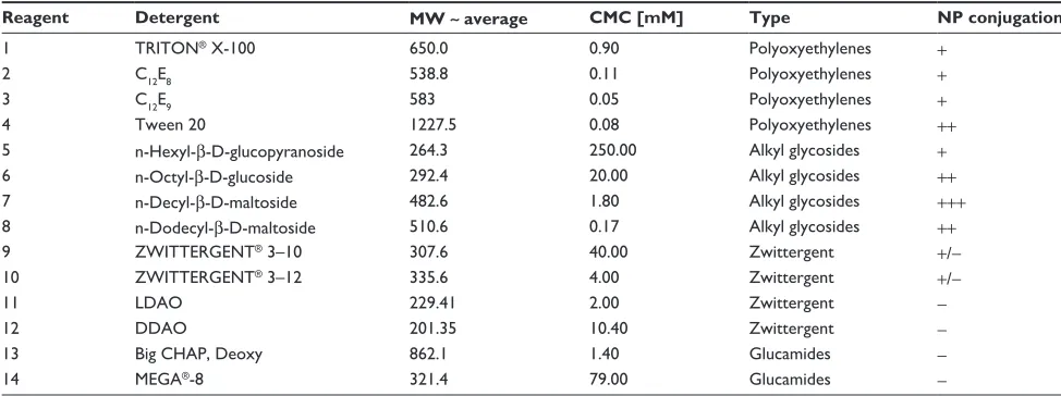
To choose the optimal detergent for coating of AuNPs
with FGF1V, yielding a monodisperse colloid preparation,
we tested 14 detergents with different molecular structures and properties representing four groups: polyoxythylenes, alkyl glycosides, zwittergents, and glucamides. The results are summarized in Table 1. In general, alkyl glucosides proved to be the most effective in the preparation of the protein-nanoparticle conjugates, with polyoxyethylenes being slightly less effective, whereas zwittergents and glucamides yielded colloids prone to aggregation. In this
particular system, n-decyl-β-D-maltoside allowed us to
produce monodisperse, stable, high-yielding gold colloids, and was used in all subsequent preparations.
Characterization of FGF1
V-AuNP
conjugates
The UV-Vis spectrum measurement confirmed that FGF1V
-AuNPs were monodisperse (Figure 2B) with a hydrodynamic diameter of 17.3 nm as established by DLS (Figure 2D). Noncoated AuNPs were approximately 9 nm in diameter,
thus the increase in the hydrodiameter of FGF1V-AuNPs
suggested the presence of covalently bound FGF1V on the
surface of nanoparticles. FGF1V-AuNPs did not show any
signs of aggregation when visualized by transmission
elec-tron microscopy (Figure 2C). Importantly, FGF1V-AuNPs
retained colloidal properties even in high salt concentra-tions, which caused aggregation of noncoated colloids (Figure 2E).
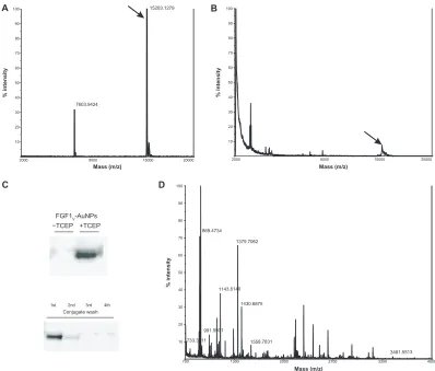
The presence of FGF1V on the surface of gold colloids
was additionally confirmed by MS and Western blot analysis.
To identify FGF1V via MS, conjugates were reduced with
high concentrations of reducing agents (0.5 M DTT or 0.1 M TCEP). The reduced sample yielded the mass spectrum of
intact FGF1V, whereas the untreated conjugate did not result
in any peaks in the range of the free FGF1 mass (Figure 3A and B). Both samples were also verified by Western blot analysis with the use of anti-FGF1 antibody confirmed by the MS analysis (Figure 3C, upper panel). For the verifica-tion of FGF1 presence on the nanoparticle surface, we have also employed a second MS approach, which involved trypsin digestion of conjugates and subsequent identifica-tion of tryptic peptides. This method revealed the presence
of FGF1V peptides in the FGF1V-AuNPs sample and
con-firmed the presence of FGF1V on the surface of conjugates
(Figure 3D).
The solution from the last washing step of the conju-gate preparation was also subjected to Western blot and
MS analyses. There was not any trace of either the FGF1V
band in immunoblotting assay, or of FGF1V detected by MS
(Figure 3C, lower panel). In addition, the absence of free
FGF1V was confirmed by the proliferation assay in NIH
3T3 cells. The last wash did not activate the FGFRs, and the downstream signaling cascades and did not stimulate NIH 3T3 fibroblast proliferation, confirming again that there was
no free FGF1V in the sample of FGF1V-AuNPs. Due to the
spectral properties of AuNPs, spectroscopic measurements of protein concentration were not possible. Therefore, the
concentration of FGF1V conjugated to the AuNPs was
esti-mated using a modified Bradford assay (Bio-Rad Protein Assay, Bio-Rad Laboratories).
Biological activity of FGF1
V-AuNPs
The biological activity of FGF1V conjugated to the AuNPs
was tested using the NIH 3T3 cell line. To establish whether the immobilized FGF1 is natively folded and retains its ability to bind to FGF receptors on the cell surface, we employed a proliferation assay. Serum-starved NIH 3T3 cells were treated with varying concentrations of FGF1 and
corresponding concentrations of FGF1V-AuNPs (with the
Dovepress
Szlachcic et al
International Journal of Nanomedicine downloaded from https://www.dovepress.com/ by 118.70.13.36 on 23-Aug-2020
same amount of FGF1V) and gold colloids alone (with the same amount of AuNPs). As shown in Figure 4A, the
colloid-bound FGF1V stimulates the proliferation of the NIH
3T3 cells at a level comparable to FGF1V. In agreement with
the proliferation assay, FGF1V-AuNPs activated all main
FGF1-induced signaling pathways (Figure 4B). Furthermore,
FGF1V-AuNPs showed a moderate proliferative effect,
pre-sumably due to the presence of a functional growth factor
on their surface. These results show that the FGF1V covalent
bond to gold colloids is properly folded, fully functional, and not cytotoxic for the cells.
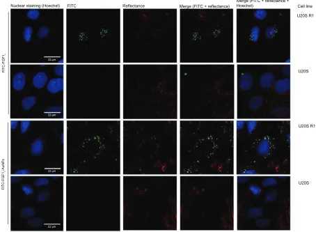
FGFR-mediated specific cell uptake
of FGF1
V-AuNPs
To determine if the FGF1V-AuNP particle conjugates can
be efficiently internalized in an FGFR-dependent manner, we employed two model cell lines: U2OS and U2OS, stably transfected with FGFR1 (U2OS R1), as described previously
by Haugsten et al.19 Untransfected U2OS cells did not express
any FGFR isoforms, and thus could serve as a negative control, as confirmed by quantitative reverse transcription
polymerase chain reaction. Fluorescently labeled FGF1V
(FITC-FGF1V) was attached to the surface of AuNPs via the
0.7
0.6
0.5
0.4
0.3
0.2
0.1
0
350 400 450 500 550 600 650 700 750
0.3
0.25
0.2
0.15
0.1
0.05
350 0.01
FGF1V-AuNPs Diam [nm] 17.33
FGF1V-AuNPs
B
D E
C
FGF1V-AuNPs AuNPs
AuNPs
FGF1V-AuNPs
AuNPs
AuNPs Diam [nm] 8.79
Size [nm]
Intensity [%
]
Intensity [%
]
0.1 1 10 100 1000
0.01 0.1 1 10 100 1000
400 450 500 550
Wavelength [nm] Wavelength [nm]
Absorbance
Absorbance
600 650 700 750
0
CYS
CYS CY S
CY
S
CYS
CYS
C
YS
CYS CYS
Detergent
A
FGF1V
AU AU AU AU
FGF1V
Figure 2 Biophysical characterization of AuNP conjugates. (A) Scheme of the two-step functionalization of AuNPs with FGF1V based on the gradual exchange of detergent
molecules for the protein. (B) UV-Vis spectrum of the synthesized gold colloids and FGF1V-coated gold colloids (FGF1V-AuNPs). (C) Transmission electron microscopy image
of gold colloid solution and AuNPs coated with FGF1V. Bar equals to 100 µm. (D) DLS analysis of AuNPs coated with FGF1V (upper panel) and synthesized unconjugated AuNPs (lower panel). (E) UV-Vis spectra of AuNPs treated with high salt concentration, coated with the FGF1V (FGF1V-AuNPs) and aggregated AuNPs without any coating agent.
Note: The observed shift of absorption maximum corresponds to nanoparticle aggregation, as the conjugates coated with FGF1V are a lot more stable.
Abbreviations: AuNP, gold nanoparticle; Au, gold; FGF1V, fibroblast growth factor 1 variant; UV-Vis, ultraviolet visible spectroscopy; DLS, dynamic light scattering.
Dovepress FGF1-gold nanoparticle conjugates target FGFR
International Journal of Nanomedicine downloaded from https://www.dovepress.com/ by 118.70.13.36 on 23-Aug-2020
protocol described earlier. The conjugates (FITC-FGF1V
-AuNPs) or free FITC-FGF1V were incubated with U2OS
and U2OS R1 cells for 30 minutes at 37°C, and they were
then analyzed with a confocal laser scanning microscope. In order to directly visualize AuNPs, we have employed the
resonance light scattering of AuNPs.28 We observed that
both fluorescently labeled free FGF1V and FGF1V-AuNPs
were internalized by FGFR1-expressing cells (U2OS R1), which was in clear contrast to the FGFR-deficient cells (U2OS). The presence of AuNPs (by reflectance mode) was detected only in the U2OS R1 cells, and they
colocal-ized with FGF1V-FITC (Figure 5). These results confirm
that FGF1V-nanoparticle conjugates were internalized in a
receptor-dependent manner.
NIR induced hyperthermia in
FGFR-positive cells treated with FGF1
V-AuNPs
Finally, we evaluated the applicability of the FGF1V
-coated nanoparticles for the targeted photothermal therapy. Untransfected U2OS cells, U2OS R1 cells highly express-ing FGFR1, and BJ cells naturally expressexpress-ing FGFR1 were
treated with FGF1V-AuNPs or AuNPs and then irradiated
with NIR light (808 nm). The level of FGFR1 expression in the BJ cells was quite high as compared to the other primary cells. Both FGFR-positive cell lines showed significantly decreased viability upon NIR irradiation (39% of the initial viability in the case of U2OS R1 cells, and 58% of the initial viability in the case of BJ cells), whereas in cell line lacking FGFR (U2OS cells) no effect of irradiation in the presence
of FGF1V-AuNPs was observed (viability equal to 95% of
untreated controls) (Figure 6A and B). Both cell lines were also exposed to 808 nm laser irradiation in the absence of
FGF1V-AuNPs, and incubated with FGF1V-AuNPs without
laser light treatment. Cells treated with uncoated AuNPs served as an additional negative control (treated and untreated with infrared light).
Laser irradiation of cells untreated with conjugates did not yield significant cytotoxicity; this was similar to cells treated with nanoparticles in the absence of laser irradiation (Figure 6A and B). This indicates that neither nanoparticles nor laser light itself could be toxic for cells and influence
their viability. On the contrary, FGF1V-AuNPs alone showed
a slight increase in viability, presumably caused by the
pro-liferation activity of the FGF1V of conjugates.
Taken together, the photothermally induced cell damage was prominent only for cell lines expressing FGFR1, which is in agreement with the receptor-specific internalization of nanoconjugates shown above (Figure 5).
Discussion
FGFRs meet the expectations for potential therapeutic tar-gets, as significant amplification of FGFRs has been identified
in various tumor types.6 Currently there are several
differ-ent strategies to develop an effective anticancer therapy of the FGFR-related tumors. They include the application of
small-molecule tyrosine kinase inhibitors,29,30 FGF ligand
traps preventing FGFs from binding to their receptors,31
and monoclonal antibodies against FGFs and FGFRs.32,33
Table 1 Detergents tested for the preparation of gold colloids coated with FGF1V
Reagent Detergent MW ∼ average CMC [mM] Type NP conjugation
1 TRITON® X-100 650.0 0.90 Polyoxyethylenes +
2 C12E8 538.8 0.11 Polyoxyethylenes +
3 C12E9 583 0.05 Polyoxyethylenes +
4 Tween 20 1227.5 0.08 Polyoxyethylenes ++
5 n-Hexyl-β-D-glucopyranoside 264.3 250.00 Alkyl glycosides +
6 n-Octyl-β-D-glucoside 292.4 20.00 Alkyl glycosides ++
7 n-Decyl-β-D-maltoside 482.6 1.80 Alkyl glycosides +++
8 n-Dodecyl-β-D-maltoside 510.6 0.17 Alkyl glycosides ++
9 ZWITTERGENT® 3–10 307.6 40.00 Zwittergent +/−
10 ZWITTERGENT® 3–12 335.6 4.00 Zwittergent +/−
11 LDAO 229.41 2.00 Zwittergent −
12 DDAO 201.35 10.40 Zwittergent −
13 Big CHAP, Deoxy 862.1 1.40 Glucamides −
14 MEGA®-8 321.4 79.00 Glucamides −
Notes: Nanoparticle conjugation efficiency was judged based on coated nanoparticle monodispersity, resistance to salt-induced aggregation, and effective replacement with
protein. (+++) denotes the detergent yielding nanoparticles with best characteristics, while (++), (+), and (+/−) correspond to detergents that were relatively less efficient, yielding less dispersed colloids. (−) indicates that the detergent does not prevent colloid aggregation during coating.
Abbreviations: FGF1V, fibroblast growth factor 1 variant; MW, molecular weight; CMC, critical micelle concentration; NP, nanoparticles; LDAO, lauryldimethylamine
N-oxide; DDAO, N,N-dimethyldodecylamine N-oxide.
Dovepress
Szlachcic et al
International Journal of Nanomedicine downloaded from https://www.dovepress.com/ by 118.70.13.36 on 23-Aug-2020
2000 8000 15000 20000
Mass (m/z) Mass (m/z)
Mass (m/z)
10 20 30 40 50 60 70 80 90 100
% intensity
10 20 30 40 50 60 70 80 90 100
% intensity
10 20 30 40 50 60 70 80 90 100
% intensity
A B
869.4734
1379.7062
1143.6140
1430.6875
733.3711 991.5501
1558.7831
3481.5513
D C
7603.9424
15203.1279
2000 8000 15000 20000
700 1300 2000 2700 3300 4000
FGF1V-AuNPs −TCEP +TCEP
1st 2nd 3rd
Conjugate wash
4th
Figure 3 Biophysical characterization of FGF1V-gold nanoparticles. (A) Mass spectrometry analysis of intact FGF1V. (B) Mass analysis of FGF1V-gold nanoparticles reduced
with TCEP. (C) Western blot analysis of conjugates and their preparation steps with anti-FGF1 antibody. Upon SDS-PAGE, separation FGF1V can be detected only after reduction by TCEP – when released from the nanoparticle surface. (D) Mass spectrometry analysis of trypsin digest of FGF1V-coated gold nanoparticles.
Note: Tryptic peptides corresponding to the FGF1V sequence are annotated.
Abbreviations: FGF1V, fibroblast growth factor 1 variant; TCEP, tris(2-carboxyethyl)phosphone; FGF1, fibroblast growth factor 1; SDS-PAGE, sodium dodecyl sulfate
polyacrylamide gel electrophoresis.
600
A B
Control 1 ng/m L
10 ng/m L
100 ng/mL1* ng/m L
10* ng/m L
100* ng/m L
1** ng/m L
10** ng/m L
100** ng/m L
Control 1 ng/mL
10 ng/mL
100 ng/mL
1* ng/mL
10*
ng/mL ng/mL100* ng/mL1** ng/mL10** 100** ng/mL
FGF1v FGF1v-AuNPs AuNPs
Phospho-Erk1,2
FGF1v FGF1v-AuNPs AuNPs
Phospho-FGFR
Phospho-PLCγ
Phospho-Akt
γ-tubulin
500
400
300
200
100
% control response
0
Figure 4 Biological activity of FGF1V-AuNP conjugates. (A) Cell proliferation in response to FGF1V, FGF1V-AuNPs, and nanoparticle colloids alone (AuNPs) measured using
Alamar Blue assay. Starved NIH 3T3 cells were incubated with the growth factor for 48 hours in the presence of 10 U/mL heparin. Equal volume of PBS was added to the control samples. Results shown are mean values from four experiments. Bars indicate +/− standard deviation. (B) Activation of signaling cascades in NIH 3T3 cells after incubation with increasing concentrations of FGF1V, FGF1V-AuNPs, and nanoparticle colloids alone (AuNPs) detected with Western blot analysis.
Notes: 1*, 10*, and 100* ng/mL AuNPs denotes the concentration of AuNPs equal to the concentration of AuNPs in 1*, 10*, and 100* ng/mL FGF1V-AuNPs.
Abbreviations: FGF1V, fibroblast growth factor 1 variant; AuNP, gold nanoparticle; PLCγ, phospholipase C-gamma; NIH 3T3, mouse embryonic fibroblast cell line;
PBS, phosphate buffered saline.
Dovepress FGF1-gold nanoparticle conjugates target FGFR
International Journal of Nanomedicine downloaded from https://www.dovepress.com/ by 118.70.13.36 on 23-Aug-2020
U20S R1 Cell line
U20S Nuclear staining (Hoechst) FITC
22 µm
22 µm
22 µm
22 µm
Reflectance
FITC-FGF
1v
FITC-FGF
1v
-AuNPs
Merge (FITC + reflectance)
Merge (FITC + reflectance + Hoechst)
U20S R1
U20S
Figure 5 Receptor-specific internalization of FGF1V or FGF1V-AuNPs visualized with confocal fluorescence microscopy.
Notes: U20S R1 and U20S cells were incubated with fluorescently labeled FGF1V-AuNPs (FITC, FGF1V-AuNPs), and fluorescently labeled FGF1V (FITC, FGF1V). AuNPs were
detected with confocal reflectance and cell nuclei were visualized with Hoechst staining. Only cells expressing FGF receptor internalized conjugates.
Abbreviations: FGF1V, fibroblast growth factor 1 variant; AuNPs, gold nanoparticles; FITC, fluorescein isothiocyanate.
In this report, we evaluate a novel approach of targeting tumors overexpressing FGFRs with their natural ligand, FGF1, conjugated with AuNPs.
To exploit FGF1 targeting potential, we designed a new variant of this growth factor suitable for therapeutic application.
Based on the formerly characterized stable FGF1 mutant,8 we
engineered a protein (FGF1V) containing three stabilizing point
mutations that overcame low stability and the relatively short half-life in vivo of the wt FGF1, Cys117Ser substitution to eliminate Cys on the protein surface, and N-terminal Cys-Gly-Gly-Gly flexible linker to enable precise site-directed
conjugation of FGF1 to gold. The engineered FGF1V was
properly folded, highly stable, and showed biological activ-ity well comparable to the wt FGF1. These properties were preserved upon conjugation to AuNPs.
FGF conjugates previously studied in the form of fusion proteins with bacterial toxins (such as diphtheria toxin and
Pseudomonas exotoxin) were shown to kill tumor cells.34,35
The main obstacle was the high immunogenicity effect
evoked by these fusions in vivo. Alternatively, the FGF1V
-AuNP conjugates that we synthesized are expected to exhibit minimal immunogenicity due to the good biocompatibility
of AuNPs in vivo.36
Gold colloids comprise a novel group of potential anti-cancer agents. Given their plasmonic absorption in the NIR range, which can be effectively converted into thermal energy, they can be used to evoke localized heating within the tis-sues leading to tumor cell death (called thermoablation or
hyperthermia).37 Due to the low permeability of NIR into the
tissues, photothermal therapies involving plasmonic AuNPs were limited to the penetration range of available lasers. However, in the 650–900 nm region where the absorption of physiological fluids and tissues is minimal, the Food and Drug Administration Class 1 (microwatt) laser can efficiently
penetrate up to 10 cm through the tissue.38 The efficiency of
AuNP-based systems can be further improved by their con-jugation with targeting molecules, and these were shown to
be suitable for therapy against solid tumors.39
Dovepress
Szlachcic et al
International Journal of Nanomedicine downloaded from https://www.dovepress.com/ by 118.70.13.36 on 23-Aug-2020
Kogot et al40 demonstrated that the FGF1 wt can be
effectively conjugated via His-tag to the CAAKA peptide passivated AuNPs of 1.5 nm; however, they did not present how coupling with the nanoparticle affected the biological activity of the growth factor. The system used in this study, which was based on much larger, 10 nm nanoparticles, presents different advantages together with different challenges to be overcome. The major benefit is that larger nanoparticles are much more effective for the photothermal therapy of cancerous cells.
The 10 nm AuNPs suitable for photothermal therapy are
much larger than the FGF1V molecule itself, and the major
difficulties to overcome during the conjugation process were the direct and unspecific hydrophobic interactions of protein and nanoparticle surfaces, leading to protein denaturation and loss of function. Using the detergent exchange approach with a panel of different detergent types, we developed and further optimized a protocol yielding nanoparticles coated
with FGF1V covalently bound to a gold surface that were
monodisperse and retained protein functionality. The
cova-lent attachment of FGF1V to AuNPs was verified by several
methods including a reduction protocol followed by MS analysis. After incubation in a highly reductive environment, conjugated proteins could be released from the nanoparticles and identified by MS. This approach provides a robust, reli-able, and versatile method to determine the proper conjuga-tion of protein to AuNPs, and should be universal and suitable for other gold-thiol based systems.
Precise targeting of cancer cells is the most crucial aspect of successful antitumor therapies. Specific ligands should
not only mark the cancerous cell, but also be specifically internalized in order to act intracellularly and not affect the
surrounding healthy cells.41 In cell culture experiments, we
showed that FGF1V conjugated to AuNPs remained properly
folded and biologically active, and that the FGF1V-AuNPs
were only internalized specifically by cells expressing
FGFRs. Thus, our report demonstrates that FGF1V is a
promising delivery agent for cancer cells expressing FGFRs. Moreover, apart from being targeted by FGF1 to the cell sur-face of FGFR-overexpressing cells, the nanoparticles could also preferentially accumulate at the tumor site due to the enhanced permeability and retention effect, achieving even higher intratumoral concentrations useful for subsequent
treatment with NIR.42,43
Finally, we showed that irradiation with NIR laser light allows for effective and specific photothermal destruction of cancer cells expressing FGF receptors on their surface upon
treatment with FGF1V-AuNPs. After single 20-minute
irradia-tion with 808 nm light, the viability of FGFR1-expressing cells decreased by about 60%. This result clearly shows the potential applicability of novel growth-factor targeted AuNPs in cancer treatment. Even though the laser wave-length used for irradiation was significantly different from the surface plasmon resonance wavelength of AuNPs, the cytotoxic effect was observed, and we believe that it was due
to the second harmonic generation described previously.44,45
Indeed, previous studies have shown the applicability of NIR irradiation with AuNPs, and that the required laser power of NIR irradiation (800 nm) was ten times lower than for
the visible (520 nm) light treatment.11 Furthermore, it was
120
U2OS R1
A U2OS
100
80
60
40
20
0
−NIR +NIR −NIR +NIR −NIR +NIR FGF1v-AuNPs AuNPs
Control
% control viability
120
BJ
B
100
80
60
40
20
0
−NIR +NIR −NIR +NIR −NIR +NIR FGF1v-AuNPs AuNPs
Control
% control viability
Figure 6 Decrease in viability of FGFR-positive cells after irradiation with NIR. (A) U20S R1 and U20S, and (B) BJ cells treated with FGF1V-AuNPs show significantly
decreased viability irradiated with NIR.
Notes: Results shown are mean values from three experiments. Bars indicate +/− standard deviation. Cells were treated with NIR (808 nm diode laser, 1.2 A) for 20 minutes,
and the visibility was measured by the MTT assay 12 hours after NIR treatment. Equal volume of PBS was added to the control samples.
Abbreviations: FGFR, fibroblast growth factor receptor; NIR, near-infrared; FGF1V, fibroblast growth factor 1 variant; AuNPs, gold nanoparticles; MTT,
3-(4,5-dimethylthiazol-2-yl)-2,5-diphenyltetrazolium bromide; PBS, phosphate buffered saline.
Dovepress FGF1-gold nanoparticle conjugates target FGFR
International Journal of Nanomedicine downloaded from https://www.dovepress.com/ by 118.70.13.36 on 23-Aug-2020
previously suggested that due to the close proximity of the nanoparticles in the endosomes, their absorption spectrum
may shift to the NIR region.46 Additionally, NIR irradiation
offers the advantage of being highly permeable in tissues
when compared to visible light.47
Our study is the first report employing engineered FGF-type ligands to the specific delivery of AuNPs to cancer cells via FGFR1-mediated endocytosis. The results presented show the potential applicability of FGF1-gold conjugates in further animal studies using FGFR-expressing xenografts, which may lead to the development of a novel anticancer therapy in the future.
Acknowledgments
This work was supported by the Wroclaw Research Center
EIT+ under the project Biotechnologies and advanced medical
technologies – BioMed (POIG 01.01.02-02-003/08-00) financed from the European Regional Development Fund (Operational Program Innovative Economy, 1.1.2).
We thank Dr Michał Majkowski from the Laboratory of Cytobiochemistry, University of Wroclaw; Dr Jorgen Wesche from the Department of Biochemistry, Institute for Cancer Research; the Norwegian Radium Hospital for help with confocal microscopy experiments; and the skillful techni-cal assistance of Anne Engen and Anne-Mari G Pedersen from the Department of Biochemistry, Institute for Cancer Research. The Norwegian Radium Hospital is gratefully acknowledged.
Disclosure
The authors report no conflicts of interest in this work.
References
1. Blanco E, Hsiao A, Ruiz-Esparza GU, Landry MG, Meric-Bernstam F, Ferrari M. Molecular-targeted nanotherapies in cancer: enabling
treatment specificity. Mol Oncol. 2011;5(6):492–503.
2. Knights V, Cook SJ. De-regulated FGF receptors as therapeutic
targets in cancer. Pharmacol Ther. 2010;125(1):105–117.
3. Haugsten EM, Wiedlocha A, Olsnes S. Roles of Fibroblast Growth
Factor Receptors in Carcinogenesis. Mol Canc Res. 2010; 8:1439–1452.
4. Penault-Llorca F, Bertucci F, Adélaide J, et al. Expression of FGF
and FGF receptor genes in human breast cancer. Int J Cancer. 1995;
61(2):170–176.
5. Heinzle C, Sutterlüty H, Grusch M, Grasl-Kraupp B, Berger W, Marian B. Targeting fibroblast-growth-factor-receptor-dependent
signaling for cancer therapy. Expert Opin Ther Targets. 2011;15(7):
829–846.
6. Wesche J, Haglund K, Haugsten EM. Fibroblast growth factors and their
receptors in cancer. Biochem J. 2011;437(2):199–213.
7. Ornitz DM, Itoh N. Fibroblast growth factors. Genome Biol. 2001;
2(3):REVIEWS3005.
8. Zakrzewska M, Krowarsch D, Wiedlocha A, Olsnes S, Otlewski J. Highly stable mutants of human fibroblast growth factor-1 exhibit prolonged
biological action. J Mol Biol. 2005;352(4):860–875.
9. El-Sayed IH, Huang X, El-Sayed MA. Selective laser photo-thermal therapy of epithelial carcinoma using anti-EGFR antibody conjugated
gold nanoparticles. Cancer Lett. 2006;239(1):129–135.
10. Huang X, Jain PK, El-Sayed IH, El-Sayed MA. Determination of the minimum temperature required for selective photothermal destruction of cancer cells with the use of immunotargeted gold nanoparticles.
Photochem Photobiol. 2006;82(2):412–417.
11. Huang X, Jain PK, El-Sayed IH, El-Sayed MA. Plasmonic
photo-thermal therapy (PPTT) using gold nanoparticles. Lasers Med Sci.
2008;23(3):217–228.
12. Dykman L, Khlebtsov N. Gold nanoparticles in biomedical applications:
recent advances and perspectives. Chem Soc Rev. 2012;41(6):
2256–2282.
13. Loo C, Lowery A, Halas N, West J, Drezek R. Immunotargeted
nanoshells for integrated cancer imaging and therapy. Nano Lett.
2005;5(4):709–711.
14. Brust M, Walker M, Bethell D, Schiffrin DJ, Whyman R. Synthesis of thiol-derivatised gold nanoparticles in a two-phase liquid-liquid
system. Journal of the Chemical Society, Chemical Communications.
1994;(7):801–802.
15. Perrault SD, Chan WC. Synthesis and surface modification of highly
monodispersed, spherical gold nanoparticles of 50–200 nm. J Am Chem
Soc. 2009;131(47):17042–17043.
16. Kimling J, Maier M, Okenve B, Kotaidis V, Ballot H, Plech A.
Turkevich method for gold nanoparticle synthesis revisited. J Phys
Chem B. 2006;110(32):15700–15707.
17. Turkevich J, Stevenson PC, Hillier J. A study of the nucleation and
growth processes in the synthesis of colloidal gold. Discuss Faraday
Soc. 1951;11:55–75.
18. Haugsten EM, Malecki J, Bjørklund SM, Olsnes S, Wesche J. Ubiquitination of fibroblast growth factor receptor 1 is required for
its intracellular sorting but not for its endocytosis. Mol Biol Cell.
2008;19(8):3390–3403.
19. Haugsten EM, Sørensen V, Brech A, Olsnes S, Wesche J. Different intracellular trafficking of FGF1 endocytosed by the four homologous
FGF receptors. J Cell Sci. 2005;118(Pt 17):3869–3881.
20. Ortega S, Schaeffer MT, Soderman D, et al. Conversion of cysteine to serine residues alters the activity, stability, and
hepa-rin dependence of acidic fibroblast growth factor. J Biol Chem.
1991;266(9):5842–5846.
21. Zakrzewska M, Krowarsch D, Wiedlocha A, Otlewski J. Design of
fully active FGF-1 variants with increased stability. Protein Eng Des
Sel. 2004;17(8):603–611.
22. Santoro MM, Bolen DW. A test of the linear extrapolation of unfolding free energy changes over an extended denaturant concentration range.
Biochemistry. 1992;31(20):4901–4907.
23. Wilson R. The use of gold nanoparticles in diagnostics and detection.
Chem Soc Rev. 2008;37(9):2028–2045.
24. Grzelczak M, Pérez-Juste J, Mulvaney P, Liz-Marzán LM. Shape
control in gold nanoparticle synthesis. Chem Soc Rev. 2008;37(9):
1783–1791.
25. Jain PK, Lee KS, El-Sayed IH, El-Sayed MA. Calculated absorption and scattering properties of gold nanoparticles of different size, shape, and composition: applications in biological imaging and biomedicine.
J Phys Chem B. 2006;110(14):7238–7248.
26. Aslan K, Lakowicz JR, Geddes CD.
Nanogold-plasmon-resonance-based glucose sensing. Anal Biochem. 2004;330(1):145–155.
27. Aslan K, Pérez-Luna VH. Quenched emission of fluorescence by ligand
functionalized gold nanoparticles. J Fluoresc. 2004;14(4):401–405.
28. Kah JC, Olivo MC, Lee CG, Sheppard CJ. Molecular contrast of EGFR expression using gold nanoparticles as a reflectance-based imaging
probe. Mol Cell Probes. 2008;22(1):14–23.
29. Byron SA, Pollock PM. FGFR2 as a molecular target in endometrial
cancer. Future Oncol. 2009;5(1):27–32.
30. Grand EK, Chase AJ, Heath C, Rahemtulla A, Cross NC. Targeting FGFR3 in multiple myeloma: inhibition of t(4;14)-positive cells by
SU5402 and PD173074. Leukemia. 2004;18(5):962–966.
Dovepress
Szlachcic et al
International Journal of Nanomedicine downloaded from https://www.dovepress.com/ by 118.70.13.36 on 23-Aug-2020
International Journal of Nanomedicine
Publish your work in this journal
Submit your manuscript here: http://www.dovepress.com/international-journal-of-nanomedicine-journal
The International Journal of Nanomedicine is an international, peer-reviewed journal focusing on the application of nanotechnology in diagnostics, therapeutics, and drug delivery systems throughout the biomedical field. This journal is indexed on PubMed Central, MedLine, CAS, SciSearch®, Current Contents®/Clinical Medicine,
Journal Citation Reports/Science Edition, EMBase, Scopus and the Elsevier Bibliographic databases. The manuscript management system is completely online and includes a very quick and fair peer-review system, which is all easy to use. Visit http://www.dovepress.com/ testimonials.php to read real quotes from published authors. 31. Zhang H, Masuoka L, Baker K, et al. FP-1039 (FGFR1:Fc), A soluble
FGFR1 receptor antagonist, inhibits tumor growth and angiogenesis. Proceedings of the AACR-NCI-EORTC International Conference Molecular Targets and Cancer Therapeutics Discovery, Biology and Clinical Applications; October 22–26, 2007; San Francisco, CA. Avail-able at http://www.aacr.org/Uploads/DocumentRepository/2007conf/ moltar/mt07_posterb.pdf.
32. Desnoyers LR, Pai R, Ferrando RE, et al. Targeting FGF19 inhibits tumor growth in colon cancer xenograft and FGF19 transgenic
hepa-tocellular carcinoma models. Oncogene. 2008;27(1):85–97.
33. Maruyama-Takahashi K, Shimada N, Imada T, et al. A neutralizing anti-fibroblast growth factor (FGF) 8 monoclonal antibody shows anti-tumor activity against FGF8b-expressing LNCaP xenografts
in androgen- dependent and -independent conditions. Prostate. 2008;
68(6):640–650.
34. Gawlak SL, Pastan I, Siegall CB. Basic fibroblast growth factor-Pseudomonas exotoxin chimeric proteins; comparison with acidic
fibroblast growth factor-Pseudomonas exotoxin. Bioconjug Chem.
1993;4(6):483–489.
35. Siegall CB, Gawlak SL, Chace DF, Merwin JR, Pastan I. In vivo activi-ties of acidic fibroblast growth factor-Pseudomonas exotoxin fusion
proteins. Bioconjug Chem. 1994;5(1):77–83.
36. Dreaden EC, Alkilany AM, Huang X, Murphy CJ, El-Sayed MA.
The golden age: gold nanoparticles for biomedicine. Chem Soc Rev.
2012;41(7):2740–2779.
37. Cherukuri P, Glazer ES, Curley SA. Targeted hyperthermia using metal
nanoparticles. Adv Drug Deliv Rev. 2010;62(3):339–345.
38. Dreaden EC, Mackey MA, Huang X, Kang B, El-Sayed MA.
Beating cancer in multiple ways using nanogold. Chem Soc Rev.
2011;40(7):3391–3404.
39. Kennedy LC, Bear AS, Young JK, et al. T cells enhance gold
nanopar-ticle delivery to tumors in vivo. Nanoscale Res Lett. 2011;6(1): 283.
40. Kogot JM, Parker AM, Lee J, Blaber M, Strouse GF, Logan TM. Analysis of the dynamics of assembly and structural impact for a histidine
tagged FGF1-1.5 nm Au nanoparticle bioconjugate. Bioconjug Chem.
2009;20(11):2106–2113.
41. Sawyers C. Targeted cancer therapy. Nature. 2004;432(7015):
294–297.
42. Greish K. Enhanced permeability and retention of macromolecular drugs in solid tumors: a royal gate for targeted anticancer nanomedicines.
J Drug Target. 2007;15(7–8):457–464.
43. Hirsjärvi S, Passirani C, Benoit JP. Passive and active tumour targeting
with nanocarriers. Curr Drug Discov Technol. 2011;8(3):188–196.
44. Lippitz M, van Dijk MA, Orrit M. Third-harmonic generation from
single gold nanoparticles. Nano Lett. 2005;5(4):799–802.
45. Nappa J, Russier-Antoine I, Benichou E, Jonin Ch, Brevet PF. Second harmonic generation from small gold metallic particles: from the dipolar
to the quadrupolar response. J Chem Phys. 2006;125(18):184712.
46. Zharov VP, Galitovskaya EN, Johnson C, Kelly T. Synergistic enhancement of selective nanophotothermolysis with gold nanoclusters:
potential for cancer therapy. Lasers Surg Med. 2005;37(3):219–226.
47. Cheong WF, Prahl SA, Welch AJ. A review of the optical properties of
biological tissues. IEEE Journal of Quantum Electronics. 1990;26(12):
2166–2185.
Dovepress
Dove
press
FGF1-gold nanoparticle conjugates target FGFR
International Journal of Nanomedicine downloaded from https://www.dovepress.com/ by 118.70.13.36 on 23-Aug-2020