University of South Carolina
Scholar Commons
Theses and Dissertations
2017
Individual Differences in Markers of Cholinergic
Signaling Correlating to Fear and Extinction
Learning
Grace C. JonesUniversity of South Carolina
Follow this and additional works at:https://scholarcommons.sc.edu/etd
Part of theBiomedical and Dental Materials Commons
This Open Access Thesis is brought to you by Scholar Commons. It has been accepted for inclusion in Theses and Dissertations by an authorized administrator of Scholar Commons. For more information, please [email protected].
Recommended Citation
Individual Differences in Markers of Cholinergic Signaling Correlating to
Fear and Extinction Learning
By Grace C. Jones Bachelor of Science Winthrop University, 2015
Submitted in Partial Fulfillment of the Requirements For the Degree of Master of Science in
Biomedical Science School of Medicine University of South Carolina
2017 Accepted by:
David D. Mott, Director of Thesis Marlene A. Wilson, Reader Alexander J. McDonald, Reader
ii
iii
Acknowledgements
iv
Abstract
Posttraumatic stress disorder (PTSD) results when individuals are exposed to a life threatening event, assault, serious injury, or other traumatic incident. Individuals with PTSD are impaired in their ability to extinguish fear memories, resulting in intrusive symptoms that impair their ability to live otherwise healthy lives. It remains unclear why some individuals exposed to traumatic events develop PTSD while others do not.
v
Coronal brain sections were processed for immunofluorescence, labeling for M1 mAChR and AChE, and imaged in order to measure extent of protein expression. Significant correlations were observed between individual’s BLA M1 mAChR densities and ability to undergo fear acquisition and ability to recall fear extinction memories. This lead to the conclusions that M1 mAChR are functioning in the BLA in the processes of fear memory acquisition and extinction memory consolidation and that high expression of M1 mAChR allows for improved ability to undergo fear memory acquisition, resulting in a deficit in fear extinction. No significant correlations were observed between BLA AChE
vi
Table of Contents
Acknowledgements ... iii
Abstract ... iv
List of Figures ... viii
List of Abbreviations ... x
Chapter 1: Introduction ... 1
A. Fear Learning and Extinction ... 1
B. Anatomy of Fear Learning and Extinction ... 4
C. Cholinergic Regulation of Fear Learning and Extinction ... 7
D. Objective, Hypothesis, and Aims ... 11
Chapter 2: Methods ... 13
A. Animal Model of Fear Conditioning and Extinction Learning ... 13
B. Tissue Preparation, Immunofluorescence, & Image Collection & Analysis ... 16
C. Statistical Analysis ... 19
Chapter 3: Results ... 21
A. Behavior Results ... 21
vii
C. Correlation between Behavior and Image Analysis ... 34
Chapter 4: Conclusions ... 43
Chapter 5: Future directions... 48
References ... 50
viii
List of Figures
Figure 2.1: Fear and Extinction Behavior Paradigm ... 14
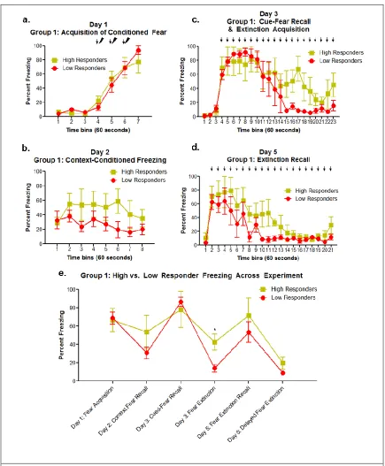
Figure 3.1: Group 1- Grouped Differences in Freezing Behavior During Fear and Extinction Paradigm ... 23
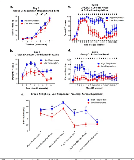
Figure 3.2: Group 2- Grouped Differences in Freezing Behavior During Fear and Extinction Paradigm ... 26
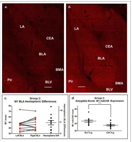
Figure 3.3: M1 mAChR Immunofluorescence ... 29
Figure 3.4: Basolateral Amygdala M1 mAChR+ Cell Density ... 30
Figure 3.5: Acetylcholinesterase Immunofluorescence ... 33
Figure 3.6: Correlation- M1 mAChR vs AChE Protein Expression Levels ... 34
Figure 3.7: Correlation- M1 mAChR expression levels vs fear learning processes ... 36
Figure 3.8: Correlation- M1 mAChR expression levels vs the extinction learning processes ... 38
Figure 3.9: Correlation- AChE expression levels vs fear learning processes ... 41
ix
Figure A.1: M1 mAChR CEA expression ... 57
Figure A.2: S1BF image collection and comparison ... 58
Figure A.3: Correlation- M1 mAChR expression levels vs contextual fear recall and delayed extinction acquisition... 59
x
List of Abbreviations
xi
PL ... Prelimbic cortex Pri ... Periform cortex* PTSD ...Posttraumatic stress disorder PV ... Parvalbumin S1BF ... Barrel field of the somatosensory cortex* TB/TBS ... Tris-buffered/tris-buffered saline solution US ... Unconditioned stimulus VAChT ...Vesicular acetylcholine transporter
* : as defined by Paxinos and Watson, The Rat Brain in Stereotaxic Coordinates, 6th revised edition, 2008
#
1
Chapter 1: Introduction
A.
Fear Learning and Extinction
Fear is the natural, seemingly instantaneous response to a stimulus that is perceived as potentially harmful or threatening. It keeps us safe. It causes soldiers to swerve when they see a bomb in the road during a battle; it causes us to reach for the phone or a frying pan when we come across a stranger sifting through our drawers in a dark kitchen; and it causes us to run screaming when we happen across a bear while walking in the woods. It keeps animals safe too. It causes a deer to run when it hears a hunter approaching; it causes a fish to swim when it senses movement in the current as a shark speeds toward it; and it causes a rat to freeze when it hears a snake slithering toward it. All of these are appropriate responses to frightening or potentially threatening situations which keep individuals safe.
2
What about when fear responses are generalized and become inappropriate? When throwing a friend a party, you don’t expect them to call the police or reach for a frying pan when their family and friends yell “surprise”. A paper bag on a city road should not cause a veteran to swerve into oncoming traffic. The fear of being presented with a potentially threatening situation should not keep an individual from being able to leave the safety of their home. These inappropriate responses to non-threatening situations are a hallmark of posttraumatic stress disorder.
3
of fear learning and fear extinction (Fendt and Fanselow, 1999; Zoladz and Diamond, 2016; Wilson and Reagan, 2016; Wilson and Fadel, 2017).
Classical or Pavlovian conditioning, a technique often used in fear learning and fear extinction animal behavioral models, is the learned association between a neutral stimulus, such as a tone (conditioned stimuli, CS) and a biologically relevant stimulus, such as food or pain (unconditioned stimuli, US). This is a crucial ability for survival and allows individuals to associate safe and harmful situations with unconditioned stimuli, and is thus conserved across higher organisms(Pavlov, 1927; for review see Milad and Quirk, 2012 and Orsini and Maren, 2012).
4
expression, which can be demonstrated by observed instances of spontaneous recovery, renewal, or reinstatement (Baldi and Bucherelli, 2015; Baldi and Bucherelli, 2010; Myers and Davis, 2007; Quirk and Mueller, 2008). There is a natural, observable variation in individual ability to undergo fear extinction, as seen commonly in individuals with PTSD (Horn et al., 2016).
This study investigates some of the suspected underlying mechanisms of fear and extinction learning, as well as the individual differences in ability to undergo fear and extinction learning. This study is unique in that no pharmacological manipulations were made and protein expression is directly correlated to freezing behavior in order to extrapolate how protein expression level relates to fear learning, extinction learning, and specific learning phase.
B.
Anatomy of Fear Learning and Extinction
Brain structures involved in the processing of fear and fear learning, including the prefrontal cortex (PFC), thalamus, hippocampus, and the amygdala, are conserved across species (Milad and Quirk, 2012). Environmental information is sent to the amygdala from the thalamus, PFC, and hippocampus (Fendt and Fanselow, 1999). This includes
5
synapse on to the BLA-CEA projection, allowing for filtering of information passed from the BLA to the CEA (Orsini and Maren, 2012). The CEA projects to the hypothalamus and brain stem, initiating behavioral and physiological responses, including freezing or running, autonomic responses, and inducing stress and startle responses (Sah and Westbrook, 2008). This initial response to threatening stimuli occurs much quicker than situational evaluation can occur, which allows individuals to respond seemingly
instantaneously in preparation for fight or flight action (Principles of Neural Science, page 1478; Milad and Quirk, 2012).
Long-term memory formation and consolidation then occurs, allowing the animal to recall details about the threatening situation, should it be presented again. Synaptic plasticity caused by associative cued-fear learning can be observed in both the LA and the BLA, shown by enhanced excitatory postsynaptic potentials, increasing synaptic plasticity between BLA and CEA fear-out-put circuits (Sah and Westbrook, 2008; Orsini and Maren, 2012). Consolidation of the fear memory in the amygdala is required for stable long-term memory storage, and requires new protein synthesis. This can be demonstrated by giving intra-amygdalar protein synthesis inhibitors after fear
conditioning, which prevents memory consolidation and subsequent recall (Maren et al. 2003).
6
Like cued-fear learning, cued-fear extinction learning relies on multiple brain regions, which connect and communicate to make a plastic network. The amygdala, PFC, and hippocampus are the major players in this network, and, while each function in acquisition, consolidation, and retrieval, each has a set of major functions. The hippocampus functions in extinction context recall, the PFC mediates extinction
consolidation, and the amygdala is thought to be where extinction memories are acquired and stored (Baldi and Bucherelli, 2015; Power et al., 2003b; Orsini and Maren, 2012). Hippocampal CA1 and ventral subiculum regions project to the LA, BLA, CEA, and the PFC. The BLA projects to each sub-region of the hippocampus and PFC. These dense reciprocal projections between the amygdala and hippocampus allow for fast and effective communication, which has been shown to be crucial for retrieval of context-aspects of extinction memories (Herry et al., 2008; Orsini and Maren, 2012). The PFC is subdivided into the prelimbic cortex (PL), which projects to the BLA and CEA, and the infralimbic cortex (IL), which projects to the basomedial amygdala (BM), ITC cells, and CEA (McDonald et al., 1996; Orsini and Maren, 2012). IL suppression of BLA through inhibitory circuits, including ITC cells, causes suppression of fear response (Quirk et al., 2003; Likhtik et al., 2008; Akirav et al., 2006; Sah and Westbrook, 2008; Orsini and Maren, 2012). Once extinction has been acquired, information about the CS, tone specifically, is relayed to the amygdala, not by the thalamus, but by the auditory cortex, indicating that after extinction there is a redistribution or rearrangement of information about the CS throughout the fear circuit (Pape and Pare, 2010; Orsini and Maren, 2012).
7
of the presented CS using contextual cues (Quirk and Mueller, 2008). The PFC, specifically the IL, has been shown to be important for retrieval of extinction memory and suppression of fear (Myers and Davis, 2007). The importance of the IL in this
process has been shown in electrophysiology and inactivation studies, where presentation of the extinguished CS, specifically, causes IL firing and BLA inactivation before
extinction retrieval results in a fear response (Herry and Garcia, 2002; Milad and Quirk, 2002; Sierra-Mercado et al., 2006). Herry et al. (2008) and Senn et al. (2014) showed that the BLA contains separate populations of cells, fear neurons and extinction neurons, which are active specifically during fear or extinction, respectively. The BLA cell population which project to the PL is involved in fear and is activated during high fear situations, whereas the cell population projecting to the IL is involved in extinction and is activated during extinction behavior (Herry et al., 2008; Senn et al., 2014). While many brain regions are critical for the learning and expression of fear and extinction memories, this study’s main focus was the amygdala, specifically the BLA due to its central role in each aspect of the fear learning and extinction process outlined above.
C.
Cholinergic Regulation of Fear Learning and Extinction
8
of various brain regions during learning in a variety of situations. It has been shown that the amygdala is the regulatory region modulating extinction learning and memory
formation occurring in other regions, and that these regions compete over control of what is learned in the processing of information (Gold, 2003). However, Thiele et al. argues in a 2013 review that the local distribution and contribution of muscarinic signaling is what determines the cognitive tasks a brain region controls, rather than the release of
acetylcholine alone. As thoroughly examined in Wilson and Fadel’s 2017 review, current evidence suggests that fear extinction learning is regulated by activation of
acetylcholine’s metabotropic, muscarinic receptors (mAChR). Current research indicates that mAChR activation is crucial for fear acquisition, consolidation, and potentially recall, as well as extinction memory consolidation, and potentially cued fear extinction acquisition (Wilson and Fadel 2017).
It has been demonstrated that there is dense cholinergic presence in the brain regions involved in fear learning outlined above. The hippocampus and amygdala, specifically the BLA, were shown by Muller, Mascagni, and McDonald to have very dense cholinergic projections originating in the basal forebrain (2011). These projections terminate heavily on pyramidal neurons of the BLA, which postsynaptically express high levels of M1 mAChR. McDonald and Mascagni (2010) demonstrated through
9
acetylcholinesterase (AChE) expression in the BLA has been shown to be some of the densest in the brain, further indicating the importance of cholinergic function in this region (Ben-Ari et al., 1977). This would lead one to assume that this region and its functions are largely modulated by cholinergic neurotransmission and cholinergic receptors.
As outlined in Wilson and Fadel’s 2017 review, multiple drug studies have determined mAChR are important for fear learning. By giving mAChR antagonists prior to fear conditioning systemically or intracerebrally, studies have indicated that mAChR function is important for acquisition of cued and contextual fear (Rudy, 1996; Young et al., 1995; Feiro and Gould, 2005; Jiang et al., 2016; Fornari et al., 2000; for review see Wilson and Fadel, 2017). Drug studies examining fear consolidation, specifically, have generated varied results: several studies indicate that mAChR activation is not crucial for cued fear consolidation (Young et al., 1995; Anagnostaras et al., 1995; Wilson and Fadel, 2017), whereas several studies have shown that mAChR antagonists decrease contextual fear consolidation (Bucherelli et al., 2006; Passani et al., 2001; Wilson and Fadel, 2017) and mAChR agonists increase contextual or cued fear consolidation (Vazdarjanova and McGaugh, 1999; Power et al., 2003a; Young and Thomas, 2014; Wilson and Fadel, 2017). Young, Bohenek, and Fanselow, however, found that administration of a mAChR inhibitor actually increased consolidation of the fear memory (1995). A recent paper by Patricio et al. (2017) found that M1 mAChR are required for context fear memory recall. Collectively, these results indicate that ACh and mAChR are important for fear
10
be. These studies indicate a necessity for further studies elucidating the function of mAChR in the fear memory processes.
Literature examining the role of mAChR in extinction learning is also analyzed in Wilson and Fadel (2017). As of 2007, only 2 studies had looked at the role of cholinergic transmission in fear extinction, so the body of work surrounding this process is much smaller (Myers and Davis, 2007). However, the studies conducted thus far indicate that ACh and mAChR are important for extinction acquisition and consolidation in multiple different brain regions. Santini et al. (2012) highlighted the importance of mAChR by injecting the non-selective mAChR inhibitor scopolamine systemically and into the IL before and after extinction learning. Systemic injections both before and immediately after extinction were shown to impair extinction consolidation, shown by poor recall of extinction memory. Intra-IL injections were shown to impair extinction when
administered before extinction learning, but not when administered after, indicating that mAChR in the IL are important for acquisition of extinction memory but not
11
conditioning paradigm, Schroeder and Packard (2004) tested the effect of systemic and intra-BLA oxotremorine on amphetamine-induced conditioned place preference
extinction consolidation. This study found that both systemic and intra-BLA treatment given post-extinction training facilitated extinction learning, further indicating that mAChR are functioning in extinction consolidation (Schroeder and Packard, 2004). Zelikowsky et al. found that post-extinction training mAChR inhibition by systemic scopolamine injection impaired rats’ ability to undergo extinction consolidation (2013). Together, these findings seem to indicate that mAChR are important in extinction learning consolidation and mAChR inhibition prevents this process, where mAChR enhancement improves consolidation. My data, along with previous work, allows for the solidification of the hypothesis that mAChR are highly functional in the BLA’s role in extinction acquisition and extinction memory consolidation. The Mott and McDonald laboratories are currently undergoing collaborative efforts to better understand
muscarinic signaling within the amygdala, and this study aids in that effort.
D.
Objective, Hypothesis, and Aims
12
(VAChT) in the basolateral amygdala and the ability of an animal to undergo extinction learning. These findings led us to propose the following hypothesis:
We hypothesized that BLA level of the cholinergic proteins M1 mAChR and AChE would positively correlate with extinction learning. Additionally, we hypothesized that expression of these proteins would demonstrate variations between individuals. To test these hypotheses, the following aims were proposed and accomplished: Aim 1: Determine if animals demonstrate grouped and individual differences when tested in fear learning and fear extinction paradigm, and Aim 2: Determine if there is a correlation between M1 mAChR and AChE expression in the BLA and individual differences in fear and extinction learning.
13
Chapter 2: Methods
The experiments conducted for this study included the generation of 2 groups of rats: one groups of 8 rats and a second group of 12 rats. A fear conditioning paradigm was used to condition fear to a US, tone, and induce fear extinction learning. Brain sections from each animal were fluorescently labeled for proteins involved in the cholinergic pathway. Labeled tissue was imaged using confocal and widefield fluorescence microscopy. The images were then analyzed and the data collected was analyzed alongside the data generated during behavioral conditioning trials.
A.
Animal Model of Fear Conditioning and Extinction
Learning
The fear conditioning and extinction paradigm used for this project was previously described in Sharko et al. (2016). The two groups of rats fear conditioned were done so separately and were slightly different and thus will be referred to and presented as
14
between individuals or groups (data not shown). Group 2 (n=12) were not exposed to ferret (predator) odor while all other paradigm parameters were kept the same between the two groups. Previous research conducted in the Wilson lab found that ferret scent exposure did not change the outcome of the observed behavioral patterns and thus it was decided not to conduct such behavioral trial on group 2 rats (unpublished data).
1.
Subjects
Two groups (referred to as group 1 and group 2) of adult (9 weeks old) male Long Evans outbred rats 175-200 grams upon arrival were used for this study (n=8 and 12, respectively). Rats were singly housed and maintained a 12-hour light-dark cycle with free access to food and water. Upon arrival, rats were handled and weighed daily (weight change data not shown) for at least one week prior to the fear conditioning to habituate to experimenter.
2.
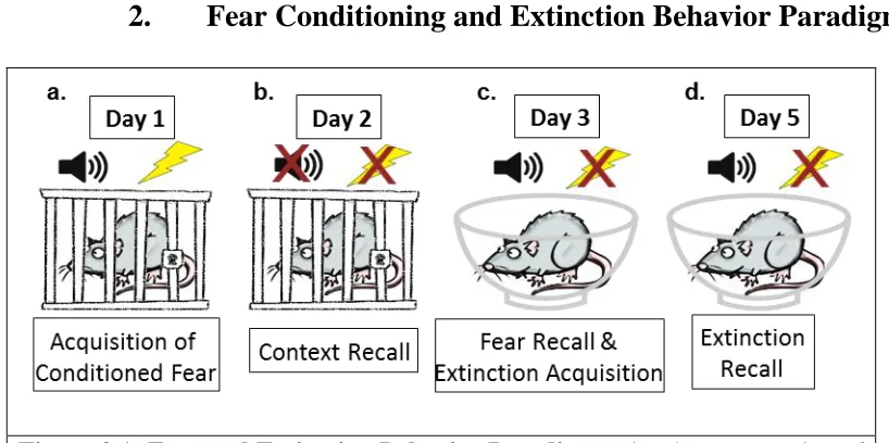
Fear Conditioning and Extinction Behavior Paradigm
Figure 2.1: Fear and Extinction Behavior Paradigm. Visual representation of the fear learning and fear extinction paradigm used in this study.
15
stainless steel rods which were connected to a shocking apparatus which delivered the foot shock (Figure 2.1.a). The shock box was inside a sound-attenuating box containing a ventilation fan and light. Unconditioned freezing behavior was recorded for the first 3 minutes of time in the box. Rats were then conditioned to the unconditioned stimulus (tone) with three 10 second tones (80db, 2kHz) co-terminating with a mild foot shock (1mA, 1 sec) at 60 second intervals. The shock box was cleaned between trials with 5% ammonium hydroxide.
Day 2: Context Recall: On day 2, 24-hours after fear acquisition, rats were placed back into the original shock box (Context A) for 8 minutes without the presentation of tone or shock to assess context conditioned freezing (Figure 2.1.b).
Day 3: Fear Recall & Extinction Acquisition: On day 3, 48-hours after fear
acquisition, rats were assessed for cue conditioned freezing and within-session extinction learning using a novel chamber (Context B) with visual and olfactory cues distinct from those of the shock box (Figure 2.1.c). Animals were brought into the testing facility in a different manner (cages carried individually as opposed to in pairs and pushed on a cart) and tested in a different testing room. Context B was a Plexiglas bowl placed in a sound-attenuating box with a ventilation fan and light, cleaned with 70% ethanol between animals, lined with bedding, and scented with lemon extract (20µL). After unconditioned freezing in the novel environment was assessed for 3 minutes, rats were presented with twenty 10 second tones (80db, 2 kHz) at 60 second intervals.
16
cues consistent with testing on day 3) and presented with twenty 10 second tones (80 db, 2kHz) to assess for fear extinction learning recall (Figure 2.1.d).
B.
Tissue Preparation, Immunofluorescence, & Image
Collection & Analysis
Two hours after the start of extinction recall on day 5, animals were anesthetized by 5% isoflurane inhalation for 5 minutes, transcardially perfused with 100mL of cold 0.1M phosphate buffered saline (pH 7.4) then 300mL cold 4% paraformaldehyde in 0.1M phosphate buffered saline (pH 7.4). Brains were immediately removed and post-fixed for 2 days in 4% paraformaldehyde in 0.1M phosphate buffered saline (pH 7.4) at 4°C. Brains were moved to 15% sucrose for 1 day and 30% sucrose until saturated. Coronal sections at 50µm were cut on a microtome and stored at -20°C in anti-freezing solution (30% ethylene glycol and 30% sucrose in 0.1M phosphate buffer, pH 7.4) until
immunofluorescence processing. One tissue section per rat was labeled for M1 mAChR and the neighboring section was labeled for AChE. Tissue sections labeled and imaged ranged from Bregma -2.05mm to Bregma -2.30mm, according to Paxinos and Watson, The Rat Brain in Stereotaxic Coordinates (2008), all of which contained anterior BLA. All tissue was processed, imaged, and analyzed blindly together.
Immunofluorescence labeling with M1 mAChR antibody was used to measure protein expression in the amygdalar complex. Tissue was washed for 10 minutes 3 times in 0.05M tris-buffered saline solution (TBS) (pH 7.6). Tissue was exposed to pre-block for 30 minutes, consisting of 0.5% Triton and 10% normal goat serum in 0.05M TBS. Tissue was washed for 5 minutes 3 times in 0.05M TBS. Tissue was incubated at room
17
(1:500; rabbit polyclonal; mAChR-M1-Rb-Af340; AB_2571791; Frontier Institute co., ltd.). Frontier Institute’s M1 mAChR antibody specificity was confirmed by Narushima et al. using an M1 knock-out brain (2007). The next day, tissue was washed for 10 minutes 3 times in 0.05M TBS, then incubated for 2 hours, protected from light, in 0.5% triton, 2% normal goat serum, goat anti-rabbit conjugated Alexa Fluor 546 secondary antibody (1:400; A-11035; Thermo Fisher). Tissue was washed for 10 minutes 2 times in 0.05 TBS and exposed to DAPI staining solution (GTX16206; Lot# 821700090;
GeneTex Inc.) for 10 minutes. Tissue was washed for 5 minutes 2 times in 0.05M TBS, 2 times for 5 minutes in 0.05M tris-buffered solution (TB) (pH 7.4), mounted on 0.5% gelatinized slides, and allowed to dry. Slides were coverslipped using ProLong Diamond Antifade Mountant (P36970; Invitrogen, Thermo Fisher) and kept flat at 4°C until imaging. Controls for antibody specificity were conducted by exposing one piece of experimental tissue only to secondary antibody, goat α-rabbit conjugated Alexa Fluor 546 secondary antibody. This tissue, imaged under identical parameters as that for
experimental tissue, showed no detectable staining (data not shown).
18
Units. Mosaic merge and maximal projection settings were optimized for each image using the accompanying LAS AF 3 software. Merged maximal projection images were used to perform all image analysis.
One neighboring tissue section from each rat was labeled for acetylcholinesterase (AChE), which allowed for both measured AChE protein expression and to identify the BLA in M1 mAChR labeled tissue, seeing that AChE cleanly and clearly labels the BLA. The labeling protocol used was identical to that outlined above, with the exception of the serum used, which here was BSA. The primary antibody used was anti-acetylcholine mouse monoclonal antibody (1:75; ZR3 clone, MA3-041, Thermo Fisher) with a chicken anti-mouse Alexa Fluor 647 (1:400; A-21463; Thermo Fisher).
All AChE image collection was conducted using an Invitrogen EVOS FL Auto cell imaging system, equipped with a Cy5 LED light cube (Thermo Fisher). Grid images were collected at 20X with Cy5 light intensity at 65%, exposure 0.1 msec, and gain set to 1.0V. Images were auto-merged by the Invitrogen EVOS FL Auto 2.0 Imaging system; merged images were used to perform all image analysis.
19
histogram mean was recorded. To generate a value of M1 mAChR expression for each animal, the histogram mean of M1 mAChR measured in the BLA was divided by the histogram mean of M1 mAChR measured in the CEA. The same was done for AChE images. All values generated for each rat are an average of each hemisphere (unless otherwise stated) on one tissue section from each animal. Only image analysis of group 2 is shown. The histogram mean values showing average pixel intensity collected and used for individual rat image analysis of M1 mAChR labeled tissue and AChE labeled tissue can be found in Table A.1 and Table A.2, respectively.
C.
Statistical Analysis
20
above 34% freezing were determined to be high responders (poor extinction learning) (n=6 per group). Statistical analysis was conducted using Graph Pad Prism (Prism 5 for Windows, version 5.02). High versus low responders in each group were compared by two-way analysis of variance (ANOVA; high vs. low freezing) with repeated measures across time bins (significance level p<0.05). Bonferroni post-tests were conducted to compare individuals over time. Graphs showing grouped high vs low responder freezing across the experiment (Figure 3.1.e, 3.2.e) were generated by taking the average time bin for each animal during each stage and comparing by two-way ANOVA (high vs low freezers) with Bonferroni post-tests to compare over time. Amygdalar nuclei protein expression was analyzed by a paired t-test (Figure 3.3.d, 3.5.e).
21
Chapter 3: Results
A.
Behavior Results
Two separate groups of rats were submitted to the fear learning and fear
extinction paradigm described above, referred to as group 1 and group 2 (analyzed and discussed separately). High and low responder groups were determined by a median split of the average percent freezing of the last 10 minutes of day 3 cue-conditioned freezing& extinction learning, grouping rats into within session extinction (low responders) versus those who did not undergo within session extinction (high responders).
1.
Group 1 Behavior Results:
22
acquisition, learning that the CS is no longer associated with the US. Group 1 shows varied cue-fear memory recall across individuals but did not show significant differences between groups (F[1,6]=0.60; p=0.698) (Figure 3.1.c, e). When examined alone, the last 10 tone presentations of cue exposure, indicating fear extinction learning, show
significant differences between high and low responders (F[1,6]=8.60; p=0.026) (Figure 3.1.c, e). Animals were grouped into high vs low responder groups based on the median split of freezing during this behavioral stage (median value = 21.77%). Group 1 rats showed individual variation during extinction recall on day 5, but did not show
significant differences between groups (F[1,6]=2.17; p=0.316) (Figure 3.1.d). Extinction recall, on day 5, can also be broken into 2 different phases: the first few tone exposures (tones 1-5, minutes 2-6) which indicate recall of the previously learned extinction memory, and the last 10 tone presentations indicating within session extinction learning for those individuals who had not yet undergone extinction learning, or extinction
23
24
2.
Group 2 Behavior Results:
Although there were observed individual differences, all rats in group 2 acquired fear on day 1 of the behavior paradigm and did not show high vs low grouped differences (F[1,10]=2.46; p=0.148) (Figure 3.2.a, e). Group 2 then shows context recall and within session context extinction on day 2, which shows grouped differences between high and low responders, with high responders showing better context recall than the low
responders (F[1,10]=8.40; p=0.0159) (Figure 3.2.b, e). Grouped differences between high and low responders on day 3 cue-conditioned freezing and extinction learning was found to be significant (F[1,10]=14.47; p=0.0035)(Figure 3.2.c). Fear recall and extinction learning can be broken into 2 different phases: the first few tone exposures (tones 1-5, minutes 5-9) which indicate cue-conditioned freezing in response to experiencing the CS in a new context, and the last 10 CS/tone presentations when the rats were undergoing within-session extinction learning, learning that the CS is no longer associated with the US. All animals in group 2 showed good cue-condition freezing, indicating both high and low responders were able to recall the CS-associated fear memory (F[1,10]=4.61;
p=0.0572)(Figure 3.2.c, e). The last 10 tone presentations of cue exposure, indicating fear extinction learning, show stark differences in group 2 split between high and low responders, indicating low responders underwent cued fear extinction learning
25
minutes of tone exposure (tones 1-5, minutes 2-6) which indicate recall of the previously learned extinction memory, and the last 10 tone presentations indicate within session extinction learning for those individuals who had not yet undergone extinction learning, or extinction learning reinforcement. Group 2 shows very clear distinctions between high and low responders extinction recall, where high responders demonstrated a recovery of the fear memory and subsequent freezing response and low responders demonstrated recall of the fear extinction memory (F[1,10]=13.23; p=0.0046) (Figure 3.2.d, e). Finally, both high and low responders undergo within session extinction learning (F[1,10]=0.32; p=0.585) (Figure 3.2.d, 3.2.e). Group 2 rats demonstrate clear grouped differences in the extinction processes, including acquisition and recall. This test does not indicate if recall differences are due to individual differences in ability to recall extinction memory or differences in ability to consolidate the extinction memory.
26
27
B.
Fluorescent Imaging Results and Analysis
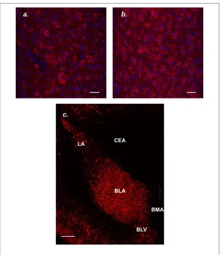
M1 mAChR labeling and distribution observed in this study was similar to that observed in McDonald and Mascagni (2010). Dense M1 mAChR cell body labeling is evident in many temporal lobe structures imaged, including the LA, BLA, BLV, and the periform cortex (Pir), where cell body labeling is absent in BMA and CEA (Figure 3.3.a, b, A.1). The M1 mAChR positive cells labeled in Figures 3.3 and 3.4 have the
28
Labeling in the BLA was primarily cell body labeling, which can be clearly seen in the higher power image showing M1 mAChR + DAPI labeling (Figure 3.4. a, b). It has been reported that approximately 85% of neurons in the basolateral nuclear complex (BLC) of the amygdala (consisting of the LA, BLA, and BM) are positive for
calcium/calmodulin protein kinase II (CaMK), a known marker of pyramidal neurons, and almost all CaMK positive/pyramidal neurons in the BLC are also M1 mAChR positive cells (McDonald, 1992; McDonald and Mascagni 2010). However, when
29
30
31
Tissue sections for M1 mAChR labeling were selected based on McDonald and Mascagni’s analysis of density of M1 mAChR labeling from rostral to caudal amygdala, with the most robust M1 mAChR immunoreactivity seen in the anterior divisions of the basolateral amygdala (2010). Tissue sections labeled and imaged ranged from Bregma -2.05mm to Bregma -2.30mm, according to Paxinos and Watson, The Rat Brain in Stereotaxic Coordinates (2008), all of which contained BLA seen to be densely labeled with M1 mAChR in McDonald and Mascagni (2010). The CEA showed some neuropil labeling and an absence of cell body labeling across all animals (Appendix A, Figure A.1). We felt confident normalizing BLA M1 mAChR intensity to CEA M1 mAChR intensity due to McDonald and Mascagni’s assertion that the majority of differences between amygdalar nuclei was due to cell body labeling, not neuropil labeling (2010).
One issue that was observed upon image collection and analysis was that overall intensity varied, not just in the amygdala, but the entire image. This begs the question as to if variations are due to overall artefactual intensity or legitimate changes in receptor expression. This issue was the motivation behind using the histogram mean BLA value divided by the mean CEA value. Control sections with no primary antibody labeling were imaged and analyzed but generate no measureable autofluorescence. Additionally,
32
mAChR expressers appear to have a uniform brightness or dimness in amygdalar images, this analysis shows that the changes observed in the BLA are greater than those seen across an entire tissue section and are thus not due to differences in perfusion or tissue processing. More controls will need to be imaged in order to further prove this finding and validate this technique and the correlation findings.
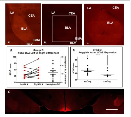
While some structures can be differentiated by examining M1 mAChR labeling alone, AChE labeling was conducted to cleanly and reliably identify temporal lobe nuclei. AChE was one of the labels used in Paxinos and Watson (2008) to differentiate between brain regions due to its clearly defined expression pattern. Amygdala AChE expression has been observed to be some of the densest in the brain, which allowed for clean distinguishing of amygdalar nuclei (Ben-Ari et al. 1997; Girgis 1980). This is useful in this study for amygdalar nuclei separation.
33
mAChR image analysis. Image 3.5.f is a confocal image showing the most drastic left vs right disparities. This begs the question as to if AChE functionality varies between left and right amygdala in each animal. Further analysis is required to resolve these issues.
34
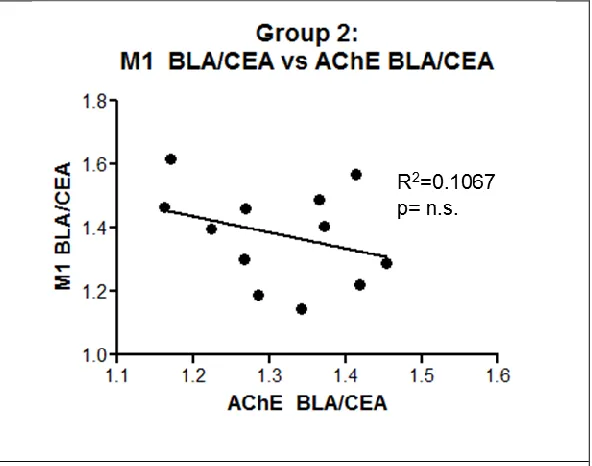
Correlation analysis between AChE and M1 mAChR was conducted to examine if there was a correlation between these two protein’s expressions of these two proteins in the BLA or if the different protein levels observed existed independent of overall
cholinergic influence. No statistical significance was observed between the two proteins expression level (F[1,10]=1.194; p>0.1) (FUGURE 3.6). This finding suggests that if significant correlations are observed, they are not due to increased cholinergic proteins, as was suggested in early studies (Power et al., 2003b; Gold, 2003).
Figure 3.6: Correlation- M1 mAChR vs AChE Protein Expression Levels. No significant correlation exists between BLA M1 mAChR expression and BLA AChE expression.
C.
Correlation between Behavior and Image Analysis
35
of measured freezing behavior in specific time bins, outlined in TABLE 3.1 (group 2 only). These time bins were also used to generate figure 3.1.e and 3.2.e.
Table 3.1: Freezing behavior representing stages of fear learning used for imaging correlation analysis
Learning processes
Stage of behavior paradigm
Time bins used for analysis of learning processes Correlation analysis Fear Acquisition Day 1: Acquisition of Conditioned Fear
Average of last 3 minutes
M1 BLA/CEA: p=0.0251 AChE BLA/CEA: p>0.05; n.s.
Context Fear Recall
Day 2: Context-Conditioned Freezing
Average of minutes 2-5
M1 BLA/CEA: p>0.05; n.s. AChE BLA/CEA: p>0.05; n.s.
Cued-Fear Recall
Day 3: Cued-Fear Recall &
Extinction Acquisition
Average of tones 2-6 (minutes 5-9)
M1 BLA/CEA: p>0.05; n.s. AChE BLA/CEA: p>0.05; n.s.
Extinction Acquisition
Day 3: Cued-Fear Recall &
Extinction Acquisition
Average of tones 10-19 (minutes 13-22)
M1 BLA/CEA: p>0.05; n.s. AChE BLA/CEA: p>0.05; n.s.
Extinction Recall
Day 5: Extinction Recall
Average of tones 1-5 (minutes 2-6)
M1 BLA/CEA: p=0.0230 AChE BLA/CEA: p>0.05; n.s.
Delayed Extinction Acquisition
Day 5 Extinction Recall
Average of tones 10-19 (minutes 11-21)
M1 BLA/CEA: p>0.05; n.s. AChE BLA/CEA: p>0.05; n.s.
1.
M1 mAChR behavior correlation results
The histogram mean values showing average pixel intensity collected and used for individual rat image analysis of M1 mAChR labeled tissue can be found in TABLE A.1. Correlation analysis between M1 mAChR expression and fear acquisition,
36
be statistically significant (F[1,10]=6.929; p=0.0251) (Figure 3.7.a.). Correlation analysis between M1 mAChR expression and contextual-fear recall, represented by the average percent freezing during minutes 2-5 of day 2, was found not to be statistically significant (F[1,10]=0.2724; p=0.6131) (Appendix A, Figure A.3.a). Correlation analysis between M1 mAChR expression and cued-fear recall, represented by the average percent freezing during minutes 5-9, capturing behavior after the first tone presentation of day 3, was found not to be statistically significant (F[1,10]=0.040; p=0.8455) (Figure 3.7.b).
Figure 3.7: Correlation- M1 mAChR expression levels vs fear learning processes. Figures a and b examine the correlation between M1 mAChR BLA expression levels and different aspects of the fear learning process, including fear acquisition, a, measured by the average percent freezing during last 3 minutes of day 1, and cued fear recall (or consolidation), b, measured by the average percent freezing during minutes 2-5 of day 3. No statistical significance was found between M1 mAChR expression and fear recall, b. Statistical significance was found between fear acquisition and M1 mAChR BLA expression, a.
37
pyramidal neurons in the BLA, allowing for inhibition of M-current which allows for synaptic plasticity to occur. A number of studies discussed previously have found similar results, reinforcing this finding (Rudy, 1996; Young et al., 1995; Feiro and Gould, 2005; Jiang et al., 2016; Fornari et al., 2000; for review see Wilson and Fadel, 2017).
The non-significant correlation observed between contextual- and cued-fear recall seems to indicate that M1 mAChR are not functioning in either consolidation or recall of the fear memory. However, several studies would disagree with this finding. A recent study by Patricio et al., found that M1 mAChR are important in the recall of contextual-fear memories (2017). Similarly, Young and Thomas found that specific M1 mAChR activation increases the consolidation of fear memories (2014). Young, Bohenek, and Fanselow, however, found that administration of a mAChR inhibitor actually increased consolidation of the fear memory (1995). Further studies are necessary to elucidate the precise function and involvement of M1 mAChR in the fear learning process.
38
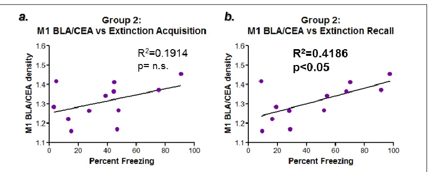
10 tone presentations of day 5, was found not to be statistically significant (F[1,10]=0.5613; p=0.4710) (Appendix A, Figure A.3.b).
Figure 3.8: Correlation- M1 mAChR expression levels vs the extinction learning processes. Figures 3.8.a and b examine the correlation between BLA M1 mAChR expression levels and different aspects of the extinction learning process, including extinction acquisition, a, measured by the average percent freezing during tones 10-19 on day 3, and extinction recall (or consolidation), b, measured by the average percent freezing during tones 1-5 on day 5. A trend was observed between extinction acquisition and M1 mAChR expression, a, where a statistically significant correlation was observed between extinction recall and BLA M1 mAChR expression.
39
memory. This study does not allow for the differentiation between consolidation and recall so this finding could, likewise, be indicating that high M1 mAChR protein
expression prevents consolidation of extinction memories. This finding is the opposite of the original hypothesis, which stated that higher M1 mAChR expression was expected to improve extinction acquisition.
This finding also contradicts previous research conducted in the Mott and Wilson labs by Joshua McElroy, a study which found that animals with higher BLA M1 mAChR protein expression were better able to undergo extinction learning (McElroy, 2016). The current study’s finding could be due to the solidity of the previously acquired fear
memory, which is enhanced by high levels of M1 mAChR protein expression in the BLA, and that more extinction training is required to allow for proper extinction memory recall in the high responding rats. Delayed extinction acquisition, measuring the within session extinction that occurs on the second round of CS exposure, could allow individuals to better acquire the extinction memory. Delayed extinction acquisition was found not to be correlated to M1 mAChR expression. This is to be expected due to the poor correlation seen between M1 expression and the initial extinction acquisition.
40
mAChR inhibition impaired rat’s ability to extinguish conditioned fear (2013). Schroeder and Packard showed that mAChR agonists improve ability to extinguish amphetamine-induced place preference (2004). These findings, along with those in the present study, paint a confusing picture of M1 mAChR involvement in fear extinction. As it currently stands, it seems safe to say that mAChR are, at the very least, important in extinction learning. My data, along with previous work, allow for the solidification of the hypothesis that M1 mAChR are highly functional in the BLA’s role in extinction
acquisition and extinction memory consolidation. The precise function and if up or down regulation of M1 mAChR would be beneficial in extinction learning, however, is
somewhat more confusing. Previous literature outlined above would seem to indicate that more mAChR functionality would mean better extinction consolidation, although not specifically speaking to which receptor subtype. This study found that higher M1 mAChR expression, specifically, indicates worse extinction consolidation.
2.
AChE behavior correlation results
41
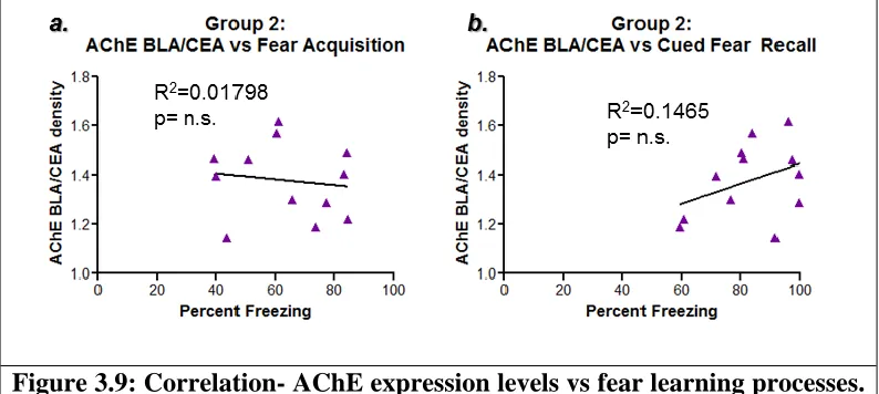
capturing behavior after the first tone presentation of day 3, was found not to be statistically significant (F[1,10]=1.716; p=0.2195) (Figure 3.9.b).
Figure 3.9: Correlation- AChE expression levels vs fear learning processes.
Figures 3.9.a and b examine the correlation between AChE expression levels and different aspects of the fear learning process, including fear acquisition, a, measured by the average percent freezing during last 3 minutes of day 1, and cued fear recall (or consolidation), b, measured by the average percent freezing during minutes 2-5 of day 3. No statistical significance was found for either analysis.
Correlation analysis between AChE expression and extinction acquisition, represented by the average percent freezing during minutes 13-22, capturing behavior through the last 10 tone presentations of day 3, was found not to be statistically significant (F[1,10]=0.6590; p=0.4358) (Figure 3.10.a). Correlation analysis between AChE expression and extinction recall, represented by the average percent freezing during the first 5 minutes after the first tone presentation (minutes 2-6) of day 5, was not found to be statistically significant (F[1,10]=0.1703 ; p=0.6885) (Figure 3.10.b).
42
through the last 10 tone presentations of day 5, was found not to be statistically significant (F[1,10]=0.5613; p=0.5914) (appendix A, Figure A.4.b).
Figure 3.10: Correlation- AChE expression levels vs Extinction Learning Processes. Figures 3.10.a and b examine the correlation between AChE expression levels and different aspects of the extinction learning process, including extinction acquisition, a, measured by the average percent freezing during tones 10-19 on day 3, and extinction recall (or consolidation), b, measured by the average percent freezing during tones 1-5 on day 5. No statistical significance was found for either analysis.
While studies have examined the role of AChE in the fear learning process, many of those studies have done so using acetylcholinesterase inhibitors or have been studies examining nicotinic receptor functioning, not AChE. This can be done because AChE inhibitors reverse nicotine withdrawal effects (Wilson and Fadel, 2017). Understanding what roles all aspects of the cholinergic system play in fear and extinction learning and memory is necessary if treatments for fear related disorders are to be developed
43
Chapter 4: Conclusions
The observable deviation of group 2 rats into two different responder groups, high and low responders was similar to that observed by Sharko et al. (2016) and McElroy (2015). Similar to these two studies, the results of this study show that rats demonstrate observable and quantifiable individual differences that go beyond behavioral differences. While grouped differences cannot be seen in ability to acquire fear, when comparing individual differences in this ability to protein expression, substantial individual differences can be observed, and these differences positively correlate to M1 mAChR protein expression. These data seem to indicate that an animal’s ability to acquire cued-fear is dependent upon M1 mAChR expression, with better cued-fear acquisition correlating to higher M1 mAChR BLA expression.
Statistically significant differences were observed between high and low responder groups in ability to acquire cued-extinction memory due to repeated CS exposure during the second half of the day 3 trial. When examining correlation between individual differences in extinction acquisition and M1 mAChR expression, a trend is visible but no statistical differences were observed. A significant correlation was
observed between M1 mAChR expression level and rats ability to recall cued-extinction memory during the first 5 tone presentations on day 5. This behavior paradigm does not indicate if this difference is due to M1 mAChR function in extinction memory
44
functioning in extinction consolidation (Boccia et al., 2009; Santini et al., 2012; Schroder and Packard, 2004; Zelikowsky et al., 2013).
The several significant correlative findings discussed implicate M1 mAChR in the initial fear acquisition and the consolidation of extinction memory. Together, these two findings seem to indicate that those individuals that had better initial fear acquisition, potentially caused by higher M1 mAChR expression levels, had worse extinction recall or consolidation. These data allow for two different interpretations: M1 mAChR function in directly inhibiting the extinction learning pathway (IL pathway), or that M1 mAChR in the BLA primarily function in strengthening the fear learning pathway (PL pathway) which inhibit extinction by making the strongly formed fear memory difficult to
overcome. If M1 mAChR are directly functioning in inhibition of the IL pathway, giving animals an M1 mAChR antagonist during extinction learning processes would
theoretically result in improved extinction learning, and a M1 mAChR agonist would inhibit extinction learning. Seeing that the opposite has been observed, where mAChR antagonists impair extinction and mAChR agonists enhance extinction, this interpretation of these data is unlikely to be correct (Boccia et al., 2009; Santini et al., 2012; Schroeder and Packard, 2004; Zelikowsky et al., 2013). However, the interpretation could be true that M1 mAChR are primarily functioning in the strengthening of fear learning pathway, creating a stronger fear memory than that of individuals with less dense BLA M1
45
Furthermore, this interpretation does not discount a role for M1 mAChR in fear
extinction, for beyond-physiological activation or inhibition of M1 mAChR in the BLA during fear extinction learning processes could still be effecting ability to undergo extinction acquisition, consolidation, or recall.
No significant correlations were observed between AChE protein expression and fear learning or extinction phases or between extent of M1 mAChR expression. This seems to indicate that overall cholinergic protein expression is not the driver of individual ability to learn or extinguish fears.
Few studies thoroughly examine fear learning and even fewer examine fear extinction. In studies that do examine these processes, there is such a myriad of
behavioral paradigms utilized, comparing any two studies can challenging. Attempting to understand the mechanisms of not only fear and emotion, but all aspects of behavior, is a relatively new aspect of neuroscience, which is itself a relatively new and unexplored field. Nevertheless, it is crucial. Understanding the mechanisms behind fear and extinction learning, a primal and complex behavioral system, would aid in our
46
expected to have similar effects in human trials. This study along with previous fear and extinction learning studies would seem to encourage the use of M1 mAChR positive allosteric modulators (PAM) in the treatment of PTSD. PAMs reversibly bind to allosteric receptor sites, causing conformation changes that result in an increase in receptor cooperativity and increasing binding of its neurotransmitter, such as ACh in the case of mAChR (Jakubik and El-Fakahany, 2010). A survivor of abuse or veteran of war seeking treatment for PTSD would be encouraged to undergo exposure therapy. During therapy sessions, individual identifiable triggers would be presented, terminating with the PAM binding M1 mAChR in the amygdala. Administration timing would be critical, seeing that the consolidation process occurs for a limited period of time. The drug would need to be given in time for it to pass the blood-brain barrier, enter the amygdala, and react with receptors at the beginning of the consolidation process. This should, in theory, allow for improved consolidation of the newly acquired extinction memory. Rodent trials should give similar results; PAM treatment immediately following extinction acquisition should cause all individuals to have a low percent freezing during extinction memory recall. Such treatment given before fear acquisition would be expected to have a similar result, seeing that preventing strong fear memory formation could cause a weaker PL fear pathway and allow for improved extinction. However, this would also not be
47
48
Chapter 5: Future Directions
Additional image collection is necessary for the solidification of the results of this study. The difficulty with which metabotropic receptors are labeled and imaged made high power confocal image collection a necessity, as opposed to widefield microscopy used for AChE labeled tissue imaging. Seeing the considerable amount of time M1 mAChR image collection takes and the cost of collection, it was first necessary to determine if significant, meaningful correlations existed between behavior and protein expression. Now that such correlation has been established, it is pertinent to continue image collection and generate amygdalar images from no fewer than 3 tissue sections for each animal in group 2. Additionally, proper controls from each animal must be
collected. This would consist of image collection of a second region unassociated with the described behavioral process, as presented and described in figure A.2. It is our belief that further image collection will strengthen the protein-behavioral correlations observed. In addition, continued EVOS image collection of AChE labeled tissue, bringing theimage collection up to at least 3 sections per animal, is also valuable for elucidating any
correlations. Additionally, analysis of hemispheric differences in AChE expression within each animal could prove to be a more valuable means for evaluating the protein’s role in the fear and extinction learning processes.
49
labs, M2 mAChR were implicated in the modulation of cholinergic terminals within the BLA (Fajardo-Serrano et al., 2017). Examining M2 mAChR expression and the PV interneuron population in behaved tissue could give valuable insight into how the interneuron population within the BLA relates to fear learning and extinction processes. Seeing that many behavioral studies utilize scopolamine, a non-selective mAChR
50
References
Akirav I, Raizel H, Maroun M. 2006. Enhancement of conditioned fear extinction by infusion of the GABA(A) agonist muscimol into the rat prefrontal cortex and amygdala. Eur J Neurosci 23:758–764.
American Psychiatric Association. 2013. Diagnostic and statistical manual of mental disorders: DSM-5. Washington, D.C: American Psychiatric Association. Anagnostaras SG, Maren S, Fanselow MS. 1995. Scopolamine selectively disrupts the
acquisition of contextual fear conditioning in rats. Neurobiol Learn Mem 64:191– 194.
Baldi E, Bucherelli C. 2010. Substantia nigra, nucleus basalis magnocellularis and basolateral amygdala roles in extinction of contextual fear conditioning in the rat. Neurobiol Learn Mem 94:199–205.
Baldi E, Bucherelli C. 2015. Brain sites involved in fear memory reconsolidation and extinction of rodents. Neurosci Biobehav Rev 53:160–190.
Ben-Ari Y, Zigmond RE, Shute CC, Lewis PR. 1977. Regional distribution of choline acetyltransferase and acetylcholinesterase within the amygdaloid complex and stria terminalis system. Brain Res 120:435–444
Boccia MM, Blake MG, Baratti CM, McGaugh JL. 2009. Involvement of the basolateral amygdala in muscarinic cholinergic modulation of extinction memory
51
Bucherelli C, Baldi E, Mariottini C, Passani MB, Blandina P. 2006. Aversive memory reactivation engages in the amygdala only some neurotransmitters involved in consolidation. Learn Mem 13:426–430.
Fajardo-Serrano A, Liu L, Mott DD, McDonald AJ. 2017. Evidence for M2 muscarinic receptor modulation of axon terminals and dendrites in the rodent basolateral amygdala: an ultrastructural and electrophysiological analysis. Neuroscience, http://dx.doi.org/10.1016/j.neuroscience.2017.06.019
Feiro O, Gould TJ. 2005. The interactive effects of nicotinic and muscarinic cholinergic receptor inhibition on fear conditioning in young and aged C57BL/6 mice. Pharmacol Biochem Behav 80:251–262.
Fendt M, Fanselow MS. 1999. The neuroanatomical and neurochemical basis of conditioned fear. Neurosci Biobehav Rev 23:743-760.
Fornari RV, Moreira KM, Oliveria MGM. 2000. Effects of the selective M1 muscarinic antagonist dicyclomine on emotional memory. Learn Memory 7:287-292. Gale GD, Anagnostaras SG, Godsil BP, Mitchell S, Nozawa T, Sage JR, Wiltgen B,
Fanselow MS. 2004. Role of the basolateral amygdala in the storage of fear memories across the adult lifetime of rats. J Neurosci 24:3810–3815. Girgis M. 1980. Acetylcholinesterase enzyme localization in the amygdala: a
comparative histochemical and ultrastructural study. Acta Anat (Basel) 106:192– 202
52
Herry C, Ciocchi S, Senn V, Demmou L, Muller C, Luthi A. 2008. Switching on and off fear by distinct neuronal circuits. Nature 454:600–606.
Herry C, Garcia R. 2002. Prefrontal cortex long-term potentiation, but not long-term depression, is associated with the maintenance of extinction of learned fear in mice. J Neurosci 22:577–583.
Horn SR, Charney DS, Feder A. 2016. Understanding resilience: New approaches for preventing and treating PTSD. Exp Neurol 284:119-132.
Jakubik J, El-Fakahany EE. 2010. Allosteric modulation of muscarinic acetylcholine receptors. Pharmaceuticals 3(9):2838-2860.
Jiang L, Kundu S, Lederman JD, Lopez-Hernandez GY, Ballinger EC, Wang S, Talmage DA, Role LW. 2016. Cholinergic signaling controls conditioned fear behaviors and enhances plasticity of cortical-amygdala circuits. Neuron 90:1057–1070. Kandel ER, Schwartz JH, Jessell TM. Siegelbaum SA, Hudspeth AJ. Principles of Neural
Science, Fifth Edition. New York:McGraw-Hill Professional Publishing, 2013. Kessler RC, Chiu WT, Demler O, Merikangas KR,Walters EE. 2005. Prevalence,
severity, and comorbidity of 12-month DSM-IV disorders in the National Comorbidity Survey Replication. Arch Gen Psychiatry 62:617–627. Likhtik E, Popa D, Apergis-Schoute J, Fidacaro GA, Pare D. 2008. Amygdala
intercalated neurons are required for expression of fear extinction. Nature 454:642–645.
53
McDonald AJ, Mascagni F, Guo L. 1996. Projections of the medial and lateral prefrontal cortices to the amygdala: a Phaseolus vulgaris leucoagglutini study in the rat. Neuroscience 71:55-75.
McDonald AJ, Mascagni F. 2010. Neuronal localization of m1 muscarinic receptor immunoreactivity in the rat basolateral amygdala. Brain Struct Funct 215:37-48. McDonald AJ. 1992. Projection neurons of the basolateral amygdala: A correlative Golgi
and retrograde tract tracing study. Brain Res Bull 28:179-185.
McElroy JR. 2016. Muscarinic Acetylcholine Receptor M1’s Impact on Fear Extinction Learning. University of South Carolina.
Milad MR, Quirk GJ. 2002. Neurons in medial prefrontal cortex signal memory for fear extinction. Nature 420:70–74.
Milad MR, Quirk GJ. 2012. Fear Extinction as a Model for Translational Neuroscience: Ten Years of Progress. Annu Rev Psychol 63:129-151.
Muller JF, Mascagni F, McDonald AJ. 2011. Cholinergic innervation of pyramidal cells and parvalbumin-immunoreactive interneurons in the rat basolateral amygdala. J Comp Neurol 519:790–805.
Myers KM, Davis M. 2007. Mechanisms of fear extinction. Molec Psychiatry 12:120– 150.
Narushima M, Uchigashima M, Fukaya M, Matsui M, Manabe T, Hashimoto K,
54
Orsini CA, Maren S. 2012. Neural and cellular mechanisms of fear and extinction memory formation. Neurosci Biobehav Rev 36:1773-1802.
Pape HC, Pare D. 2010. Plastic synaptic networks of the amygdala for the acquisition, expression, and extinction of conditioned fear. Physiol Rev 90:419–463. Passani MB, Cangioli I, Baldi E, Bucherelli C, Mannaioni PF, Blandina P. 2001.
Histamine H3 receptor-mediated impairment of contextual fear conditioning and in vivo inhibition of cholinergic transmission in the rat basolateral amygdala. Eur J Neurosci 14:1522–1532.
Patricio RR, Soares JCK, Oliveria MGM. 2017. M1 muscarinic receptors are necessary for retrieval of remote context fear memory. Phsyiol Behav 169:202-207. Pavlov IP. 1927. Conditioned reflexes: an investigation of the psychological activity of
the cerebral cortex. Oxford University Press, London.
Paxinos G, Watson C. 2008. The Rat Brain in Stereotaxic Coordinates, 6th revised edition. Academic Press; New York.
Power AE, McIntyre CK, Litmanovich A, McGaugh JL. 2003a. Cholinergic modulation of memory in the basolateral amygdala involves activation of both m1 and m2 receptors. Behav Pharmacol 14:207–213.
Power AE, Vazdarjanova A, McGaugh JL. 2003b. Muscarinic cholinergic influences in memory consolidation. Neurobiol Learn Mem 80:178–193.
55
Quirk GJ, Mueller D. 2008. Neural mechanisms of extinction learning and retrieval. Neuropsychopharmacology 33:56–72.
Rudy JW. 1996. Scopolamine administered before and after training impairs both contextual and auditory-cue fear conditioning. Neurobiol Learn Mem 65:73–81. Sah P, Faber ESL, Lopez De Armentia M, Power J. 2003. The amygdaloid complex:
anatomy and physiology. Physiol Rev 83:803-834.
Sah P, Westbrook RF. 2008. Behavioral neuroscience: the circuit of fear. Nature 454:589-590.
Santini E, Sepulveda-Orengo M, Porter JT. 2012. Muscarinic receptors modulate the intrinsic excitability of infralimbic neurons and consolidation of fear extinction. Neuropharmacology 37:2047-2056.
Schroeder JP, Packard MG. 2004. Facilitation of memory for extinction of drug-induced conditioned reward: role of amygdala and acetylcholine. Learn Mem 11:641-647. Senn V, Wolff SBE, Herry C, Grenier F, Ehrlich I, Grundermann J, Fadok JP, Muller C,
Letzkus JJ, Luthi A. 2014. Long-range connectivity defines behavioral specificity of amygdala neurons. Neuron 81:428-437.
Sharko AC, Fadel JR, Kaigler KF, Wilson MA. 2016. Activation of Orexin/Hypocretin Neurons is Associated with Individual Differences in Cued Fear Extinction. Physiol Behav. http://dx.doi.org/10.1016/j.physbeh.2016.10.008
56
Tamelian TL, Jaycox LH, 2008. Invisible Wounds of War: Psychological and Cognitive Injuries, Their Consequences, and Services to Assist Recovery. Santa Monica, CA.
Thiele A. 2013. Muscarinic Signaling in the Brain. Annu Rev Neurosci 36:271-94. Vazdarjanova A, McGaugh JL. 1999. Basolateral amygdala is involved in modulating
consolidation of memory for classical fear conditioning. J Neurosci 19:6615– 6622.
Wilson MA, Fadel J R. 2017. Cholinergic Regulation of Fear Learning and Extinction. J Neurosci Res 95:836-852.
Wilson MA, Reagan LP. 2016. Special Issue: New Perspectives in PTSD. Exp Neurol 284:115-118.
Young MB, Thomas SA. 2014. M1-muscarinic receptors promote fear memory consolidation via phospholipase C and the M-current. J Neuroscience 34:1570-1578.
Young SL, Bohenek DL, Fanselow MS. 1995. Scopolamine impairs acquisition and facilitates consolidation of fear conditioning: differential effects for tone vs context conditioning. Neurobiol Learn Mem 63:174-180.
Zelikowsky M, Hast TA, Bennett RZ, Merjanian M, Nocera NA, Ponnusamy R, Fanselow MS. 2013. Biol Psychiatry 73:345-352.
57
Appendix A: Supplemental Data and Figures
58
59
Figure A.3: Correlation- M1 mAChR expression levels vs contextual fear recall and delayed extinction acquisition. Figures a and b examine the correlation between M1 mAChR BLA expression levels and different aspects of the fear learning and extinction process. In figure a, context recall values were generated by the average percent freezing during minutes 2-5 of day 2. No statistical significance was found between M1 mAChR expression and context recall. In figure b, delayed extinction acquisition values were generated by the average percent freezing during minutes 11-21 of day 5. No statistical significance was found between M1 mAChR BLA expression and delayed extinction acquisition.
60
Table A.1: The histogram average pixel intensity of M1 mAChR labeled tissue with hemispheres averaged
Designated Rat Number Averaged M1 intensity: entire image Averaged M1 intensity: LA + BLA
Averaged M1 intensity: BLA Averaged M1 intensity: CEA Averaged M1 intensity: BLA/CEA
186 41.59 49.47 51.88 38.00 1.36
187 38.19 43.03 43.94 34.69 1.27
188 30.02 33.67 35.68 26.00 1.37
189 47.50 52.13 53.09 37.43 1.42
190 29.83 33.29 35.04 30.17 1.16
191 37.78 42.94 45.51 33.90 1.34
192 48.01 52.01 53.46 42.14 1.27
193 32.20 35.28 37.54 32.11 1.17
194 59.73 69.78 72.11 49.59 1.45
195 33.49 40.01 40.77 33.31 1.22
196 40.84 44.75 48.95 34.62 1.41
197 39.91 42.89 44.68 34.76 1.28
Table A.2: The histogram average pixel intensity of AChE labeled tissue with hemispheres averaged
Designated Rat Number Averaged AChE intensity: entire image Averaged AChE intensity: LA + BLA
Averaged AChE intensity: BLA Averaged AChE intensity: CEA Averaged AChE intensity: BLA/CEA
186 69.47 76.53 83.60 56.12 1.49
187 55.80 61.09 63.94 49.69 1.29
188 39.50 45.28 48.50 34.55 1.40
189 33.91 36.27 37.73 30.89 1.22
190 40.12 46.13 49.24 33.64 1.46
191 31.70 32.78 34.89 30.14 1.16
192 37.41 43.62 47.52 32.80 1.45
193 39.79 49.65 54.34 33.65 1.61
194 32.32 36.94 39.38 30.56 1.29
195 36.29 41.42 43.94 31.41 1.40
196 35.79 46.77 51.43 32.78 1.57