University of South Carolina
Scholar Commons
Theses and Dissertations
2016
Cdk8 and Cdk19 as Novel Regulators of BMP4
Induced EMT in Cancer
Anne Elizabeth Serrao University of South Carolina
Follow this and additional works at:https://scholarcommons.sc.edu/etd
Part of theChemistry Commons
Recommended Citation
Cdk8 and Cdk19 as Novel Regulators of BMP4 Induced EMT in Cancer
by
Anne Elizabeth Serrao
Bachelor of Arts
Washington & Jefferson College, 2013
Submitted in Partial Fulfillment of the Requirements
For the Degree of Master of Science in
Chemistry
College of Arts and Sciences
University of South Carolina
2016
Accepted by:
Mythreye Karthikeyan, Director of Thesis
Maksymilian Chruszcz, Reader
Abstract
Bone morphogenetic protein 2/4 (BMP2/4), members of the transforming growth
factor β (Tgf-β) superfamily, play a dichotomous role in the progression of
cancers through cell growth suppression and the enhancement of tumorigenesis
in a variety of cancers. Cyclin dependent kinase 8 (CDK8), a protein kinase that
regulates gene transcription, is involved in a variety of cancers including breast
and pancreatic cancers as well as in mediating BMP2 signaling via its canonical
Smad1 pathway. Our studies show that BMP4 induced EMT signals primarily
though Smad1 and that Yap1, a transcriptional co-activator, is necessary for
BMP4 induced EMT. Using a highly specific ATP competitive inhibitor of Cdk8
and its twin kinase Cdk19, Senexin B, we find that blocking the kinase activity of
Cdk8/19 decreases BMP4 transcription of genes associated with EMT and
inhibits BMP4 induced invasion. By separately reducing the expression of Cdk8
and Cdk19, we show for the first time that these kinases have both distinct and
overlapping functions. Our findings suggest that Cdk8 and Cdk19, along with
Yap1, are crucial for dictating BMP4/Smad1 responses during EMT and each of
these factors could serve as a selective therapeutic target to block BMP4
Table of Contents
Abstract ... iii
List of Figures... v
Chapter 1: Introduction ... 1
Chapter 2: Results ... 5
Chapter 3: Discussion ... 27
Chapter 4: Materials and Methods ... 30
References ... 34
Appendix A: OvCa429 Data ... 37
Appendix B: Panc1 Data ... 51
Appendix C: CT26 Data ... 63
Appendix D: Py2T Data ... 70
List of Figures
Figure 2.1 BMP4 Induced EMT is Suppressed on Compliant Substrates ... 13
Figure 2.2 Figure 2.2 (Figure 2.1 continued) ... 14
Figure 2.3 BMP4 Induced EMT is Driven by Smad1 ... 15
Figure 2.4 Figure 2.4 Continued ... 16
Figure 2.5 Yap1 Enhances BMP4 Induced EMT ... 17
Figure 2.6 Figure 2.5 Continued ... 18
Figure 2.7 The Kinase Activity of Cdk8/19 Enhances BMP4 Induced EMT ... 19
Figure 2.8 Figure 2.7 Continued ... 20
Figure 2.9 Cdk8/19 Enhance BMP4 EMT and Exhibit Differential Effects ... 21
Figure 2.10 Figure 2.9 Continued ... 22
Figure 2.11 Supplementary Figure 1 ... 23
Figure 2.12 Supplementary Figure 1 (continued) ... 24
Figure 2.13 Supplementary Figure 2 ... 25
Figure 2.14 Supplementary Figure 3 ... 26
Figure A.1 Effect of BMP on Transcript Levels & Migration ... 38
Figure A.2 Effects of BMP on Smad Signaling ... 39
Figure A.3 Role of Alk3/6 & Alk5/7 on BMP4 Induced EMT Transcript Levels ... 40
Figure A.7 Effects of Matrix Rigidity on BMP4 Induced EMT and Apoptosis ... 44
Figure A.8 Effect of Matrix Rigidity on BMP Induced EMT/Receptor Levels ... 45
Figure A.9 Effect of Yap5SA on BMP Induced EMT ... 46
Figure A.10 Effects of SnxB on BMP4 Induced EMT ... 47
Figure A.11 Effects of shCdk8 and shCdk19 on BMP4 Induced EMT ... 48
Figure A.12 Effect of Wnt3a and Tgf-β1/2 on EMT ... 49
Figure A.13 Effects of R3 and Wnt3a on EMT ... 50
Figure B.1 Effects of BMP on Migration ... 52
Figure B.2 Role of BMP on Integrins and Smad Signaling ... 53
Figure B.3 Effect of Blebbistatin and Y27632 on Yap Localization ... 54
Figure B.4 Effects of Matrix Rigidity on BMP4 Induced EMT 1 ... 55
Figure B.5 Effects of Matrix Rigidity on BMP4 Induced EMT 2 ... 56
Figure B.6 Effects of Matrix Rigidity on BMP4 induced EMT 3 ... 57
Figure B.7 Effect of SnxB on BMP4 Induced EMT IF ... 58
Figure B.8 Effect of BMP4, Snx, shCdk8, and shCdk9 on Cell Proliferation ... 59
Figure B.9 Effect of shCdk8 (kinase dead) on BMP4 Induced EMT ... 60
Figure B.10 Effects of Wnt3a on BMP4/Tgfβ-1 Induced EMT... 61
Figure B.11 Effects of Matrix Rigidity on Cell Stiffness ... 62
Figure C.1 Effect of SnxB on CT26 Cell Invasion ... 64
Figure C.6 Effects of SnxB, shCdk8, shSmad4, & shB-catenin on Invasion 3 ... 69
Figure D.1 Effects of SnxB on Tgf-β1 Induced Invasion. ... 71
Figure D.2 Effects of SnxB on Tgf−β1 and BMP4 Induced EMT ... 72
Figure E.1 Effects of BMP4 on EMT in OvCa4 Cells ... 74
Figure E.2 Effects of BMP4 on Smad Signaling in OvCa433 Cells. ... 75
Figure E.3 Effects of SnxB on Tgf-β1 Induced EMT in NMuMg Cells ... 76
Figure E.4 Effects of SnxB on Tgf-β1 Induced EMT in NMuMg Cells ... 77
List of Abbreviations
ALK ... Activin Receptor-like Kinase
BMP ... Bone Morphogenetic Protein
BMPR ... Bone Morphogenetic Protein Receptor
CDK ... Cyclin Dependent Kinase
ECM ... Extracellular Matrix
EMT ... Epithelial to Mesenchymal Transition
SnxB... Senexin B
Tgf-β ... Transforming Growth Factor β
Chapter 1 Introduction
Epithelial cancer cells from a primary tumor can become metastatic by
undergoing epithelial to mesenchymal transition (EMT). Activation of EMT is
characterized by a loss of markers, such as e-cadherin and cell-cell adhesions,
and a gain in mesenchymal markers, such as the transcriptional markers Snail
and Slug and changes in the cytoskeletal structure, which can lead to an invasive
phenotype [1]. EMT induced invasion largely contributes to the advanced
metastases of cancers [2, 3].
Bone morphogenetic proteins 2 and 4 (BMP2/4) share an 83% homology
in amino acid sequence, and comprise the largest subgroup of the transforming
growth factor beta (TGF-β) superfamily [4]. The role of TGF-β1 in EMT has been
widely characterized, however, the role of BMP2/4, particularly BMP4,in EMT
has been less studied [5]. BMP2/4 canonically signal through the Smad1/5/8
proteins, which form a complex with the co-Smad, Smad4, that translocate to the
nucleus to regulate the transcription of target genes [6] [7]. BMP2/4 have also
been shown to signal independently of the canonical Smad pathway, such as
through Smad2/3, RhoA/ROCK, TAK1/p38, and PI3-kinase [7-10]. BMPs
regulate a variety of processes, including differentiation, cell proliferation, and
tumor suppressor and a tumor promoter [13-16]. In breast cancer, increased
BMP4 lead to a decrease in metastasis to lung and bone, as well as lower BMP4
levels have been associated with higher grade cancers [16]. On the other hand,
BMP4 overexpression can lead to increased cell invasiveness and alterations in
cell morphology characteristic of EMT in a variety of cancers, including ovarian,
pancreatic, and colon cancers [13-15].
Cyclin dependent kinase 8 (Cdk8) is a protein kinase that is part of the
Mediator complex and regulates gene transcription by its association with RNA
polymerase II [17]. Cdk8 regulation of gene transcription has a role in tumor
formation and has been established as an oncogene in various cancers,
including pancreatic and colon cancers and melanoma [18-20]. Cdk8 has also
been shown to increase proliferation, growth, and migration in breast cancer [21,
22]. Cdk8 is involved in agonist-induced linker phosphorylation, and the lack of
agonist-induced linker phosphorylation has been shown to lead to decreased
Smad1 transcriptional responses [23]. Despite Cdk8’s emerging role in cancer
and having a crucial role in Smad1 linker phosphorylation, its role in Smad1
dependent EMT has yet to be elucidated.
YAP (Yes-associated protein) is a transcriptional co-activator that
enhances BMP signaling through its interaction with Smad1 [23, 24]. YAP has
with Smad1 enhances BMP-induced Smad1 gene expression responses of Id1,
Id2, and Id3 [23].
During cancer progression, the ECM can become remodeled and result in
an increase in matrix stiffness [28, 29]. Because cellular function is controlled by
its physical surroundings, this change in matrix has an effect on cell behavior,
with a decreased ability to develop stress fibers and a change in gene expression
[30, 31]. Similarly, cell stiffness is altered during cancer progression. During BMP
induced EMT, cancer cells decrease in stiffness, leading to increased cell
invasiveness [32]. Yap activity is affected by matrix rigidity, with a compliant
matrix resulting in a decrease in Yap nuclear localization and Yap inactivation
[33]. This link between BMP and YAP could play a crucial role in BMP induced
EMT on varying substrate stiffness.
In this study we show that BMP4 induces EMT in ovarian (OvCa429),
pancreatic (Panc1), and breast (Py2T) cancer cells through the reorganization of
e-cadherin and actin, the up-regulation of Snail and Slug, and increased
invasion. Additionally, we show that BMP4 induced EMT is dependent on
Smad1. We show that EMT and Smad1 localization are both dependent on
matrix rigidity, with a more compliant matrix resulting in decreased EMT and
inhibition of Smad1 nuclear localization. We find that BMP4 enhances Yap
nuclear translocation and Yap enhances Smad1 dependent BMP4 induced EMT
and invasion. In addition, we find that a compliant matrix inhibits BMP4 induced
recruitment of the Smad-Yap interaction and are critical for for BMP4 induced
Chapter 2
Results
Matrix rigidity and mechanosensitive YAP1 dictate BMP4 induced EMT Smad1
dependent responses.
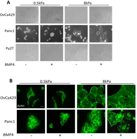
We have recently demonstrated that BMP4 induced EMT alters cell
stiffness properties and response to mechanical force [32]. To test the effects of
matrix rigidity on BMP induced EMT, we used three cell line models—OvCa429
cells, Panc1 cells, and Py2T murine breast cancer cells—that underwent EMT in
response to BMP4 based on alterations to E-cadherin localization (Figure 2.11A)
and the actin cytoskeleton (Figure 2.11B), changes in the EMT- transcription
factors (Figure 2.12C), and increased invasion in response to BMP4 (Figure
2.12D-E). Using these three cell lines we first examined cellular morphology on
fibronectin conjugated polyacrylamide gels of elastic modulus 0.5kPa (soft), 8kPa
(medium) (Methods), or glass (high stiffness). Consistent with prior studies on the
role of rigidity on cellular phenotypes [34, 35], we find that all three cancer cell
lines exhibited differences in morphology on the 8kPa (medium) substrate versus
0.5kPa soft substrates (Figure 2.1 A,B), with cells on the soft substrates being
rounded and forming clusters, regardless of treatment. Alternatively, cells on the
a more polygonal-like appearance. Additionally, cells on the substrates 8kPa
showed similar morphology to cells cultured on plastic and/ or glass (data not
shown).
To examine the changes associated with BMP4 induced EMT, we treated
the cells on the different substrates and examined cell morphology using actin
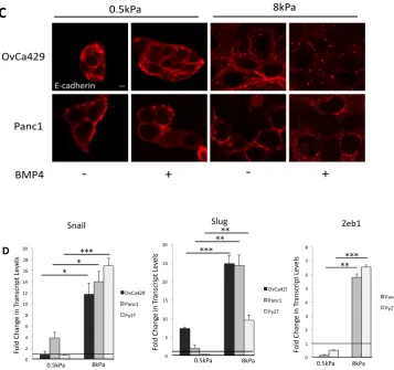
along with E-cadherin localization (Figure 2.1B Figure 2.2C). We find that
OvCa429 and Panc1 cells treated with BMP4 for 5 and 3 days, respectively, on
the 8kPa showed similar actin morphology and E-cadherin localization as cells
cultured on glass for 5 and 3 days for OvCa429 and Panc1, respectively. (Figure
2.1B, Figure 2.2C and Figure 2.11A-B), while cells on the 0.5kPa substrates
showed no change in actin morphology or E-cadherin localization. E- cadherin
remained tightly localized around cell- cell junctions along with cortical actin
(Figure 2.1B, Figure 2.2C), in contrast to on stiffer substrates where E-cadherin
appeared more scattered and diffuse, along with actin showed significant stress
fibers (Figure 2.11B, Figure 2.12C). To test if these changes were associated
with rapid induction of EMT associated transcriptional factors Snail, Slug, and
Zeb1, we treated the cells on the different substrates with BMP4 for 24 hours,
after the cells were plated on the substrates for at least 12 hours. We find that
each of the three cell lines upregulated a subset of the examined transcription
to significantly lesser extents, in response to BMP4, as compared to the 8kPa
substrates (Figure 2.2D).
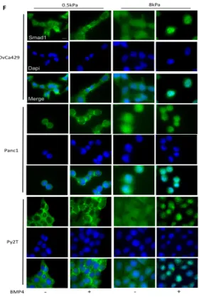
To examine if this was due to a defect in mounting a signaling response to
BMP4 on 0.5Kpa gels, we examined if Smad1 activation was altered on the
substrates with varying rigidities. Phosphorylation of Smad1 was determined by
treating cells with exogenous BMP4 after allowing the cells to grow for 24 hours
on the different substrates. No significant difference in Smad1 activation (Figure
2.3E) was observed on the substrates with different rigidities indicating that
change in response was not at the level of signal reception. Since no significant
change in Smad1 phosphorylation was observed, we tested if Smad1 nuclear
translocation in response to BMP4 was altered on 0.5kPa soft substrates. We
examined Smad1 and the co-Smad Smad4 localization by treating the cells with
BMP4 on the 0.5kPa and 8kPa substrates. We find that both Smad1 and Smad4
were excluded from the nucleus in compliant gels (0.5Kpa) in all three models,
while BMP4 induced Smad1 and Smad4 localization to the nucleus occurs
robustly in more rigid substrates at the 1 hr time point (Figure 2.4F for Smad1
and Figure 2.13B for Smad4) and at longer time points as well (data not shown).
These data suggest that lower transcriptional response to BMP4 on soft
substrates may be due to reduced nuclear Smad localization.
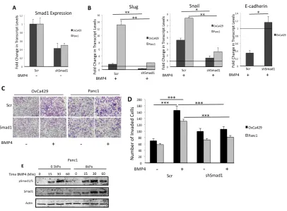
Since BMP4 can activate both Smad1 and non-Smad pathways [7], we
confirmed that cancer EMT in response to BMP4 was largely driven by a Smad1
[36], and performed shRNA to Smad1 on BMP4 induced EMT. Dorsomorphin
significantly reduced BMP4 induced increase in Snail and Slug transcript levels
(Figure 2.13A) and two siRNA constructs (Figure 2.3A) (Methods) to Smad1
significantly reduced BMP4 induced Snail and Slug transcription and suppressed
BMP4 induced cell invasion (Figure 2.3 B,C,D). While these data do not rule out
possible contributions of non-Smad pathway or Smad2/3 involvement [7], they
confirm Smad1’s significant role in mediating BMP4 induced EMT in the different
cancer cell line model systems and suggest that failure to mount a Smad1
response on substrates of 0.5kPa rigidity may lead to a failure to mount an EMT
response as well.
To define the mechanistic impact of substrate rigidity on BMP4 induced
EMT, we hypothesized a role for the mechanosensitive Yap1 [33] that besides
being regulated by the Hippo pathway can also interact with Smad1 in a BMP
dependent manner [23]. To test a direct role for Yap1, we used two independent
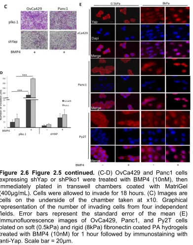
shRNAs to Yap1 in the two human cell lines Panc1 and OvCa429 (Figure 2.5A).
We find that BMP4 induced EMT transcription factors Snail and Slug, were
suppressed 2.5 and 9 fold in OvCa429 and Panc1 shYap cells, respectively,
compared to control cells (Figure 2.5B). Similarly, shYap1 reduced BMP4
induced cell invasion in OvCa429 and Panc1 cells by 10 and 14 fold, respectively
of Yap1 to facilitate EMT. We find that BMP4 increases and induces the nuclear
translocation of Yap1 on rigid substrates even in cells that have cell- cell contact
(8kPa, Figure 2.6E). However, BMP4 does not increase the amount of Yap1 in
the nucleus on soft substrates—where BMP4 was unable to induce EMT (Figure
2.6E). These Yap1 shRNA findings, together with a role for Smad1 in BMP4
mediated EMT reduction, suggest a role for nuclear Yap1 in mediating Smad1
induced EMT.
The kinase activities of Cdk8 and Cdk19 are required for BMP4 induced EMT
Our data strongly indicate that BMP4 induced EMT is driven by nuclear
Smad1 and Yap1. However, Yap interaction with Smad1 and its ability to mediate
Smad1 responses is dependent on the activity of the mediator kinase CDK8 in
the nucleus that phosphorylates the Smad1 linker region resulting in recruitment
of Yap1 [23]. Therefore, we tested a role for CDK8 in BMP4 induced EMT. To do
this, we used the specific ATP competitive inhibitor of CDK8 and its isoform
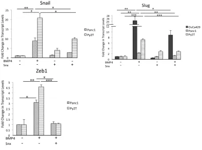
CDK19, Senexin B [38]. Inhibition of the kinase activity of the twin kinases
CDK8/19 using Senexin B robustly suppressed BMP4 induced increase in the
transcription factor Slug in OvCa429, Panc1, and Py2T EMT models and Snail
and Zeb1 in Panc1 and Py2T cells without significantly altering baseline
transcript levels (Figure 2.7A). Senexin B also lead to a suppression of BMP4
induced invasion in all three cell lines (Figure 2.8C,D and Figure 2.14A).
Moreover this suppression was dose dependent (Figure 2.14 3A). Since our
phosphorylation has been shown to be required for Yap interaction with Smad1
in the nucleus [23], we examined if blocking the kinase activity of CDK8/19 with
Senexin B dampened BMP4 induced Yap nuclear translocation. Indeed, Senexin
B treatment dampened Yap1 nuclear localization in response to BMP4 albeit to
different extents in Py2T, Panc1, and OvCa429 cells (Figure 2.8B). These results
demonstrate that the kinase activities of CDK8/19 that link Yap1 to Smad1 may
be necessary for BMP4 induced EMT likely through the recruitment of Yap1 for
mediating BMP4 induced EMT.
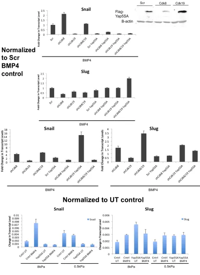
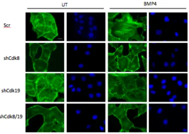
Cdk8/Cdk19 are required for EMT.
Since Senexin B targets the kinase activity of both CDK8 and CDK19, we
asked if the effects of Senexin B were primarily due to CDK8, or if CDK19 could
compensate for CDK8. To test the separate roles of CDK8 and CDK19, we
reduced the expression of each CDK8 and CDK19 (Figure 2.9A) using 2
independent shRNA’s for CDK8 and CDK19 (shRNA 1 to the coding region and
shRNA2 to the 3’ UTR region) were used in the studies. Reducing the expression
of CDK8 and CDK19 individually reduced BMP4 induced Smad1 linker
phosphorylation (Figure 2.9B) suggesting that both CDK8 and CDK19 could
compensate for each other’s loss. Consistent with the effects of Senexin B on
reduced nuclear translocation of Yap1 in response to BMP4, reducing the
factors Snail and Slug. We find that shRNA to CDK8 and CDK19 resulted in a
suppression of BMP4 induced Snail and Slug in both Panc1 and OvC429 cells
(Figure 2.9C, Figure 2.14B) (Figure 2.10D Panc1: shCdk8 1.5 and 3 fold,
shCdk19 2.5 and 1.5 fold, Snail and Slug, respectively. Figure 2.14B OvCa429:
shCdk8 2 fold and 2 fold, shCdk19 N.S. and 2 fold, Snail and Slug, respectively).
These data indicate that while Cdk8 and Cdk19 have overlapping functions in
mediating Smad1 linker phosphorylation (Figure 2.9B), they also have distinct
functions in regulating transcription of individual genes. Finally, to examine if
reducing Cdk8 and Cdk19 expression also impact BMP4 induced invasion in the
different EMT models, we examined EMT induced invasion in OvCa429 and
Panc1 cells. We find that while loss of Cdk8 did not affect baseline invasion
(Figure 2.10F), shCdk8, reduced BMP4 induced invasion by 2 fold in both
OvCa429 and Panc1 cells and shCdk19 reduced BMP4 induced invasion by 4
fold and 3 fold in OvCa429 and Panc1, respectively.
Cdk8 and Cdk19 expression correlate with EMT genes in ovarian cancer
Given that our in vitro studies indicate that CDK8 and CDK19 expression
regulated EMT in ovarian cancer cell lines, we next assessed whether the
expression of these genes correlated with common EMT markers in human high
grade serous ovarian tumors. To address this question we examined RNAseq
data from 283 high grade serous ovarian tumors (Data Not Shown) from the
Cancer Genome Atlas (TCGA) project [39]. We first confirmed that positive
positive correlation, ranging from 0.38 to 0.78, with the expression of each other
EMT marker (data not shown). We further confirmed the significant negative
correlation between CDH1, expression ofwhich is lost during EMT, and SNAI2,
TWIST1 and ZEB1; SNAI1, ZEB2, and TWIST2 were negatively correlated but
only trended towards significance. Consistent with our in vitro results, expression
of neither CDK8 nor CDK19 was negatively correlated with CDH1. However,
CDK8 expression had a significant positive correlation (Pearson Correlation: 0.14
– 0.24) with SNAI1 (p=5.3E-04), SNAI2 (p=5.2E-04), ZEB1 (p=0.0051), TWIST1
(p=0.0173), and TWIST2 (p=5.1E-05) while CDK19 had a significant positive
correlation (Pearson Correlation: 0.16 - 0.22) with ZEB1 (p=2.0E-04) and
TWIST1 (p=0.0089). Coupled with our in vitro data, these results suggest that
CDK8, and to a lesser extend CDK19, expression is significantly associated with
Figure 2.1 BMP4 Induced EMT is Suppressed
on Compliant Substrates. (A) Phase contrast
Figure 2.2 (Figure 2.1 continued) C) Scale bars 20µm. (D) Real time quantitative PCR showing the levels of the
Figure 2.3 BMP4 Induced EMT is Driven by Smad1 (A-B) Real time quantitative PCR showing expression fold change of Smad1 (A) and fold change in transcript levels, relative to the untreated control, for Slug, Snail, and E-cadherin (B) in OvCa429 and Panc1 cells expressing shSmad1 or shScr after being treated with BMP4 (10nM) for 24hrs and 12 hours,
respectively. Values are normalized to their untreated control (Black horizontal bars). Error bars represent the standard error of the mean. *p<0.05, **p<0.01. (C-D) OvCa429 and Panc1 cells expressing shSmad1 or shScr cells were treated with BMP4 (10nM), then immediately plated in transwell chambers coated with MatriGel (400µg/mL). Cells were allowed to invade for 18 hours. Images (C) are cells on the underside of the chamber taken at x10. Images are representative of two independent experiments done in triplicate.
Graphical representation of the number of invading cells from four
independent fields. Error bars represent the standard error of the mean. *** p<0.001. (E) Time course of Smad1/5 phosphorylation in Panc1 cell lines treated with BMP4 (10nM) plated on soft (0.5kPa) and rigid (8kPa) fibronectin coated polyacrylamide (PA) hydrogels. Lysates were analyzed by
Figure 2.4 Figure 2.3 Continued. (F) Immunofluorescence images of OvCa429, Panc1, and Py2T cells plated on soft (0.5kPa) and rigid (8kPa) fibronectin coated PA
Figure 2.5 Yap1 Enhances BMP4 Induced EMT. (A-B) Real time quantitative PCR showing Yap1 fold changes in the
Figure 2.8 Figure 2.7 continued. (B) Immunofluorescence images of OvCa429, Panc1, and Py2T cells pre-treated for 30 minutes with SnxB (5µM), then treated with BMP4 (10nM) for 24 hours followed by immunostaining with Yap. Scale bars = 20µm (C) OvCa429 and Panc1 cells were pre-treated with SnxB (5µM) for 30 minutes, then treated with BMP4 (10nM), and then immediately plated in transwell chambers coated with MatriGel (400µg/mL). Cells were allowed to invade for 18 hours. Graphical representation of the number of invading cells from four independent fields. Error bars represent the standard error of the mean.
B C
Figure 2.9 Cdk8/19 Enhance BMP4 EMT and Exhibit Differential
Effects. (A) Real time quantitative PCR showing the levels of Cdk8 and
Cdk19 in OvCa429 and Panc1 cells. (B) OvCa429 and Panc1 cells with stable shRNA Cdk8 and Cdk19 knockdown were treated with BMP4 (10nM) for 1 hour, analyzed by immunoblotting with the indicated
Figure 2.11. Supplementary Figure 1. (A) OvCa429 and Panc1 cells treated with BMP4 (10nM) for
indicated times. Lysates were analyzed by immunoblotting with indicated antibodies. (B)
OvCa429 and Panc1 cells treated with BMP4 (10nM) for 5 and 3 days, respectively, followed by
Figure 2.12 (Figure 2.11 Supplementary Fig 1 continued): (C) Real time quantitative PCR showing the levels of the transcription factors Snail, Slug, and E-cadherin in OvCa429 and Panc1 cells treated with BMP4 (10nM) for 24 and 12 hours, respectively. Graphs are
Figure 2.14. Supplementary Figure 3 (A) OvCa429 and Panc1 cells were pre-treated with SnxB with increasing concentrations from 250nM, 500nM, 1µM, and 5µM for 30 minutes, then treated with BMP4 (10nM), and then
Chapter 3
Discussion
The role of BMP4 in the progression of cancer has been conflicting given
that it has been shown to increase cell invasiveness, migration, and metastasis in
some cancers [13-15, 40], however, has been shown to suppress growth and
found in lower levels in other cancers [16, 41]. Determining the signaling pathway
and specific factors involved in BMP4 induced EMT could help identify under
what conditions BMP4 acts as a tumor suppressor or promoter. Previous studies
have shown that BMP4 is able to signal through Smad1 and Smad2 [7]. Our
studies confirm that BMP4 induced Smad1 activation prompts EMT in ovarian,
pancreatic, and breast cancer cell models (Figure 2.3B-D, Fig 2.13). The present
study shows the requirement of the transcriptional regulators Cdk8/19, as well as
the transcriptional co-activator Yap1 in Smad1 dependent BMP4 induced EMT in
ovarian, pancreatic, and breast cancer cell line models. We also find that Yap1,
Cdk8, and Cdk19 are each responsible for the increases in the EMT associated
transcription factors Snail and Slug along with the increase in cell invasion
associated with BMP4 signaling. Consistent with previous studies, our data show
that Cdk8 is necessary for the linker phosphorylation of Smad1 [23], along with
regulation of BMP4 induced EMT, but they may be able to compensate for one
another.
Our study shows for the first time the use of Senexin B, the specific small
molecule inhibitor of Cdk8 and Cdk19, in the involvement of suppressing
transcriptional activation of EMT transcription factors Snail and Slug with robust
effects on suppressing cell invasion, thus further confirming the role of Cdk8 and
Cdk19 involvement in BMP4 induced EMT. These findings on the effects of
Senexin B also have broader implications for the use of Senexin B as a potential
therapeutic strategy for suppressing metastasis. Cdk8 has been shown to be an
oncogene and leads to cancer progression in a variety of cancers including
pancreatic, colon, and breast cancers and melanoma [18-22]. In addition to the
BMP4/Smad1 signaling pathway, Cdk8 has also been shown to be involved in
other signaling pathways, including Wnt/β-catenin, Tgf-β, and Notch [18, 23, 42].
The diverse role of Cdk8 indicates that Senexin B may be able to be used as a
selective approach to improve cancer treatment.
We find that softer substrates dampen the effects of BMP4 on the EMT
associated transcription factors, Snail and Slug, along with inhibiting E-cadherin
dislocation from the junctions. This is similar to previous studies on matrix rigidity
with Tgfβ−1, where decreased matrix rigidity reduced the levels of Tgfβ−1
stiffness have shown that a decreased rigidity resulted in Yap exclusion from the
nucleus [33]. In addition to Yap exclusion from the nucleus with softer substrates,
we found that BMP4 is unable to override the mechanical control on Yap1, as
Yap1 remains excluded from the nucleus on soft substrates. Cdk8/19, however,
remained localized in the nucleus regardless of matrix rigidity (data not shown).
Because matrix rigidity reduced the effects of EMT, while inhibiting Smad1 and
Yap1 activity, this data could suggest that Smad1 and Yap1 are necessary for
the activity of Cdk8/19 during BMP4 induced EMT. The particular mechanism by
which Cdk8/19, Yap1, and Smad1 interact to drive BMP4 induced EMT had yet
to be established. Future studies to examine the interplay among these factors
Chapter 4
Materials and Methods
Cell Lines and Culture Conditions
OvCa429 cells were obtained from Duke Gynecology/Oncology Bank, Panc1
cells were from ATCC, and Py2T cells were a kind gift from Gerhard Christofori
(University of Basel). Panc1 and Py2T cells were cultured in DMEM containing
L-glutamine, 10% fetal bovine serum (FBS), and 100 U of penicillin-streptomycin.
OvCa429 cells were cultured in RPMI containing L-glutamine, 10% fetal bovine
serum (FBS), and 100 U of penicillin-streptomycin.
Softwells
For rigidity experiments, cells were plated on 0.5kPa (soft) or 8kPa (rigid)
substrates (Matrigen, Softslip 12 well and Softwell 6 well) that were coated with
10ug/mL fibronectin (Cultrex® #3420-001-01) in 1X PBS for one hour. All cells
were allowed to sit on substrates for at least 12 hours before BMP4 treatment.
For Smad1 (1 hour treatment) and Yap1 (24 hour treatment) localization
experiments on Softslip 12 well, 50000 cells were plated on 8kPa substrates and
100000 cells were plated on 0.5kPa substrates. For E-cadherin and actin
respectively. For time course experiments on Softwell 6 well, 200000 cells were
plated on 8kPa substrates and 300000 cells were plated on 0.5kPa substrates.
For qPCR experiments on Softwell 6 well, plated 50000 on 8kPa and 200000
cells on 0.5kPa for OvCa429 cells (5 day treatment with re-treatment at day 3),
80000 on 8kPa and 200000 cells on 0.5kPa for Panc1 cells (3 day treatment),
and 70000 cells on 8kPa and 200000 cells on 0.5kPa for Py2T cells (2 day
treatment).
Antibodies, Reagents, Plasmids, and Lentiviral Infections
Antibody E-cadherin (BD Biosciences #610181), B-Actin (Sigma #A2228) was
from Sigma. pSmad1 Ser206 (#5753), pSmad1/5/8 (#9511), Smad1 (#6944), and
Smad4 (#9515) antibodies were from Cell Signaling. Senexin B was from Senex.
For Smad1 knockdown, OvCa429 and Panc1 cells were infected with 100MOI
siRNA adenovirus construct, generously provided by Maria Trojanowska.
Lipofectamine 2000 (Life Technologies #11668019). Constructs with the shRNA
to Yap was obtained from Sigma with the following sequences:
Yap:0000107265:
CCGGCCCAGTTAAATGTTCACCAATCTCGAGATTGGTGAACATTTAACTGGG
TTTTTG and
Yap:0000107268:CCGGGACCAATAGCTCAGATCCTTTCTCGAGAAAGGATCT
GAGCTATTGGTCTTTTTG. For Yap knockdown, OvCa429 and Panc1 cells
were infected at a 1:8 ratio of the Yap shRNA lentiviral construct. The target
OvCa429 and Panc1 cells were infected at a 1:4 ratio of the Cdk8 and Cdk19
shRNA lentiviral construct. OvCa429 and Panc1 cells were selected in puromycin
at 3µg and 5µg, respectively. All shRNA lentiviral constructs were generously
provided by Serena Altilia (University of South Carolina). The knockdown was
confirmed by qPCR (sequences in Table 1).
Quantitative Polymerase Chain Reaction
Panc1, OvCa429, and Py2T cells were treated for 12, 24, and 48 hours,
respectively. In experiments where SenexinB was used, cells were pre-treated
with SenexinB for 30 minutes. Total RNA was isolated from cells using Trizol
reagent (Invitrogen, Carlsbad, CA). 1ug of RNA was reverse transcribed to cDNA
using 5x iScript Reverse Transcription Supermix (Bio-Rad #1708840) and the
Sso Advanced Universal SYBR Green Supermix (Bio-Rad #1725271). qRT-PCR
primer sequences are listed in Table 1.
MatriGel Invasion Assays
Invasion assays were performed using 24-well transwells (Greiner Bio-One;
ThinCertsTM, 24 well 8.0um) coated with 400ug/ml MatriGel (BD Biosciences
#3248404). Cells (50,000) in serum free media were plated in upper chamber,
and allowed to invade for 18 hours toward serum media in lower chamber. Filters
were stained with Three Step Stain (Richard-Allan Scientific). The filters were
Immunofluorescence
In experiments where SenexinB was used, cells were pre-treated with SenexinB
for 30 minutes. Cells were fixed in 4% paraformaldehyde, permeabilized in 0.1%
TX-100, and blocked with 1% BSA PBS. Cells were incubated in primary
antibody (1:200) for an hour, followed by incubation with Alexa Fluor 488 or 594
second antibody (LifeTechnologies). After washing, cells were stained with
4’6-diamidino-2-phenylindole (Roche). Imaging was performed using an Olympus
IX81 motorized inverted microscope. Imaging was performed using Imaging on
References
1. Kalluri, R. and R.A. Weinberg, The basics of epithelial-mesenchymal transition. J Clin Invest, 2009. 119(6): p. 1420-8.
2. Lengyel, E., Ovarian cancer development and metastasis. Am J Pathol, 2010. 177(3): p. 1053-64.
3. Li, D., et al., Pancreatic cancer. Lancet, 2004. 363(9414): p. 1049-57. 4. Kawabata, M., T. Imamura, and K. Miyazono, Signal transduction by bone
morphogenetic proteins. Cytokine Growth Factor Rev, 1998. 9(1): p. 49-61.
5. Lamouille, S., J. Xu, and R. Derynck, Molecular mechanisms of epithelial-mesenchymal transition. Nat Rev Mol Cell Biol, 2014. 15(3): p. 178-96. 6. Ross, S. and C.S. Hill, How the Smads regulate transcription. Int J
Biochem Cell Biol, 2008. 40(3): p. 383-408.
7. Holtzhausen, A., et al., Novel bone morphogenetic protein signaling through Smad2 and Smad3 to regulate cancer progression and development. FASEB J, 2013.
8. Wang, Y.K., et al., Bone morphogenetic protein-2-induced signaling and osteogenesis is regulated by cell shape, RhoA/ROCK, and cytoskeletal tension. Stem Cells Dev, 2012. 21(7): p. 1176-86.
9. Pal, A., et al., CCN6 modulates BMP signaling via the Smad-independent TAK1/p38 pathway, acting to suppress metastasis of breast cancer.
Cancer Res, 2012. 72(18): p. 4818-28.
10. Gamell, C., et al., BMP2 induction of actin cytoskeleton reorganization and cell migration requires PI3-kinase and Cdc42 activity. J Cell Sci, 2008. 121(Pt 23): p. 3960-70.
11. Richter, A., et al., BMP4 promotes EMT and mesodermal commitment in human embryonic stem cells via SLUG and MSX2. Stem Cells, 2014. 32(3): p. 636-48.
12. Wang, R.N., et al., Bone Morphogenetic Protein (BMP) signaling in development and human diseases. Genes Dis, 2014. 1(1): p. 87-105. 13. Theriault, B.L., et al., BMP4 induces EMT and Rho GTPase activation in
human ovarian cancer cells. Carcinogenesis, 2007. 28(6): p. 1153-62. 14. Gordon, K.J., et al., Bone morphogenetic proteins induce pancreatic
16. Cao, Y., et al., BMP4 inhibits breast cancer metastasis by blocking myeloid-derived suppressor cell activity. Cancer Res, 2014. 74(18): p. 5091-102.
17. Gold, M.O. and A.P. Rice, Targeting of CDK8 to a promoter-proximal RNA element demonstrates catalysis-dependent activation of gene expression.
Nucleic Acids Res, 1998. 26(16): p. 3784-8.
18. Xu, W., et al., Mutated K-ras activates CDK8 to stimulate the epithelial-to-mesenchymal transition in pancreatic cancer in part via the Wnt/beta-catenin signaling pathway. Cancer Lett, 2015. 356(2 Pt B): p. 613-27. 19. Firestein, R., et al., CDK8 is a colorectal cancer oncogene that regulates
beta-catenin activity. Nature, 2008. 455(7212): p. 547-51.
20. Kapoor, A., et al., The histone variant macroH2A suppresses melanoma progression through regulation of CDK8. Nature, 2010. 468(7327): p. 1105-9.
21. Li, X.Y., et al., siRNA-mediated silencing of CDK8 inhibits proliferation and growth in breast cancer cells. Int J Clin Exp Pathol, 2014. 7(1): p. 92-100. 22. Li, X.Y., et al., MiRNA-107 inhibits proliferation and migration by targeting
CDK8 in breast cancer. Int J Clin Exp Med, 2014. 7(1): p. 32-40.
23. Alarcon, C., et al., Nuclear CDKs drive Smad transcriptional activation and turnover in BMP and TGF-beta pathways. Cell, 2009. 139(4): p. 757-69. 24. Aragon, E., et al., A Smad action turnover switch operated by WW domain
readers of a phosphoserine code. Genes Dev, 2011. 25(12): p. 1275-88. 25. Lamar, J.M., et al., The Hippo pathway target, YAP, promotes metastasis
through its TEAD-interaction domain. Proc Natl Acad Sci U S A, 2012. 109(37): p. E2441-50.
26. Zhao, H., et al., E2A suppresses invasion and migration by targeting YAP in colorectal cancer cells. J Transl Med, 2013. 11: p. 317.
27. Hao, Y., et al., Tumor suppressor LATS1 is a negative regulator of oncogene YAP. J Biol Chem, 2008. 283(9): p. 5496-509.
28. Levental, K.R., et al., Matrix crosslinking forces tumor progression by enhancing integrin signaling. Cell, 2009. 139(5): p. 891-906.
29. Seewaldt, V., ECM stiffness paves the way for tumor cells. Nat Med, 2014. 20(4): p. 332-3.
30. Lessey, E.C., C. Guilluy, and K. Burridge, From mechanical force to RhoA activation. Biochemistry, 2012. 51(38): p. 7420-32.
31. Jaalouk, D.E. and J. Lammerding, Mechanotransduction gone awry. Nat Rev Mol Cell Biol, 2009. 10(1): p. 63-73.
32. Osborne, L.D., et al., TGF-beta regulates LARG and GEF-H1 during EMT to impact stiffening response to force and cell invasion. Mol Biol Cell, 2014.
35. Tilghman, R.W., et al., Matrix rigidity regulates cancer cell growth and cellular phenotype. PLoS One, 2010. 5(9): p. e12905.
36. Yu, P.B., et al., Dorsomorphin inhibits BMP signals required for
embryogenesis and iron metabolism. Nat Chem Biol, 2008. 4(1): p. 33-41. 37. Piccolo, S., S. Dupont, and M. Cordenonsi, The Biology of YAP/TAZ:
Hippo Signaling and Beyond. Physiol Rev, 2014. 94(4): p. 1287-1312. 38. Porter, D.C., et al., Cyclin-dependent kinase 8 mediates
chemotherapy-induced tumor-promoting paracrine activities. Proc Natl Acad Sci U S A, 2012. 109(34): p. 13799-804.
39. Hoadley, K.A., et al., Multiplatform analysis of 12 cancer types reveals molecular classification within and across tissues of origin. Cell, 2014. 158(4): p. 929-44.
40. Ampuja, M., et al., The impact of bone morphogenetic protein 4 (BMP4) on breast cancer metastasis in a mouse xenograft model. Cancer Lett, 2016.
41. Fang, W.T., et al., Downregulation of a putative tumor suppressor BMP4 by SOX2 promotes growth of lung squamous cell carcinoma. Int J Cancer, 2014. 135(4): p. 809-19.
42. Fryer, C.J., J.B. White, and K.A. Jones, Mastermind recruits CycC:CDK8 to phosphorylate the Notch ICD and coordinate activation with turnover.
Figure A.10: Effects of SnxB on BMP4 Induced EMT (A) E-cadherin (B) Actin (A-B) Expt 3-19
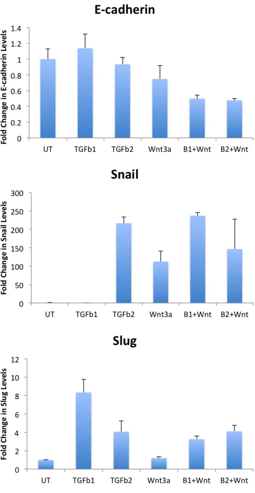
Figure A.12: Effect of Wnt3a and Tgf-β1/2 on
0 5 10 15 20 25 30
pLKO.1 UT pLKO.1
BMP pLKO.1 Snx pLKO.1 Snx BMP Cdk8- UT Cdk8- BMP
Fo ld C h an ge in In va si on
0 0.2 0.4 0.6 0.8 1 1.2
0hr 6hrs 12hrs 18hrs 24hrs
%
C
lo
su
re
Effects of SnxB on BMP4 Induced
Migration
pLKO UT pLKO BMP4 pLKO Snx pLKO Snx+BMP4 Cdk8- UT Cdk8- BMP4Figure D.1: Effects of SnxB on Tgf-β1
Figure E.4: Effects of SnxB on Tgf-β
Figure E.5: Effects of SnxB on Tgf-β Induced FN Invasion in NMuMg Cells.

![Poly[[diaquatris[μ4 (p phenylenedioxy)diacetato]didysprosium(III)] dihydrate]](data:image/gif;base64,R0lGODlhAQABAIAAAP///wAAACH5BAEAAAAALAAAAAABAAEAAAICRAEAOw==)