EXAFS Studies of Ruthenium Based Double Perovskite
ALaMnRuO
6
(A = Ca, Sr, Ba)
Rubina Shaheen, Javaid Bashir, Muhammad Nasir Khan*
Physics Division, PINSTECH, Islamabad, Pakistan. Email: *[email protected]
Received August 25th, 2011; revised November 19th, 2011; accepted December 9th, 2011
ABSTRACT
Room and liquid nitrogen temperatures Mn K-edge extended X-ray fine structure (EXAFS) studies were carried out on powder samples of ALaMnRuO6 (A = Ca, Sr, Ba) perovskites. The EXAFS analysis of Mn K-edge spectra showed
MnO6 octahedra are distorted. Among the three perovskites, the polyhedral distortion is highest for SrLaMnRuO6 ( =
1.4 × 10⁻³). For CaLaMnRuO
6 ( = 0.5 × 10⁻³) and BaLaMnRuO6, ( = 0.7 10–3), the smaller values of distortion
pa-rameters indicate that the MnO6 octahedra are relatively regular and undistorted in the latter two systems. In
SrLaMnRuO6, MnO6 distortion appears to be of orthorhombic (Q2) type whereas for CaLaMnRuO6 and BaLaMnRuO6,
the distortion is of tetragonal or Q3 type. The Mn-O distances as determined from the EXAFS measurements, increases
with the increase in the size of the divalent cation. No structural changes occurred in the local environment around Mn atom when the temperature was lowered from room to liquid nitrogen temperature.
Keywords: Oxides; Ceramics; EXAFS; Crystal Structure
1. Introduction
The ideal perovskite structure with stoicheometry AMO3,
where A is typically a large, low oxidation state cation, M is a smaller transition metal or lanthanide cation, has cubic symmetry with space group Pm m3 [1]. The ca-pacity of perovskite structure to incorporate almost all the elements in the periodic table except for noble gas elements, beryllium and phosphorus has resulted in ma-terials exhibiting range of properties including dielectric, piezoelectric, ferroelectric, optical, superconducting and magnetoresistive properties [2]. The perovskite structure is viable to wide variations in compositions from the ideal formula AMO3. These variations can be achieved
by isomorphous substitutions at A or M sites, and cati-onic and anicati-onic deficiencies. These can be represented as 1 x x 3, 1 x x 3. The physical properties of perovskites are shown to depend on the doping levels at the A or M sites [3-6]. For 50% substitution, such mixed system are classified as double perovskites with the formula AA′MM′O6 or A2MM′O6 as reported by
Anderson et al. (1993) in reference[7]. The lattice distor-tion and local structure plays an important role in the magnetoelectric transport properties of perovskite man-ganites, in particular its effect on the colossal magnetore-sistive (CMR) properties [8-10].
A A MO AM M O
Ruthenates perovskites has attracted immense inter-est of the physicist and chemist over the past few years [11-15]. This surge of interest in ruthenates is because of their rich variety of magnetic states exhibited by these materials. For example, SrRuO3 is a 4d ferromagnetic
metal [14] while CaRuO3 is metallic but not magnetic;
the double perovskite Sr2YRuO6 is an antiferromagnetic
insulator; and the layered perovskite Sr2RuO4 is
super-conducting [15]. The magnetic properties of double pero- vskite ruthenate oxides depends on the nature of MM′
distribution over the octahedral sites [16-18].
Though the crystal structures of ALaMnRuO6 (A = Ca,
Sr, Ba) are well established [19-24]. However, no infor-mation is available about the local structure around the manganese in these materials. In our earlier publications, we have reported crystal structure as well as the oxida-tion state of Mn in SrLaMnRuO6, BaLaMnRuO6 and
CaLaMnRuO6 [21-23]. Room temperature local strucutre
around Mn in SrLaMnRuO6 has also been reported [21].
In the present paper, we now report the local structure around Mn in CaLaMnRuO6 (CLMRO), and BaLa-
MnRuO6 (BLMRO) at room and liquid nitrogen
tem-peratures. In addition we also report the loal structure in SrLaMnRuO6 (SLMRO) at liquid nitrogen temperature.
2. Experimental
The single-phase powder samples of ALaMnRuO6 (A=
Ca, Sr, Ba) were synthesized by conventional solid-state reaction method as reported earlier in [21]. X-ray diffrac-tion revealed that all the samples have orthorhombic symmetry (space group Pbnm), with the tilting scheme
a–a–b+ which results in the unit cell dimension of a ×
a × 2a (where a is the pseudocubic unit cell parame-ter).
Room and liquid nitrogen temperatur Mn K-edge X- ray absorption spectroscopy experiments, which include both XANES and EXAFS techniques, were performed at the XAFS beam line at ELETTRA Synchrotron Radia-tion Source, Trieste, Italy. Well-dispersed powder sam-ples in cyclohexane were deposited on porous mem-branes. The amount of the sample was pre-calculated to obtain optimum absorption jump (μ 1). X-ray beam energies were tuned using a double crystal, focusing Si (111) monochromator crystals. The incident (Io) and
transmitted (It) beam intensities were recorded using
ionization chambers filled with appropriate gases. Data were collected in three regions: 1) the pre-edge region with a step size of 5 eV to allow pre-edge background calculation and subtraction; 2) the XANES region from 30 eV below the edge to 50 eV above the absorption edge with a step size of 0.3 eV; and 3) the EXAFS region upto 7050 eV. All the edges were recorded at least twice. Raw data were corrected and converted to k-space, sum- med and background subtracted to yied the EXAFS func-tion (k) using the EXCALIB and EXBACK Programs [25]. The edge profiles were separated from the EXAFS data and, after subtraction of the linear pre-edge back-ground, normalized to the edge step. The data were weighted by k3, where k is the photoelectron wavevector,
to enlarge the oscillations at high k. The data were fitted using the non-linear square minimisation program EX-CURV92 [26] which calculates the theoretical function using the fast curved wave theory [27]. Phase shifts corrections were calculated using Xalpha exchange and ground potentials.
3. Results and Discussion
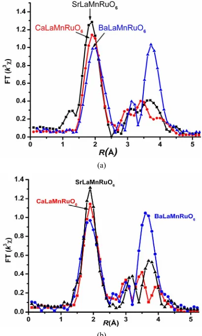
Figure 1(a)-(b) represents the modulus of Fourier Trans-form (FT) extracted from the XAS data at 300 K and 80 K by measuring Mn K-edge EXAFS spectra for the three systems. The k3-weighted (k) data were Fourier
trans-formed (FT) in the k range of 2.5 - 12 Å–1 using a
Guas-sian window. Well defined peaks corresponding to the different coordination shells around the Mn absorbing atom are visible upto about 5 Å. The main intense peak at about 1.9 Å in the FT is associated with the MnO6
oc-tahedron. Further peaks around 3 - 4 Å in the FT are at-tributed to Mn-A/La and Mn-O-Mn interactions [8,28].
The first peak of FT, corresponding to the Mn-O, oc-tahedra are identical for all the materials except for a
(a)
[image:2.595.320.525.86.427.2](b)
Figure 1. EXAFS fitting results (a) at room temperature and (b) liquid nitrogen temperature; experimental points (dots) and best fit (─) for comparison of three systems.
slight upward shift that indicates gradual increase of the Mn-O distances with increasing size of A atom. However, there are marked differences corresponding to Mn-A/La and Mn-O-Mn interactions as we move from CaLaMn- RuO6 to BaLaMnRuO6. The shape of these peaks are
found to be dependent either on the crystallographic symmetry or the different scattering power of the diva-lent cations. Since, laboratory X-ray diffraction studies have revealed that all of these materials have the same orthorhombic symmetry (space group Pbnm) [21-23], these differences can only be attributed to the different scattering powers of A cations. For all three samples, nearly constant amplitudes of Mn-O peaks at 300 K and 80 K indicates that no structural changes occur in the local environment around the Mn as the temperature is lowered.
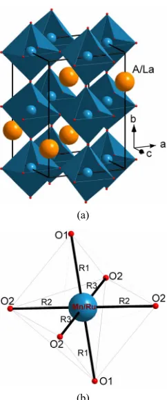
Previous neutron diffraction [19,20] and synchrotron X-ray diffraction studies [21,23] have shown that the structure of ALaMnRuO6 is orthorhombic with distorted
Mn/RuO6 octahedra (Figure 2). Owing to the Jahn Teller
(a)
(b)
Figure 2. (a) Schematic representation of the orthorhombic Pbnm crystal structure of ALaMnRuO6 in which A/La (A = Ca, Sr, Ba) atoms; (b) Ru/MnO6 octahedron.
of MnO6 produces three Mn-O bonds with equal bond
distances, long (l), medium (m) and short (s) bonds [29, 30]. In case of CLMRO and BLMRO, the MnO6
octahe-dra are quite regular and the spread in the bond lengths is small [22,23] whereas in SLMRO the bondlengths in the basal plane are further split into two Mn-O bonds of equal bond lengths and the structure is considerably dis-torted [21].
Extended X-ray absorption fine structure (EXAFS) spectroscopy provides structural information about a sample by way of the analysis of its X-ray absorption spectrum. In order to extract structural information from experimental spectra, a simple analytical expression that relates the EXAFS signal to the structural parameters is required. An EXAFS analytical expression like the one suggested by Teo [31] is:
2 2 2
2
sin 2
j k
j j j j
j
j
N f k e kR k
k
kR
where k is the electron wave vector, Njis the number of
atoms in the j-th coordination shell, δj(k) is the phase shift, fj(k) is the scattering amplitude, σj is the Debye
Waller factor in terms of a Gaussian broadening resulting from thermal and static disorder and Rjis the M-O bond
length in the Mn-O octahedral (Figure 2(b)). Various
structural parameters can be extracted by an approximate Fourier analysis of the normalized EXAFS term (k) [32].
In all of these perovskites oxides, the Mn ions are sur-rounded by six oxygen atoms forming an octahedron (Figure 2(b)). In cubic, undistorted perovskite structure, all the Mn-O bond distance are equal, i.e., R1 = R2 = R3.
As both La and A cations are distorted from the central position in the simple cubic unit cell, making their near- neighbor oxygen environment complicated. In addition to these distortions, presence of JT distortion is also present. The six otherwise equal, Mn-O bond lengths in the tilted octahedral consequently split into pairs of unequal bond length. Distortion caused by the Jahn-Teller effect in pe- rovskites usually involves four of the octahedral bonds (bonds involving planar oxygen, R2 and R3) contracting
and two of the octahedral bonds (apical oxygen) leng- thening which gives an elongated octahedral shape.
Different models were employed for fitting the expe- rimental EXAFS data. In the first model (2 + 2 + 2 model), we have used three subshells each with a coor-dination number NMn-O = 2 with unequal Mn-O bond-
lengths. Subsequently we employed models with two sub shells, one corresponding to a Mn-O coordination num-ber NMn-O = 4 at shorter distance and the other
corre-sponding to NMn-O = 2 at longer distance (4 + 2 model); a
model with bimodal distribution of Mn-O bonds with three longer Mn-O bonds and three shorter Mn-O bonds (3 + 3 model) and a model with one Mn-O sub shell with
NMn-O = 6. Owing to strong correlation between
coordi-nation number (N) and Debye-Waller factor (σ2), only the
distance (R) and σ2 were refined. Since electronic
proper-ties mainly depend on the nearest neighbours, we are focusing only on the first shell analysis.
For SLMRO, as reported previously[21] the best fit to EXAFS data were obtained for 2 + 2 + 2 model. In case of CLMRO and BLMRO, the best fits were obtained for 4 + 2 model, i.e. four short and two long bonds. The re-manining models lead to relatively high values of Debye Waller Factors without any significant improvement in the fit. Typical fitted FT spectra at room temperature are shown in Figure 1(a). The summary of the fitted struc-tural parameters are listed in Table 1. The calculated in- teratomic distances are in good agreement with values obtained from the crystallographic measurements[19-23]. This suggests that the local structure as determined from EXAFS mathches the average structure as determined from neutron or Synchrotron X-ray diffraction. The main result is that the Mn-O distances increases with the in-creasing size of the divalent cation A in agreement with the shifts observed in the first peak of the FT. The sli- ghtly higher values of Debye-Waller factors suggests a site to site variation of the bond lengths in the MnO6
[image:3.595.112.233.80.370.2]Table 1. EXAFS parameters of the first oxygen coordination shell at Mn K-edge. R is the interatomic Mn-O distance, N being the coordination number and 2 is the Debye-Waller factor. The errors on radial distances are 0.01 Å whereas on DW factors the error is 0.001 Å2.
CaLaMnRuO6 SrLaMnRuO6 BaLaMnRuO6
R(Å) 2 10–2 (Å2) R(Å) 2 10–2 (Å2) R(Å) 2 10–2 (Å2) Shell
N
300 K 80 K 300 K 80 K N 300 K 80 K 300 K 80 K N 300 K 80 K 300 K 80 K
Mn-O 4 1.92 1.90 0.6 0.4 2 1.87 1.85 0.9 1.4 4 1.97 1.95 0.1 0.1
Mn-O 2 2.01 1.99 0.9 0.8 2 1.95 1.95 0.6 0.4 2 2.08 2.07 0.3 0.2
Mn-O - - - 2 2.05 2.02 1.4 1.8 - - -
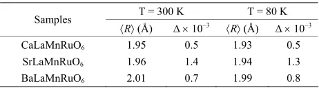
urements of Garanado et al. [19]. As the temperature is lowered, there is little change in the average bond dis-tances and hence the distortion parameters.
There are two types of distortion modes associated with the JT distortion: Q2 and Q3 which are defined in
terms of long (l), medium (m) and short (s) Mn-O bonds as:
4. Conclusion
2 2 2
Q l s
3 2 2 6
Q m l s We have carried out Mn-K-edge X-ray Absorption spec-troscopy measurements to study valence state of Mn and
local structure around it in single phase ALaMnRuO6
perovskites prepared through solid state technique. The Mn-O distances as determined from the EXAFS meas-urements, increases with the increasing size of the diva-lent cation. No structural changes occur in the local en-voirnment around Mn as the temperature is lowered from room to liquid nitrogen temperature.
with l, s being the Mn-O distances in the ab plane and m
is out of plane Mn-O bond length. Q3 is a tetragonal
whereas Q2 is orthorhombic distortion [29]. In the tetra-
gonal or Q3 distortion the in-plane bonds extend and out
of the plane bond shortens or vice versa implying that one bond length is larger than the other two. On the other hand, in the orthorhombic or Q2 distortion, the two in
plane bond lengths (Mn-O2 ) split more symmetrically
around the out of plane Mn-O1 bond length. Examination
of the bond lengths derived from the XAFS data reveals that in SLMRO, MnO6 distortion appears to be of
ortho-rhombic (Q2) type whereas for CLMRO and BLMRO, the
distortion is of tetragonal or Q3 type.
5. Acknowledgements
Two of the authors (JB and RS) are grateful to the Abdus Salam International Center for Theoretical Physics (AS- ICTP) for the provision of financial support under the ICTP-ELETTRA user programme. The authors are also grateful to Dr. Luca Olivi for his skilful help in conduct-ing the experiments at the XAFS beam line at the ELETTRA Synchrotron Radiation Source, Trieste, Italy. The MnO6 polyhedral distortion parameter ,
fre-quently observed for eg level degenerated systems such
as Mn3+ ( 3 1 2g g
t e ) containing perovskites is defined as
2
1
1 N n
n n
d d
N d
REFERENCES
[1] R. H. Mitchell, “Perovskites: Modern and Ancient,” Al-maz Press, Thunder Bay, Canada, 2002.
where d is average Mn – O bond distance. The distor-tion parameters of ALaMnRuO6 are listed in Table 2.
Among the three perovskites, the polyhedral distortion is the highest for SLMRO. For CLMRO and BLMRO, the smaller values of distortion parameters indicate that the MnO6 octahedra are relatively regular and undistorted.
This result is consistent with the neutron diffraction meas-
[2] B. Raveau, “The Perovskite History: More than 60 Years of Research from the Discovery of Ferroelectricity to Colossal Magnetoresistance via High Tc Superconducti- vity,” Progress in Solid State Chemistry, Vol. 35, No. 2-4, 2007, pp. 171-173.
doi:10.1016/j.progsolidstchem.2007.04.001
[3] K. Kuepper, M. C. Falub, K. C. Prince, V. R. Galakhov, I. O. Troyanchuk, S. G. Chiuzbaian, M. Matteucci, D. Wett, R. Szargan, N. A. Ovechkina, Y. M. Mukovskii and M. Neumann, “Electronic Structure of A- and B-Site Doped Lanthanum Manganites: A Combined X-Ray Spectro-scopic Study,” The Journal of Physical Chemistry B, Vol. 109, No. 19, 2005, pp. 9354-9361.
doi:10.1021/jp044447w Table 2. MnO6 Polyhedral distortion parameter () for
ALaMnRuO6 as determined from EXAFS parameters. R is the average Mn-O bond length.
T = 300 K T = 80 K Samples
R (Å) 10–3 R (Å) 10–3
CaLaMnRuO6 1.95 0.5 1.93 0.5
SrLaMnRuO6 1.96 1.4 1.94 1.3
BaLaMnRuO6 2.01 0.7 1.99 0.8
[image:4.595.57.288.667.731.2]pp. 147-150. doi:10.1038/33017
[5] P. M. Woodward, T. Vogt, D. E. Cox, A. Arulraj, C. N. R. Rao, P. Karen and A. K. Cheetham, “Influence of Cation Size on the Structural Features of Ln1/2A1/2MnO3 Perovs- kites at Room Temperature,” Chemistry of Materials, Vol. 10, No. 11, 1998, pp. 3652-3665.
doi:10.1021/cm980397u
[6] J. Guan, S. E. Dorris, U. Balachandran and M. Liu, “The Effects of Dopants and A: B Site Nonstoichiometry on Properties of Perovskite-Type Proton Conductors,”
Jour-nal of the Electrochemical Society, Vol. 145, No. 5, 1998,
pp. 1780-1786. doi:10.1149/1.1838557
[7] M. T. Anderson, K. B. Greenwood, G. A. Taylor and K. R. Poeppelmeier, “B-Cation Arrangements in Double Pero- vskites,” Progress in Solid State Chemistry, Vol. 22, No. 3, 1993, pp. 197-233.
doi:10.1016/0079-6786(93)90004-B
[8] D. M. A. Melo, F. M. M. Borges, R. C. Ambrosio, P. M. Pimentel, C. N. da Silva Jr. and M. A. F. Melo, “XAFS Characterization of La1−xSrxMnO3δ Catalysts Prepared
by Pechini’s Method,” Chemical Physics, Vol. 322, No. 3, 2006, pp. 477-484. doi:10.1016/j.chemphys.2005.09.008 [9] A. J. Millis, B. I. Shraiman, R. Mueller, “Dynamic Jahn-
Teller Effect and Colossal Magnetoresistance in La1-xSrx-
MnO3,” Physical Review Letters, Vol. 77, No. 1, 1996, pp.
175-178. doi:10.1103/PhysRevLett.77.175
[10] V. Krishnan, G. Bottaro, S. Gross, L. Armelao, E. Ton-dello and H. Bertagnolli, “Structural Evolution and Effects of Calcium Doping on Nanophasic LaCoO3 Pow- ders Prepared by Non-Alkoxidic Sol-Gel Technique,”
Journal of Materials Chemistry, Vol. 15, No. 20, 2005, pp.
2020-2027. doi:10.1039/b502470f
[11] E. Quarez, F. Abraham and O. Mentré, “Synthesis, Crys- tal Structure and Characterization of New 12H Hexagonal Perovskite-Related Oxides Ba6M2Na2X2O17 (M=Ru, Nb, Ta, Sb; X=V, Cr, Mn, P, As)” Journal of Solid State
Chemistry, Vol. 176, No. 1, 2003, pp. 137-150.
doi:10.1016/S0022-4596(03)00379-7
[12] R. O. Bune, M. V. Lobanov, G. Popov, M. Greenblatt, C. E. Botez, P. W. Stephens, M. Croft, J. Hadermann and G. V. Tendeloo, “Crystal Structure and Properties of Ru-Stoi- chiometric LaSrMnRuO6,” Chemistry of Materials, Vol. 18, No. 10, 2006, pp. 2611-2617. doi:10.1021/cm052371q [13] J.-W. G. Bos and J. P. Attfield, “Structural, Magnetic, and
Transport Properties of (La1+xSr1-x)CoRuO6 Double Pero- vskites,” Chemistry of Materials, Vol. 16, No. 9, 2004, 1822-1827. doi:10.1021/cm0497733
[14] A. Callaghan, C. W. Moeller and R. Ward, “Magnetic In- teractions in Ternary Ruthenium Oxides,” Inorganic Che-
mistry, Vol. 5, No. 9, 1966, pp. 1572-1576.
doi:10.1021/ic50043a023
[15] Y. Maeno, H. Hashimoto, K. Yoshida, S. Nishizaki, T. Fujita, J. G. Bednorz and F. Lichtenberg, “Superconduc- tivity in a Layered Perovskite without Copper,” Nature, Vol. 372, No. 6506, 1994, pp. 532-534.
doi:10.1038/372532a0
[16] M. P. Attfield, P. D. Battle, S. K. Bollen , S. H. Kim, A. Powell and M. Workman, “Structural and Electrical Stu-
dies of Mixed Copper/Ruthenium Oxides and Related Com- pounds of Zinc and Antimony,” Journal of Solid State
Chemistry, Vol. 96, No. 2, 1992, pp. 344-359.
doi:10.1016/S0022-4596(05)80268-3
[17] P. D. Battle, T. C. Gibbs, C. W. Jones and F. Studer, “Spin-Glass Behavior in Sr2FeRuO6 and BaLaNiRuO6: A Comparison with Antiferromagnetic BaLaZnRuO6,” Jour-
nal of Solid State Chemistry, Vol. 78, No. 2, 1989, pp.
281-293. doi:10.1016/0022-4596(89)90109-6
[18] P. D. Battle, C. W. Jones and F. Studer, “The Crystal and Magnetic Structures of Ca2NdRuO6, Ca2HoRuO6, and Sr2ErRuO6,” Journal of Solid State Chemistry, Vol. 90, No. 2, 1991, pp. 302-312.
doi:10.1016/0022-4596(91)90147-A
[19] E. Granado, Q. Huang, J. W. Lynn, J. Gopalakrishnan and K. Ramesha, “Crystal Structures and Magnetic Order of La0.5+dA0.5-dMn0.5+eRu0.5-eO3 (A = Ca, Sr, Ba): Possible Orbital Glass Ferromagnetic State,” Physical Review B, Vol. 70, No. 21, 2004, pp. 214416.1-214416.7.
[20] R. I. Das, J.-Q. Yan and J. B. Goodenough, “Ruthenium Double Perovskites: Transport and Magnetic Properties,”
Physical Review B, Vol. 69, No. 9, 2004, pp. 094416.1-
094416.16.
[21] J. Bashir, R. Shaheen and M. N. Khan, “Structural Chara- cterization of SrLaMnRuO6 by Synchrotron X-Ray Pow- der Diffraction and X-Ray Absorption Spectroscopy,”
Solid State Sciences, Vol. 10, No. 5, 2008, pp. 638-644.
doi:10.1016/j.solidstatesciences.2007.10.011
[22] K. Shahzad, R. Shaheen, M. Nasir Khan and J. Bashir, “Crystal Structure and Valence State of Mn in BaLa- MnRuO6,” Journal of Physics D: Applied Physics, Vol. 41, No. 17, 2008, p. 175413.
doi:10.1088/0022-3727/41/17/175413
[23] M. N. Khan , R. Shaheen and J. Bashir, “Neutron, Syn- chrotron X-Ray Diffraction and X-Ray Absorption Stud-ies of CaLaFeMnO6 Double Perovskite,” Solid State
Sci-ences, Vol. 10, No. 11, 2008, pp. 1634-1639.
doi:10.1016/j.solidstatesciences.2008.02.007
[24] S. S. Pillai, S. N. Jammalamadaka and P. N. Santhosh, “Magnetotransport Properties of Ba2MnRuO6 and LaBa- MnRuO6,” Magnetics, IEEE Transactions, Vol. 43, No. 6, 2007, pp. 3076-3078. doi:10.1109/TMAG.2007.892568 [25] N. Binstead, J. W. Campbell, S. J. Gurman and P. C. Ste-
phenson, “SERC Daresbury Laboratory EXCALIB and EXBACK Programs,” 1990.
[26] N. Binstead, J. W. Campbell, S. J. Gurman and P. C. Ste- phenson, “SERC Daresbury Laboratory EXCURV92 Pro- gramme,” 1991.
[27] S. J. Gurman, N. Binsted and I. Ross, “A Rapid, Exact- Curved-Wave Theory for EXAFS Calculations,” Journal
of Physics C: Solid State Physics, Vol. 17, No. 1, 1984, p.
143.
[28] T. Shibata, B. A. Bunker and J. F. Mitchell, “Local Dis- tortion of MnO6 Clusters in the Metallic Phase of La1-x-
SrxMnO3,”Physical Review B, Vol. 68, No. 2, 2003, pp.
024103.1-024103.10.
Chemistry of Solids, Vol. 10, No. 2-3, 1959, pp. 87-98. doi:10.1016/0022-3697(59)90061-7
[30] G. Matsumoto, “Study of (La1−xCax)MnO3. I. Magnetic Structure of LaMnO3,” Journal of the Physical Society of
Japan, Vol. 29, No. 3, 1970, pp. 606-615.
doi:10.1143/JPSJ.29.606
[31] B. K. Teo, “EXAFS: Basic Principles and Data Ana-
lysis,” Springer Verlag, New York, 1986.