The role of ADAMTS-1, -4 and -5 in multiple sclerosis.
GIBREL, Gehan G.F.
Available from Sheffield Hallam University Research Archive (SHURA) at:
http://shura.shu.ac.uk/20683/
This document is the author deposited version. You are advised to consult the publisher's version if you wish to cite from it.
Published version
GIBREL, Gehan G.F. (2012). The role of ADAMTS-1, -4 and -5 in multiple sclerosis. Doctoral, Sheffield Hallam University (United Kingdom)..
Copyright and re-use policy
j u u a m a n y csnu s l oerV iC Q S
! Collegiate Learning Centre j Collegiate Crescent Campus
Sheffield S10 2BP
102 083 180 4
ProQuest Number: 10702778
All rights reserved
INFORMATION TO ALL USERS
The quality of this reproduction is dependent upon the quality of the copy submitted.
In the unlikely event that the author did not send a com plete manuscript and there are missing pages, these will be noted. Also, if material had to be removed,
a note will indicate the deletion.
uest
ProQuest 10702778
Published by ProQuest LLC(2017). Copyright of the Dissertation is held by the Author.
All rights reserved.
This work is protected against unauthorized copying under Title 17, United States C ode Microform Edition © ProQuest LLC.
ProQuest LLC.
789 East Eisenhower Parkway P.O. Box 1346
The Role of ADAMTS-1, -4 and -5 in
Multiple Sclerosis
Gehan G.F. Gibrel
A thesis submitted in partial fulfilment of the requirements of
Sheffield Hallam University
Dedication
This thesis is dedicated
To
Abstract
ADAMTS (a disintegrin and metalloproteinase with thrombospondin motifs)-1, -4 and - 5 are secreted enzymes which are members of the glutamyl endopeptidases (GEPs) group of ADAMTSs. These enzymes break down chondroitin sulphate proteoglycans (CSPGs) which are key components of brain extracellular matrix (ECM). In multiple sclerosis (MS), CSPG breakdown by ADAMTSs may enable axonal regeneration or conversely it may lead to alterations of the ECM, allowing influx of inflammatory cells promoting tissue damage.
ADAMTS-1, -4 and -5 mRNA expression was studied by quantitative real-time PCR (qRT-PCR) using the Taqman method in SHSY-5Y and SK-N-DZ human neuroblastoma cells, undifferentiated or differentiated to a more neuronal phenotype using retinoic acid (RetA). Modulation by pro-inflammatory cytokines ((interleukin-1 IL-1) or tumour necrosis factor (TNF)), which are involved in the pathogenesis of MS, was also studied. As ADAMTS-1 was the most abundant ADAMTS in the neuronal cell lines, it was investigated at its protein level in both cell lines by Sodium Dodecyl Sulphate-Polyacrylamide Gel Electrophoresis (SDS-PAGE) with western blotting. Furthermore, the presence of ADAMTS-1 at its mRNA and protein levels was confirmed by the small interfering RNA (siRNA) technique in SHSY-5Y cells. Cryostat sections of normal and MS central nervous system (CNS) tissue white matter, obtained from the UK Multiple Sclerosis Tissue Bank, were used to determine the localisation of V0/V2 neoepitopes of versican, derived by ADAMTS cleavage, using immunohistochemistry.
SHSY-5Y and SK-N-DZ cells expressed mRNA for ADAMTS-1, -4 and -5. ADAMTS- 1 expression was significantly increased on cellular differentiation with RetA in SHSY- 5Y cells. Its expression was confirmed at the mRNA and protein level. IL-1 P and TNF had no effect on ADAMTS mRNA expression in SHSY-5 Y cells. However, ADAMTS- 1 mRNA expression was upregulated by IL-1 p in differentiated SK-N-DZ and there was also a significant increase in ADAMTS-4 mRNA expression with TNF treatment. ADAMTS-mediated versican breakdown, as determined immuohistochemically by versican (V0/V2) neoepitopes expression, was increased in MS brain tissue compared to normal brain tissue.
In conclusion, ADAMTS-1, -4 and -5 were constitutively expressed in SHSY-5Y and SK-N-DZ neuronal cells. Modulation by the cytokines tested was seen in the SK-N-DZ cells. From these in vitro studies, neuronal ADAMTSs in the CNS may have a potential role in MS pathogenesis. However further investigation is needed on primary neuronal cells and CNS to elucidate the role of neuronal ADAMTSs and their contribution in MS.
Acknowledgements
I take this opportunity to express my gratitude to the people who have been instrumental in the successful completion of this project.
I wish to express my gratitude to my supervisor, Dr. Rowena Bunning who was abundantly helpful and offered invaluable assistance, support and guidance. Deepest gratitude is also due to the members of the supervisory committee, Dr. Alison Cross, Dr. Gail Haddock and Dr. David Buttle without their knowledge, assistance and insightful criticisms this study would not have been successful.
I would also like to thank Prof. Nicola Woodroofe whose steadfast advice and help of this project was greatly and deeply appreciated. Thanks also to Dr. Neil Cross for imparting his knowledge of siRNA technology and Dr. Christine Le Maitre for her advice on statistical analysis.
I am also extremely grateful to The UK Multiple Sclerosis Tissue Bank for providing me with human brain tissues, utilised in this study.
I am indebted to my father and mother for their care and love. As typical parents, they worked industriously to support me and spared no effort to provide the best possible environment for me to grow up and learn. Their encouragement and constant source of support helped me to achieve and reach this stage. I wish to express my gratitude to my remarkable husband Dr. Ali Elfaitori; for his understanding, endless patience and support, through the duration of my project. Special thanks also to my brothers and sisters, their love was the energy and support for getting over hard times during this project.
Contents
Dedication... i
Abstract... ii
Acknowledgements... iii
Contents... iv
List of Figures...xi
List of Tables...xiv
Abbreviations...xv
Chapter 1 ... 1
Introduction...1
1.1 The Central Nervous System (CNS)... 2
1.1.1 The Blood brain barrier (BBB)... 2
1.1.2 Neurons... 5
1.1.3 Glial Cells... 5
1.1.4 The CNS Extracellular Matrix (ECM)...6
1.1.4.1 Previous Studies on CSPGs Proteolysis...11
1.2 Multiple Sclerosis... 14
1.2.1 Clinical course of M S... 14
1.2.2 Epidemiology of MS... 14
1.2.3 Aetiology and Immunopathogensis of M S...16
1.2.3.1 Toll-like Receptors (TLRs) in MS Immunopathogenesis... 22
1.3 Cytokines in M S... 23
1.3.1 The Interleukin-1 (IL-1) Family... 24
1.3.2. Tumour Necrosis Factor (TNF)... 25
1.4 ADAM-17...26
1.4.1 ADAM-17 and the CNS...27
1.5.2. The Domain Structure of the ADAMTS Proteins... 29
1.5.2.1 The Signal Peptide and Prodomain... 29
1.5.2.2 The Metalloproteinase Domain (Catalytic Domain)...30
1.5.2.3 The Disintegrin-like Domain...30
1.5.2.4 The Cysteine-rich Domain (CRD)... 32
1.5.2.5 The Spacer Domain... ... 32
1.5.2.6 The Thrombospondin (TSP) - Repeats... 33
1.5.3 ADAMTS-1, -4 and -5 and Brain ECM Breakdown... 33
1.5.4 Studies of ADAMTS-1, -4 and -5 in MS... 39
1.6 Tissue Inhibitors of Metalloproteinases (TIMPs)...41
1.7 The Aims and Objectives of this Study... 42
Chapter 2 ...44
Materials and Methods...44
2.1 Materials...45
2.2 Cell Culture...45
2.2.1. Human neuroblastoma SHSY-5Y and SK-N-DZ cell lines...45
2.2.2. Cryopreservation of Neuroblastoma Cells... 54
2.3 Characterisation of SHSY-5Y and SK-N-DZ Neuroblastoma Cell Lines with a Neuronal Marker Neurofilament L (NF L)...56
2.4 Treatment with Cytokines...57
2.5 SHSY-5Y and SK-N- DZ Cell Differentiation with Retinoic Acid... 57
2.6 RNA and Protein Extraction... :... 58
2.6.1 Sample Preparation... 58
2.6.2 RNA Extraction...58
2.6.3 Protein Extraction... 59
2.7 Agarose Gel Electrophoresis of RNA...59
2.8 Bicinchoninic Acid (BCA) Protein Assay...60
2.9.1 Introduction...60
2.9.2 The Amplification Plot...61
2.9.3 Cycle Threshold (Ct) ...61
2.9.4 TaqMan Probes... 64
2.9.5 cDNA Synthesis and qRT-PCR... 66
2.9.6 Housekeeping Genes for qRT-PCR Validation... 67
2.10 Sodium Dodecyl Sulphate-Polyacrylamide Gel Electrophoresis (SDS-PAGE) and Western Blotting...68
2.10.1 General Principle... 68
2.10.2 SDS-PAGE and Western Blotting Procedure to Determine ADAMTS-1 Protein Expression in SHSY-5Y... 69
2.10.2.1 Optimisation of Abeam and Triple Point Antibodies to Detect ADAMTS-1 Protein by Western Blotting in Human Neuroblastoma Cell Lines... 69
2.10.2.2 Optimisation of Santa Cruz Biotechnology Antibody to Detect ADAMTS-1 Protein by Western Blotting in Human Neuroblastoma Cell Lines... 70
2.10.2.3 Antibody Detection (Enhanced Chemiluminescence (ECL) and SIGMAFAST 3,3' Diaminobenzidine (DAB)) Tablets... 71
2.10.2.4 Measurement of the Molecular Weight...72
2.10.2.5 Stripping for Re-probing Western Blots...72
2.11 Small Interfering RNA (siRNA) Mediated Gene Silencing for ADAMTS-1 Knockdown in SHSY-5Y Cells...75
2.11.1 General Principle... 75
2.11.2 Cell Plating... 75
2.11.3 Transfection... 78
2.11.3.1 Optimisation of Transfection...78
2.12.1 Human Brain Tissue...80
2.13 Scoring of MS Lesions...80
2.13.1 Haematoxylin and Eosin (H&E) / Oil-Red O Staining (ORO)... 80
2.14 Immunohistochemistry for Versican (V0/V2) Neoepitopes in MS and Normal CNS Tissue... 83
2.14.1 Immunofluorescence for Versican (V0/V2) Neoepitopes... 83
2.14.2 Immunofluorescence for Markers von Willebrand factor (vWF), Human Leucocyte Antigen DR (HLA-DR) and Myelin Oligodendrocyte Glycoprotein (MOG) in Human Brain Tissue... 84
2.14.3 Dual Immunofluorescence...85
2.15 Expression of ADAMTS-1 Protein in Human Brain Tissue by Western Blotting 86 2.16 Expression of Versican (V0/V2) Neoepitopes in Human Brain Tissue by Western Blotting...86
2.17 Statistical Analysis... 87
Chapter 3 ... 89
ADAMTS-1, -4 and -5 Expression and Modulation in Neuroblastoma Cell Lines...89
3.1 Introduction...90
3.1.1 Objectives...91
3.2 Results... 91
3.2.1 Characterisation of the Neuroblastoma Cell Lines...91
3.2.2. Neuroblastoma Cell Lines Differentiation with Retinoic Acid (RetA)...93
3.3 Expression of ADAMTS-1, - 4 and -5 mRNA in SHSY-5 Y Cells...93
3.3.1 Sample Preparation for qRT-PCR... 93
3.3.2 qRT-PCR Validation, Normalisation and Housekeeping Genes...96
3.3.4 Effect of Pro-inflammmatory Cytokines on ADAMTS-1, -4, and -5 , ADAM-17
and TIMP-3 mRNA Expression Levels in SHSY-5Y Cells... 99
3.3.5 Comparison of ADAMTS-1, -4, -5 and TIMP-3 mRNA Expression in Cytokine Treated SK-N-DZ Cells with RetA Differentiation... 104
3.3.6 Effect of Pro-Inflammmatory Cytokines on ADAMTS-1, -4, -5 and TIMP-3 mRNA expression levels (qRT-PCR) in Differentiated (+RetA) SK-N-DZ Cells...104
3.4 Discussion...108
3.4.1 ADAMTS-1,-4, -5 and TIMP-3 Expression in Neuroblastoma Cell lines...108
Chapter 4 ... I l l Expression of ADAMTS-1 Protein...I l l 4.1 Introduction... 112
4.1.1 Objectives... 113
4.2. Results... 113
4.2.1 Optimisation of Abeam and Triple Point Biologies Antibodies to Detect ADAMTS-1 Protein by Western Blotting... 113
4.2.2 Optimisation of Santa Cruz Biotechnology Antibody to Detect ADAMTS-1 Protein by Western Blotting in Human Neuroblastoma Cell Lines... 115
4.2.3 Determination of ADAMTS-1 Santa Cruz Biotechnology Antibody Specificity.... 115
4.2.3.1 ADAMTS-1 Santa Cruz Biotechnology Antibody and Blocking Peptide...115
4.2.4 ADAMTS-1 siRNA Knockdown in the SHSY-5Y Neuroblastoma Cell Line...119
4.2.4.1 Optimisation of Transfection...119
4.2.4.2 ADAMTS-1 siRNA Knockdown in the SHSY-5Y Neuroblastoma Cell Line Determined at the mRNA (qRT-PCR) and Protein (Western Blotting) Levels... 119
4.3 Discussion... 126
4.3.1 The Optimisation of Antibodies and the Detection of ADAMTS-1...126
Chapter 5 ... 128
5.1 Introduction... 129
5.1.1 Versican (V0/V2) Neoepitopes in Human Brain Tissues...131
5.1.1 Objectives... 133
5.2 Results... 135
5.2.1 Re-characterisation of the Human Brain Tissues (Blocks)... 135
5.2.3 Human Brain Tissues Classification According to HLA-DR Immunostaining 144 5.2.3.1 Expression of HLA-DR and Versican (V0/V2) Neoepitopes in Human Brain Tissues...145
5.2.4 vWF and Versican (V0/V2) Neoepitopes in Human Brain Tissues...151
5.2.5 Expression of MOG and Versican (V0/V2) Neoepitopes in Human Brain Tissuesl55 5.3 Expression of ADAMTS-1 and Versican (V0/V2) Neoepitopes in Human Brain Tissues by Western blotting... 159
5.3.1 Protein Extracts from Human Brain Tissues... 159
5.3.2 Expression of Versican (V0/V2) Neoepitopes in Human Brain Tissue by Western Blotting...162
5.4 Discussion... 166
Chapter 6 ...170
General Discussion...170
6.1 In Vitro Studies...171
6.1.1 Expression and Modulation of ADAMTS-1, -4 and -5 in Human Neuroblastoma Cell Lines...171
6.1.2 Expression and Modulation of ADAM-17 and TIMP-3 in Human Neuroblastoma Cell Lines...173
6.2 Antibody Optimisation and ADAMTS-1 Expression (Western Blotting)... 174
6.3 Post mortem Studies...7.... 175
6.4 Future Work...180
6.5 Conclusion...181
Chapter 7 ... 183
References... 183
Appendix 1...203
Amplification Plots for cDNA from SHS Y-5 Y Cells...203
Appendix II... 206
Amplification Plots for cDNA from SK-N-DZ Cells...206
Appendix III...208
Modules, Publications and Presentations...208
Appendix IV ...212
List of Figures
Figure 1.1: Cell to cell interactions within blood brain barrier...4
Figure 1.2: A schematic view of the ECM...9
Figure 1.3: A schematic presentation of the lectican family members in CNS development and adult brain...10
Figure 1.4: The basic four lectican family members in humans... 13
Figure 1.5: The clinical course of MS... 15
Figure 1.6: Immunopathogenesis in MS... 19
Figure 1.7: Domain structure of ADAMTS proteins... 31
Figure 1.8: Schematic representation of the domain structure of ADAMs and ADAMTSs... 34
Figure 1.9: Aggrecan cleavage sites...37
Figure 1.10: A schematic representation of the brain ECM in normal and degraded ECM ... 38
Figure 1.11: Immunohistochemical confocal microscope image of rat CNS tissue following transient middle cerebral artery occlusion (tMCAo)...40
Figure 2.1: qRT-PCR amplification plot...63
Figure 2.2: TaqMan Probes Action... 65
Figure 2.3: Protein detection on western blots...73
Figure 2.4: Principle of siRNA method...76
Figure 2.5: Human brain tissues blocks... 81
Figure 3.1: Characterisation of the neuroblastoma cell lines (SHSY-5 Y and SK-N-DZ) without RetA used in this study...92
Figure 3.3: RNA extraction from SHSY-5Y cells... 95
Figure 3.4: Validation of housekeeping genes (qRT-PCR) for normalising mRNA expression in SHSY-5Y cells... 97
Figure 3.5: ADAMTS-1, -4 and -5 mRNA expressions in SHSY-5Y cells... ...100
Figure 3.6: ADAMTS-1, -4 and -5 mRNA expression in SHSY-5Y cells following cytokine treatment...101
Figure 3.6 (continued): ADAM-17 and TIMP-3 mRNA expression in SHSY-5Y cells following cytokine treatment... 103
Figure 3.7: ADAMTS-1, -4, -5 and TIMP-3 mRNA expression in SK-N-DZ cells differentiated with RetA...105
Figure 3.8: ADAMTS-1, -4 and -5 mRNA expression in RetA differentiated SK-N-DZ cells following cytokine treatment... 106
Figure 4.1: ADAMTS-1 protein optimisation by western blotting in SHSY-5Y cells.. ... 114
Figure 4.2: Detection of ADAMTS-1 by western blotting...117
Figure 4.3: Western blot analysis of ADAMTS-1 antibody specificity... 118
Figure 4.4: Transfection reagent efficiencies optimisation...121
Figure 4.5: qRT-PCR of the ADAMTS-1 and GAPDH after siRNA knockdown in SHSY-5Y cells... 123
Figure 4.6: Western blotting of ADAMTS-1 and GAPDH...124
Figure 4.7: IOD of the ADAMTS-1 and GAPDH in SHSY-5Y cells... 125
Figure 5.1: A schematic diagram of the versican (V0/V2) core glycoprotein species.. ... 134
Figure 5.3: Versican (V0/V2) neoepitope and negative controls in human brain tissue.
...140
Figure 5.4: Versican (V0/V2) neoepitopes scoring in human brain tissue... 142
Figure 5.5: Versican (V0/V2) neoepitopes in normal control brain tissue.. ... 143
Figure 5.6: Negative controls... 147
Figure 5.7: HLA-DR expression in human brain tissues...148
Figure 5.8: Expression of versican (V0/V2) neoepitopes and HLA-DR immunoreactivity in an active MS lesion and control tissue...149
Figure 5.9: Expression of versican (V0/V2) neoepitopes in a perivascular region in lesional MS tissue... 152-153 Figure 5.10: Expression of versican (V0/V2) neoepitopes in normal control brain tissue ...154
Figure 5.11: Expression of MOG in normal control and lesional MS tissues...156
Figure 5.12: Expression of versican (V0/V2) neoepitopes and MOG in lesional MS, NAWM and NC tissues...157
Figure 5.13: ADAMTS-1 protein expression in human brain tissue... 160
Figure 5.14: Densitometry of the ADAMTS-1...161
Figure 5.15: Western blot optimisation of detection of versican (V0/V2) neoepitopes in human brain...163
Figure 5.16: Western blots illustrating ADAMTS enzyme cleavage in post mortem tissue... 164
Figure 5.17: Densitometry of the 64 kDa versican neoepitope band in protein extracts from human brain tissue samples...165
List of Tables
Table 1.1: ADAMTS-1, -4 and -5 functions...36
Table 2.1: Materials utilised in this study... 46-53 Table 2.2: The details of neuroblastoma cell lines and media composition used in this study... 55
Table 2.3: The details of gene expression assays used in this study...67
Table 2.4: Details of the primary and secondary antibodies used in western blotting for detecting ADAMTS-1...74
Table 2.5: Details of the primary and secondary antibodies used in immunohistochemistry of human brain...88
Table 4.1: Transfection reagents efficiencies in the siRNA experiment in SHSY-5Y... 122
Table 5.1: Transcriptional versican isoforms in normal human tissues... 132
Table 5.2: Case Material...136
Table 5.3: Summary of case details in this study... .137
Abbreviations
ADAM A disintegrin and metalloproteinase
ADAMTS A disintegrin and metalloproteinase with throi
APCs Antigen presenting cells APP Amyloid precursor protein BBB Blood brain barrier
BCA Bicinchoninic acid CAM Cell adhesion molecule
cDNA Complementary deoxyribonucleic acid CESs Cerebral endothelial cells
CNS Central nervous system
cs
Chondroitin sulphateCSPGs Chondroitin sulphate proteoglycans CT Cycle threshold
DAB Diaminobenzidine
DAMPs Damage-associated molecular patterns
DAPI 4',6-diamidino-2-phenylindole dH20 Distilled water
DMEM Dulbecco's modified eagle's medium dNTPs Deoxynucleotide triphosphates dsRNA Double-stranded RNA
DTT Dithiothreitol
EAE Experimental autoimmune encephalomyelitis EBV Epstein-Barr virus
ECL Enhanced chemiluminescence ECM Extracellular matrix
EDTA Ethylenediaminetetraacetic acid EGF Epidermal growth factor
ER Endoplasmic reticulum EtBr Ethidium bromide F Fluorophore
FRET Forster Resonance Energy Transfer GAGs Glycosaminoglycans
GAPDH Glyceraldehyde-3 -phosphate dehydrogenase GEP Glutamyl endopeptidase
GF Growth factor
GHAP Glial hyaluronate binding protein H&E Haematoxylin and eosin
HIFCS Heat-inactivated foetal calf serum
ICC Immunocytochemistry IHC Immunohistochemistry IL-1 a Interleukin-1 alpha IL-1 p Intrleukin-1 beta IL-2 Interleukin-2 IL-6 Interleukin-6 INFy Interferon gamma
IOD Integrated optical density LDS Lithium dodecyl sulphate LPS Lipopolysaccharide LT Lymphotoxin M Monoclonal
MAG Myelin-associated glycoprotein MBP Myelin basic protein
MHC II Major histocompatability complex class II MMP Matrix metalloproteinase
MOG Myelin oligodendrocyte glycoprotein MOPs Morpholino propanesulfonic acid MW Molecular weight
mRNA Messenger ribonucleic acid MS Multiple sclerosis
NAWM Normal appearing white matter NC Normal control
N FL Neurofilament light NFDM Non-fat dried milk Ng Nanogram
O/N Overnight ORO Oil red 0 P Polyclonal
PAMPs Pathogen-associated molecular patterns PBS Phosphate buffer saline
PC Proprotein convertase PFA Paraformaldehyde PGs Proteoglycans PP Primary progressive PR Progressive relaping
PRRs Pattern recognition receptors PTGS Post-transcriptional gene silencing
Q
QuencherqRT-PCR Quantitative real time polymerase chain reaction RARs Retinoic acid receptors
RNAi Ribonucleic acid interference RNAP-II Ribonucleic acid polymerase II RR Relapse-remitting
rRNA Ribosomal ribonucleic acid RT Reverse transcription SBB Sudan Black B
SDS Sodium dodecyl sulphate
SDS-PAGE Sodium dodecyl sulphate polyacrylamide gel electrophoresis SEM Standard error of the mean
SFM Serum free medium siRNA Small interfering RNA SP Secondary progressive
SVMPs Snake venom metalloproteinases TACE TNF-alpha converting enzyme
TBE Tris-boric acid-ethylenediaminetetraacetic acid TE Trypsin-ethylenediaminetetraacetic acid TEP Triethyl phosphate
TGF Transforming growth factor Th T-helper cell
TEMP Tissue inhibitor of metalloproteinase TJ Tight junctions
TLRs Toll-like receptors
tMCAo Transient middle cerebral artery occlusion TNF Tumour necrosis factor
TSP Thrombospondin
Chapter 1
1.1 The Central Nervous System (CNS)
The CNS consists of the brain and spinal cord and is a complex well vascularised system that transmits electrical signals i.e. triggering neural functions of communication. From histological studies the nervous system can be divided into two major cell types: nerve cells (neurons) and supporting cells called glia. The function of neurons is electrical signalling and their function differs from glia. Glia do not participate directly in synaptic interactions and electrical signalling, they are supportive for neuronal cells. They also provide essential resources for repair of damaged CNS by promoting re-growth of damaged neurons and enable the CNS environment to be tightly regulated in order to permit uninterrupted and efficient neural chemistry (Purves
et al., 2008).
1.1.1 The Blood Brain Barrier (BBB)
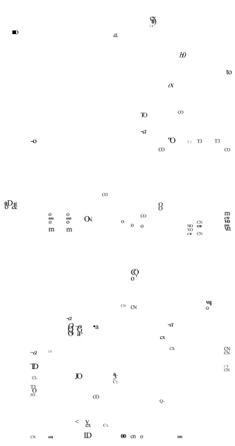
The BBB separates vascular components from the brain parenchyma, impeding the entry of detrimental materials. In normal physiological conditions it maintains homeostasis, maintaining essential proteins and ionic balances and allowing the entrance of essential nutrients. It also prevents access by immune cells. However, disruption of the BBB in the CNS may lead to neurological damage. The potential source of damage comes from the immune system, with the entry of activated lymphocytes into the CNS. The immune system is equipped with numerous effector mechanisms and can greatly alter the homeostasis and function of the CNS. Autoimmunity and pathogenic infectious agents can all result in acute or chronic inflammation within the CNS which subsequently leads to demyelination and axonal loss as in multiple sclerosis (Purves et al., 2008).
~50-100 times tighter than peripheral micro-vessels as a result of complex tight junctions (TJs) produced by the interaction of several transmembrane proteins. Transmembrane proteins occludins, are main contributors to the TJs along with claudins and the junctional adhesion molecules. Cytoplasmic proteins e.g. zonula occludens protein-1 and -2 link the transmembrane proteins to the actin cytoskeleton allowing paracellular transport to be modulated in response to different stimuli (Huber et al., 2001, Ballabh et
0 • • •
al., 2004). Despite an estimated total surface area of between 10-20 m of capillaries in the human brain, the TJs make the brain practically inaccessible for polar molecules unless they are transferred by transport pathways at the BBB that regulate the microenvironment of the brain (Pardridge et al., 1990).
There are also adherens junctions, which stabilise cell-cell interactions in the junctional zone. Large molecules such as antibodies, lipoproteins, proteins and peptides can also be transferred to the central compartment by receptor-mediated transcytosis or non specific adsorptive-mediated transcytosis. The receptors for insulin, low-density lipoprotein and iron transferrin are all involved in transcytosis.
M icroglia
Dlood Tight
junction Ligand
R eceptor
Endothelial cell
Pericyte Sm ooth m uscle
D
Basal lamina
Neuron
Neuron A strocyte
1.1.2 Neurons
A typical neuron has four distinct parts; cell body (soma), which is the main part and has all the necessary components of the cell, e.g. nucleus containing the DNA, endoplasmic reticulum (ER), ribosomes and mitochondria. The dendrites are the afferent thin structures of the neurons, frequently arranged around the neuronal cell body in the form of dendritic branches. They act to conduct electrochemical signals received from other neuronal cells to the cell body from which the dendrites project. At the other end of the soma a long unique extension, the axon 1 pm in diameter and up to 1 m long in humans, conducts the signal away from the soma. The axons of many neurons have myelin wrapped around them to form the myelin sheath. This is formed by either of two types of glial cell Schwann cells which ensheath axons of peripheral neurons and oligodendrocytes which insulate those of CNS neurons. Along the myelinated axons, gaps in the sheath known as nodes of Ranvier occur at evenly-spaced intervals. The myelination enables efficient and rapid electrical impulse propagation. The demyelination of axons is what causes the multitude of neurological symptoms found in diseases such as MS (Siegel et al., 2006).
In the human brain generally, there are three types of neurons depending on their functions. Motor neurons or multipolar neurons control muscle contractions by carrying messages from the CNS to the muscles or glands. Motor neurons include spinal motor neurons, pyramidal neurons and Purkinje cells. The second type are sensory neurons or bipolar neurons which carry signals from the body's sense receptors, as in the eye and ear to the CNS. The third type are the intemeurons these neurons communicate with the spinal cord and with the skin or muscle which connect sensory and motor neurons e.g. dorsal root ganglia cells (Squire et al., 2003).
1.1.3 Glial Cells
in glutamate (as a neurotransmitter) uptake, whereby they detoxify it, converting it to glutamine (Farina et al., 2007). There is also increasing evidence to suggest astrocytes have potential roles in immunity e.g. acting as antigen presenting cells to CD4+ helper/inducer cells in vitro by expressing major histocompatibility complex class II (MHC II) antigens. Also they can synthesise and secrete cytokines e.g. IL-1, TNF and interferon gamma (INFy). Microglia are macrophage-like cells resident within the CNS. Microglia are derived from bone marrow stem cells and early during development they occupy the CNS and remain quiescent as a resident macrophage population (Federoff, 1995). These cells can act as antigen presenting cells (APCs) and may have proinflammatory effector functions such as secreting cytokines following activation by inflammatory stimuli (Kreutzberg, 1996, Shrikant and Benveniste, 1996). Activated microglia have been shown to be present within demyelinating lesions and to phagocytose myelin debris and express MHC II, suggesting microglia may play an important role in the pathogenesis of MS (Loughlin et al., 1993, Aloisi et al., 2000).
1.1.4 The CNS Extracellular Matrix (ECM)
Structurally, CSPGs are highly anionic macromolecules, as a result of sulphate and carboxyl groups in their polysaccharide GAG side chains. The GAGs are key components of the brain ECM. They are involved in binding cations, e.g. potassium, calcium and water also regulating the movement of molecules through the matrix and cell adhesion and growth (Bellail et al., 2004). Dermatan sulphate PGs are also present. In addition, the ECM contains tenascin-C and -R (Toole, 2000, Toole, 2004).
CNS ECM is deficient in collagen, fibronectin and laminin, the brain has a soft consistency when compared to cartilage. The ECM has an important role in physiological processes in the brain such as development (migration of neuronal and glial precursor cells), repair, proliferation and cell signalling (Bellail et al., 2004). It has been reported that during maturation of the CNS, distinctive changes in the composition of the molecules of the ECM (lecticans, tenascins and link proteins in brain) occur (Milev et al., 1998, Hirakawa et al., 2000). In the developing ECM brain components are neurocan, the versican VI splice variant, tenascin-C and. link protein, while brevican, versican V2, aggrecan, tenascin-R and link proteins are characteristic components of the adult brain ECM (Figure 1.3) (Pesheva et al., 1989, Yamaguchi, 1996, Schmalfeldt et al., 1998). Also predominantly expressed in the mature nervous system are aggrecan, tenascin-N and brain link proteins (Hirakawa et al., 2000, Bekku
et al., 2003, Neidhardt et al., 2003). Brevican and tenascin-R are considered as specific CNS molecules. Similarly, neurocan and tenascin-C are representatives of juvenile brain ECM, both proteins decreasing significantly in brain after the first postnatal week (Rauch et al., 1991, Dorries and Schachner, 1994).
members, brevican with sulphated glycolipids and neurocan with members of cellular adhesion molecules (Viapiano and Matthews, 2006).
Neuron
brain
developm ent
late
brain link proteins
versican V 2 n
N-terminal (hyaluronan-binding) domain
C> C-terminal (C -ty p e lectin-containing) domain
I chondroitin sulfate chains
t keratan sulfate chains ~1 OO m
cartilage link protein
cartilage link protein
hyaluronan
versican V I
early tenascin-C
aggrecan
Furthermore, alternative splicing of versican results in transcripts that encode four variants: VO, VI, V2 and V3 (Zimmermann and Ruoslahti, 1989, Ito et al, 1995, Zako
et al., 1995). All four isoforms have distinct amino and carboxy terminal globular domains (G1 and G3) (Figure 1.4). The G1 domain contains hyaluronan and link protein binding sites. The versican isoforms differ in numbers of the GAG chain binding sites with VO containing both GAGa and GAGP domains, VI containing only GAGP, V2 containing only GAGa, and the V3 isoform lacks the entire central domain including both GAG a and p domains: the G1 domain is directly followed by the G3 domain (Zako et al., 1995). Lecticans, aggrecan and versican are expressed by a wide range of tissues (aggrecan by cartilage and CNS and versican by connective tissue, blood vessels, brain, kidney and cartilage). CNS-specific proteoglycans are brevican and neurocan (Viapiano and Matthews, 2006).
Immunohistochemical studies demonstrated alterations in the composition of the ECM in various types of MS plaques. In active and chronic active MS lesions that are characterised by a massive influx of inflammatory cells, a decreased immunoreactivity of chondroitin and dermatan sulphate proteoglycans was observed. In active lesions, white matter-associated proteoglycans accumulate in macrophages, suggesting that chondroitin and dermatan sulphate proteoglycans are phagocytosed together with myelin or myelin breakdown products. Accumulation of CSPGs around lesions contribute to their formation of a barrier to axonal growth (Sobel and Ahmed, 2001). Also, Back et al, (2005) observed accumulation of hyaluronan in MS lesions (Back et al., 2005).
1.1.4.1 Previous Studies on CSPGs Proteolysis
also section 1.5.1). ADAMTS-1, -4 and -5 dependent degradation of aggrecan has been demonstrated (Abbaszade et al., 1999, Kuno et al., 2000, Tortorella et al., 2000). ADAMTS-5 is a key enzyme in the breakdown of aggrecan in arthritic cartilage (Glasson et al., 2005, Stanton et al., 2005). ADAMTS-1, -4 and -9 have been reported to mediate versican proteolysis in different tissues including aorta and brain (Sandy et al., 2001, Somerville et al., 2003, Westling et al., 2004). Versican can control several cellular processes such as adhesion, proliferation, apoptosis, migration and invasion via the highly negatively-charged chondroitin/dermatan sulfate side chains and by the interactions of the G1 and G3 domains with other proteins (LeBaron et al., 1992, Wight,
2002).
ADAMTS-1 and ADAMTS-4 have been shown to cleave versican VI in the GAG-P binding domain at Glu441-Ala442 resulting in a 70 kDa fragment in human aorta (Sandy
et al., 2001). Furthermore, Westling et al, 2004 have demonstrated that versican V2 in human brain tissue can be cleaved by ADAMTS-4 at the Glu405-Gln406 site within the GAG-a binding domain to produce a 64 kDa fragment (Westling et al., 2004). Also it has been found that versican (V0/V1) proteolysis via ADAMTS leads to a 70 kDa fragment that is essential for the formation and differentiation of endocardial cushion mesenchyme (Kern et al., 2007). Another study by McCulloch et al, (2009) suggests that ADAMTS-mediated proteolysis of versican is important in limb development and the generated fragment could regulate interdigital web regression (McCulloch et al.,
Aggrecan Versican VO Versican V1 Versican V2 Neurocan Brevican Versican V3
COOH
COOII
nhjJ Iwl G1 Region
•Wyr-cooH G3 Region — — Chondroitin Sulfate Hyaluronan
-* X ADAMTS-Cleavage Site <3^ Proteolytic Cleavage Site
(N eurxan)
ju u Core 3rotein Portions used
— for Artibody Production
1.2 Multiple Sclerosis
Multiple sclerosis (MS) is a chronic, inflammatory, demyelinating and neurological disorder which affects mainly the white matter of the CNS (Bebo et al., 1999). MS is a complex disease and can have a personal, social and economic load on the society, which is estimated ~ £1 million per MS patient (Orton et a l, 2006).
1.2.1 Clinical course of MS
The majority of MS patients present with relapse-remitting (RR) symptoms, which starts with sporadic attacks and then periods of remission with partial or complete recovery. Symptoms may resolve completely, remission periods can last for months or years. Secondary progressive (SP) MS develops in more than half of RR patients. This will usually occur 15-20 years after the onset of the disease and involves fewer attacks and incomplete recovery. The primary progressive (PP) MS accounts for about 15% of MS cases in the UK and involves a progressive accumulation of disability without remission periods. Finally, the progressive relapsing (PR) disease is a very rare (< 1%) condition where symptoms steadily get worse from the beginning of the disease. Figure 1.5 shows the clinical course of MS types (Lublin and Reingold, 1996).
1.2.2 Epidemiology of MS
pOce
t/3
Time
Relapsing- remitting
Time
Secondary Progressive
pOCS
VI
Time
Primary Progressive
Sia
i k
Time
Progressive-relapsing
There is a latitudinal gradient in prevalence, independent of genetic factors, with rates increasing moving south or north away from the equator. The prevalence of MS varies considerably, being highest in northern Europe, New Zealand, Australia, North America and Canada. Latitudinal gradients are even described within the UK, with the highest rates being observed in Scotland and Northern Ireland. Asia, Africa and South America that lie on the equator have low levels of MS (5-10 per 100,000) (Compston et al.,
1998). Migration studies demonstrate that those who emigrate from an area of low- prevalence to an area of high-prevalence remain at low risk if they move after 15 years of age (Alter et al., 1966).
As the geographical distribution of MS prevalence is increased when approaching the poles, this highlighted the interest in low vitamin D levels as a risk factor for developing MS (Acheson and Bachrach, 1960). Recently, clinical observations and experimental work in vitro and in experimental autoimmune encephalomyelitis (EAE) animal models of MS has been reported, which showed that limited exposure to sunlight, which relates to vitamin D synthesis, has been associated consistently with an increased risk of developing MS (Ascherio et al., 2010, Burton et al., 2010, Solomon and Whitham, 2010). The prospect of a potential tool such as vitamin D supplements to prevent MS is tempting, yet challenging to investigate in an intervention study since it would require a huge population to measure any effect on MS incidence. However, vitamin D supplementation has not only been proposed to prevent MS, but also to attenuate disease activity of MS (Goldberg et al., 1986).
1.2.3 Aetiology and Immunopathogensis of MS
and the degree of exposure to microorganisms in early life (hygiene hypothesis) (Ponsonby et al., 2005) as well as personal UVR exposure (van der Mei et al., 2003, Lucas et al., 2011) are well-established environmental risk factors that significantly alter the risk of developing MS.
Treatment to reduce the severity of the disease has been improved substantially but there is not yet a cure. An immune reaction to a viral infection where viral proteins are similar to self proteins may cause a cross reaction to myelin self proteins, molecular mimicry. This may occur early in life, in the periphery, and may initiate an autoimmune disease process with a genetic susceptibility. Activated antigen-specific T cells and B cells cross the BBB and infiltrate the perivascular space (Hemmer et al., 2006).
Figure 1.6 B shows a schematic view of the immunopathology of the MS lesion. A number of immune and CNS cell types are involved in lesion development and repair. T cells, B cells and macrophages infiltrate the lesion. CD4+ T cells are located in the perivascular cuff. The antigen-specific T cells, guided by chemoattractants, infiltrate the lesion in CNS. These cells become reactivated by antigens in association with MHC II presented on dendritic cells, B cells, microglial cells and macrophages, and locally released cytokines and chemokines attracting macrophages to the lesions and their distribution throughout the lesion is reflected in the expression pattern of MHC molecules. Macrophages release proinflammatory cytokines (IL-6 and TNF) and toxic molecules (nitric oxide). MHC class I is expressed by all cells in the inflammatory surroundings of the CNS (Neumann et al., 1995, Dandekar et al., 2001). CD8+ T cells infiltrate the parenchyma and, as well as secreting inflammatory mediators, they directly attack cells expressing MHC class I such as neurons and oligodendrocytes (Hemmer et al, 2006).
remyelination is usually incomplete and loss may be irreversible. Overall, the extent of inflammation, neurodegeneration and remyelination is heterogeneous between patients (Hemmer et al., 2006).
Studies in EAE mainly concentrated on the roles played by CD4+ T cells in MS pathogenesis. It is now becoming clear that CD8+ T cells also may play a significant role in the disease. It was observed that CD8+ T cell numbers were significantly higher than those of CD4+ T cells in MS lesions at all stages of MS (Lucchinetti et al., 2000). Numbers of myelin-reactive CD8+ T cells in the peripheral blood of people with MS were significantly higher than in individuals without MS (Crawford et al., 2004). Another study by Skulina et al, (2004) showed the persistence of clonally expanded CD8+ T cells in MS lesions (Skulina et al., 2004).
The mechanism of the BBB breakdown in MS is uncertain but it is thought that the exposure of the endothelium to proinflammatory cytokines such as INFy, IL-lp and TNF disturbs the BBB by disorganising cell-cell junctions and enhances leukocyte endothelial adhesion and migration and increases expression of MHC II (Minagar and Alexander, 2003).
Figure 1.6: Immunopathogenesis in MS. Reprinted by permission from Macmillan Publishers Ltd: Nature Reviews Neuroscience (Hemmer et al.,
2006.
A
AHered
immune
system
Infectious agont?
Peripheral
activation
Blood-braln Infiltration of Activation of Iccal barrier p a ssa g e Immune celts Immune ceils
Periphery
Inflam m ation
CNS
R epalr/reorganizaticn Su sceptib ility
/
CD-I * T cells
Macrophage*'
SLJ. j y Myelin O /'‘Oligodendrocytes
Dendritic
cell
Normal w hite m atter
Besting microglia
It has been shown that the release of MMPs may facilitate the infiltration of activated immune cells into the CNS parenchyma by breaking down proteins, glycoproteins and PGs present in the basal lamina including type IV collagen and heparan sulphate proteoglycan of the BBB (Maeda and Sobel, 1996). Following migration into the CNS, activated T cells initiate an inflammatory process triggered by specific antigen. Immunological effector mechanisms include activation of microglia and infiltrating macrophages by T cell cytokines which may lead to ECM damage and demyelination. Recognition of myelin proteins e.g. myelin-associated glycoprotein (MAG), myelin basic protein (MBP) and myelin oligodendrocyte glycoprotein (MOG) as foreign leads to myelin damage (demyelination) and subsequently axonal damage and loss (Grigoriadis et al., 2004). This damage causes a large variety of symptoms such as vision impairment, bladder or bowel dysfunction, motor symptoms (eg. weakness and spasticity), sensory symptoms (e.g. numbness and dysaethesia), tremor and ataxia and other symptoms such as fatigue and cognitive impairment.
1.2.3.1 Toll-like Receptors (TLRs) in MS Immunopathogenesis
Normally, when pathogens invade the CNS, the innate immune system provides a rapid but relatively nonspecific response to neutralise the infection via Toll-like receptors (TLRs). Due to this moderately nonspecific response, local tissue injury can occur (Lehnardt et al., 2003). If infection persists, adaptive immunity, which involves T and B cell activation, can provide a delayed but more specific response that is typically less destructive to the host tissue. An exception to this theory is with autoimmune disease where adaptive immunity promotes extensive host tissue damage. In general, however, the immune system provides a response that is progressively more specific to pathogens and less destructive to host tissue. An innate immune response is also provoked in non- infectious CNS injury or disease, including neurodegenerative diseases (Zhang et al., 2005, Boillee et al., 2006, Yoshiyama et al., 2007, Giunta et al., 2008), stroke (Cao et
al., 2007, Lehnardt et al., 2007, Tang et al., 2007), spinal cord trauma (Kigerl et al.,
2007), spinal nerve damage (Kim et al., 2007) and tumour infiltration (Hussain et al.,
2006, Curtin et al., 2009).
Innate immune activation in MS is indistinguishable from that associated with microbial exposure. The rapidity of resident immune cell activation is similar, for example occurring within 5 minute of spinal cord injury (Pineau and Lacroix, 2007) and within 8 minute of lipopolysaccharide (LPS) exposure (Clark et al., 2006). The underlying stimulus is that injured tissue or cells release, secrete, or synthesise molecules associated with damage, that communicate the presence of injury to the innate immune system.
presenting cells (APCs) to T cells to recognize antigens (Takeda and Akira, 2005, Hacker et al., 2006). These receptors represent a key molecular link between tissue injury, infection, and inflammation. Thus, TLRs play an important role in linking the innate to the adaptive immune response (Bell et ah 2005).
DAMPs include endogenous molecules released by activated cells and ECM molecules. These molecules which are generated upon tissue injury activate TLRs. One of the first endogenous TLR activators recognised was the heat shock protein 60 (HSP60), which was shown to induce cytokine synthesis and to induce an inflammatory response. TLR activators that have been associated with MS are HSPs and high-mobility group box (HMGB1) protein. HMGB1 protein, originally described as a DNA-binding protein that facilitates transcription, can also be released extracellularly during acute inflammatory responses. Exposure of neutrophils, monocytes, or macrophages to HMGB1 results in an enhanced expression of proinflammatory cytokines (Park et al., 2004). Other TLR activators linked to MS are ECM molecules such as tenascin-C, versican and fragments of hyaluronan which may promote an inflammatory response (Piccinini and Midwood,
2010).
1.3 Cytokines in MS
et al., 1992). Thl cells activate macrophages and microglia to produce proinflammatory cytokines (IL-1 and TNF) and toxic molecules such as nitric oxide.
Activated astrocytes, due to their ability to secrete cytokines, play a significant role in triggering and maintaining immune responses in the brain. IL-1 and TNF modulate apoptosis of CNS cells and lymphocyte differentiation and infiltration (Kim, 1996). The Thl cytokines, lymphotoxin (LT), INFy and IL-2 are upregulated in MS. In contrast, T- cells producing Th2 cytokines (IL-4, IL-5, IL-6 and IL-10) are non encephalitogenic and possess the capacity to induce resistance to EAE (Ramirez and Mason, 2000). An exception to this, reported by Lafaille et al (1997), was that MBP-specific Th2 cells have the capacity to cause EAE in immunodeficient mice (Lafaille et al., 1997). Other findings, obtained with both EAE models and people with MS indicate the involvement of IL-17, produced by the Thl 7 cells another subset of T-helper cells, which in addition, toThl cells are thought to be critical in MS (Hofstetter et al., 2009). In MS increased numbers of mononuclear cells have been shown to express IL-17 mRNA in the peripheral blood (Matusevicius et al., 1999). Also these cells have been shown to migrate across the BBB (Kebir et al., 2007). People with MS, have also been shown to have elevated IL-17 levels in their cerebrospinal fluid (Ishizu et al., 2005) and IL-17 protein is restricted to active areas of MS lesions (Tzartos et al., 2008).
1.3.1 The Interleukin-1 (IL-1) Family
Rivest, 2007). Pro-IL-lp must be cleaved by the enzyme caspase 1, to produce the active form and allow cellular release (Thomberry et al., 1992). The third member of the IL-1 family is IL-1 receptor antagonist (IL-lra). It blocks IL-1 action by binding to IL-1 receptors but does not induce any intracellular signal (Gibson et al., 2004). In normal physiological conditions in the CNS, IL-1 p is expressed at low concentrations, but it is upregulated in response to pathological CNS injuries (Giulian et al., 1989). Hauser et al., (1990) investigated the cytokines IL-1 p, TNF, and IL-6 by specific radioimmunoassays in the cerebrospinal fluid (CSF) of people with MS and other neurologic diseases. There was an increase in IL-1 p in people with active MS compared with other neurologic diseases (Hauser et al., 1990). IL-1 p also has been reported to be increased in the CSF of MS patients and EAE animals (Wang and Shuaib, 2002). Chronic expression of IL-1 in rat brain results in extensive demyelinating lesions, mimicking MS and IL-ra decreases disease progression in EAE.
1.3.2. Tumour Necrosis Factor (TNF)
Tumor necrosis factor (TNF) is a protein produced by monocytes, macrophages and a wide variety of other cell types in response to other cytokines. In contrast, lymphotoxin (LT) is a 25-kDa glycoprotein but it is lymphokine cytokine. Both TNF and LT are homologous (28%) in their amino acid sequences and formerly these were known as TNFa and TNFP respectively. As they have common cell surface receptors, it is thought that all cellular responses mediated through TNF are also mediated by LT (Chaturvedi
et al., 1994).
TNF can be synthesised in the CNS by microglia, astrocytes, and some populations of neurons (Lieberman et al., 1989, Morganti-Kossman et al., 1997, Chung et al., 2005). TNF has been suggested as an important mediator of MS pathogenesis (Wajant et al.,
TNF binds to two receptors (TNFRI [p55] and TNFRII [p75]), which initiates intracellular signalling (Viviani et al., 2004). The resultant gene expression modulation can contribute to inflammation and hence TNF is described as a pro-inflammatory cytokine. A study done by Hauser et al. (1990) shown that TNF was found more frequently in active MS than in other neurologic diseases. Gregersen et al, (2000) demonstrated that TNF was localised to microglial cells in mice post-ischaemia (Gregersen et al., 2000).
TNF is expressed by macrophages, astrocytes, microglia and endothelial cells in chronic and active MS. Furthermore, MS studies demonstrate that there is a correlation between TNF concentration and severity of the disease (Matusevicius et al., 1996).
1.4 ADAM-17
The ADAMs (a disintegrin and metalloproteinases) are a family of transmembrane and secreted proteins with important roles in regulating cell phenotype via their effects on cell adhesion, migration, proteolysis and signalling. The ADAMs belong to the M12B adamalysin protease subfamily in the MEROPS classification (Rawlings et al., 2008), which contains the closely related snake venom reprolysins and the ADAMTSs. ADAMs are multi-domain proteins with a pro-domain, metalloprotease, disintegrin, cysteine-rich, epidermal growth factor (EGF)-like, transmembrane and cytoplasmic tail domains. The functional ADAM metalloproteinases are involved in "ectodomain shedding " of growth factors, cytokines, receptors and adhesion molecules. Proteins can be cleaved and thereby released (shed) from the plasma membrane by these proteases or sheddases. TACE or ADAM-17 is a well known sheddase which is involved in releasing soluble TNF from its membrane-bound precursor (pro-TNF) (Moss et al.,
ADAM-17 is responsible for the cleavage of a wide variety of substrates involved in inflammation including transforming growth factor-alpha (Pro-TGFa), p75 (Peschon et al., 1998) and p55 TNF receptors (Reddy et al., 2000), the chemokine (fractalkine) (Garton et al., 2001) and amyloid precursor protein (Garton et al., 2001). The release of TNF from different cells occurs in response to injury or infection and plays an important role in the adaptive immune response to these conditions. However, TNF can also be produced in excess and can cause tissue damage. Generation of soluble TNF by ADAM-17 cleavage has been found to have a pathogenic role in CNS inflammatory responses involving MS (Kieseier et al., 2003, Plumb et al., 2006).
1.4.1 ADAM-17 and the CNS
Within the normal CNS, ADAM-17 expression has been observed in astrocytes and endothelial cells using double indirect immunofluorescence and confocal microscopy (Goddard et al., 2001). Plumb et al (2006) observed expression of ADAM-17 in activated macrophage/microglia and parenchymal astrocytes in MS white matter (Plumb
et al., 2005, Plumb et al., 2006). Its expression is upregulated in active MS lesions and in EAE. Expression levels correlated with disease activity indicating that it may be involved in disease pathogenesis (Plumb et al., 2005, Plumb et al., 2006). ADAM-17 expression by astrocytes and activated macrophage/microglia may therefore allow shedding of TNF producing soluble proinflammatory TNF. ADAM-17 cleavage of TNF has several proinflammatory effects including increased endothelial cell attachment of inflammatory cells via upregulated adhesion molecules, chemotaxis, migration and BBB disruption (Dobbie et al., 1999). Alternatively, it may induce a beneficial immune response by causing apoptosis of auto-reactive T cells as seen in experiments on the MS animal model EAE (Probert and Akassoglou, 2001, Weishaupt et al., 2004).
1.5 ADAMTSs
belonging to the same subfamily (B) of metzincin proteins as ADAMs, the adamalysins according to MEROPS database (Rawlings et al., 2010). ADAMTS-1 was first described by Kuno et al, (1997) identified as a novel murine complementary DNA (cDNA) expressed in a cachexigenic adenocarcinoma cell line. There are 19 known ADAMTSs identified in humans, numbered 1-20 (ADAMTS-5 protein is the same as ADAMTS-11) (Figure 1.7) and their known functions include cleavage of the ECM proteoglycans (aggrecan, versican and brevican), collagen processing, organogenesis, anti-angiogenesis and blood coagulation homoeostasis.
1.5.1 ADAMTS Subgroups
ADAMTS proteins can be divided into seven divisions, according to their structural characteristics and activities (targeted substrates) (Jones and Riley, 2005). The members of ADAMTS-1, -4, -5, -8, -9 and -15 forms a subgroup that is able to cleave the major cartilage proteoglycan aggrecan from which they derived the name aggrecanases. They also have a more general name, which is hyalectanases due to their ability to cleave substrates of the hyalectan (lectican) family of proteoglycans, including aggrecan, versican and brevican (Gao et al., 2002). They have GEP activity, cleaving peptide bonds at the carboxyl end of the glutamate residue. The GON-ADAMTSs are ADAMTS-9 and -20. ADAMTS-20 shares its modular arrangement with that of the long isoform of ADAMTS-9, having 14 C-terminal thrombospondin (TSP)-repeats and a gon domain (Somerville et al., 2003, Llamazares et al., 2003). Moreover, though ADAMTS-9 is classified as a GON-ADAMTS with ADAMTS-20, it too is able to cleave aggrecan at the Glul771-Alal772 bond and versican at the Glu441-Ala442 (Somerville et al., 2003).
1.5.2. The Domain Structure of the ADAMTS Proteins
The main difference between ADAMs and ADAMTS is that ADAMs are transmembrane proteins while the ADAMTSs are secreted proteins, which bind to the ECM. ADAMTSs consist of prodomain, metalloprotease and disintegrin domains, but lack the ADAMs’ transmembrane domain. The thrombospondin motif(s), present on the C-terminal side of the disintegrin domain of ADAMTS also distinguishes them from ADAMs (Figure 1.8) and are thought to function, with the disintegrin domain, in binding to the ECM.
The molecular structure of the ADAMTS proteins can be subcategorized into domains. The ADAMTSs are synthesized as inactive zymogens. From the N- to the C-terminus, they each consist of: a signal peptide, a prodomain, a metalloproteinase catalytic domain, a disintegrin-like domain, a central thrombospondin (TSP)-repeat, a cysteine-rich domain, a spacer domain and a variable number of C-terminal thrombospondin (TSP)- repeats, which range from 14 C-terminal repeats in the case of ADAMTS-20 to none in the case of ADAMTS-4 (Stocker et al, 1995, Kaushal and Shah, 2000).
1.5.2.1 The Signal Peptide and Prodomain
The prodomain of human and mouse ADAMTS-5 contains three potential furin
• 'ico 9f^9
recognition sites (Hurskainen et al., 1999) within a multi basic sequence RRRRR at the activation site. Although the predicted activating cleavage site at R - S has been detected in an in vitro expression system (Zeng et al., 2006), cleavage at this site has not been confirmed in vivo. Whether proprotein convertases other than furin can activate ADAMTS-5 and whether removal of the prodomain is required for its secretion has yet to be resolved (Wang et al., 2004).
1.5.2.2 The Metalloproteinase Domain (Catalytic Domain)
All ADAMTSs have a Zn-binding peptidase consensus HEXXH, catalytic domain which contains a zinc-binding active site similar to that in the ADAMs, which distinguishes the ADAMs and ADAMTSs from other metalloproteinases (reviewed in Porter et al., 2005).
1.5.2.3 The Disintegrin-Iike Domain
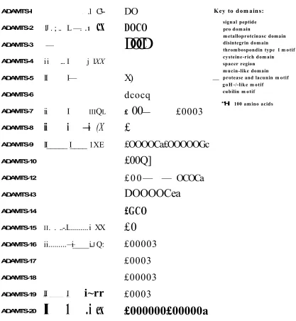
ADAMTS-I ADAMTS-2 ADAMTS-3 ADAMTS-4 ADAMTS-5 ADAMTS-6 ADAMTS-7 ADAMTS-8
A DA MTS-9
ADAMTS-10 ADAMTS-12 ADAMTS-I3 ADAMTS-14 ADAMTS-15 ADAMTS-16 ADAMTS-17 ADAMTS-18 ADAMTS-19 ADAMTS-20
. .1
C3--1J . ; .. L —. .1
cx
—
ii ... I j IXX
II I—
ii I iiiQl
ii i —i
(X
II_____ I____ 1XE
ii. . ..-.I...i XX
ii...—i____i... jQ:
II I
I I ... L...
i~rr
II 1 . i ex
DO
DOCO
DOCD
Key to dom ains:
X)
dcocq
£
00
—£
£0003
£OOOOCa£OOOOOGc
£00Q]
£00— — OCOCa
DOOOOCea
£GCO
£0
£00003
£0003
£00003
£0003
£000000£00000a
_signal peptide pro domain
metalloprotcinasc domain disintegrin domain
thrombospondin type 1 m otif cysteine-rich domain spacer region mucin-like domain protease and lacunin m otif goH -/-like m otif
cubilin m otif
“Hi 100 amino acids
[image:52.612.59.476.75.527.2]1.5.2.4 The Cysteine-rich Domain (CRD)
This domain is a cysteine rich sequence containing ten cysteine residues. Little is known of the function of this domain. However, the expression of various domain- deletion constructs of murine ADAMTS-1 showed that the CRD-spacer sequence is a functional ECM-binding domain (Kuno and Matsushima, 1998).
1.5.2.5 The Spacer Domain
There are no common structural features in this region and it is variable in length. ADAMTSs may undergo an extracellular C-terminal processing in this domain, which may have a significant effect on enzyme activity, localization and substrate specificity. ADAMTS-4 undergoes a C-terminal truncation to generate two isoforms with a distinct reduction in affinity of binding to their substrate (Flannery et al., 2002). Efficient aggrecanase activity requires the presence of GAGs attached to the aggrecan core protein. Flannery et al, (2002) demonstrated that full-length ADAMTS-4 (~ 68 kDa) undergoes autocatalytic C-terminal truncation to generate two distinct isoforms (-5 3 kDa and 40 kDa), which showed a clear reduction in affinity of binding to sulphated GAGs. ADAMTS-4 without the spacer region was 53 kDa (Gendron et al., 2007).The C-terminal spacer domains also affect binding of full-length ADAMTS-4 to sulphated GAGs (Flannery et al., 2002).
1.5.2.6 The Thrombospondin (TSP) - Repeats
The major difference between ADAMTSs, MMPs and ADAMs is the ability of ADAMTSs to bind to the ECM (Kuno and Matsushima, 1998, Somerville et al., 2003, Kashiwagi et al., 2004). They possess a well conserved thrombospondin (TSP)-repeat, homologous to the type I repeat of thrombospondins 1 and 2. The anti-angiogenic activity of ADAMTS-1 and -8 is thought to be mediated through their TSP-repeats (Porter et a l, 2005). It has been found that ADAMTS-4 cannot cleave GAG-free aggrecan. Kuno and Matsushima, (1998) demonstrated that ECM binding was mediated through the central and C-terminal TSP-repeats and the spacer region, and that sulphated GAGs were probably binding sites (Kuno and Matsushima, 1998).
With the exception of ADAMTS-4, which has no C-terminal TSP-repeats, all ADAMTSs have between 1 and 14 TSP-repeats, C-terminal to the spacer region (Figure 1.7). C-terminal TSP-repeats are more variable in sequence than the central TSP-repeats. For murine ADAMTS-1 it was demonstrated that C-terminal TSP-repeats had a significant role in binding to heparin (Kuno and Matsushima, 1998). The TSP-repeats of the C-terminus are arranged in groups separated by a short linked sequence between the groups such as in ADAMTS-9 and ADAMTS-20 or a mucin-like domain as in ADAMTS-7 and ADAMTS-12 (Somerville et al, 2004). Other types of domains may be C-terminal to the TSP-repeats groups; ADAMTS-9 and -20 contain gon-1 domains, containing ten conserved cysteine residues (Somerville et al., 2003). ADAMTS-6, -7, - 10,-12,-16,-17,-18 and -19 contain a protease and lacunin domain with six conserved cysteine residues (Nardi et a l, 1999). Additional C-terminal domains are present in ADAMTS-13 which contains two cubilin domains, (Zheng et al., 2001) (Figure 1.7).
1.5.3 ADAMTS-1, -4 and -5 and Brain ECM Breakdown
ADAM
ADAMTS-1
Signal peptide
Prodomain
Catalytic domain
Disintegrin-like domain
Cysteine-rich domain EGF repeat
Transmembrane domain
Cytoplasmic domain
TSP-repeats Spacer region
Furin recognition site
They also have been demonstrated to cleave the major cartilage proteoglycan aggrecan, and have been termed ‘aggrecanases’ (Porter et al., 2005). ADAMTS-1, -4 and -5 also cleave other lectican family members and have other functions as shown in Table 1.1. ADAMTS-1 can cleave both aggrecan and versican. Kuno et al, (2000) showed that ADAMTS-1 cleaves aggrecan in vitro and that the spacer region is necessary for this. N-terminal sequence analysis of the cleavage product revealed that the chondroitin sulphate attachment domain of aggrecan was cleaved (Kuno et al., 2000).
ADAMTS-4 and -5 are the most extensively studied of the aggrecanases. Previous studies have shown that both enzymes can cleave aggrecan (Ilic et al., 2000) but ADAMTS-4 can also cleave brevican and versican. The main aggrecanase cleavage site of ADAMTS-4 and -5 is Glu373-Ala374 but there are also four other sites in the GAG attachment regions at Glul480-Glyl481, Glul667-Glyl668, Glul771-Alal772 and Glul871-Leul872 (Sugimoto et al., 1999, Tortorella et al., 2000) (Figure 1.9). In addition, ADAMTS-5 exhibited an additional site of cleavage in the region spanning residues Glyl481 and Glul667, representing a unique cleavage of ADAMTS-5 (Tortorella et al., 2002). Nakamura et al, (2000) demonstrated that ADAMTS-4 cleaves brevican at only one site Glu395-Ser396. Furthermore, Nakada et al (2005) showed that glioblastoma cells transfected with ADAMTS-4 and -5 produced brevican cleavage products, whereas ADAMTS-1 and un-transfected cells displayed no cleavage (Nakada
et al., 2005).
Table 1.1: ADAMTS-1, -4 and -5 functions
ADAMTS-1 METH-1/
Aggrecanase-3
Inflammatory response, anti-angiogenic activity, organ morphogenesis, aggrecan and versican cleavage.
ADAMTS-4 Aggrecanase-1
Aggrecan, brevican, versican cleavage also cleaves fibromodulin, decorin, carboxymethylated transferrin and anti- angiogenic activity
ADAMTS-5 Aggrecanase-2/
ADAMTS 11
Aggrecan cleavage
r
341
E G
373
G
1771 1480
1667
*
E S
G Q Q
L
G 1871
Link Protein
Keratan Sulphate
Rich Region Chondroitin Sulphate Rich Region
Hyaluronan
[image:58.612.87.504.111.488.2]A) Brain Extracellular matrix B) ADAMTS mediated degradation of CSPGs
ADAMTS ADAMTS
O - /fr Lecticans
Tenascin-R
Hyluronan
1.5.4 Studies of ADAMTS-1, -4 and -5 in MS
Previous studies have shown that MMPs contribute in CNS inflammatory disorders (Yong et al., 1998, Yong, 1999). However, more recent studies have shown that other peptidases such as ADAMTSs are expressed by CNS tissue and modulated in inflammatory CNS disorders (Cross et al., 2006b, Haddock et al., 2006). MS is an inflammatory demyelinating disorder of the CNS and is thought to involve cytokines and proteases. Only a limited number of studies have demonstrated that ADAMTS-1, -4 and -5 are expressed in the CNS. These ADAMTSs are thought to have a significant role in the CNS during the disease process due to their ability to cleave CSPGs, which are essential molecules in the CNS ECM.
1.6 Tissue Inhibitors of Metalloproteinases (TIMPs)
Metalloproteinase activity is regulated by a group of physiological inhibitors (TIMP-1, - 2, -3 and -4), the tissue inhibitors of metalloproteinases (TIMPs) (Amour et al., 1998, Yu et al., 2000, Borland et al., 1999). TIMPs are proteins of between 21 and 34 kDa, with twelve conserved cysteine residues. The proteins are folded into two domains, with all the TIMPs containing a conserved binding site for proteinases, in the domain responsible for the inhibitory activity, the N-terminal domain (Lambert et al, 2004). TIMP-3 has been shown to be the only member of the TIMP family able to effectively inhibit the actions of ADAM-17 (Amour et al., 1998). It is also known as the main inhibitor of ADAMTSs, although TIMP-1 and TIMP-2 are inhibitory at higher concentrations (Hashimoto et al, 2001). TIMP-3 is the only TIMP known to bind to the ECM (via GAGs) (Yu et al., 2000), suggesting it has the potential to inhibit ADAMTSs in cartilage or brain.
1.7 The Aims and Objectives of this Study
Hypothesis
• Neuronal ADAMTS-1, -4 and -5 expression is modulated in response to proinflammatory cytokines in MS.
• Increased ADAMTS activity in