Polymicrogyria
Jill E. Thompson, Mauricio Castillo, David Thomas, Michelle M. Smith, and Suresh K. Mukherji
From the Departments of Radiology (J.E.T., M.C., M.M.S., S.K.M.) and Pathology (D.T.), University of North Carolina School of Medicine, Chapel Hill
The termpolymicrogyria refers to the ab-normal macroscopic appearance of a portion of the cerebral cortex characterized by too many small convolutions, simulating Moroc-can leather. Commonly, polymicrogyria is composed of small irregular gyri without in-tervening sulci or with inin-tervening sulci oblit-erated and bridged by fusion of their superfi-cial cellular layers (particularly the molecular
one). Occasionally, polymicrogyria may
have many small or widened gyri separated by shallow sulci (microsulci) (1). Histologi-cally, polymicrogyric cortex is composed of a heterogeneous collection of neurons and de-rangement of the normal six-layered lamina-tion (2). Because of the macroscopic and histologic variations found in polymicrogyria, the termnonlissencephalic cortical dysplasia
may be more appropriate.
Clinical History
The patient was a 20-year-old woman with a history of asthma and long-standing focal motor seizures; she was found dead in her bed. Six months earlier, the patient had had magnetic resonance (MR) imaging study of the brain at an outside institution (Fig 1). This study showed an area of dysplastic cortex
involving the right posterior and superior temporal lobe and extending into the parietal region. The ipsilateral sylvian fissure was prominent. Superficial to the dysplastic cor-tex, abnormal vessels, believed to be related to prominent veins, were present. The under-lying white matter was normal. Postmortem examination revealed hyperinflated lungs with abundant mucus in the bronchial tree. No other specific organ abnormalities were identified. Examination of the brain showed a cavum septum pellucidum, normal-sized ventricles, an open right sylvian fissure, and abnormal sulcation of the right frontal, insu-lar, lateral parietal, and temporal regions (Fig 1D and E). The abnormal cerebral cortex was characterized by shallow sulcation. The cer-ebellum was normal in size. After fixation, the brain was sectioned into 5- to 10-mm slices in the coronal position in order to match the coronal MR images as much as possible. Sec-tions were made of the abnormal areas and stained with hematoxylin-eosin and Nissl stain. At low power, the abnormal cortex showed shallow sulci, which were fused in the majority of their course (Fig 1F). Nissl stain showed no normal lamination and complete neuronal disorganization (Fig 1G and H). The findings were compatible with polymicrogyria of the unlayered type. The cause of death of the patient was assumed to be related to the seizures.
Discussion
The bilateral cerebral vesicles appear at approximately 35 days of gestation and later Address reprint requests to Dr M. Castillo, Radiology CB 7510,
University of North Carolina, Chapel Hill, NC 27599-7510.
Index terms: Brain, abnormalities and anomalies; Brain, gyri; Ra-diologic-pathologic correlations
AJNR 18:307–312, February 1997 0195-6108/97/1802–0307 ©American Society of Neuroradiology
Fig 1. A 20-year-old woman with polymicrogyria.
A, Coronal T1-weighted MR image (400/15/2 [repetition time/echo time/excitations]) shows wide right sylvian fissure (S). The cortex (p) in the right parietal region is slightly thick and lacks sulci, suggesting a dysplasia. The right cerebral hemisphere is small.
B, Proton density–weighted MR image (4000/19/1) shows thick insular cortex (p). Note prominent vessels in right sylvian fissure, which may be either disorganized branches of the middle cerebral artery or uncondensed veins.
C, Axial T2-weighted MR image (4000/90/1) shows posterior continuation of the right sylvian fissure, which is lined with thickened cortex (p). Note anomalous venous drainage into uncondensed cortical veins. There is scalloping of the inner table of the skull caused by pulsations of cerebrospinal fluid. No signal intensity abnormalities were present in the white matter.
D, Diagram illustrating the cortical dysplasia at the same level asAandEshows thickened dysplastic cortex (P) and a wide right sylvian fissure (S). The square (3) denotes the region from which the microscopic specimens inFthroughHwere obtained.
E, Fixed brain section. Coronal slice corresponding toAshows dysplastic cortex (p). The right cerebral hemisphere is smaller than the left one and the ipsilateral sylvian fissure (S) is wide.
F–H, Microscopy.
F, Low-power view shows very shallow sulci (arrowheads). The pial vessels (small arrows) are deep in the abnormal cortex, following the expected course of the sulci, which are fused in this specimen. Note that surrounding these vessels there is slight cellular paucity, which corresponds to the molecular layer (layer 1) (hematoxylin-eosin, magnification310).
G, Low-power view of different area of the same specimen again shows superficial arteries (arrowheads) in shallow cortical sulci, which are fused thereafter at the level of the molecular layers. Note the penetrating arteries and veins (small arrows) at the expected course of the sulci (Nissl, magnification310).
become the cerebral hemispheres. These vesicles, which have uniformly thin walls, are connected in the midline by the lamina ter-minalis. On the subependymal aspect of these vesicles, layers of tightly packed imma-ture pluripotential cells develop (the germinal matrix). The germinal matrix is separated from the pia mater by a superficial acellular zone called the marginal layer. The cells within the germinal matrix give origin to neu-roblasts that migrate outwardly to rest even-tually in the cortex. This migration begins at approximately 7 weeks of gestation and is completed between 24 and 40 weeks of ges-tation. Neurons, which will form the cortex, migrate along radial glia to their final
desti-nation. The radial cells are initially
pseudostratified bipolar immature cells origi-nating from primitive neuroepithelium. The processes of the radial glia, glial fibrillary acidic protein (GFAP), are vimentin positive and extend from the walls of the ventricles to the pia mater. These structures, known as
radial glial fibers, are essential because they provide a scaffold on which the young neu-rons migrate outwardly. Neuronal migration is fed by active proliferation of cells from the germinal matrix. Neuronal migration occurs in waves. The highly populated cortex is sep-arated from the germinal matrix by the inter-mediate layer, which contains concentric waves and parallel roles of migrating neu-rons. This intermediate layer progressively increases in size, loses all neurons, and even-tually becomes the white matter. Neuronal migration occurs in an “inside out” sequence; that is, the cells of the deepest cortical layer (layer 6) migrate early, followed by cells of layers 5, 4, 3, and 2. Cells that eventually rest in layer 1 (the molecular layer) are an excep-tion to this rule, arriving first at their final superficial cortical destination. Neuroblast formation stops at about 100 days of gesta-tion but migragesta-tion continues. The germinal matrix involutes completely by the end of the first year of life. A remnant of the germinal matrix can be seen in premature and some term newborns at the level at which the cau-date nuclei and thalami are close together (the so-called caudothalamic groove or notch).
A unique feature of the human neocortex is the presence of a temporary superficial
gran-ular layer under the pia. Although this feature disappears by 27 to 30 weeks of gestation, areas of retained superficial granular layers can normally be found in the cortex of the temporal lobes and basal cortex of the frontal lobes throughout life. All cortical layers un-dergo special organization, establishing syn-aptic contacts with local and distant neurons; these contacts contribute to the normal hori-zontal and columnar stratification of the layers. Inhibition of neuronal migration or organi-zation can result in a dysplastic cortex. Dysplastic cortex occurs as a result of a de-structive, toxic metabolic event, or of chro-mosomal deletion anomalies. Polymicrogyria also results from ischemic insults to the de-veloping brain as seen in cases of schizen-cephaly. Polymicrogyria has been reported to occur at the periphery of porencephalies, presumably because of prenatal infarcts (3). Also, the location of a polymicrogyric cortex often corresponds to well-defined arterial ter-ritories such as the middle cerebral distribu-tion (4). Prenatal infecdistribu-tion with cytomegalo-virus results in meningitis, encephalitis, and ependymitis, and affects the regions of the developing brain with greater cellular prolif-eration, such as the germinal matrix. Addi-tionally, cytomegalovirus can damage either the radial glial fibers, resulting in abnormal migration, or the established molecular layer, resulting in cortical disorganization. In pa-tients with confirmed prenatal infection with cytomegalovirus, the brain-neighboring re-gions of polymicrogyria can contain viral in-clusions, foci of necrosis, calcifications, het-erotopias, and infarctions (3). Therefore, several simultaneously occurring mecha-nisms, including direct cell loss, loss of integ-rity at the pial– glial border,
hypoxia-isch-emia, and other local vascular insults
It is agreed that polymicrogyria most likely results from insults to the developing brain in the late migrational period or after neuronal migration has stopped. The incidence of polymicrogyria is unknown but most patients with polymicrogyria have seizures and are mentally disabled. Histologically, there are several schema that attempt to classify polymicrogyria. Brain injury occurring during the early second trimester of pregnancy (12 to 17 weeks) has been associated with unlay-ered polymicrogyria; injury occurring later (18 to 24 weeks of gestation) may result in
layered polymicrogyria(1). Norman et al (3) subdivide the above histologic classification as follows: unlayered polymicrogyria can have (a) unlayered cortex, in which a molec-ular layer is present with a single band of unlayered neurons, resulting in an appear-ance of looping back and forth, or (b) poorly laminated cortex, in which four individual lay-ers are not distinctly evident; layered polymi-crogyria can have (a) four-layered cortex, in which a molecular layer is present, and there is a second layer of unlaminated neurons ar-ranged in a sinuous band with two underlying layers of horizontal, unlaminated neurons, and the outermost of these two layers, layer 3, is cell poor; or (b) parallel four-layered cortex, in which there are the same four lay-ers as above, but all are horizontal and par-allel each other.
What is clear from the above schema is that there are two types of polymicrogyria, one in which there is no neuronal organization (as in the case here shown) and another in which the cortex is laminated but contains only four layers instead of the normal six. Injury during the postmigrational period typ-ically leads to the classic four-layered cortex with associated laminar necrosis. The basic cytoarchitectonic abnormality is that of isch-emic laminar necrosis predominating in layer 5, resulting in a cell-sparse layer. Superficial to this, layers 4, 3, and 2 are normal. It is important to remember that, although the classic description of polymicrogyria is of a four-layered cortex, each case is unique; the appearance may vary from place to place even in the same patient (3).
The clinical presentation of patients with polymicrogyria is variable and depends on the severity of involvement and its location.
Most patients present with seizures, usually focal motor, and developmental delay. More than 80% of patients with cortical dys-plasias have seizures. (A surgical resection of cortical dysplasias can be beneficial in alleviating or diminishing seizures.) This high incidence of seizures is probably at-tributable to the presence of lesions in cor-tical layer 5 (which is a source of epilepsy) and in layer 4, which normally inhibits input onto layer 5 (1). Diffuse polymicrogyria re-sults in microcephaly, hypotonicity, and in-fantile seizures with marked developmental delay. This constellation of features can also be caused by congenital cytomegalo-virus infection. Patients with large cortical dysplasias can also have congenital hemi-plegia contralateral to the abnormal cortex. In the most severe form of cortical dyspla-sia, agyria, microcephaly, decerebrated posture, and severe motor retardation are typical (8). Patients with the syndrome of bilateral perisylvian cortical dysplasia present with pseudobulbar palsy, epilepsy, and bilateral motor dysfunction. Rarely, pa-tients with polymicrogyria (even those with extensive dysplasias) maintain near-nor-mal cognitive abilities.
MR imaging is the method of choice for the evaluation of patients with suspected cortical dysplasias. With MR imaging, neuronal mi-gration disorders may be classified as (a) fo-cal, including polymicrogyria and unclassifi-able cortical dysplasias, schizencephaly (always associated with dysplastic cortex of the polymicrogyric type), and macrogyria; (b) hemispheric, including hemimegalen-cephaly and megalenhemimegalen-cephaly; or (c) diffuse, including bilateral perisylvian dysplasia (polymicrogyria), band heterotopias,
lissen-cephaly-pachygyria complex, and
sub-ependymal heterotopias.
subarachnoid space and associated underly-ing parenchymal atrophy.
Although traditionally it has been accepted that polymicrogyria has the appearance of pachygyria on MR studies, this observation is probably related to the fact that initial de-scriptions were based on thick sections (5 mm or larger), which do not have the spatial resolution to resolve the characteristics of the dysplastic cortex. Polymicrogyria can be clearly appreciated on MR images with thin (1.0- to 1.5-mm-thick) sections and three-dimensional heavily T1-weighted or
gradient-echo images (2, 4). One advantage of the latter technique is that multiplanar reforma-tion is readily achieved and aids in the con-firmation of dysplastic cortex. In polymicro-gyria, the superficial cortex may have a “bumpy” appearance and its inner surface (at the gray–white matter interface) a corrugated appearance. On MR imaging, the dysplastic cortex is typically isointense to normal cor-tex. Although polymicrogyria can involve any part of the brain, the region of the sylvian fissure is most commonly affected (Fig 2). This observation and the fact that the lips of schizencephalies are lined by dysplastic cor-tex have erroneously led some authors to
la-bel some polymicrogyrias as
“schizen-cephaly type I” (9).
Generally, polymicrogyria does not extend into the ventricle (as schizencephaly does) and is separated from it by a band of white matter. In most perisylvian polymicrogyrias, the fissure is deep and extends farther back than normal. Calcifications in zones of corti-cal dysplasias are found in fewer than 5% of cases (2). Associated anomalous venous drainage is a relatively common finding. Anomalous veins are more common when there is an infolding of dysplastic cortex and should not be confused with a true
arterio-venous malformation. These anomalous
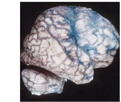
[image:5.587.50.246.274.439.2]veins are probably caused by lack of conden-sation of the cortical veins. The term uncon-densed cortical veinsrefers to persistence of the tributaries of the embryonic dural plexus, which will later coalesce to form the normal superficial cortical veins and dural sinuses. If Fig 2. Deep cleft (sylvian fissure) related to
polymicro-gyria. Lateral view of fixed brain shows deep right sylvian fissure, which extends abnormally superiorly and posteriorly. Note the abnormal course of the sylvian branches of the mid-dle cerebral artery caused by lack of temporal and parietal opercula. Cortical veins are not condensed at this level. These findings are similar to those shown in Figure 1B. Along the sides of the abnormal sylvian fissure, there are no normal sulci.
Fig 3. Diffuse polymicrogyria.
A, Axial T2-weighted MR image (4000/90/1) in a newborn with dif-fuse polymicrogyria shows incom-plete sulcation throughout the brain, multiple areas of thick cortex (par-ticularly in the right hemisphere), and a close-lip schizencephaly ( ar-rowheads) involving the left occipi-tal region. Note slightly corrugated appearance (small arrows) in the inner border of the polymicrogyric cortex.
[image:5.587.49.394.537.731.2]any doubt exists, MR angiography usually confirms the diagnosis by showing lack of arterial feeders. Large areas of dysplastic cor-tex occasionally engulf portions of the sub-arachnoid space that on MR imaging can simulate cysts within a mass. If these dyspla-sias are not recognized as such and biopsy is done, the pathologist might misinterpret them as ganglion cell tumors. Proton MR spectroscopy can play a role in these cases by establishing that metabolites (namely choline, creatine, andN-acetylaspartate) are in concentrations very similar to those of nor-mal brain (10).
The white matter underlying polymicro-gyria can also be abnormal. As many as 20% of patients show hyperintensity in underlying white matter on T2-weighted images (2). This hyperintensity can be present at birth or develop with age. It can be secondary to gli-osis, probably caused by ischemia or poor, delayed, or absent myelination. Additionally, the gray–white matter interface can be indis-tinct by MR imaging in cases of cortical dys-plasias. On MR imaging, diffuse polymicro-gyria appears very similar to pachypolymicro-gyria (Fig 3).
Positron emission tomography and
single-photon emission computed tomography
(SPECT) have been used to detect abnormal local metabolism and perfusion related to the presence of cortical dysplasias. Positron emission tomography has been shown to be sensitive in locating focal areas of cortical dysplasia, heterotopias, and other migration abnormalities corresponding to surface elec-trographic location of epileptogenic regions (11). SPECT can help identify cortical dys-plasias by demonstrating hypoperfusion
dur-ing the interictal phase and hyperperfusion during seizures (12).
References
1. Barkovich AJ, Gressens P, Evard P, et al. Formation, matura-tion, and disorders of brain neocortex.AJNR Am J Neuroradiol
1992;13:423– 446
2. Barkovich AJ. Destructive brain disorders of childhood. In: Barkovich AJ, ed. Pediatric Neuroradiology. 2nd ed. New York, NY: Raven Press; 1995;107–176
3. Norman MG, McGillivray BC, Kalousek DK, et al. Neuronal migration diseases and cortical dysplasia. In: Norman MG, McGillivray BC, Kalousek DK, et al, eds.Congenital Malforma-tions of the Brain. New York, NY: Oxford University Press; 1995;223–279
4. Raybaud C, Canto-Moreira N, Girard N, et al. Polymicrogyria: MR appearance and its relationship to the fetal development of the cortex and its microvasculature.Int J Neuroradiol1995;2: 161–170
5. Sugama S, Kusano K. Monozygous twin with polymicrogyria and normal co-twin.Pediatr Neuroradiol1994;11:62– 63 6. Larroche JC, Girard N, Narcy F, et al. Abnormal cortical plate
(polymicrogyria), heterotopias and brain damage in monozy-gous twins.Biol Neonate1994;65:343–352
7. Kuzniecky R, Andermann F, Guerrini R, et al. Congenital bi-lateral perisylvian syndrome: study of 31 patients. Lancet
1993;341:608 – 612
8. Friede RL. Dysplasias of the cerebral cortex. In: Friede RL, ed.
Developmental Neuropathology. 2nd ed. New York, NY: Springer-Verlag; 1989;330 –346
9. Castillo M, Mukherji SK. Destructive, ischemic, and vascular diseases. In: Castillo M, Mukherji SK, eds. Pediatric Head, Neck, and Spine. Philadelphia, Pa: Lippincott-Raven; 1996; 145–190
10. Castillo M, Kwock L, Mukherji SK. Clinical applications of proton MR spectroscopy.AJNR Am J Neuroradiol1996;17: 1–15
11. Chugani HT. The use of positron emmission tomography in the clinical assessment of epilepsy.Semin Nucl Med1992;22: 247–253