Eucapnic voluntary hyperpnoea and
exercise-induced vocal cord dysfunction
Julie Turmel,1Simon Gagnon,2Mélanie Bernier,3Louis-Philippe Boulet1
To cite:Turmel J, Gagnon S,
Bernier M,et al. Eucapnic
voluntary hyperpnoea and exercise-induced vocal cord
dysfunction.BMJ Open Sport
Exerc Med2015;1:e000065. doi:10.1136/bmjsem-2015-000065
▸ Prepublication history for
this paper is available online. To view these files please visit the journal online (http://dx.doi.org/10.1136/ bmjsem-2015-000065).
Accepted 17 November 2015
1Centre de recherche de
l’Institut universitaire de
cardiologie et de pneumologie de Québec (CRIUCPQ), Québec, Canada
2Centre Hospitalier
Universitaire de Québec, Québec, Canada
3Clinique d’orthophonie
Québec, Québec, Canada
Correspondence to Dr Louis-Philippe Boulet; [email protected]
ABSTRACT
Introduction:Exercise-induced bronchoconstriction
(EIB) is a common condition in endurance athletes. Exercise-induced vocal cord dysfunction (EIVCD) is a frequent confounder of EIB. The diagnosis of EIVCD may be challenging and can be missed as the problem is often intermittent and may only occur during intense exercise. Eucapnic voluntary hyperventilation (EVH) is the best test to detect EIB. This pilot study aimed to assess if EVH could be helpful in the diagnosis of EIVCD associated or not to EIB in athletes.
Methods:A nasolaryngoscopy was performed during
a 6 min EVH test, in 13 female athletes suspected to have VCD, aged 21±7 years. Image analysis was conducted by two Ear Nose and Throat surgeons in random order.
Results:During the EVH, three athletes showed
incomplete paradoxical vocal cords movement, without inspiratory stridor. However, 12 athletes showed marked supraglottic movement without inspiratory stridor. In two athletes, this supraglottic movement was severe, one showing a marked collapse of the epiglottis with an almost complete obstruction of the larynx by the arytenoid cartilage mucosa. In 3 of the 12 athletes with supraglottic movement, severe vibration of the mucosa covering the arytenoid cartilages was also observed.
Conclusions:EVH challenge in athletes can provide
information on various types of glottic and supraglottic obstruction in reproducing laryngeal movements during hyperventilation. Our findings make us suggest that exercise induced upper airway obstructions should be named: Exercise-induced laryngeal obstruction (EILO). Then, EILO should be divided in three categories: supraglottic, glottic (EIVCD) and mixed (glottic and supraglottic) obstruction.
BACKGROUND
Endurance athletes frequently report exercise-induced respiratory symptoms and show a high prevalence of exercise-induced bronchoconstriction (EIB).1–4 EIB is described as a transient narrowing of the airway during or, most often, after exercise.5–7 Symptoms of EIB may include dyspnoea, phlegm production, chest tightness, shortness of breath, wheezing and cough. However, other clinical entities can produce similar symptoms.8 9
Exercise-induced vocal cord dysfunction (EIVCD) is a frequent confounder of EIB. EIVCD is the most common cause of upper airway obstruction during exercise.10 11 It may present either as an abnormal adduc-tion of vocal cords, during inspiraadduc-tion or early expiration. Symptoms of EIVCD are often intermittent, but recurrent and include sensations of throat tightness, inspiratory stridor, cough and/or choking.12 It fre-quently presents itself as a noisy breathing and dyspnoea that occur at any level of exer-tion and symptoms are characterised by sudden onset and rapid resolution with the exercise cessation. Contrary to asthma, EIVCD has no refractory period and some athletes can experience repeated episodes immediately on restarting physical activity.13
A prevalence of EIVCD ranging from 5 to 27% in patients referred for exercise-induced dyspnoea has been reported14–17 and a similar incidence was reported among ath-letes.10 18–24 EIVCD is often associated with EIB (>50%)25 26and mostly occurs in adoles-cents11 and young adults (20–40-years-old), with a predominance in females.16 17 25 EIVCD does not seem sport-specific,13 but tends to be more common in athletes partici-pating in outdoor (8.3%) than indoor sports (2.5%).27
Summary box
▪ The eucapnic voluntary hyperventilation chal-lenge in athletes can provide information on various types of glottic and supraglottic obstruc-tion in reproducing laryngeal movements during hyperventilation.
▪ The supraglottic movement induced by hyper-ventilation can be severe in some athletes, showing a marked collapse of the epiglottis with an almost complete obstruction of the larynx by the arytenoid cartilage mucosa.
▪ Exercise-induced laryngeal obstruction should be divided in three categories: supraglottic, glottic (exercise-induced vocal cord dysfunction) and mixed (glottic and supraglottic) obstruction.
Open Access Research
copyright.
on May 7, 2020 by guest. Protected by
The aetiology of EIVCD is unclear. It may be asso-ciated with chronic rhinosinusitis,28 gastroesophageal reflux (GER),29 30 sleep apnoea and asthma.8 31 Moreover, many factors can trigger paradoxical vocal cord movement such as inhalation of allergens, pollu-tants, cold and dry air or exercise.28 32 In athletes, anxiety relative to the pressure of high expectations may subconsciously lead to laryngeal closure and chocking.10
EIVCD can be suspected by aflattening of the inspira-tory flow-volume loop, airway obstruction and/or inspiratory stridor.3 However, no technique has shown good sensitivity and specificity33–35 and it is still difficult to diagnose EIVCD.36
Direct visualisation of the upper airway by fiberoptic laryngoscopy is the gold standard for making a diagnosis of VCD or EIVCD.33 37 It can show an adduction of vocal cords during inspiration, reveal evidence of upper airway inflammation or pharyngeal erythema and also exclude other pathologies.38 However, performing a resting laryngoscopy in a currently asymptomatic athlete may not reveal paradoxical motion of vocal cords. Bronchoprovocation test with methacholine or exercise have been used to trigger VCD34 39 and laryngoscopy during exercise can also be performed.40 41 However, exercise laryngoscopy is complex to be performed in many clinics and it can be difficult to reproduce the intense physical and emotional setting of athletes in an artificial environment.42 Thus, new methods to evaluate EIVCD are needed.
Eucapnic voluntary hyperpnoea (EVH) is the best test to detect increased airway responses to exercise and support the diagnosis of EIB.43 The advantage of this test is that it can be done in the laboratory without exer-cising while it reproduces the high minute ventilation that occurs during exercise. We report the results of a pilot study which aims to assess if EVH may be useful to identify EIVCD in athletes with a high degree of suspi-cion of such problem.
METHODS Participants
Out of 352 athletes regularly followed at our centre, 41 (12%) had a confirmed or suspected diagnosis of EIVCD of whom 13 agreed to participate in the study. Six had a confirmed diagnosis of EIVCD, based on the following criteria: (1) presence of breathing difficulties during exercise with the presence or not of an inspira-tory stridor, (2) symptoms appear suddenly during exer-cise, (3) symptoms stops or diminish quickly when the exercise is stopped, (4) adduction of the vocal cords during inspiration, visualised by laryngoscopy. Athletes were aged between 14 and 35 years and trained in a competitive sport. Written informed consent was obtained from each participant and/or their parents or guardians before inclusion in the study. The protocol was approved by the local Ethics Committee.
Study design
All participants attended the laboratory on two occa-sions. On a first visit, a physical examination was per-formed followed by a medical questionnaire regarding health condition, family history of disease, medication and experience in sport. In addition, questionnaires about GER symptoms and airway sensory hyperactivity were filled by the participants. A methacholine inhal-ation test was then performed with aflow-volume curve, performed at baseline and at maximal fall in forced expiratory volume in 1 s (FEV1). Finally, if not done in the past 2 years, allergy skin prick tests were done. If a methacholine challenge had been performed in the last year, these results were used for this study.
On a second visit, a nasolaryngoscope was properly installed after local anesthesia and a resting laryngos-copy was performed by the Ear Nose and Throat surgeon (ENT). Thereafter, the pre-EVH flow-volume curve was performed and the 6 min EVH test was done, with continuous video recording laryngoscopy.
Questionnaires
A questionnaire on present and past history of respira-tory symptoms, medical conditions, as well as a record-ing of the type and frequency of exercise performed was filled. Particular attention was devoted to the description of symptoms during exercise.
Reflux symptoms index
The Reflux Symptoms Index (RSI), a self-administered nine-item instrument, was used to assess for laryngophar-yngeal reflux.44 A score greater than 9, on a maximal score of 45, is considered abnormal, suggesting laryngo-pharyngeal reflux.
Chemical sensitivity scale for sensory hyper-reactivity
The chemical sensitivity scale for sensory hyper-reactivity (CSS-SHR) questionnaire was used to quantify the affect-ive and behavioural consequences of odour intoler-ance.45 Selected from a larger number of items about odour intolerance, the CSS-SHR questionnaire consists of 11 statements/questions that are particularly sensitive in discriminating participants with an airway sensory hyper-reactivity syndrome from control participants.46 47 The sum of all 11 items makes up the individual’s total CSS-SHR score (range from 1 to 55 points; a score≥43 points indicates severe odour intolerance).
Flow volume curves
The FEV1and forced vital capacity (FVC) were assessed from flow-volume curves performed according to the American Thoracic Society (ATS) specifications using an ATS-approved spirometer.48 Predicted values were derived from Knudsonet al.49The best of three reprodu-cible measurements was used for analysis.
Open Access
copyright.
on May 7, 2020 by guest. Protected by
Methacholine inhalation test
The tidal-breathing method described by Juniperet al50 was used to determine airway hyper-responsiveness (AHR) to methacholine. After baseline measurements of FEV1and FVC, each participant inhaled 0.9% saline followed by doubling concentrations of methacholine between 0.03 and 16 mg/mL to obtain a 20% decrease in FEV1 (PC20). Methacholine aerosols were generated from a Wright nebuliser with an output of 0.13 mL/min and were inhaled for 2 min at 5 min intervals. FEV1was measured at 30 and 90 s after each inhalation and every 2 min until it started to improve. An acceptable-quality FEV1 was obtained at each time point; otherwise the FEV1 manoeuvre was repeated. AHR was defined as a PC20≤16 mg/mL.
Allergy skin test
Each participant had an allergy skin test unless per-formed within the past 2 years, to determine his atopic status. Skin-prick tests were performed with a battery of 26 common airborne allergens. Normal saline and hista-mine were used as negative and positive controls, respect-ively. Skin weal diameters were recorded at 10 min as the mean of two perpendicular measurements. A positive response was defined as a skin weal diameter≥3 mm.
Nasolaryngoscopy
Five minutes before the laryngoscopy, the nasal cavity was anaesthetised with topical lidocaine (1%) and xylometazo-line (0.1%) applied by cotton-tipped swabs in the nares. The posterior pharynx was not anaesthetised intentionally in order to avoid any anaesthesia of the vocal cords. A flexible video-naso-pharyngo-laryngoscope (VNL-1170K/ 1171K, VNL-1070STK/1570STK, VNL-1190STK, Pentax Medical, Mississauga, Ontario, Canada) was directed to the posterior pharynx several centimetres above the glottis in order to prevent stimulation and adduction of the vocal cords.
Observation of the vocal cords was first made at rest before EVH challenge. The participant was asked to make the sound ‘E,’ cough and breathe rapidly for approximately 15 s each. The possible adduction of the supraglottic structures and vocal cords were assessed during the EVH challenge at each inspiratory phase of the respiratory cycle, at the same level of ventilation for 6 min.
Eucapnic voluntary hyperpnoea
The EVH test was performed according to the method described by Anderson and Brannan.43 Briefly, partici-pants inhaled a dry-air mixture containing 21% O2, 5% CO2 and the balance with N2 at room temperature for 6 min. The target ventilation was 30 times the FEV1 according to the baseline FEV1. To get a valid test for the diagnosis of EIB, the participant must ventilate at least 21 times the baseline FEV1. FEV1 was measured before the test and at 3, 5, 10 and 15 min after the EVH test. At each time interval, FEV1 was measured twice,
and if there was a >10% difference between the two measurements, a third FEV1 was performed. After the test, the highest of the two reproducible values was used to calculate the maximal decrease in FEV1. A maximum post-EVH fall in FEV1of >10% from baseline value sus-tained during at least 5 min or observable at two con-secutive time points was considered positive for EIB.
Presence of respiratory symptoms (wheeze, cough, dys-pnoea, choking sensation, chest tightness and stridor) and their severity, during the EVH, were also recorded on a modified Borg scale (0–10).
Interobserver evaluation
All video recordings were reviewed by two experienced ENT investigators. Each data set was anonymised and presented to the observers in random order. Observers performed their evaluation separately and they then combined their assessments and agreed on afinal evalu-ation for each participant. Movement at the glottic and the supraglottic level were assessed at rest and during the 6 min EVH. The findings were rated as: (1) vocal cord adduction (yes/no), (2) vocal cord adduction (complete/incomplete), (3) supraglottic movement (yes/no), (4) severity of the supraglottic movement (slight/moderate/severe), (5) vibration of the supraglot-tic movement (yes/no), (6) respiratory distress (yes/ no), (7) audible inspiratory stridor (yes/no), (8) laryn-geal anatomy (normal/abnormal) and (9) larynlaryn-geal function (normal/abnormal).
RESULTS
Participants’characteristics
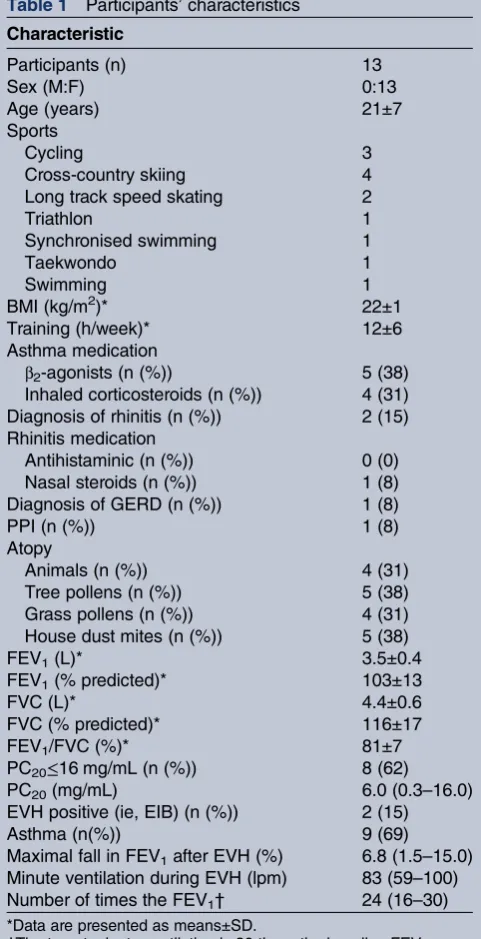
The participants’ characteristics are summarised in
table 1. The mean age of the athletes was 21±7 years. On average, they trained for 12±6 h per week. Six athletes were sensitised to common airborne allergen and nine had a diagnosis of asthma. Only two athletes reported rhinitis and one had GER disease (GERD) and was treat with proton pump inhibitors (PPI).
Questionnaires
All athletes reported respiratory symptoms (wheeze, cough, dyspnoea, chest tightness and phlegm) and inspiratory stridor during exercise with a mean Borg score for the severity of symptoms of 5±3. In all athletes, these symptoms disappeared a few minutes after the ces-sation of the exercise. In four of the athletes these symp-toms affected their exercise performance and occurred a mean of 2±2 times per week.
The RSI showed a mean score of 5.3 (0–20), on a maximal possible score of 45. Only three athletes had an abnormal score, that is, more than 9, suggesting laryngo-pharyngeal reflux. The sensitivity index to chemical agents and odours showed a mean score of 2616–34on a maximal possible score of 55. None of the athletes had an abnormal score that is, more than 43, which would have suggested airway hypersensitivity syndrome.
Open Access
copyright.
on May 7, 2020 by guest. Protected by
Spirometry findings
Ten athletes performed methacholine challenge, as part of the study, and among them none demonstrated flattering of the inspiratory curve of the flow volume loop. Inspiratory curve of theflow volume loop, before and after the EVH challenge, was also normal in all participants.
Laryngoscopic findings
All athletes had normal laryngeal anatomy and normal laryngeal function at rest. One athlete showed opening of the upper oesophageal sphincter a few times during the EVH challenge.
None of the athletes had audible inspiratory stridor or respiratory distress during the EVH challenge. Three athletes showed incomplete adduction of the vocal cords movement. However, 12 athletes showed mild supraglot-tic movement. In two athletes, this supraglotsupraglot-tic move-ment was severe, one showing a marked epiglottis posterior collapse with an almost complete obstruction of the larynx by the arytenoid cartilage mucosa. In three athletes with supraglottic movement (1 mild and 2 severe), marked vibration of the arytenoid cartilage mucosa was also observed.
The mean minute ventilation during the EVH was 83 (59–100) L/min, which correspond to 2416–30 times the baseline FEV1. Ten athletes reported respiratory symp-toms (wheeze, cough, dyspnea, chest tightness and phlegm) during the test with a mean Borg score for the severity of symptoms of 3±1. However, none of the ath-letes reported choking sensation or a sensation of tigh-tening of the throat, but three felt like they had an inspiratory stridor during the test.
DISCUSSION
Our study suggests that videolaryngoscopy during an EVH challenge may not be the most appropriate test to induce VCD. However, this test can discriminate between types of exercise-induced laryngeal obstruction (EILO): supraglottic dysfunction (obstruction), vocal cords dysfunction (EIVCD) or mixed dysfunctions (glottic and supraglottic), in reproducing laryngeal movements during hyperventilation that resting laryn-goscopy does not reveal. Thus, the EVH challenge may serve as a complementary tool in the diagnosis of patients with exercise-induced respiratory symptoms, as it is important to differentiate these conditions from EIB, their treatments are different.51 52
Christensenet al53also showed that EVH testing can be used to induce EILO, and has a potential diagnostic applicability. As a result of these observations, we can think that the inspiratory stridor reported by athletes par-ticipating in this study, during field exercise, could be due not only to EIVCD. It could be also attributable to larger and floppy arytenoid cartilage mucosa that com-bined with hyperventilation may result in inspiratory col-lapse of the supraglottic structures. That could eventually cause complete obstruction of the larynx such as laryngo-malacia observed in infants. For those athletes with a severe supraglottic involvement, a laser supraglottoplasty might be of some interest as suggested by Maatet al.52
[image:4.595.45.286.56.526.2]Although some athletes clearly have EIVCD or asthma, both conditions may coexist. In half of the participants with EIVCD, EIB or asthma is also present54 and up to Table 1 Participants’characteristics
Characteristic
Participants (n) 13
Sex (M:F) 0:13
Age (years) 21±7
Sports
Cycling 3
Cross-country skiing 4
Long track speed skating 2
Triathlon 1
Synchronised swimming 1
Taekwondo 1
Swimming 1
BMI (kg/m2)* 22±1
Training (h/week)* 12±6
Asthma medication
β2-agonists (n (%)) 5 (38) Inhaled corticosteroids (n (%)) 4 (31) Diagnosis of rhinitis (n (%)) 2 (15) Rhinitis medication
Antihistaminic (n (%)) 0 (0) Nasal steroids (n (%)) 1 (8) Diagnosis of GERD (n (%)) 1 (8)
PPI (n (%)) 1 (8)
Atopy
Animals (n (%)) 4 (31)
Tree pollens (n (%)) 5 (38)
Grass pollens (n (%)) 4 (31) House dust mites (n (%)) 5 (38)
FEV1(L)* 3.5±0.4
FEV1(% predicted)* 103±13
FVC (L)* 4.4±0.6
FVC (% predicted)* 116±17
FEV1/FVC (%)* 81±7
PC20≤16 mg/mL (n (%)) 8 (62)
PC20(mg/mL) 6.0 (0.3–16.0)
EVH positive (ie, EIB) (n (%)) 2 (15)
Asthma (n(%)) 9 (69)
Maximal fall in FEV1after EVH (%) 6.8 (1.5–15.0) Minute ventilation during EVH (lpm) 83 (59–100) Number of times the FEV1† 24 (16–30)
*Data are presented as means±SD.
†The target minute ventilation is 30 times the baseline FEV1.
Airway hyper-responsiveness to methacholine (PC20≤16 mg/mL)
and/or maximal fall in FEV1after EVH≥10%; BMI, body mass
index; EIB, exercise-induced bronchoconstriction; EVH, eucapnic voluntary hyperpnoea; FEV1, forced expiratory volume in 1 s;
FVC, forced vital capacity; maximal fall in FEV1after EVH,
expressed as mean(range); PC20, concentration of inhaled
methacholine causing a 20% decrease in the forced expiratory volume in 1 s, expressed as geometric mean (range); GERD, gastroesophageal reflux disease; PPI, proton pump inhibitors. Open Access
copyright.
on May 7, 2020 by guest. Protected by
30% of patients with asthma have coexistent VCD.25 In our study, 9 (69%) of the athletes had asthma.
The larynx and the vocal cords are innervated by sensory receptors which can be stimulated by many irri-tants.55 56 VCD can be due to glottic closure reflex induced by repetitive irritant exposure to which athletes are often exposed in their training environment.28 For example, chlorine inhalation causing VCD has been described and deserves special consideration in swim-mers.57 Several other inhaled irritants such as strong odours, perfumes, ammonia and particles matter have been identified as causative agents in VCD.28 58For this reason, we used the CSS-SHR in our study in order to identify if some athletes had odour intolerance suggest-ing airway sensory hyper-reactivity syndrome that could possibly cause VCD. However, we did not observe abnor-mal CSS-SHR score in this study.
Furthermore, GER and laryngopharyngeal reflux (LPR), associated or not with symptoms, are frequent in endurance athletes and can trigger EIVCD.28 59 These conditions can cause laryngeal lesions such as oedema, erythema and granulomas to the glottic and supraglottic structures.60 In our study, we evaluated, through RSI score, if athletes had GER symptoms. Only three athletes had GER symptoms which is not surprising as many patients with irritant-induced VCD will not report clas-sical symptoms of GER,40 but which is not in keeping with another study reporting that 84% of patients with VCD had abnormal RSI scores.27 Thesefindings may be explained by the fact that our participants are younger. In addition, previous report showed that 66–95% of patients with VCD have laryngeal findings, such as inflammation, consistent with GER.38 59 However, in our study, none of the athletes exhibited laryngeal fi nd-ings suggesting GER or LPR, but one athlete was treated with PPI.
Isolated flattering of the inspiratory curve of the flow volume loop while the patient is symptomatic is consist-ent with extrathoracic obstruction, which is the most commonly described abnormality in VCD.34 However, flow volume loop abnormalities are uncommonly noted on spirometry10 and particularly in athletes, as their symptoms occur generally only during high-intensity exercise, making the usefulness of spirometry confl ict-ing.26 61 In our study, none of the athletes showed abnormal inspiratory loop before or after the EVH test, neither during the methacholine challenge. Furthermore, bronchoprovocation tests such as metha-choline provide inconsistent results in regard to change in flow volume curve appearance. Only 40–50% of patients in whom VCD is highly suspected will have symptoms with methacholine provocation.62 63 For this reason, the appearance of theflow volume loops should not influence the decision to perform or not laryngoscopy.64
In our study, we did not observe EIVCD during the EVH challenge in athletes suspected or already having such diagnosis, which may be explained by many
reasons. EVH is done with dry air at room temperature, which does not reproduce every environmental condi-tions such as temperature, humidity, as well as the pollu-tants or allergens contained in air during field training. Moreover, EIVCD is more often observed during compe-tition possibly due to the stress and anxiety of a competi-tive event, as a relationship between VCD and psychological stress has been shown.59 EIVCD can also be related to psychological disorders or to personality traits.65 In addition, athletes may become used to the EVH challenge, as all athletes had already done this test before. Thus, they probably had less apprehension and stress in regard to that test. Finally, some athletes may not have sufficient ventilation during the EVH test, to trigger VCD. The performance during the EVH test is effectively influenced by individual factors such as atti-tude and expectations. Thus, the motivation and the effort to achieve the target ventilation may vary.
CONCLUSION
Reported exercise-induced respiratory symptoms are common in endurance athletes, but these symptoms are often treated as EIB and upper airway obstruction are not often considered and therefore rarely diagnosed. Patients with EIVCD are frequently misdiagnosed as having poorly controlled EIB and their response to asthma treatment is poor.66 Failure to diagnose EIVCD may lead to unnecessary healthcare utilisation, inappro-priate medication use25 67 and hospitalisation.68 In ath-letes, suboptimal performance and discontinuation of sports are unfortunate additional consequences of misdiagnosis.
Although EIVCD was not specifically diagnosed using EVH, by reproducing the hyperventilation-induced laryngeal movement, the EVH allowed identifying laryn-geal obstructions, which are not always observed during a resting nasolaryngoscopy. The exercise-induced inspiratory stridor reported by athletes may be associated with EIVCD or movements of different severities of the supraglottic structures. The latter observations lead us to suggest that EILO can be divided into subcategories: supraglottic obstruction, glottic obstruction (VCD) or mixed obstruction (glottic and supraglottic).
Actually, we need to find a method to quantify the larynx obstruction observed in EILO, in order to object-ively assess test results and to define criteria for diagnos-tic purpose, to evaluate effects of treatment and to further assess which patients might benefit from reduc-tion surgery. Maat et al69 had previously proposed a scoring system which is rather subjective and allowed large interobserver variability. Christensen et al53 have created software (EILOMEA) to calculate specific dis-tances and areas in the larynx from a still frame of the laryngoscopic recording, but this software need to be validated.
Acknowledgements The authors thank all the athletes who participated in the study, Mrs Kristi Huff, National Territory Manager-ENT at Pentax Medical for
Open Access
copyright.
on May 7, 2020 by guest. Protected by
the loan of equipment, Dr Jean-Philippe Vézina for his help in the images analysis, as well as the Conseil des Sports de Haut Niveau de Québec (CSHNQ) for their support in the accomplishment of this project.
Competing interests Disclosure of potential conflicts of interest of L-PB. L-PB consider to have no conflict of interests but wish to declare what can be
perceived as‘potential’conflicts of interests Advisory Boards:
GlaxoSmithKline, Novartis. Conferences (honoraria): AstraZeneca, GlaxoSmithKline, Merck, Novartis. Sponsorship for investigator-generated research: AstraZeneca, GlaxoSmithKline, Merck Frosst, Schering. Sponsorship for research funding for participating in multicenter studies: most of these studies are performed in the context of the Canadian Investigative
Collaboration with the NCE: AllerGen, Altair, Amgen, Asmacure, AstraZeneca, Boehringer-Ingelheim, Genentech, GlaxoSmithKline, Novartis, Ono Pharma, Pharmaxis, Schering, Wyeth. Support for the production of educational materials: AstraZeneca, GlaxoSmithKline, Merck Frosst, Boehringer-Ingelheim, Novartis. Governmental: Adviser for INNESS, the Quebec National Health Institute, Member of the Quebec Workmen Compensation Board Respiratory Committee. Organisational: Chair of the Canadian Thoracic Society Respiratory Guidelines Committee, Chair of the Global Initiative for Asthma (GINA) Guidelines Dissemination and Implementation Committee.
Patient consent Obtained.
Ethics approval This study was approved by the Institut Universitaire de
Cardiologie et de Pneumologie de Québec’s Ethics Committee.
Provenance and peer review Not commissioned; internally peer reviewed.
Data sharing statement No additional data are available.
Open Access This is an Open Access article distributed in accordance with the Creative Commons Attribution Non Commercial (CC BY-NC 4.0) license, which permits others to distribute, remix, adapt, build upon this work non-commercially, and license their derivative works on different terms, provided the original work is properly cited and the use is non-commercial. See: http:// creativecommons.org/licenses/by-nc/4.0/
REFERENCES
1. Turmel J, Poirier P, Bougault V,et al. Cardiorespiratory screening in elite endurance sports athletes: the Quebec study.Phys Sportsmed
2012;40:55–65.
2. Wilber RL, Rundell KW, Szmedra L,et al. Incidence of
exercise-induced bronchospasm in Olympic winter sport athletes.
Med Sci Sports Exerc2000;32:732–7.
3. Heir T, Oseid S. Self-reported asthma and exercise-induced asthma symptoms in high-level competitive cross-country skiers.Scand J Med Sci Sports1994;4:128–33.
4. Bougault V, Turmel J, St-Laurent J,et al. Asthma, airway inflammation and epithelial damage in swimmers and cold-air athletes.Eur Respir J2009;33:740–6.
5. Anderson SD, Silverma M, Walker SR. Metabolic and ventilatory changes in asthmatic patients during and after exercise.Thorax
1972;27:718–25.
6. Anderson SD, McEvoy JD, Bianco S. Changes in lung volumes and airway resistance after exercise in asthmatic subjects.Am Rev Respir Dis1972;106:30–7.
7. McFadden ER, Nelson JA, Skowronski ME,et al. Thermally induced asthma and airway drying.Am J Respir Crit Care Med1999;160:221–6. 8. Weiss P, Rundell K. Imitators of exercise-induced
bronchoconstriction.Allergy Asthma Clin Immunol2009;5:7. 9. Weinberger M. Exercise induced dyspnoea: if not asthma, then
what.Arch Dis Child2006;91:543–4.
10. McFadden ER, Zawadski DK. Vocal cord dysfunction masquerading as exercise-induced asthma—a physiologic cause for“choking” during athletic activities.Am J Respir Crit Care Med
1996;153:942–7.
11. Doshi DR, Weinberger MM. Long-term outcome of vocal cord dysfunction.Ann Allergy Asthma Immunol2006;96:794–9. 12. Newsham KR, Klaben BK, Miller VJ,et al. Paradoxical vocal-cord
dysfunction: management in athletes.J Athl Train2002;37:325–8. 13. Brugman SM, Simons SM. Vocal cord dysfunction: don’t mistake it
for asthma.Phys Sportsmed1998;26:63–85.
14. Seear M, Wensley D, West N. How accurate is the diagnosis of exercise induced asthma among Vancouver school children?Arch Dis Child2005;90:898–902.
15. Abu-Hasan M, Tannous B, Weinberger M. Exercise-induced dyspnea in children and adolescents: if not asthma then what?Ann Allergy Asthma Immunol2005;94:366–71.
16. Morris MJ, Deal LE, Bean DR,et al. Vocal cord dysfunction in patients with exertional dyspnea.Chest1999;116:1676–82. 17. Morris MJ, Grabach VX, Deal LE. Vocal cord dysfunction:
aetiologies and treatment.Clin Pulm Med2006;13:73–86. 18. Smith MS. Acute psychogenic stridor in an adolescent athlete
treated with hypnosis.Pediatrics1983;72:247–8.
19. Kivity S, Bibi H, Schwarz Y,et al. Variable vocal cord dysfunction presenting as wheezing and exercise-induced asthma.J Asthma
1986;23:241–4.
20. Liistro G, Stanescu D, Dejonckere P,et al. Exercise-induced laryngospasm of emotional origin.Pediatr Pulmonol1990;8:58–60. 21. Balasubramaniam SK, O’Connell EJ, Sachs MI,et al. Recurrent
exercise-induced stridor in an adolescent.Ann Allergy1986;57:243, 87–8.
22. Wood RP II, Jafek BW, Cherniack RM. Laryngeal dysfunction and pulmonary disorder.Otolaryngol Head Neck Surg1986;94:374–8. 23. Gallivan GJ, Hoffman L, Gallivan KH. Episodic paroxysmal
laryngospasm: voice and pulmonary function assessment and management.J Voice1996;10:93–105.
24. Lakin RC, Metzger WJ, Haughey BH. Upper airway obstruction presenting as exercise-induced asthma.Chest1984;86:499–501. 25. Newman KB, Mason UG, Schmaling KB. Clinical features of vocal
cord dysfunction.Am J Respir Crit Care Med1995;152:1382–6. 26. Rundell KW, Spiering BA. Inspiratory stridor in elite athletes.Chest
2003;123:468–74.
27. Cukier-Blaj S, Bewley A, Aviv JE,et al. Paradoxical vocal fold motion: a sensory-motor laryngeal disorder.Laryngoscope
2008;118:367–70.
28. Perkner JJ, Fennelly KP, Balkissoon R,et al. Irritant-associated vocal cord dysfunction.J Occup Environ Med1998;40:136–43. 29. Balkissoon R. Vocal cord dysfunction, gastroesophageal reflux
disease, and nonallergic rhinitis.Clin Allergy Immunol
2007;19:411–26.
30. Bucca C, Rolla G, Scappaticci E,et al. Extrathoracic and intrathoracic airway responsiveness in sinusitis.J Allergy Clin Immunol1995;95(1 Pt 1):52–9.
31. Weiler JM, Bonini S, Coifman R,et al. American academy of allergy, asthma & immunology work group report: exercise-induced asthma.
J Allergy Clin Immunol2007;119:1349–58.
32. Vertigan AE, Theodoros DG, Gibson PG,et al. The relationship between chronic cough and paradoxical vocal fold movement: a review of the literature.J Voice2006;20:466–80.
33. Ibrahim WH, Gheriani HA, Almohamed AA,et al. Paradoxical vocal cord motion disorder: past, present and future.Postgrad Med J
2007;83:164–72.
34. Perkins PJ, Morris MJ. Vocal cord dysfunction induced by methacholine challenge testing.Chest2002;122:1988–93. 35. Jain S, Bandi V, Officer T,et al. Role of vocal cord function and
dysfunction in patients presenting with symptoms of acute asthma exacerbation.J Asthma2006;43:207–12.
36. Backer V. Not all who wheeze have asthma.Breathe2010;7:17–22. 37. Christopher KL. Understanding vocal cord dysfunction: a step in the
right direction with a long road ahead.Chest2006;129:842–3. 38. Patel NJ, Jorgensen C, Kuhn J,et al. Concurrent laryngeal
abnormalities in patients with paradoxical vocal fold dysfunction.
Otolaryngol Head Neck Surg2004;130:686–9.
39. Heimdal JH, Roksund OD, Halvorsen T,et al. Continuous laryngoscopy exercise test: a method for visualizing laryngeal dysfunction during exercise.Laryngoscope2006;116:52–7. 40. Wilson JJ, Theis SM, Wilson EM. Evaluation and management of
vocal cord dysfunction in the athlete.Curr Sports Med Rep
2009;8:65–70.
41. Wilson JJ, Wilson EM. Practical management: vocal cord dysfunction in athletes.Clin J Sport Med2006;16:357–60. 42. Fallon KE. Upper airway obstruction masquerading as exercise
induced bronchospasm in an elite road cyclist.Br J Sports Med
2004;38:E9.
43. Anderson SD, Brannan JD. Methods for“indirect”challenge tests including exercise, eucapnic voluntary hyperpnea, and hypertonic aerosols.Clin Rev Allergy Immunol2003;24:27–54.
44. Belafsky PC, Postma GN, Koufman JA. Validity and reliability of the reflux symptom index (RSI).J Voice2002;16:274–7.
45. Nordin S, Millqvist E, Löwhagen O,et al. A short Chemical Sensitivity Scale for assessment of airway sensory hyperreactivity.
Int Arch Occup Environ Health2004;77:249–54.
46. Nordin S, Palmquist E, Bende M,et al. Normative data for the chemical sensitivity scale for sensory hyperreactivity: the Open Access
copyright.
on May 7, 2020 by guest. Protected by
Västerbotten environmental health study.Int Arch Occup Environ Health2013;86:749–53.
47. Nordin S, Millqvist E, Lowhagen O,et al. The Chemical Sensitivity Scale: Psychometric properties and comparison with the noise sensitivity scale.J Environ Psychol2003;23: 359–67.
48. Gardner RM. Standardization of spirometry: a summary of recommendations from the American Thoracic Society—the 1987 Update.Ann Intern Med1988;108:217–20.
49. Knudson RJ, Lebowitz MD, Holberg CJ,et al. Changes in the normal maximal expiratory flow-volume curve with growth and aging.
Am Rev Respir Dis1983;127:725–34.
50. Juniper EF, Cockcroft DW, Kolendowicz R.Histamine and methacholine inhalation tests: tidal breathing method; laboratory procedure and standardization. AB Draco, 1994.
51. Sullivan MD, Heywood BM, Beukelman DR. A treatment for vocal cord dysfunction in female athletes: an outcome study.
Laryngoscope2001;111:1751–5.
52. Maat RC, Roksund OD, Olofsson J,et al. Surgical treatment of exercise-induced laryngeal dysfunction.Eur Arch Otorhinolaryngol
2007;264:401–7.
53. Christensen P, Thomsen SF, Rasmussen N,et al. Exercise-induced laryngeal obstructions objectively assessed using EILOMEA.Eur Arch Otorhinolaryngol2010;267:401–7.
54. Newman KBD, SN. Vocal cord dysfunction: masquerader of asthma.
Semin Respir Crit Care Med1994;15:162–7.
55. Altman KW, Simpson CB, Amin MR,et al. Cough and paradoxical vocal fold motion.Otolaryngol Head Neck Surg2002;127: 501–11.
56. Sant’Ambrogio G, Sant’Ambrogio FB. Role of laryngeal afferents in cough.Pulm Pharmacol1996;9:309–14.
57. Allan PF, Abouchahine S, Harvis L,et al. Progressive vocal cord dysfunction subsequent to a chlorine gas exposure.J Voice
2006;20:291–6.
58. de la Hoz RE, Shohet MR, Bienenfeld LA,et al. Vocal cord dysfunction in former World Trade Center (WTC) rescue and recovery workers and volunteers.Am J Ind Med2008;51:161–5. 59. Powell DM, Karanfilov BI, Beechler KB,et al. Paradoxical vocal cord
dysfunction in juveniles.Arch Otolaryngol Head Neck Surg
2000;126:29–34.
60. Good JT Jr., Rollins DR, Curran-Everett D,et al. An index to objectively score supraglottic abnormalities in refractory asthma: learning, validation, and significance.Chest2014;145:486–91. 61. Garcia de la Rubia S, Pajaron-Fernandez MJ, Sanchez-Solis M,
et al. Exercise-induced asthma in children: a comparative study of free and treadmill running.Ann Allergy Asthma Immunol
1998;80:232–6.
62. Hite PR, Greene KA, Levy DI,et al. Injuries resulting from bungee-cord jumping.Ann Emerg Med1993;22:1060–3.
63. Kenn K, Schmitz M. Vocal cord dysfunction, an important differential diagnosis of severe and implausible bronchial asthma.Pneumologie
1997;51:14–18.
64. Watson MA, King CS, Holley AB,et al. Clinical and lung-function variables associated with vocal cord dysfunction.Respir Care
2009;54:467–73.
65. Maschka DA, Bauman NM, McCray PB Jr.,et al. A classification scheme for paradoxical vocal cord motion.Laryngoscope1997;107 (11 Pt 1):1429–35.
66. Christopher KL, Wood RP, Eckert RC,et al. Vocal-cord dysfunction presenting as asthma.N Engl J Med1983;308:1566–70.
67. Sim TC, McClean SP, Lee JL,et al. Functional laryngeal obstruction: a somatization disorder.Am J Med1990;88:293–5.
68. Mikita J, Parker J. High levels of medical utilization by ambulatory patients with vocal cord dysfunction as compared to age- and gender-matched asthmatics.Chest2006;129:905–8.
69. Maat RC, Røksund OD, Halvorsen T,et al. Audiovisual assessment of exercise-induced laryngeal obstruction: reliability and validity of observations.Eur Arch Otorhinolaryngol2009;266:1929–36.
Open Access
copyright.
on May 7, 2020 by guest. Protected by