R E V I E W
Open Access
Vocal cord dysfunction: a review
Neha M. Dunn
1*, Rohit K. Katial
2and Flavia C. L. Hoyte
2Abstract
Vocal cord dysfunction (VCD) is a term that refers to inappropriate adduction of the vocal cords during inhalation and sometimes exhalation. It is a functional disorder that serves as an important mimicker of asthma. Vocal cord dysfunction can be difficult to treat as the condition is often underappreciated and misdiagnosed in clinical practice. Recognition of vocal cord dysfunction in patients with asthma-type symptoms is essential since missing this diagnosis can be a barrier to adequately treating patients with uncontrolled respiratory symptoms. Although symptoms often mimic asthma, the two conditions have certain distinct clinical features and demonstrate specific findings on diagnostic studies, which can serve to differentiate the two conditions. Moreover, management of vocal cord dysfunction should be directed at minimizing known triggers and initiating speech therapy, thereby minimizing use of unnecessary asthma medications. This review article describes key clinical features, important physical exam findings and commonly reported triggers in patients with vocal cord dysfunction. Additionally, this article discusses useful diagnostic studies to identify patients with vocal cord dysfunction and current management options for such patients.
Keywords: Vocal cord dysfunction, Paradoxical vocal fold movement, Vocal cord, Asthma-comorbidity
Introduction
Vocal cord dysfunction (VCD) is a term that refers to in-appropriate adduction of the vocal cords during inhalation and sometimes exhalation [1]. It is a functional disorder that serves as an important mimicker of asthma. Con-comitant vocal cord dysfunction and asthma are seen in a high degree of patients, up to 50 % of patients in some studies [2]. Recognition of vocal cord dysfunction in pa-tients with asthma-type symptoms is often missed and can be a barrier to adequately treating patients with uncon-trolled respiratory symptoms.
Review
Historical background
Vocal cord dysfunction was first described clinically in 1842 as dysfunction of the laryngeal muscles sometimes seen in hysterical women [3]. This condition was first vi-sualized during laryngoscopy in 1869 by MacKenzie, who made the diagnosis in “hysteric” patients [4]. In 1902, Sir William Osler described VCD as a disorder af-fecting both the inspiratory and expiratory phases of the respiratory cycle in the textbook The Principles and Practice of Medicine[5]. VCD was next described in the
medical literature 70 years later, in 1974, by Patterson and colleagues in a 33 year old woman with 15 hospitali-zations for what they termed“Munchausen’s stridor”[6]. Since then, more than 70 terms have been used to de-scribe abnormal movement of the true vocal cords. Today, the two most commonly encountered terms in medical lit-erature are paradoxical vocal fold motion (PVFM) and vocal cord dysfunction. For the purpose of this article, we will use the term vocal cord dysfunction (VCD) to refer to the group of conditions that encompasses all of these terms.
Clinical presentation
The true incidence of VCD is unknown, but is likely un-derappreciated in clinical practice. VCD was initially only thought to exist in the context of psychological ill-ness or hysteria; however, over the past several decades, it has been recognized to occur outside of psychological illness and affect a broader patient base [1]. Brugman and colleagues investigated 1530 patients with VCD and found that 65 % were adults above 19 years of age, with a broad overall age range from 0.02 to 82 years. The me-dian age range was 36.5 years in adults and 14 years in pediatric patients [7]. In addition, there tends to be a fe-male predominance among patients with VCD. Brugman * Correspondence:[email protected]
1National Jewish Health, University of Colorado, Denver, CO, USA Full list of author information is available at the end of the article
and colleagues found a 3:1 female predominance among their patients, while Morris and colleagues studied 1161 patients in another literature review and found a 2:1 fe-male predominance [8].
The clinical presentation of vocal cord dysfunction is widely variable, ranging from no symptoms to mild dys-pnea to acute-onset respiratory distress that can mimic an asthma attack [9]. Often, symptoms are periodic and have been refractory to prior prescribed medical therapy, such as asthma medications [1]. In a review of 1020 pa-tients with VCD, Morris and Christopher found that symptoms were chronic in 860 patients (85 %) and acute in 151 patients (15 %) [10]. Patient-reported symptoms include air hunger, sensation of choking, chest tightness, chest pain, difficulty swallowing, globus sensation, inter-mittent aphonia or dysphonia, neck or chest retractions, fatigue and throat clearing. Many of these sensations can elicit fear, panic and anxiety, which can further worsen respiratory symptoms [1]. Many studies have described patients with VCD who have concomitant cough [11, 12]. One theory by Vertigan and colleagues proposed that chronic cough and VCD are different manifestations of a single underlying condition. They proposed a model of chronic cough and VCD on a continuum with pure cough at one end and pure VCD at the other with some combin-ation of the two in the middle [13].
Patients with VCD are often misdiagnosed as having refractory asthma, which can lead to increased health care costs. Newman and colleagues studied 95 patients with VCD and found that these patients were misdiag-nosed for an average of 4.8 years before being diagmisdiag-nosed with VCD. During this time, they were treated with medi-cations for severe asthma, sometimes including daily pred-nisone, and required multiple ER visits, hospitalizations, and even intubation in 28 % of patients [11]. Traister and colleagues performed a study comparing 59 patients with asthma, 43 patients with asthma and VCD and 89 patients with VCD alone. They found that 42.4 % of all VCD sub-jects had been previously misdiagnosed with asthma for an average of 9 years. Those patients with coexisting VCD and asthma or asthma alone had increased health care usage compared to patients with VCD alone. Interestingly, patients with VCD alone who were misdiagnosed as having asthma had increased medication and health care usage compared to patients with VCD who were not given the diagnosis of asthma. This suggests that the main mor-bidity associated with VCD may lie in its ability to mimic asthma [14].
Physical examination can help to differentiate patients with VCD or asthma. Patients often point to or grab their throat when describing their respiratory symptoms [15–17]. Rather than helping symptoms, patients often report that metered dose or powder inhalers can trigger or exacerbate symptoms, whereas nebulized medications
tend to provide relief [16]. During an acute attack, VCD often presents with stridor, tachypnea, hoarseness, dys-phonia, cough, tugging of the neck or upper chest mus-cles and a look of anxiety or distress [17]. A patient’s noisy breathing can be reported on physical examination as “stridor” or “wheeze”. Patients may appear to be in extremis during an episode and may have complaints out of proportion to objective findings [18]. Many case re-ports have described that patients with VCD who require intubation are easy to ventilate, with quick resolution of symptoms and normal airway pressures, followed gener-ally by extubation within 24 h [18].
The differential diagnosis for VCD is broad and in-cludes any disorder with episodic dyspnea, cough and wheezing. There are many mimickers of VCD, with asthma historically on the top of the list. A broad differ-ential is listed in Table 1 and is important as VCD can often mimic or coexist with many of these other condi-tions [1, 17].
Disease mechanism and triggers
Vocal cord dysfunction is due to transient obstruction of the upper airway associated with paradoxical adduction (closure) of the vocal folds (cords) and can occur during one or both stages of the respiratory cycle [1].
The larynx functions to provide protection of the lower airway, respiration, and phonation, all of which are regulated partially by involuntary brainstem reflexes. The protective function of the larynx is strictly reflexive, whereas the other two functions can be initiated volun-tarily [19]. Pulmonary protection is mediated by the glottic closure and cough reflexes to protect the lower airway from noxious inhaled stimuli and aspiration of foreign material during respiration [16, 19]. The cough reflex is usually initiated by an adverse stimulus trigger-ing one of the many sensory receptors of the larynx [16]. Normally, the vocal cords abduct (open) widely during inhalation, just before the onset of inspiratory flow, reach-ing a maximum width at mid-inspiration. Durreach-ing exhal-ation, vocal cord movement varies significantly between individuals but generally adducts between 10 and 40 % of the aperture from end inspiration until approximately two third of vital capacity is expelled [20].
fold paresis from prolonged intubation, and even idio-pathic vocal fold paralysis can all cause abnormal vocal fold movement and symptoms similar to those seen in VCD [18]. However, these organic causes are distinguished by the fact that they do not generally create intermittent paroxysms of vocal cord adduction but rather varying degrees of fairly consistent abnormal movement of the vocal folds.
VCD episodes frequently begin and end abruptly, so specific triggers are not always identified [1, 22, 23]. Self-reported triggers include upper respiratory infections, occupational exposures, talking, laughing, singing, acid re-flux, cough, foods, physical exertion, exercise, post nasal drip, weather changes, emotional stressor, odors, strong
scents and other airborne irritants [12, 24]. Some patients even report a priming effect where they initially have a single trigger but eventually develop multiple triggers that were previously benign [25, 26]. Christopher and Morris classify triggers for vocal cord dysfunction into exertional, psychological and irritant categories [18].
Exercise as a trigger
Exertional VCD can be caused by maximal exercise or athletic competitions but can also be seen during routine exercise [18]. Exercise-induced VCD can be seen in many patients who are highly competitive, in elite athletes, and in active duty military personnel who are required to exer-cise regularly. Exerexer-cise was initially recognized as a cause of VCD in 1984 in a 33-year-old female competitive run-ner who developed wheezing during exercise. She was treated for 10 years for exercise-induced asthma, but upon further evaluation her methacholine challenge testing was negative and her post exercise flow-volume loops showed characteristic flattening of the inspiratory limb [27]. In 1996, McFadden described seven elite athletes who re-ported a“choking”sensation during exercise but had nor-mal baseline pulmonary function testing (PFTs) and negative bronchoprovocation testing. On spirometry they had the characteristic flattening of the post exercise flow-volume loop [28]. A study of active duty military patients with exertional dyspnea found that 12 % of the patients had VCD triggered by exercise [29].
Psychological triggers
As evidenced by the initial terminology used to describe VCD, including“hysteric croup”,“Munchausen’s stridor” and “emotional laryngeal wheezing”, initial reports of VCD emphasized the dominant underlying psychological disorders in these patients. It is still thought that psycho-logical stimuli can trigger VCD, including anxiety dis-order, stress, depression, somatoform disdis-order, conversion disorder, psychiatric illness, history of sexual abuse, and mass psychogenic illness [18]. In a case series published in 1983 of five patients with“uncontrolled”asthma and dra-matic wheezing all found to have VCD, a psychiatrist per-formed personality testing prior to any physiologic or laryngoscopic studies and established a psychiatric diagno-sis in four of the five patients [8]. In a review by Lacy and McManis in the late 1990s, 45 of 48 patients had a psychi-atric condition, including conversion disorder (52 %), major depression (13 %), factitious disorder (10 %), obsessive-compulsive disorder (4 %) or adjustment dis-order (4 %) [30]. While some studies have suggested that VCD may be the result of conversion disorder, not all pa-tients with VCD have an underlying psychiatric ill-ness [31–33]. A recent prospective study evaluating psychological disorders in 45 patients with VCD dem-onstrated a classic conversion profile on Minnesota
Table 1Differential diagnosis of laryngeal movement disorders [17, 24]
VCD
Psychogenic Somatoform disorder, conversion disorder, abuse, anxiety disorder, depression, Munchausen syndrome, malingering
Exercise Exercise
Irritant Extrinsic (chemical irritants, olfactory stimuli)
Intrinsic (GERD, laryngopharyngeal reflux rhinitis/ post nasal drip, sinusitis)
Laryngospasm Intubation, airway manipulation, IgE mediated, nocturnal aspiration
Vocal cord paresis/ paralysis
Prolonged intubation, recurrent laryngeal or vagus nerve damage during chest or thyroid surgery, idiopathic
Infectious Epiglottis, bronchiolitis, laryngotracheobronchitis (croup), laryngitis, pharyngeal abscess, diphtheria, pertussis, laryngeal papillomatosis
Rheumatologic Rheumatoid cricoarytenoid arthritis, relapsing polychondritis, laryngeal sarcoidosis
Neoplastic Head and neck malignancy, cystic hygroma, hemangioma, rhabdomyosarcoma, teratoma, lymphoma, papilloma
Endocrine Thyroid goiter
Traumatic Laryngeal injury or fracture, thermal injury, upper airway hemorrhage, caustic ingestion
Allergic Angioedema, anaphylaxis, exercised induced anaphylaxis
Neurologic Brainstem stem compression, upper motor neuron injury, lower motor neuron injury, tic disorders, multiple sclerosis, postpolio syndrome, multiple system atrophy, myasthenia gravis, Parkinson disease, respiratory spasmodic dysphonia, traction on the recurrent laryngeal nerve, adductor laryngeal breathing dystonia
Pulmonary Asthma, exercise induced bronchoconstriction, chronic obstructive pulmonary disease, foreign body aspiration, hyperventilation syndrome, pulmonary embolus
Congenital Laryngomalacia, laryngeal cleft, intrathoracic vascular ring, subglottic stenosis, laryngeal web
Multiphasic Personality Inventory-2 testing in 40 % of pa-tients, but 25 % of patients had no evidence of psycho-pathology [34]. Some investigators actually suggest that depression and anxiety are often seen in these patients as a result of their chronic respiratory illness rather than as the cause of their condition [20, 31, 33].
Irritant triggers
Irritant triggers can be intrinsic, such as gastroesophageal reflux disease or rhinitis, or extrinsic, including chemical irritants and olfactory and even visual stimuli [18]. One theory of vocal cord dysfunction involves laryngeal hyper-responsiveness and accentuation of the glottic closure re-flex caused by these intrinsic or extrinsic triggers [35]. The sensory receptors that mediate the cough and glottic clos-ure reflexes in the larynx, trachea and larger airways can be stimulated directly or indirectly via olfactory nerve stimula-tion or direct stimulastimula-tion of sensory nerve endings. This stimulation leads to closure of the vocal folds, and this re-flex may be accentuated in patients with VCD [15, 16]. Dis-eases such as postnasal drip, gastroesophageal reflux disease, pharyngitis, laryngitis and sinusitis can lead to la-ryngeal inflammation and hyperresponsiveness [36–39].
Diagnosis and testing
The first step in diagnosis of vocal cord dysfunction in-volves a careful history and physical exam looking for characteristic features of vocal cord dysfunction. Diagno-sis of vocal cord dysfunction can be identified with the use of laryngoscopy, ideally performed after a broncho-provocation challenge. It can also be suggested by the appearance of the flow-volume loop obtained through spir-ometry or pulmonary function testing as well as through impulse oscillometry, although the latter is not as readily available [40].
Assessment of symptoms
Characteristic features of vocal cord dysfunction are de-scribed in the clinical presentation section. Table 2 in-cludes a list of relevant questions to discuss with patients to guide diagnosis and differentiate vocal cord dysfunction from other etiologies. Fowler and colleagues recently pro-posed a 12-item questionnaire, called the VCDQ, as a valid tool for symptom monitoring in patients with VCD. This questionnaire, which incorporates features of many questions listed in Table 2, showed improvement in scores after speech therapy. While this scoring system is new and has not yet been studied in large populations, it may serve as a novel way to assess severity of disease and monitor re-sponse to therapy in such patients [41].
Traister and colleagues developed a useful scoring sys-tem to help distinguish VCD from asthma called the Pittsburgh VCD index. This simple, valid and easy to use clinical tool assigns patients a weighted score based on
symptoms of throat tightness (score of 4) and dysphonia (score of 2), the absence of wheezing (score of 2) and the presence of odors as a trigger for symptoms (score of 3). A cutoff of≥4 yielded an 83 % sensitivity and 95 % specificity for the diagnosis of VCD. Upon application to a population with known VCD, this scoring system cor-rectly diagnosed VCD in 77.8 % of patients. Of course, since many patients have coexistent VCD and asthma, further diagnostic tests should be performed if a strong suspicion for asthma exists [42].
Flexible laryngoscopy
Endoscopic examination with direct visualization of the vocal folds via flexible, transnasal fiber-optic laryngos-copy during an acute attack is the gold standard for diagnosis of VCD [8, 11, 31]. The presence of inspiratory adduction is key to making the diagnosis [18]. Brugman found complete inspiratory vocal fold adduction at mid-inspiration was the most common laryngoscopy finding in 66 % of adult and pediatric patients with known VCD [7]. Figure 1a shows images from the laryngoscopy of a patient with paradoxical vocal fold adduction during mid-inspiration, whereas Fig. 1b shows laryngoscopy im-ages from the same patient following successful speech therapy.
The endoscopic examination of patients with VCD is frequently normal when patients are symptom-free. Pa-tients should be instructed to perform various maneu-vers during laryngoscopy including sniff, sequential
Table 2Pertinent questions for evaluation of VCD [41]
1. Do you feel like your symptoms are confined to your throat or upper chest?
2. Do you feel like there is a restriction in your throat or upper chest preventing you from getting air past a certain point?
3. Do you have shortness of breath when breathing in?
4. Do you have a sudden onset of your attacks?
5. Do you a sensation of something in your throat you are unable to clear?
6. Does your voice change when you have an attack?
7. Do you feel your breathing is loud or noisy during attacks?
8. Do specific triggers cause your attacks?
9. Do you feel your symptoms have not been understood correctly?
10. Do you have difficulty with light pressure, such as tight clothes or bending your neck?
11. Are your attacks impacting your social life?
12. Do asthma medications help?
13. Do use of your asthma inhalers sometimes make symptoms worse?
14. Do you ever feel lightheaded or dizzy during attacks?
15. Do you have numbness or tingling in your hands, feet or lips with attacks?
phonation, normal breathing, panting and repetitive deep breaths in order to fully evaluate vocal cord move-ment [8, 18, 20]. A forced expiratory and inspiratory vital capacity maneuver that simulates generation of a flow-volume loop may be helpful in discovering abnor-mal vocal cord movement [18]. VCD occurring exclu-sively during expiration is uncommon and should not be confused with glottic narrowing that occurs in asth-matics to allow for intrinsic positive end-expiratory pres-sure (PEEP) [8].
Bronchoprovocation challenge
Performing a laryngoscopy immediately after a bronchial challenge can help determine whether a patient has asthma, VCD, or both [1]. Patients with VCD often show inappropriate vocal fold movement during inspiration or expiration when laryngoscopy is performed immediately following a bronchoprovocation challenge with metha-choline. However, patients who are asymptomatic may show normal vocal fold movement. Therefore, a negative laryngoscopy in an asymptomatic patient does not rule out VCD [15]. Nonetheless, bronchial provocation with methacholine has a high negative predictive value and can be helpful in ruling out the diagnosis of asthma [43]. In patients with a compelling history who fail to react to methacholine, an irritant challenge under close observa-tion to a known trigger or provocaobserva-tion with exercise may be indicated to elicit symptoms [19, 20, 28, 35].
Pulmonary function testing
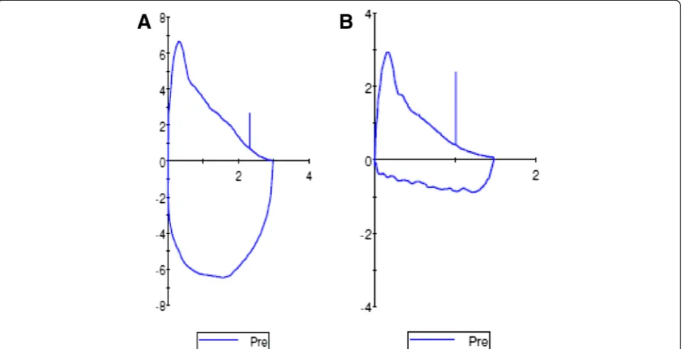
A reported characteristic finding in VCD is a highly vari-able, non-reproducible and abnormally shaped inspiratory loop on spirometry consistent with a variable extrathor-acic obstruction, as described by Miller and Hyatt [15, 44]. Morris et al. reviewed 1500 cases of VCD in the published literature and found that 28 % of reported VCD patients had flow-volume loop truncation on spirometry [8]. As
seen in Fig. 2, the inspiratory flow loop can show flatten-ing, truncation, and/or saw tooth pattern in patients with VCD, either during an acute VCD attack or even when pa-tients are asymptomatic [15, 16, 20]. Blunting of both in-spiratory and expiratory loops, on the other hand, is consistent with a fixed obstruction and should be further evaluated radiographically or endoscopically for a fixed process rather than a functional disorder [20].
Another useful measure in pulmonary function testing is the FEF50/FIF50 ratio, which is usually less than one in normal individuals. In patients with inspiratory VCD, this ratio is usually greater than 1 because truncation of the inspiratory loop reduces the FIF50. For patients with VCD and concomitant expiratory obstruction or comor-bid asthma, however, this ratio may be difficult to inter-pret [8, 16, 45].
Impulse oscillometry
Komarow and colleagues found that impulse oscillome-try (IOS) exhibits a characteristic impedance pattern in patients with VCD, verified by laryngoscopy. This may offer a rapid, noninvasive adjunct to diagnose patients with VCD [40] The main drawback to this type of test-ing, however, is the impulse oscillometers are not readily available.
Treatment and management
Despite the typically benign and self-limited episodes, VCD can lead to severe symptoms, the impression of impending respiratory failure, and even emergent intub-ation or tracheostomy. Management of VCD often re-quires a multidisciplinary approach involving the primary care physician, pulmonologist, allergist, otolaryngologist, gastroenterologist, neurologist, psychiatrist or psycholo-gist, speech patholopsycholo-gist, and athletic trainer [16, 46].
The management of VCD, especially in the acute set-ting, requires establishing the correct diagnosis. In the
absence of impending respiratory failure, performing a laryngoscopy while symptomatic is rapid, safe and in-formative in most patients. Once the diagnosis is estab-lished, treatment should be aimed at acutely relieving airway obstruction [18]. Asthma medications, including inhaled bronchodilators and corticosteroids, should be used only if the diagnosis is unclear, but patients may have minimal response to them [18]. Once the diagnosis of VCD is confirmed, the first step is to reassure patients that the condition is benign and self-limited. While the use of medications can be attempted, effective long-term therapy requires psychosocial support, speech therapy and even biofeedback.
Patient education
Patient education is a crucial component of treatment. Knowledge of normal physiology and functional abnor-malities causing symptoms can help patients accept their diagnosis and gain control over this disorder. Allowing patients to view their laryngoscopy findings often en-hances understanding and acceptance. Patients previ-ously misdiagnosed with asthma instead of VCD should have unnecessary medications discontinued gradually and under the care of their physician [8, 10, 33].
Medications
Sedation with benzodiazepines has proven successful in some patients, especially when underlying anxiety is a contributing factor [8, 12]. Heliox, a helium/oxygen
mixture that leads to a reduction of air density, decreases turbulent flow, and reduces work of breathing, has shown favorable responses in acute VCD episodes and has even demonstrated a sustained response after discontinuation in many cases [10]. An invasive and rarely used treatment modality is laryngeal injection of botulinum toxin type A, which prevents acetylcholine release at nerve endings, leading to chemical denervation and paralysis of the vocal fold in the open position [8, 9]. While this has been suc-cessfully used to treat spasmodic dystonia [33], a review by Morris and colleagues in 2006 found only 9 reported cases of botulinum toxin used to treat VCD [8]. Given the risk associated with paralyzing vocal cords in an open pos-ition, it should be reserved for patients refractory to all other therapies or those considering tracheostomy [8, 9].
Speech therapy and psychotherapy
The most common long-term treatment is speech ther-apy and psychotherther-apy. Speech therther-apy consists of a de-tailed assessment of patient’s symptoms and triggers, followed by a comprehensive treatment that is tailored to the individual patient [17]. Patients are educated about the pathophysiology of VCD, are provided supportive counseling, and are educated about suppression of laryn-geal abusive behaviors (i.e., cough and throat clearing), voice therapy, respiratory retraining, and desensitization to specific irritants. They are taught various breathing techniques, known as quick-release techniques, which act to rapidly release the vocal folds from the paradoxical
movement responsible for symptoms of VCD. These exer-cises focus on pursed-lip breathing using abdominal sup-port, with a focus on relaxation. Patients are encouraged to practice this technique with 5 repetitions 20 times per day to assist with laryngeal relaxation and retraining and to ensure that patients can respond automatically when acutely symptomatic [17]. Patient progress can be followed clinically or with more objective approaches, such as the VCDQ described above, which has shown improve-ment in scores after speech therapy [41]. In addition to speech therapy, patients may benefit from psychological counseling, which lacks a systematic study but may be warranted in those whose VCD is related to an underlying psychiatric condition [33, 47].
Biofeedback
Biofeedback and hypnosis have also shown some benefit in patients with VCD [47–50]. McFadden and Zawakski found that four of nine patients with exercise induced VCD had rapid resolution of symptoms after basic bio-feedback [28]. In another study, the use of hypnosis in 29 VCD patients showed improvement in 31 % and resolution in another 38 % [51].
Conclusions
Vocal cord dysfunction can be difficult to treat as the condition is often underappreciated and misdiagnosed in clinical practice. Although symptoms often mimic asthma, VCD and asthma have certain distinct clinical features and specific findings on diagnostic studies, which can serve to differentiate the two conditions. Early recognition and ac-curate diagnosis of vocal cord dysfunction can prevent im-proper treatment and, therefore, minimize escalated health care costs. The Pittsburgh VCD index and the VCDQ may serve as useful tools to differentiate asthma and VCD and to measure symptom improvement after treatment. While speech therapy is currently the mainstay of treatment, bio-feedback and pharmacotherapy have been successful in se-lect patients. Since intrinsic and extrinsic triggers can exacerbate laryngeal hyper-responsiveness, a focus on min-imizing such triggers can also serve to improve symptoms. Further studies are needed to investigate novel therapies for refractory patients.
Abbreviations
VCD:Vocal cord dysfunction; PFT: Pulmonary function testing; FVL: Flow volume loops; VCDQ: Vocal cord dysfunction questionnaire; PEEP: Positive end expiratory pressure; IOS: Impulse oscillometry.
Competing interests
The authors declare that they have no competing interests.
Authors’contributions
NMD participated in literature review, data analysis and did the majority of the writing for the article and completed the submission process. FCLH performed data analysis and interpretation, performed critical revision of the article and final approval of the published version. RKK performed
critical analysis and revision of the article and final approval of the published version. All authors read and approved the final manuscript.
Author details
1National Jewish Health, University of Colorado, Denver, CO, USA.2National Jewish Health, Denver, CO, USA.
Received: 23 May 2015 Accepted: 11 September 2015
References
1. Hoyte FC. Vocal cord dysfunction. Immunol Allergy Clin N Am. 2013;33(1):1–22. doi:10.1016/j.iac.2012.10.010.
2. Low K, Lau KK, Holmes P, Crossett M, Vallance N, Phyland D, et al. Abnormal vocal cord function in difficult-to-treat asthma. Am J Respir Crit Care Med. 2011;184(1):50–6. doi:10.1164/rccm.201010-1604OC.
3. Dunglison RD. The practice of medicine Lea and Blanchard. 1842. 4. Mackenzie M. The use of the laryngoscope in disease of the throat. 2nd ed.
Philadelphia: Lindsay and Blakiston; 1869.
5. Hysteria WO. The principles and practice of medicine. 1902. 6. Patterson R, Schatz M, Horton M. Munchausen’s stridor: non-organic
laryngeal obstruction. Clin Allergy. 1974;4(3):307–10.
7. Brugman S. The many faces of vocal cord dysfunction: what 36 years of literature tell us. Am J Respir Crit Care Med. 2003;167(7):A588. 8. Morris MJ, Allan PF, Perkins PJ. Vocal cord dysfunction: etiologies and
treatment. Clin Pulm Med. 2006;13(2):73–86.
9. Maillard I, Schweizer V, Broccard A, Duscher A, Liaudet L, Schaller MD. Use of botulinum toxin type A to avoid tracheal intubation or tracheostomy in severe paradoxical vocal cord movement. Chest. 2000;118(3):874–7. 10. Christopher KL, Wood 2nd RP, Eckert RC, Blager FB, Raney RA, Souhrada JF.
Vocal-cord dysfunction presenting as asthma. N Engl J Med. 1983;308(26):1566–70. doi:10.1056/nejm198306303082605.
11. Newman KB, Mason 3rd UG, Schmaling KB. Clinical features of vocal cord dysfunction. Am J Respir Crit Care Med. 1995;152(4 Pt 1):1382–6. doi:10.1164/ajrccm.152.4.7551399.
12. Andrianopoulos MV, Gallivan GJ, Gallivan KH. PVCM, PVCD, EPL, and irritable larynx syndrome: what are we talking about and how do we treat it? J Voice. 2000;14(4):607–18.
13. Vertigan AE, Theodoros DG, Gibson PG, Winkworth AL. The relationship between chronic cough and paradoxical vocal fold movement: a review of the literature. J Voice. 2006;20(3):466–80. doi:10.1016/j.jvoice.2005.08.001. 14. Traister R, Fajt ML, Whitman-Purves E, Anderson WC, III, Petrov A. A retrospective
analysis comparing subjects with isolated and co-existent vocal cord dysfunction and asthma. J Allergy Clin Immunol. 131(2):AB63. doi:10.1016/j.jaci.2012.12.889. 15. Balkissoon R. Occupational upper airway disease. Clin Chest Med.
2002;23(4):717–25.
16. Altman KW, Simpson CB, Amin MR, Abaza M, Balkissoon R, Casiano RR. Cough and paradoxical vocal fold motion. Otolaryngol Head Neck Surg. 2002;127(6):501–11. doi:10.1067/mhn.2002.127589.
17. Hicks M, Brugman SM, Katial R. Vocal cord dysfunction/paradoxical vocal fold motion. Prim Care. 2008;35(1):81–103. doi:10.1016/j.pop.2007.09.005. vii. 18. Christopher KL, Morris MJ. Vocal cord dysfunction, paradoxic vocal fold motion,
or laryngomalacia? Our understanding requires an interdisciplinary approach. Otolaryngol Clin N Am. 2010;43(1):43–66. doi:10.1016/j.otc.2009.12.002. viii. 19. Sasaki CT, Weaver EM. Physiology of the larynx. Am J Med. 1997;103(5A):9S–18. 20. Newman KB, Dubester SN: Vocal Cord Dysfunction: masquerader of asthma.
Seminars in Respiratory and Crit Care Med 1994; 15: 161–7.
21. Maschka DA, Bauman NM, McCray Jr PB, Hoffman HT, Karnell MP, Smith RJ. A classification scheme for paradoxical vocal cord motion. Laryngoscope. 1997;107(11 Pt 1):1429–35.
22. Barnes SD, Grob CS, Lachman BS, Marsh BR, Loughlin GM. Psychogenic upper airway obstruction presenting as refractory wheezing. J Pediatr. 1986;109(6):1067–70.
23. Vlahakis NE, Patel AM, Maragos NE, Beck KC. Diagnosis of vocal cord dysfunction: the utility of spirometry and plethysmography. Chest. 2002;122(6):2246–9.
24. Morrison M, Rammage L, Emami AJ. The irritable larynx syndrome. J Voice. 1999;13(3):447–55.
26. Cairns-Pastor C. Condition has name, but still unsettling. Tampa: Tribune; 2003. 27. Lakin RC, Metzger WJ, Haughey BH. Upper airway obstruction presenting as
exercise-induced asthma. Chest. 1984;86(3):499–501.
28. McFadden Jr ER, Zawadski DK. Vocal cord dysfunction masquerading as exercise-induced asthma. a physiologic cause for“choking”during athletic activities. Am J Respir Crit Care Med. 1996;153(3):942–7. doi:10.1164/ ajrccm.153.3.8630577.
29. Morris MJ, Deal LE, Bean DR, Grbach VX, Morgan JA. Vocal cord dysfunction in patients with exertional dyspnea. Chest. 1999;116(6):1676–82.
30. Lacy TJ, McManis SE. Psychogenic stridor. Gen Hosp Psychiatry. 1994;16(3):213–23. 31. Perkner JJ, Fennelly KP, Balkissoon R, Bartelson BB, Ruttenber AJ, Wood 2nd
RP, et al. Irritant-associated vocal cord dysfunction. J Occup Environ Med. 1998;40(2):136–43.
32. Harbison J, Dodd J, McNicholas WT. Paradoxical vocal cord motion causing stridor after thyroidectomy. Thorax. 2000;55(6):533–4.
33. Mathers-Schmidt BA. Paradoxical vocal fold MotionA tutorial on a complex disorder and the speech-language Pathologist’s role. Am J Speech Lang Pathol. 2001;10(2):111–25.
34. Weiss TM. Vocal cord dysfuction: Paradoxical vocal fold motion. The University of Texas Medical Branch. http://www.utmb.edu/otoref/Grnds/Vocal-Cord-2001-07/Vocal-Cord-pic-2001-07-M.pdf. Accessed May 12, 2015.
35. Ayres JG, Gabbott PL. Vocal cord dysfunction and laryngeal hyperresponsiveness: a function of altered autonomic balance? Thorax. 2002;57(4):284–5.
36. Bucca C, Rolla G, Brussino L, De Rose V, Bugiani M. Are asthma-like symptoms due to bronchial or extrathoracic airway dysfunction? Lancet.
1995;346(8978):791–5.
37. Thach BT. Reflux associated apnea in infants: evidence for a laryngeal chemoreflex. Am J Med. 1997;103(5A):120S–4.
38. Orenstein SR. An overview of reflux-associated disorders in infants: apnea, laryngospasm, and aspiration. Am J Med. 2001;111(Suppl 8A):60S–3. 39. Loughlin CJ, Koufman JA, Averill DB, Cummins MM, Kim YJ, Little JP, et al.
Acid-induced laryngospasm in a canine model. Laryngoscope. 1996;106(12 Pt 1):1506–9.
40. Komarow HD, Young M, Nelson C, Metcalfe DD. Vocal cord dysfunction as demonstrated by impulse oscillometry. J Allergy Clin Immunol Pract. 2013;1(4):387–93. doi:10.1016/j.jaip.2013.05.005.
41. Fowler SJ, Thurston A, Chesworth B, Cheng V, Constantinou P, Vyas A, et al. The VCDQ - a questionnaire for symptom monitoring in vocal cord dysfunction. Clin Exp Allergy. 2015. doi:10.1111/cea.12550.
42. Traister RS, Fajt ML, Landsittel D, Petrov AA. A novel scoring system to distinguish vocal cord dysfunction from asthma. J Allergy Clin Immunol Pract. 2(1):65–9. doi:10.1016/j.jaip.2013.09.002.
43. Crapo RO, Casaburi R, Coates AL, Enright PL, Hankinson JL, Irvin CG, et al. Guidelines for methacholine and exercise challenge testing-1999. This official statement of the American thoracic society was adopted by the ATS board of directors, july 1999. Am J Respir Crit Care Med. 2000;161(1):309–29. doi:10.1164/ajrccm.161.1.ats11-99.
44. Miller RD, Hyatt RE. Evaluation of obstructing lesions of the trachea and larynx by flow-volume loops. Am Rev Respir Dis. 1973;108(3):475–81. 45. Goldman J, Muers M. Vocal cord dysfunction and wheezing. Thorax.
1991;46(6):401–4.
46. Sandage MJ, Zelazny SK. Paradoxical vocal fold motion in children and adolescents. Lang Speech Hear Serv Sch. 2004;35(4):353–62. 47. Anbar RD. Hypnosis, Theodore Roosevelt, and the patient with cystic
fibrosis. Pediatrics. 2000;106(2 Pt 1):339–40.
48. Nahmias J, Tansey M, Karetzky MS. Asthmatic extrathoracic upper airway obstruction: laryngeal dyskinesis. N J Med. 1994;91(9):616–20.
49. Smith MS. Acute psychogenic stridor in an adolescent athlete treated with hypnosis. Pediatrics. 1983;72(2):247–8.
50. Caraon P, O’Toole C. Vocal cord dysfunction presenting as asthma. Ir Med J. 1991;84(3):98–9.
51. Anbar RD. Hypnosis in pediatrics: applications at a pediatric pulmonary center. BMC Pediatr. 2002;2:11.
Submit your next manuscript to BioMed Central and take full advantage of:
• Convenient online submission
• Thorough peer review
• No space constraints or color figure charges
• Immediate publication on acceptance
• Inclusion in PubMed, CAS, Scopus and Google Scholar
• Research which is freely available for redistribution
![Table 1 Differential diagnosis of laryngeal movementdisorders [17, 24]](https://thumb-us.123doks.com/thumbv2/123dok_us/420596.2039548/3.595.57.291.109.558/table-differential-diagnosis-laryngeal-movementdisorders.webp)
![Table 2 Pertinent questions for evaluation of VCD [41]](https://thumb-us.123doks.com/thumbv2/123dok_us/420596.2039548/4.595.306.534.100.367/table-pertinent-questions-for-evaluation-of-vcd.webp)