ISSN Online: 2168-5444 ISSN Print: 2168-5436
DOI: 10.4236/ijmpcero.2018.73025 Jul. 18, 2018 308 Int. J. Medical Physics, Clinical Engineering and Radiation Oncology
Robustness Evaluation of a Novel Proton Beam
Geometry for Head and Neck Patients Treated
with Pencil Beam Scanning Therapy
Sheng Huang
1,2, Haoyang Liu
1,3, Jiajian Shen
4, Huifang Zhai
1, Maura Kirk
1, Brett Hartl
1,
Alexander Lin
1, James McDonough
1, Stefan Both
5, Haibo Lin
1,2*1Department of Radiation Oncology, University of Pennsylvania, Philadelphia, PA, USA 2New York Proton Center, New York, NY, USA
3New Jersey Proton Therapy Center, Somerset, NJ, USA 4Mayo Clinic, Phoenix, AZ, USA
5University Medical Center Groningen, Groningen, The Netherlands
Abstract
Background: To evaluate the robustness of head and neck treatment using pro-ton pencil beam scanning (PBS) technique with respect to range uncertainty (RU) and setup errors (SE), and to establish a robust PBS planning strategy for fu-ture treatment. Methods and Materials: Ten consecutive patients were planned with a novel proton field geometry (combination of two posterior oblique fields and one anterior field with gradient dose match) using single-field uniform dose (SFUD) planning technique and the proton plans were dosimetrically compared to two coplanar arc VMAT plans. Robustness of the plans, with respect to range uncertainties (RU = ± 3% for proton) and setup errors (SE = 2.25 mm for proton and VMAT), in terms of deviations to target coverage (CTV D98%) and OAR doses (max/mean), were evaluated and compared for each patient under worst case scenarios. Results: Dosimetrically, PBS plans pro-vided better sparing to larynx (p = 0.005), oral cavity (p < 0.001) and contra-lateral parotid (p = 0.004) when compared to VMAT. CTV D98% variations were higher from SE than from RU for proton plans (−1.1% ± 1.3 % vs −0.4% ± 0.7% for nodal CTV and −1.4% ± 1.2 vs −0.4% ± 0.5% % for boost CTV). Overall, the magnitudes of variation of CTV D98% to combined SE and RU were found to be similar to the impact of the SE on the VMAT plans (−1.6% ± 1.9% vs −1.7% ± 1.4% for nodal CTV and −1.9% ± 1.6% vs −1.3% ± 1.5% for boost CTV). Compared to VMAT, a larger range of relative dose deviations were found for OARs in proton plans, but safe doses were maintained for cord (41.8 ± 3.6 Gy for PBS and 41.7 ± 3.9 Gy for VMAT) and brainstem (35.2 ± 8.4 Gy for PBS and 36.2 ± 5.1 Gy for VMAT) in worst case scenarios. Conclu-How to cite this paper: Huang, S., Liu,
H.Y., Shen, J.J., Zhai, H.F., Kirk, M., Hartl, B., Lin, A., McDonough, J., Both, S. and Lin, H.B. (2018) Robustness Evaluation of a Novel Proton Beam Geometry for Head and Neck Patients Treated with Pencil Beam Scanning Therapy. International Journal of Medical Physics, Clinical Engineering and Radiation Oncology, 7, 308-322.
https://doi.org/10.4236/ijmpcero.2018.73025
Received: May 11, 2018 Accepted: July 15, 2018 Published: July 18, 2018
Copyright © 2018 by authors and Scientific Research Publishing Inc. This work is licensed under the Creative Commons Attribution International License (CC BY 4.0).
http://creativecommons.org/licenses/by/4.0/
DOI: 10.4236/ijmpcero.2018.73025 309 Int. J. Medical Physics, Clinical Engineering and Radiation Oncology sions: Compared to VMAT, proton plans containing three SFUD fields with superior-inferior gradient dose matching had improved sparing to larynx, contralateral parotid and oral cavity, while providing similar robustness of target coverage. Evaluation of OAR dose robustness showed higher sensitivi-ties to uncertainsensitivi-ties for proton plans, but safe dose levels were maintained for cord and brainstem.
Keywords
Head and Neck, Robustness, Proton Therapy, Pencil Beam Scanning
1. Introduction
The main goal of modern radiotherapy is to optimize the delivery of radiation dose such that the target is fully sterilized while maximally sparing the sur-rounding healthy tissue and organs. Especially in head and neck (HN) cancers, this goal is challenging to achieve because organs at risk (OAR) are generally proximal to target volumes. Huguenin et al. had a quality of life study for HN patients received radiotherapy and revealed the highest morbidity was probably the result of using large radiation fields [1]. Therefore, reduction in irradiated volume and dose to local organs may lower the risk and severity of radiation in-duced complications [2]. Advanced conventional delivery techniques such as in-tensity modulated radiation therapy (IMRT) and volumetric modulated arc therapy (VMAT) have improved the outlook for OAR for HN patients in the past several decades [3]. Proton therapy has the potential to further improve OAR sparing and reduce integral dose resulting from the intrinsic physics of the proton beam [4].
Proton pencil beam scanning (PBS) technique is the current state of art of mod-ern proton therapy. Generally, PBS dose can be shaped using either single-field uniform dose (SFUD) or intensity modulated proton therapy (IMPT) planning techniques. In SFUD, often called single-field optimization, fields are optimized simultaneously so that each field uniformly covers the whole target. In IMPT, fields are also optimized simultaneously, but each field does not uniformly cover the whole target and only the sum of all fields provides adequate and homoge-neous target coverage, similar to IMRT. Although the flexibility of IMPT allows for creation of more complex dose distributions than SFUD, which improves OAR sparing in practice, concerns such as high sensitivity to uncertainties and lack of robustness for IMPT still exist [5]. In contrast, SFUD plans are consi-dered to be more robust than IMPT due to less dose modulation and lower complexity of dose distributions for each individual field [6].
DOI: 10.4236/ijmpcero.2018.73025 310 Int. J. Medical Physics, Clinical Engineering and Radiation Oncology range uncertainties (RU) arise from conversion of computed tomography (CT) Hounsfield units (HU) to proton stopping power, CT artifacts, fluctuation of proton energy, anatomy change and so on. Typically, RU from ±3% to ±3.5% of HU is taken into consideration for routine clinical practice [7]. Uncertainties re-lated to motion should be considered as well when evaluating proton therapy plans, especially for PBS treatments due to interplay effect [8]. As all of these uncertainties may result in significant deviations between planned and delivered dose distributions [9], it is very important to evaluate the robustness of proton therapy plans, especially for newly developed clinical protocols.
Several previous studies have investigated the robustness of proton therapy for HN cancer compared to photon techniques. Barten et al. studied OAR sparing and plan robustness for PBS proton planning techniques including SFUD and IMPT with more fields and compared these with VMAT photon plans. They reached conclusion that IMPT plans has improved OAR sparing but were less robust than SFUD and VMAT and robustness of the MFO plans did not increase with more fields [6]. Van der Voort et al. derived a “robustness recipe” for mi-nimax robust optimization of IMPT treatment plans to ensure adequate CTV coverage for HN cancer patient relative to systematic setup, random setup and range errors [10]. Malyapa et al. evaluated the robustness of IMPT plans to setup and range uncertainties for HN patient and created a robustness database, which allows the identification of future plans that may benefit from alternative plan-ning approaches to improve robustness [7]. Van de Water et al. quantified the impact of the degree of robustness against setup and range errors on OAR dose and normal tissue complication probabilities in IMPT proton therapy for HN cancer patient and concluded that minimizing setup errors should be given a higher priority than minimizing range errors [11].
In this work, a novel HN proton treatment planning geometry containing three SFUD fields (two posterior (oblique) fields and one anterior field, inversed “Y” shape) using superior-inferior gradient dose matching is presented. The do-simetrical advantages and plan robustness of this approach are first time eva-luated relative to two-Arc VMAT plans, in terms of target and OAR dose levels.
2. Materials and Methods
2.1. Patient Group, Simulation and Contouring
DOI: 10.4236/ijmpcero.2018.73025 311 Int. J. Medical Physics, Clinical Engineering and Radiation Oncology
Figure 1. (a)-(c) Target volumes: PTV and CTV for HN planning of three axial cuts;
three proton beams are indicated in (b) as 0, 195 and 160 degree. (d)-(f) Dose colorwash (30% dose threshold for all) image of VMAT plan of three axial cuts. (g)-(i) Dose color-wash image of proton plan of three axial cuts: upper targets with two posterior fields (g), lower targets with anterior field (i) and junction area with all three fields (h). (j) Dose co-lorwash of sagittal view of VMAT plan. (k) Dose coco-lorwash of sagittal view of proton plan. (l) Dose colorwash with PBSTV overlapped at junction area; dose profiles indicated by the red lines are presented in Figure 2.
2.2. Treatment Planning Techniques
VMAT (Figures 1(d)-(f)) treatments were optimized with two full arcs using 6 MV photons in Eclipse (Varian Medical System, Palo Alto, CA) treatment plan-ning system (TPS). Dose calculation was performed using the anisotropic ana-lytical algorithm (Eclipse AAA v11.030) with grid size of 2.5 mm.
DOI: 10.4236/ijmpcero.2018.73025 312 Int. J. Medical Physics, Clinical Engineering and Radiation Oncology
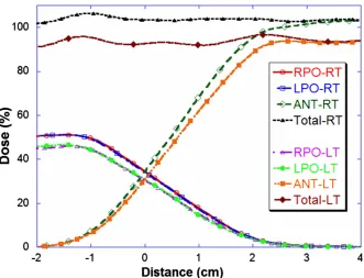
Figure 2. Dose profiles for individual field and the sum of all fields at the junction area
indicated in Figure 1(l). Two dose levels of 60 Gy and 54 Gy are prescribed to PTV60 on patient’s right (RT) side and PTV 54 on patient’s left (LT) side. RPO-RT: right posterior oblique field at PTV60 junction area; LPO-RT: left posterior oblique field at PTV60 junc-tion area; ANT-RT: anterior field at PTV60 juncjunc-tion area; Total-RT: dose sum from all three fields at PTV60 junction area. RPO-LT: right posterior oblique field at PTV54 junc-tion area; LPO-LT: left posterior oblique field at PTV54 juncjunc-tion area; ANT-LT: anterior field at PTV54 junction area; Total-LT: dose sum from all three fields at PTV54 junction area.
DOI: 10.4236/ijmpcero.2018.73025 313 Int. J. Medical Physics, Clinical Engineering and Radiation Oncology need a couch shift between fields during treatment. This removes the potential setup errors through couch shifts during treatment.
Our proton system has minimal deliverable energy of 100 MeV, which trans-lates to a range of 7.7 cm WET. To cover the shallow portion of targets, a thick couch (WET~7.35 cm) and an anterior bolus (WET~7.16 cm) served as an al-ternative solution to machine range shifter for posterior and anterior fields re-spectively [13], so that sufficient coverage to the shallow portion of the target could be maintained. Online patient positioning was completed via alignment of bony anatomy around target volumes using two orthogonal kilo-voltage (kV) based imaging guidance. The anterior bolus is placed in position after imag-ing-guided patient alignment. The vertical position of the bolus is indexed rela-tive to couch surface to maintain a reproducible air gap between bolus and pa-tient. As large air gaps could broaden the proton spot size [14], air gaps were kept as small as possible. Typically, the achieved air gap is 10 cm or below.
2.3. Robustness Evaluation
Worst case scenarios for SE were simulated for both proton and VMAT plans with simultaneous shifts in all three directions (8 combinations) similar to the method used in the study by Barten et al. [6]. As all patients were immobilized using a thermoplastic mask, a shift of 2.25 mm [7], which satisfied an 85% con-fidence level [15], was applied in three directions. The influence of RU was also investigated by introducing ±3% errors on the HU values in proton planning CT (2 combinations) and was considered as negligible in VMAT planning. SE and RU were also combined (SREs: 16 combinations) for proton plans to evaluate the plan robustness in the worst-case scenario for both sources of uncertainty combined. Loss of target coverage and deviations to OAR doses were reported. For any given patient, multiple instances of overdose to critical organs, such as cord and brainstem, relative to their dose constraints, or a loss of CTV cover-age > 5% triggered re-evaluation and potential re-optimization of the plan.
3. Results
3.1. Dosimetric Evaluation and Comparison
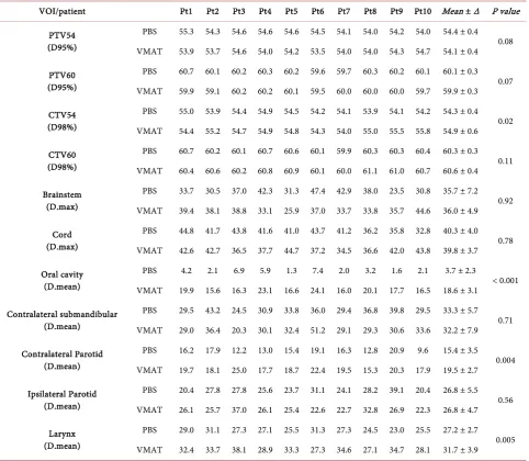
Table 1 summarizes the dosimetric results of proton and VMAT plans for 10
DOI: 10.4236/ijmpcero.2018.73025 314 Int. J. Medical Physics, Clinical Engineering and Radiation Oncology
Table 1. Dosimetric parameters in Gy for targets and OARs in 10 patients planned with proton PBS and VMAT. Mean and
stan-dard deviation value of 10-patient results and the p-value based on a two tailed student t test are also listed.
VOI/patient Pt1 Pt2 Pt3 Pt4 Pt5 Pt6 Pt7 Pt8 Pt9 Pt10 Mean ± ∆ P value
PTV54 (D95%)
PBS 55.3 54.3 54.6 54.6 54.6 54.5 54.1 54.0 54.2 54.0 54.4 ± 0.4 0.08 VMAT 53.9 53.7 54.6 54.0 54.2 53.5 54.0 54.0 54.3 54.7 54.1 ± 0.4
PTV60 (D95%)
PBS 60.7 60.1 60.2 60.3 60.2 59.6 59.7 60.3 60.2 60.1 60.1 ± 0.3 0.07 VMAT 59.9 59.1 60.2 60.2 60.1 59.5 60.0 60.0 60.0 59.7 59.9 ± 0.3
CTV54 (D98%)
PBS 55.0 53.9 54.4 54.9 54.5 54.2 54.1 53.9 54.1 54.2 54.3 ± 0.4 0.02 VMAT 54.4 55.2 54.7 54.9 54.8 54.3 54.0 55.0 55.5 55.8 54.9 ± 0.6
CTV60 (D98%)
PBS 60.7 60.2 60.1 60.7 60.6 60.1 59.9 60.3 60.3 60.4 60.3 ± 0.3 0.11 VMAT 60.4 60.6 60.2 60.8 60.9 60.1 60.0 61.1 61.0 60.7 60.6 ± 0.4
Brainstem (D.max)
PBS 33.7 30.5 37.0 42.3 31.3 47.4 42.9 38.0 23.5 30.8 35.7 ± 7.2 0.92 VMAT 39.4 38.1 38.8 33.1 25.9 37.0 33.7 33.8 35.7 44.6 36.0 ± 4.9
Cord (D.max)
PBS 44.8 41.7 43.8 41.6 41.0 43.7 41.2 36.2 35.8 32.8 40.3 ± 4.0 0.78 VMAT 42.6 42.7 36.5 37.7 44.7 37.2 34.5 36.6 42.0 43.8 39.8 ± 3.7
Oral cavity (D.mean)
PBS 4.2 2.1 6.9 5.9 1.3 7.4 2.0 3.2 1.6 2.1 3.7 ± 2.3
< 0.001 VMAT 19.9 15.6 16.3 23.1 16.6 24.1 16.0 20.1 17.7 16.5 18.6 ± 3.1
Contralateral submandibular (D.mean)
PBS 29.5 43.2 24.5 30.9 33.8 36.0 29.4 36.8 39.8 29.5 33.3 ± 5.7 0.71 VMAT 29.0 36.4 20.3 30.1 32.4 51.2 29.1 29.3 30.6 33.6 32.2 ± 7.9
Contralateral Parotid (D.mean)
PBS 16.2 17.9 12.2 13.0 15.4 19.1 16.3 12.8 20.9 9.6 15.4 ± 3.5 0.004 VMAT 19.7 18.1 25.0 17.7 18.7 22.4 19.5 15.3 20.3 17.9 19.5 ± 2.7
Ipsilateral Parotid (D.mean)
PBS 20.4 27.8 27.8 25.6 23.7 31.1 24.1 28.2 39.1 20.4 26.8 ± 5.5 0.56 VMAT 26.1 25.7 37.0 26.1 25.4 22.6 22.7 32.8 26.9 22.3 26.8 ± 4.7
Larynx (D.mean)
PBS 29.0 31.1 27.3 27.1 25.5 31.3 27.3 24.5 23.0 25.5 27.2 ± 2.7
0.005 VMAT 32.4 33.7 38.1 28.9 33.3 27.3 34.6 27.1 34.7 28.1 31.7 ± 3.9
Note: VOI: volume of interest; D.max: maximal dose; D.mean: mean dose; Pt: patient; ∆: standard deviation.
proton plan. Clinically acceptable maximum dose constraints to cord and brainstem were also achieved for all plans.
In terms of OAR sparing, proton plans significantly improved the mean dose to oral cavity (p < 0.001), larynx (p = 0.005) and contralateral parotid (p = 0.004) compared to VMAT plans. Sparing of OARs such as cord, brainstem, contrala-teral submandibular and ipsilacontrala-teral parotid was patient specific and mostly relied on the relative location between organs and target volumes, as well as their shape along the beam direction and the level of overlaps between structures.
3.2. Robustness Evaluation
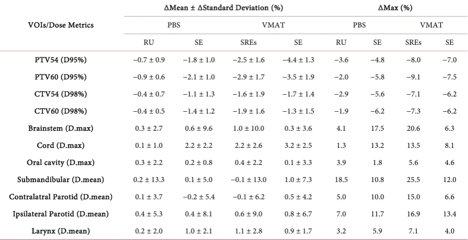
Table 2 summarizes the mean, standard deviation, and range of dose variations
DOI: 10.4236/ijmpcero.2018.73025 315 Int. J. Medical Physics, Clinical Engineering and Radiation Oncology
Table 2. Mean, standard deviation, minimum and maximum of dose variations in uncertainty comparison (between original plan
and plan with SEs, RU and SREs) in 10 HN patients for proton and VMAT plans.
VOIs/Dose Metrics
∆Mean ± ∆Standard Deviation (%) ∆Max (%)
PBS VMAT PBS VMAT
RU SE SREs SE RU SE SREs SE
PTV54 (D95%) −0.7 ± 0.9 −1.8 ± 1.0 −2.5 ± 1.6 −4.4 ± 1.3 −3.6 −4.8 −8.0 −7.0 PTV60 (D95%) −0.9 ± 0.6 −2.1 ± 1.0 −2.9 ± 1.7 −3.5 ± 1.9 −2.0 −5.8 −9.1 −7.5 CTV54 (D98%) −0.4 ± 0.7 −1.1 ± 1.3 −1.6 ± 1.9 −1.7 ± 1.4 −2.9 −5.6 −7.1 −6.2 CTV60 (D98%) −0.4 ± 0.5 −1.4 ± 1.2 −1.9 ± 1.6 −1.3 ± 1.5 −1.9 −6.2 −7.3 −6.2
Brainstem (D.max) 0.3 ± 2.7 0.6 ± 9.6 1.0 ± 10.0 0.3 ± 3.6 4.1 17.5 20.6 6.3
Cord (D.max) 0.1 ± 1.0 2.2 ± 2.2 2.2 ± 2.6 3.2 ± 2.5 1.3 13.2 13.5 8.1
Oral cavity (D.max) 0.3 ± 2.2 0.2 ± 0.8 0.4 ± 2.2 0.1 ± 3.3 3.9 1.8 5.6 4.6
Submandibular (D.mean) 0.2 ± 13.3 0.1 ± 5.0 −0.1 ± 13.0 1.0 ± 7.3 18.5 10.8 25.5 12.0 Contralatral Parotid (D.mean) 0.1 ± 3.7 −0.2 ± 5.4 −0.1 ± 6.2 0.5 ± 4.2 5.0 10.0 15.0 6.6
Ipsilateral Parotid (D.mean) 0.4 ± 5.3 0.4 ± 8.1 0.6 ± 9.0 0.8 ± 6.7 7.0 11.7 16.9 13.4
Larynx (D.mean) 0.2 ± 2.0 1.0 ± 2.1 1.1 ± 2.8 0.9 ± 1.7 3.2 5.9 7.1 4.0
Note: percentage (%) was normalized to 60 Gy for OARs and to their corresponding prescription doses to target structures. RU: range uncertainties; SE: setup errors; SREs: combination of range uncertainties and setup errors; ∆max (%) presents the worst-case scenario: maximal reduction for target coverage and maximal dose increment for OARs.
and SREs for proton plans, and only SE for VMAT. The average variations of target coverage (CTV and PTV) caused by RU alone were smaller than those from SE alone for proton cases. The overall worst-case scenario coverage devia-tions were found to be comparable for proton (SREs) and VMAT (SE) plans: −1.6% ± 1.9% versus −1.7% ± 1.4% for CTV54, −1.9% ± 1.6% versus −1.3% ± 1.5% for CTV60, −2.5% ± 1.6% versus −4.4% ± 1.3% for PTV54 and −2.9% ± 1.7% versus −3.5% ± 1.9% for PTV60. More than 5% drops in D98% of CTV54 was found for 3 proton (−6.0%, −6.8% and −7.1%) and 1 photon patient (−6.2%), and more than 5% decreases in D98% of CTV60 was found for 3 pro-ton (−5.1%,−6.9% and −7.3%) and 2 phopro-ton patients (−6.1% and −6.2%), which was also considered to be similar across the two modalities. No multiple in-stances of 5% or more loss in CTV D98% were found for any single patient.
DOI: 10.4236/ijmpcero.2018.73025 316 Int. J. Medical Physics, Clinical Engineering and Radiation Oncology such as brainstem and cord, showed dominant sensitivity to SE, rather than RU. The dose variations caused by SE, RU and SREs were 0.3% ± 2.7%, 0.6% ± 9.6%, 1.0% ± 10% for brainstem and 0.1% ± 1.0%, 2.2% ± 2.2%, 2.2% ± 2.6% for cord, respectively.
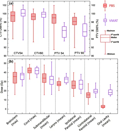
Figure 3 presents boxplots of robustness metrics under worst case scenarios
for target coverage and dose to OARs. The legend indicates maximum, 75th
per-centile, median, 25th percentile and minimum values of each metric from top to
bottom. Although the box values (between 75th and 25th percentile) of CTV 98%
were lower for proton cases, overall range of variations were about the same, and the difference was ~1%. However, the box values of PTV D95% were higher for proton cases. Larger variations on D95%, especially for PTV54 were observed. While larger dose variations were found for brainstem and cord, safe dose to
Figure 3. Comparison of dose variations on D98% of CTV54, CTV60 (a) and OARs (b) for proton and
DOI: 10.4236/ijmpcero.2018.73025 317 Int. J. Medical Physics, Clinical Engineering and Radiation Oncology cord and brainstem were generally maintained [16], as shown in Figure 3.
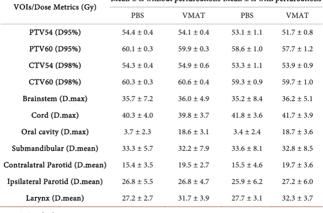
Table 3 presented the direct comparison of dosimetric metrics with and
without considering the perturbations of RU and SE. In general, it showed very similar mean and deviation on dose metrics of CTV and PTV between PBS and VMAT. The mean doses to all OARs with perturbations were very similar to their mean planning dose while the deviation increased as expected. The dose to cord were 41.8 ± 3.6 Gy for PBS and 41.7 ± 3.9 Gy for VMAT with perturbations and the dose to brainstem were 35.2 ± 8.4 Gy for PBS and 36.2 ± 5.1 Gy for VMAT, which were also about the same range.
4. Discussion
This study presented a novel field geometry for HN patients receiving proton PBS treatment. The results of treatment planning comparisons between proton SFUD PBS delivery technique and VMAT photon radiotherapy for HN were reported, focusing on target coverage, OAR sparing and plan robustness for plans generated using commercial TPS (without robust optimization). The planning method containing two PBS posterior-oblique fields (POF) was first developed and used to treat HN patients at our institution. Considering the shape of typical HN target volumes, the selection of POF was used to improve the sparing of normal organs anterior to the target volumes, and reduce their treatment related toxicities. In contrast to VMAT plans, this goal could be achieved for most of the proton HN patients when there was no direct overlap between organs and target volumes. However, verification plans based on repeated CT scanning showed potentially large coverage variations to nodal volumes
Table 3. Mean and standard deviation of target and OAR dosimetric metrics with and
without considering the perturbations of RU and SE in 10 HN patients for proton and VMAT plans.
VOIs/Dose Metrics (Gy) Mean ± ∆ without perturbations Mean ± ∆ with perturbations
PBS VMAT PBS VMAT
PTV54 (D95%) 54.4 ± 0.4 54.1 ± 0.4 53.1 ± 1.1 51.7 ± 0.8 PTV60 (D95%) 60.1 ± 0.3 59.9 ± 0.3 58.6 ± 1.0 57.7 ± 1.2 CTV54 (D98%) 54.3 ± 0.4 54.9 ± 0.6 53.3 ± 1.1 53.9 ± 0.9 CTV60 (D98%) 60.3 ± 0.3 60.6 ± 0.4 59.3 ± 0.9 59.7 ± 1.0 Brainstem (D.max) 35.7 ± 7.2 36.0 ± 4.9 35.2 ± 8.4 36.2 ± 5.1 Cord (D.max) 40.3 ± 4.0 39.8 ± 3.7 41.8 ± 3.6 41.7 ± 3.9 Oral cavity (D.max) 3.7 ± 2.3 18.6 ± 3.1 3.4 ± 2.4 18.7 ± 3.6 Submandibular (D.mean) 33.3 ± 5.7 32.2 ± 7.9 33.6 ± 8.1 32.8 ± 8.5 Contralatral Parotid (D.mean) 15.4 ± 3.5 19.5 ± 2.7 15.5 ± 4.6 19.7 ± 3.6 Ipsilateral Parotid (D.mean) 26.8 ± 5.5 26.8 ± 4.7 25.9 ± 6.2 27.2 ± 6.0 Larynx (D.mean) 27.2 ± 2.7 31.7 ± 3.9 27.7 ± 3.1 32.3 ± 3.7
DOI: 10.4236/ijmpcero.2018.73025 318 Int. J. Medical Physics, Clinical Engineering and Radiation Oncology located in the lower neck [17]. These nodal targets were typically shallow, from an anterior beam direction. Therefore, POF had to travel a longer beam path to reach them. Loose tissues on the back of the neck can fold differently from day to day and it was a challenge to achieve the goal of reproducibility for this loose tissue, especially for larger/older patients. Neither surface guidance with optical images, nor kV images during patient setup, are capable of flagging this change of tissue shape on the back of the patient’s neck, which could significantly im-pact the POF proton beam path, and translate into range errors and reduced target coverage. However, it also showed consistency and robustness of the POF to target coverage and OAR sparing for volumes superior to the thyroid notch. Therefore, a revised planning method was proposed to separate the target vo-lume into two sets, depending on their locations relative to the thyroid notch, and to add an anterior field to cover the lower target set [18]. Gradient dose matching was applied to create a “soft dose buffer” between two abutting field sets to reduce planning complications and dose uncertainties associated with the junction. The revised method has become our standard approach for proton treatment of HN patients.
One novelty of this work originates from the employment of gradient dose matching for the anterior and posterior field target sets in HN planning with a PBS technique. Different from the field matching in craniospinal irradiation, all fields here share the same isocenter in HN proton therapy and no couch shift is required between fields during treatment. Along with the fact that patient’s head and neck area is immobilized by a thermoplastic mask, that effectively limits the patient’s intrafractional movement along craniocaudal direction, so variations in daily setup should result in a wash-out effect along the junction [19], and large systematic setup errors are restricted for HN treatment through the use of accu-rate online imaging systems. According to the mathematic model for gradient field match developed by Lin [12], given a systematic setup error of 2 mm, maximal ~5% dose errors are expected at junction of 4 cm using gradient dose match. Thus, a junction length of 4 cm (shown in Figure 2) is considered to be reasonable to maintain a safe dose to critical organs, such as cord, and uniform coverage to target volumes in the junction area by adding the wash-out effect due to daily setup variations.
uncertain-DOI: 10.4236/ijmpcero.2018.73025 319 Int. J. Medical Physics, Clinical Engineering and Radiation Oncology ties, so more normal tissue volume distal to the PTV receives the prescribed dose. However, Proton could provide significant sparing when no direct overlap occurs, such as for patient 6. Moreover, critical organs such as brainstem and spinal cord are placed laterally to the field to avoid unpredicted dose uncertain-ties caused by range uncertainty and enhanced radiobiological effect at the distal end of the Bragg peak [20]. It is easy to understand why setup uncertainties do-minate the dose variations for these laterally positioned organs, as indicated in this study. Maximal cord dose between 45 Gy and 50 Gy was found for two pa-tients (1 and 3) in a specific combination of SE and RU in their robustness study. Concerns on safe cord dose are reduced not only because minimal (<0.5cc) vo-lume received dose of 45 Gy and above, these values represent the cord doses in the worst case scenario (consider SE as systematic errors through treatment course) and daily setup variation is ignored. Moreover, bony landmarks proxim-al to these organs, such as cervicproxim-al vertebrae, are generproxim-ally considered for patient setup to confirm the accuracy of patient alignments.
The robustness evaluation in this study is limited to SE and RU. The effect of patient’s inter- and intra-fractional shifts, which could potentially smear the dose deviations predicated by the uncertainty study [21], were not investigated. With modern imaging guided therapy, the risk of 2.25 mm SE in all three direc-tions simultaneously is low, especially for HN cases with immobilization devices and special attention given to precise patient positioning. Further research is needed to investigate the dose uncertainties due to anatomic changes using on-line volumetric imaging [22] or repeating CT scans [23], as well as variation in relative biological effectiveness [24]. It would also be desirable to explore robust optimization algorithms during plan optimization to simultaneously strive for a robust plan with optimal OAR sparing [25] [26]. Furthermore, with the devel-opment of fast and accurate dose calculation algorithms [27] and advanced on-line imaging analysis tools [28], application of adaptive proton therapy on HN treatment should also be considered.
5. Conclusion
This study presented proton PBS treatments with a novel proton beam geometry of three SFUD fields for HN patients. The robustness of the proton plans was assessed based on RU and SE, and was compared to VMAT results. It demon-strated that a proton plan with three SFUD fields can lead to robustness similar to that of VMAT, even without robust optimization algorithms, while providing significant sparing to larynx, contralateral parotid and oral cavity.
Acknowledgements
DOI: 10.4236/ijmpcero.2018.73025 320 Int. J. Medical Physics, Clinical Engineering and Radiation Oncology
References
[1] Huguenin, P.U., Taussky, D., Moe, K., Meister, A., Baumert, B., Lutolf, U.M., et al. (1999) Quality of Life in Patients Cured from a Carcinoma of The head and Neck by Radiotherapy: The Importance of the Target Volume. International Journal of Rad-iation Oncology • Biology • Physics, 45, 47-52.
https://doi.org/10.1016/S0360-3016(99)00128-5
[2] Deasy, J.O., Moiseenko, L., Marks, L., Chao, J., Nam, J. and Eisbruch, A. (2010) Ra-diotherapy Dose-Volume Effects on Salivary Gland Function. International Journal of Radiation Oncology • Biology • Physics, 76, S58-S63.
https://doi.org/10.1016/j.ijrobp.2009.06.090
[3] Bar Ad, V., Lin, H.B., Hwang, W.T., Deville, C., Dutta, P.R., Tochner, Z., et al. (2012) Larynx-Sparing Techniques Using Intensity-Modulated Radiation Therapy for Oropharyngeal Cancer. Medical Dosimetry, 37, 383-386.
https://doi.org/10.1016/j.meddos.2012.02.004
[4] Ahn, P.H., Lukens, J.N., Teo, B.K.K., Kirk, M. and Lin, A. (2014) The Use of Proton Therapy in the Treatment of Head and Neck Cancers. The Cancer Journal, 20, 421-426. https://doi.org/10.1097/PPO.0000000000000077
[5] Unkelbach, J., Bortfeld, T., Martin, B.C. and Soukup, M. (2009) Reducing the Sensi-tivity of IMPT Treatment Plans to Setup Errors and Range Uncertainties via Proba-bilistic Treatment Planning. Medical Physics, 36, 149-163.
https://doi.org/10.1118/1.3021139
[6] Barten Danique, L.J., Tol Jim, P., Dahele, M., Slotman Ben, J. and Verbakel Wilko, F.A.R. (2016) Comparison of Organ-at-Risk Sparing and Plan Robustness for Spot-Scanning Proton Therapy and Volumetric Modulated Arc Photon Therapy in Head-and-Neck Cancer. Medical Physics, 42, 6589-6598.
https://doi.org/10.1118/1.4933245
[7] Malyapa, R., Lowe, M., Bolsi, A., Lomax, A.J., Weber, D.C. and Albertini, F. (2016) Evaluation of Robustness to Setup and Range Uncertainties for Head and Neck Pa-tients Treated with Pencil Beam Scanning Proton Therapy. International Journal of Radiation Oncology • Biology • Physics, 95, 154-162.
https://doi.org/10.1016/j.ijrobp.2016.02.016
[8] Yu, J., Zhang, X.D., Liao, L., Li, H., Zhu, R., Park, P.C., et al. (2016) Motion-Robust Intensity-Modulated Proton Therapy for Distal Esophageal Cancer. Medical Phys-ics, 43, 1111-1118. https://doi.org/10.1118/1.4940789
[9] Liu, W. and Mohan, R. (2012) Robustness Evaluation and Robust Optimization of IMPT Plans Based on Per-Voxel Standard Deviation of Dose Distributions. Medical Physics, 39, 3850. https://doi.org/10.1118/1.4735715
[10] van der Voort, S., van de Water, S., Perko, Z., Heijmen, B., Lathouwers, D. and Hoogeman, M. (2016) Robustness Recipes for Minimax Robust Optimization in In-tensity Modulated Proton Therapy for Oropharyngeal Cancer Patients. Internation-al JournInternation-al of Radiation Oncology • Biology • Physics, 95, 163-170.
https://doi.org/10.1016/j.ijrobp.2016.02.035
[11] van de Water, S., van Dam, I., Schaart, D.R., Al-Mamgani, A., Heijmen, B.J. and Hoogeman, M.S. (2016) The Price of Robustness; Impact of Worst-Case Optimiza-tion on Organ-at-Risk Dose and ComplicaOptimiza-tion Probability in Intensity-Modulated Proton Therapy for Oropharyngeal Cancer Patients. Radiotherapy and Oncology, 120, 56-62.
https://doi.org/10.1016/j.radonc.2016.04.038
DOI: 10.4236/ijmpcero.2018.73025 321 Int. J. Medical Physics, Clinical Engineering and Radiation Oncology
Supine Craniospinal Irradiation Using a Proton Pencil Beam Scanning Technique Without Match Line Changes for Field Junctions. International Journal of Radiation Oncology • Biology • Physics, 90, 71-78.
https://doi.org/10.1016/j.ijrobp.2014.05.029
[13] Lin, H., Shen, J., McDonough, J., Tochner, Z. and Both, S. (2015) Should the Ma-chine-Related Range Shifter (MRS) Be Replaced by a Patient-Related Range Shifter (PRS) to Preserve the Scanning Beam Dosimetric Advantages? Medical Physics, 42, 3495-3496. https://doi.org/10.1118/1.4925055
[14] Both, S., Shen, J., Kirk, M., Lin, L., Tang, S., Alonso-Basanta, M., et al. (2014) De-velopment and Clinical Implementation of a Universal Bolus to Maintain Spot Size during Delivery of Base of Skull Pencil Beam Scanning Proton Therapy. Interna-tional Journal of Radiation Oncology • Biology • Physics, 90, 79-84.
https://doi.org/10.1016/j.ijrobp.2014.05.005
[15] Lowe, M., Albertini, F., Aitkenhead, A., Lomax, A.J. and MacKay, R.I. (2016) In-corporating the Effect of Fractionation in the Evaluation of Proton Plan Robustness to Setup Errors. Physics in Medicine & Biology, 61, 413-429.
https://doi.org/10.1088/0031-9155/61/1/413
[16] Taheri-Kadkhoda, Z., Bjork-Eriksson, T., Nill, S., Wilkens, J.J., Oelfke, U., Johans-son, K.A., et al. (2008) Intensity-Modulated Radiotherapy of Nasopharyngeal Car-cinoma: A Comparative Treatment Planning Study of Photons and Protons. Radia-tion Oncology, 3, 4.https://doi.org/10.1186/1748-717X-3-4
[17] Liang, X., Lin, A., Tang, S., Zhai, H., Kirk, M., Kalbasi, A., et al. (2014) Proton PBS Plan Design and Robustness Evaluation for Head and Neck Cancer. International Journal of Radiation Oncology • Biology • Physics,90, S944.
[18] Apinorasethkul, O.K.M., Teo, K., Swisher-McClure, S., Lukens, J.N. and Lin, A. (2017) Pencil Beam Scanning Proton Therapy vs. Rotational Arc Radiation Thera-py: A Treatment Planning Comparison for Postoperative Oropharyngeal Cancer.
Medical Dosimetry, 42, 7-11.https://doi.org/10.1016/j.meddos.2016.09.004
[19] van Kranen, S., van Beek, S., Rasch, C., van Herk, M. and Sonke, J.J. (2009) Setup Uncertainties of Anatomical Sub-Regions in Head-and-Neck Cancer Patients after Offline Cbct Guidance. International Journal of Radiation Oncology • Biology •
Physics,73, 1566-1573.https://doi.org/10.1016/j.ijrobp.2008.11.035
[20] Matsumoto, Y., Matsuura, T., Wada, M., Egashira, Y., Nishio, T. and Furusawa, Y. (2014) Enhanced Radiobiological Effects at the Distal End of a Clinical Proton Beam: In Vitro Study. Journal of Radiation Research, 55, 816-822.
https://doi.org/10.1093/jrr/rrt230
[21] Lin, L., Souris, K., Kang, M., Glick, A., Lin, H., Huang, S., et al. (2017) Evaluation of Motion Mitigation Using Abdominal Compression in the Clinical Implementation of Pencil Beam Scanning Proton Therapy of Liver Tumors. Medical Physics, 44, 703-712.https://doi.org/10.1002/mp.12040
[22] Kurz, C., Nijhuis, R., Reiner, M., Ganswindt, U., Thieke, C., Belka, C., et al. (2016) Feasibility of Automated Proton Therapy Plan Adaptation for Head and Neck Tu-mors Using Cone Beam CT Images. Radiation Oncology, 11, 64.
https://doi.org/10.1186/s13014-016-0641-7
[23] Li, H., Zhang, X., Park, P., Liu, W., Chang, J., Liao, Z., et al. (2015) Robust Optimi-zation in Intensity-Modulated Proton Therapy to Account for Anatomy Changes in Lung Cancer Patients. Radiotherapy and Oncology, 114, 367-372.
https://doi.org/10.1016/j.radonc.2015.01.017
Clini-DOI: 10.4236/ijmpcero.2018.73025 322 Int. J. Medical Physics, Clinical Engineering and Radiation Oncology
cally Applicable Monte Carlo-Based Biological Dose Optimization for the Treat-ment of Head and Neck Cancers with Spot-Scanning Proton Therapy. International Journal of Radiation Oncology • Biology • Physics,95, 1535-1543.
https://doi.org/10.1016/j.ijrobp.2016.03.041
[25] Van der Voort, S., van de Water, S., Perko, Z., Heijmen, B., Lathouwers, D. and Hoogeman, M. (2016) Robustness Recipes for Minimax Robust Optimization in In-tensity Modulated Proton Therapy for Oropharyngeal Cancer Patients. Internation-al JournInternation-al of Radiation Oncology • Biology • Physics,95, 163-170.
https://doi.org/10.1016/j.ijrobp.2016.02.035
[26] Stützer, K., Lin, A., Kirk, M. and Lin, L. (2017) Superiority in Robustness of Multi-field Optimization over Single-Field Optimization for Pencil-Beam Proton Therapy for Oropharynx Carcinoma: An Enhanced Robustness Analysis. International Journal of Radiation Oncology • Biology • Physics,99, 738-749.
https://doi.org/10.1016/j.ijrobp.2017.06.017
[27] Souris, K., Lee, J.A. and Sterpin, E. (2016) Fast Multipurpose Monte Carlo Simula-tion for Proton Therapy Using Multi- and Many-Core CPU Architectures. Medical Physics, 43, 1700-1712. https://doi.org/10.1118/1.4943377