CASE REPORT
OCULAR PROSTHESIS: AN ESTHETIC SOLUTION
*Kiran Kumar H.S (MDS), Sudhakara Bhat G (MDS), Sanjayagouda Patil (MDS)
Sankar Madhavan (MDS) and Nahush V. Chaudhari (MDS)
Department of Prosthodontics, Sri Hasanamba Dental College and Hospital,
Hassan, Karnataka, India
ARTICLE INFO ABSTRACT
The first feature of the face to be noticed is the eyes. Loss of eye due to any reason will cause psychological stress to the patient. Replacement of missing eyewould definitely restore the patient’s psychological comfort and social well-being. Many techniques of fabrication of ocular prosthesis are described in this literature requiring artistic expertise. A simple and time saving technique of fabrication of ocular prosthesis without sacrificing the primary objective is presented in this article.
Copyright ©2014 Kiran Kumar H.S. et al. This is an open access article distributed under the Creative Commons Attribution License, which permits unrestricted use, distribution, and reproduction in any medium, provided the original work is properly cited.
INTRODUCTION
Eyes play an important role in aesthetics, expression, and social well-being of a person. The disfigurement associated with this loss can cause not only significant physical problems but also emotional problems (Lubkinand Sloan1990. Rehabilitation of such patients both emotionally and prosthetically is really a phenomenal task (Bindhoo and Aruna2011). Hence to promote both physical and psychological healing and to improve social acceptance, replacement of the lost eye as soon as possible by ocular prosthesis is necessary (Chin et al., 2006). Ocular prostheses are either ready-made (stock) or custom-made. They may be made of either glass or methyl methacrylate resin. Glass is not the material of choice because it is subject to breakage and surface deterioration from contact with orbital fluids. Methyl methacrylate resin is superior to other ocular prosthetic materials in tissue compatibility, esthetic capabilities, durability and color permanence, adaptability of form, cost, and availability. The fabrication of a definitive ocular prosthesis should begin as soon as the socket has healed. Prosthetic rehabilitation is enhanced if an implant can be placed in the orbit to provide an attachment for the rectus muscles, which can impart motion, coordinated with the natural eye (Cain 1982). However the placement of an ocular implant is not always possible or feasible.
*Corresponding author: Kiran Kumar H.S (MDS),
Department of Prosthodontics, Sri Hasanamba Dental College and Hospital, Hassan, Karnataka, India.
Patients in this situation can be treated with custom-made ocular prostheses that have been adapted to accommodate specific situations. A custom ocular prosthesis allows infinite variations during construction. The close adaptation to the tissue bed uses the full potential to produce movement. Voids that collect mucus and debris, which can irritate mucosa and act as a potential source of infection are minimized. The optimum cosmetic and functional results of a custom ocular prosthesis enhance the patient’s rehabilitation to a normal lifestyle.
A sequence of steps of construction of ocular prostheses is outlined, and the critical areas of fabrication and artistic techniques employed in the successful prosthetic treatment are described in this article. The following clinical report of the patient demonstrates treatment with standard technique that can create functional and esthetically pleasing results
Case report
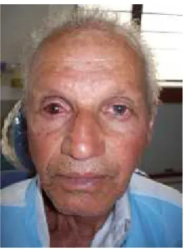
A 60-year-old male patient (Figure-1) referred to Sri Hasanamba Dental College and Hospital by an ophthalmologist for replacement of his right eye. A 5 year old ready-made ocular prosthesis was placed by an ophthalmologist following an accident. At the time of examination, it was observed that the whole eyeball was surgically excised, but the muscles at the base of the socket were intact. The existing prosthesis had poor retention, did not support the eyelids well and did not match the opposite eye. The patient complained of poor aesthetics and water collection followed by headache hence requested anew
ISSN: 0975-833X
International Journal of Current Research
Vol. 6, Issue, 09, pp.8854-8859, September, 2014
INTERNATIONAL JOURNAL OF CURRENT RESEARCH
Article History:
Received 16thJune, 2014
Received in revised form 17thJuly, 2014
Accepted 10thAugust, 2014
Published online 30thSeptember, 2014
Key words:
Ocular prosthesis, Maxillofacial prosthesis, Eye prosthesis.
CASE REPORT
OCULAR PROSTHESIS: AN ESTHETIC SOLUTION
*Kiran Kumar H.S (MDS), Sudhakara Bhat G (MDS), Sanjayagouda Patil (MDS)
Sankar Madhavan (MDS) and Nahush V. Chaudhari (MDS)
Department of Prosthodontics, Sri Hasanamba Dental College and Hospital,
Hassan, Karnataka, India
ARTICLE INFO ABSTRACT
The first feature of the face to be noticed is the eyes. Loss of eye due to any reason will cause psychological stress to the patient. Replacement of missing eyewould definitely restore the patient’s psychological comfort and social well-being. Many techniques of fabrication of ocular prosthesis are described in this literature requiring artistic expertise. A simple and time saving technique of fabrication of ocular prosthesis without sacrificing the primary objective is presented in this article.
Copyright ©2014 Kiran Kumar H.S. et al. This is an open access article distributed under the Creative Commons Attribution License, which permits unrestricted use, distribution, and reproduction in any medium, provided the original work is properly cited.
INTRODUCTION
Eyes play an important role in aesthetics, expression, and social well-being of a person. The disfigurement associated with this loss can cause not only significant physical problems but also emotional problems (Lubkinand Sloan1990. Rehabilitation of such patients both emotionally and prosthetically is really a phenomenal task (Bindhoo and Aruna2011). Hence to promote both physical and psychological healing and to improve social acceptance, replacement of the lost eye as soon as possible by ocular prosthesis is necessary (Chin et al., 2006). Ocular prostheses are either ready-made (stock) or custom-made. They may be made of either glass or methyl methacrylate resin. Glass is not the material of choice because it is subject to breakage and surface deterioration from contact with orbital fluids. Methyl methacrylate resin is superior to other ocular prosthetic materials in tissue compatibility, esthetic capabilities, durability and color permanence, adaptability of form, cost, and availability. The fabrication of a definitive ocular prosthesis should begin as soon as the socket has healed. Prosthetic rehabilitation is enhanced if an implant can be placed in the orbit to provide an attachment for the rectus muscles, which can impart motion, coordinated with the natural eye (Cain 1982). However the placement of an ocular implant is not always possible or feasible.
*Corresponding author: Kiran Kumar H.S (MDS),
Department of Prosthodontics, Sri Hasanamba Dental College and Hospital, Hassan, Karnataka, India.
Patients in this situation can be treated with custom-made ocular prostheses that have been adapted to accommodate specific situations. A custom ocular prosthesis allows infinite variations during construction. The close adaptation to the tissue bed uses the full potential to produce movement. Voids that collect mucus and debris, which can irritate mucosa and act as a potential source of infection are minimized. The optimum cosmetic and functional results of a custom ocular prosthesis enhance the patient’s rehabilitation to a normal lifestyle.
A sequence of steps of construction of ocular prostheses is outlined, and the critical areas of fabrication and artistic techniques employed in the successful prosthetic treatment are described in this article. The following clinical report of the patient demonstrates treatment with standard technique that can create functional and esthetically pleasing results
Case report
A 60-year-old male patient (Figure-1) referred to Sri Hasanamba Dental College and Hospital by an ophthalmologist for replacement of his right eye. A 5 year old ready-made ocular prosthesis was placed by an ophthalmologist following an accident. At the time of examination, it was observed that the whole eyeball was surgically excised, but the muscles at the base of the socket were intact. The existing prosthesis had poor retention, did not support the eyelids well and did not match the opposite eye. The patient complained of poor aesthetics and water collection followed by headache hence requested anew
ISSN: 0975-833X
International Journal of Current Research
Vol. 6, Issue, 09, pp.8854-8859, September, 2014
INTERNATIONAL JOURNAL OF CURRENT RESEARCH
Article History:
Received 16thJune, 2014
Received in revised form 17thJuly, 2014
Accepted 10thAugust, 2014
Published online 30thSeptember, 2014
Key words:
prosthesis, emphasizing his wish not to wear tinted glasses. It was decided to fabricate a custom acrylic ocular prosthesis to attend to all the problems of the current prosthesis. After a thorough examination of the ocular defect, the treatment was initiated.
Figure 1. Patient with enucleated right eye
Treatment Plane
Ocular prosthesis can be ready-made (stock) or custom made. Custom made eye have some advantages including better mobility, even distribution of pressure due to equal movement thereby reducing incidence of ulceration, improved fit, comfort and adaptation, improved facial contours and esthetics. Also, custom eye enhance tissue health by reducing potential stagnation space at the prosthetic tissue interface (Shenoy and Venkat Ratna 2007; Owand Amrith1997. So, considering all these benefits it was decided that a custom made ocular prosthesis would be the best prosthetic option to meet the needs of the patient. Also to get better esthetics and color matching without following the conventional, cumbersome and technique sensitive method of iris button painting, use of matching stock eye shell was planned for this particular case.
Procedure
The procedure of fabrication involves the following steps
1. Preparation of the patient 2. Making an impression 3. Preparation of the mold 4. Preparation of the wax pattern 5. Wax pattern try-in with iris button
6. Master cast preparation and acrylisation procedure 7. Care and maintenance of ocular prosthesis 8. Follow up
Preparation of the patient
The patient was informed about the procedure of impression making in detail before the start of the procedure. Patient was advised to keep the contra lateral normal eye open with
focussing the eye on a predetermined fixed point straight ahead during the procedure.
Making an impression
A thin layer of petroleum jelly should be applied on the eyelashes and around the eye socket to prevent the impression material from sticking to the eye lashes; then both the eye lashes should be retracted with the help of left hand index and thumb finger. Light body polyvinyl siloxane impression material was injected slowly into the socket (Figure- 2), Then both the fingers were released slowly and the patient was instructed to keep the contra lateral eye open. A predetermined point was fixed straight ahead and the patient was instructed to concentrate at that point till the material reached its setting time. The upper and lower eyelids were gently retracted and the impression was gently removed. The impression then was washed with cold water. Then the excess material was cut using a sharp BP blade (Figure- 3).
[image:3.595.363.507.284.474.2]Figure 2. Impression procedure
Figure 3. Tissue surface of impression
Preparation of the mold
The cast can be poured in two steps. In first step, upper third of the impression is immersed in dental stone, which is prefixed on a flat platform. After it sets, keyholes were made on the stone platform to aid in alignment in the further steps (Figure-4). In the second step, boxing is done around the first layer.
[image:3.595.344.526.506.642.2]Separating media is then applied and dental stone is poured into the remaining impression. After the mold is set, two halves of the cast are separated and the impression is removed carefully.
Figure 4. Stone cast
Preparation of the wax pattern
The layers of the mold are separated. Separating media is then applied over the mould. Modeling wax is melted and poured in two half’s of the cast and both the half’sare closed and placed in cold water till the wax hardens.
Two half’s of the mouldare then separated and the wax pattern is carefully removed. Sharp ridges and undesirable irregularities are eliminated to form a convex surface which resembles the eye shell (Figure- 5).
Figure 5. Wax pattern
Wax pattern try-in with iris button
The prepared wax pattern is then inserted into the patient orbital socket to check for proper contour and bulk. Prefabricated iris button selected according to the patient’s normal eye is inserted in the wax model. It is then rechecked forpatient’s natural eyesymmetry (Figure- 6) by trial and error
method. Central placement of the iris and marking the corneal plane is done to achieve symmetry of the two eyes.
[image:4.595.45.280.112.287.2]Various methods have been used to achieve symmetry:
Figure 6. Try-in with iris button
1. Interpupillary distance: Once the wax model has been made, wax solvent is used to smooth the surface. This wax model is inserted in the patient’s eye socket. After making it symmetrical with respect to its position and plane, the interpupillary distance is marked with a non-toxic marker.
2. Hirschberg’s test: In absence of gross asymmetry of the orbit, the base for the ocular prosthesis can be made with the help of white acrylic which is then inserted into the eye socket. The light reflex is kept at the centre of the model.
3. Inscribing a circle: On the white acrylic base, based on the ocularist’s judgment a circle is inscribed in the centre corresponding to the fellow eye.
4. Iris corneal buttons: This is the most difficult of the various methods described above. However, this gives the best cosmetic result. The iris button is inserted in the wax model using the carving wax and hot metal spatula. The symmetry was assessed using trial and error method.
Once the soft tissue contour and the location of the iris were satisfactory the pattern was removed carefully and the sclera color was selected.
Master cast preparation and acrylisation procedure
[image:4.595.43.287.459.641.2]of iris and sclera colour, iris position, iris size and pupil size. Finally, post-insertion instruction’s were advised to the patient along with the technique of removal and insertion of the prosthesis.
[image:5.595.74.258.178.352.2]The esthetic and comfort of custom-made ocular prosthesis is far better than the stock prosthesis. It is a recommended form of treatment. This procedure is simple, economical and will increase the self-confidence of the patient (Puranik et al., 2013; Thirunavukkarasu et al., 2014; Shrivastava et al., 2013).
Figure 7. Wax pattern invested
Figure 8. After treatment
Care and maintenance of ocular prosthesis
If there was any irritation or abnormal discharge in the eye the patient should report immediately.
The prosthesis should be cleaned before insertion. Never clean the prosthesis with a cloth, abrasive soap, or toothpaste. The prosthesis is best cleaned using a mild soap, baby shampoo or contact lens cleaning solutions with wet hands. All soap must be rinsed from the prosthesis and hands before reinsertion of the prosthesis. As the prosthesis is made of an acrylic plastic, it should never be soaked in alcohol, gasoline or bleach. Do not
attempt to sterilize the prosthesis in an autoclave. In the office, prosthesis can be disinfected in a cold sterilization media such as cidex. Contact lens cleaning solution can also be used for cleaning ocular prosthesis.
Excessive mucous secretions can occur when wearing an ocular prosthesis. Conditions such as head colds, winds dust, allergies and dirty hands can cause considerable secretion. Regular rinsing of the prosthesis with a contact lens cleaning solution can cause usual dislodge any surface deposits.
The warm wet face cloth will soften the secretion allowing you to remove it by wiping inward toward the nose. (Do not wipe outward, because this could rotate the prosthesis out of position, or cause it to fall out of the cavity)
Always carry a pocket pack of tissue and use it when necessary to remove any secretion from the prosthesis or lids. The average amount of wiping is three to five times a day. Avoid the use of a handkerchief or bare fingers If the socket is dry lubricant eye drops can be used. Prosthesis to be removed and placed in water in night.
Follow up
There is no definite answer as to how long the prosthesis will last. Unlike the old style glass prosthesis, modern plastic prosthesis is durable and is resistant to breakage. It is recommended that the patient should be reviewed on a yearly basis to check for the condition and fit of the ocular prosthesis. Patient’s wereinformed to report immediately if they had any complaints related to prosthesis, if not yearly for a follow up. Three years follow up of the present case has been done without any post insertion problems.
DISCUSSION
Anecdotal reports and relics from ancient civilizations indicate that the restoration of ocular defects may have existed for thousands of years. The earliest known examples of restorations date to the fourth dynasty (2613-2494 B.C.) in Egypt excavation of tombs provided evidence of eye replacement by usingprecious stones, earthenware, enameled bronze, copper and gold in the shrunken sockets (Roberts1971).In the 16th century, Pare´ fabricated an ocular prosthesis ‘‘emblepharon’’ made of gold or silver (Gibson1955; Gray1976; Martin and Clodious 1979.Pare´ also used glass and porcelain for eyes, which was a great step forward and resulted in the use of the shell type of pattern rather than spheres (Gibson1955; Dyer 1980). Till IInd world war glass was the popular material then it was difficult to obtain glassor glass eyes from Germany. Methyl methacrylate, which had already replaced vulcanite as a denture base material, seemed to be a good replacement material. A definitive technique for fabricating artificial eyes using acrylic resin was developed by the United States naval dental and medical schools and was published in 1944 (Murphey and Schlossberg 1944) .Unlike glass eyes, the acrylic resin eyes were solid. The material was lightweight, easy to fit and adjust, unbreakable, translucent, easily fabricated, had intrinsic and extrinsic coloring capabilities, and was inert to the socket secretions (Dyer 1980. The dental-prosthetic influence in the development of this prosthesis accounts for the ocular
[image:5.595.88.240.388.582.2]prosthesis being fitted from an impression of the eye socket rather than by the traditional empirical method (Allen and Webster 1969; Maloney 1979). Several techniques have been used in fitting and fabricating artificial eyes. Empirically fitting a stock eye, modifying a stock eye by making an impression of the ocular defect (Taicher et al., 1985) and the custom eye technique are the most commonly used techniques (Benson 1977; Miller 1996). Artropoulou used digital imaging (Artopoulou et al., 2006) in the fabrication of the ocular prostheses presents several advantages compared to the conventional oil paint and monopoly iris painting technique (Firtell et al., 1975). The digital image provides acceptable esthetic results because it closely replicates the patient’s iris with minimal color adjustments and modifications. The fabrication of a custom acrylic resin eye provides more esthetic and precise results because an impression establishes the defect contours, and the iris and the sclera are custom fabricated and painted. Customized stock ocular prosthesis gives an acceptable aesthetic result for rehabilitation of geriatric patients (Kamble et al.,2013).
Conclusion
Fabrication of ocular prosthesis has been known to human’s since immemorial time. The first attempt was by the use of a glass eye “ambrosepaire.” Prosthetic rehabilitation fulfills aesthetic as well as psychological requirements for a patient. A correctly placed ocular prosthesis should maintain its orientation when the patient is looking straight ahead and restore the normal opening of eye, support the eyelids, restore a degree of movement, and be adequately retained and esthetically pleasing. A standard technique can produce excellent result for patients. However because of the extreme individual variation and diverse nature ocular injuries, certain patients would benefit more from custom made ocular prosthesis that are modified to their individual needs than the ready-made prosthesis.
This procedure may be time consuming and entail a trial and error approach, but the esthetic and functional results justify the extra effort. Using prefabricated eye and silicon instead of acrylic resin has advantages such as reduce treatment time, light-weight prosthesis and increased simplicity which makes this method an alternative for fabrication of ocular prosthesis.
Informed consent
A written consent was taken from the patient before starting the treatment. They were also informed regarding the various treatment options and the publishing protocols.
Acknowledgements
We express our deep sense of gratitude and profound thanks to my respected teacher Dr. Sudhakara Bhat.G, Professor and Head of the Department of Prosthodontics, Sri Hasanamba Dental College and Hospital, Hassan for her constant encouragement, guidance and support in every aspect of this study. We are grateful to Dr. S Ravindra, Principal, Sri Hasanamba Dental College and Hospital, Hassan for providing
us with all the college facilities to carry out this study successfully.
Disclosure of interests
None
Contribution to authorship
Dr. Kiran Kumar H Swas the principal investigator for the study. We are grateful to our respected teacher Dr. Sudhakara Bhat G, professor and Head, Department of Prosthodontics for her relentless help and suggestions for the study at all times. Dr. Sankar Madhavan for his help in the case analysis and support.
REFERENCES
Allen L and Webster HE.1969. Modified impression method of artificial eye fitting. Am J Ophthalmol, 67:189-218. Artopoulou II, Montogomery PC, Wesley PJ and Lemon
JC.2006. Digital imaging in the fabrication of ocular prosthesis. J Prosthet dent, 95:327-330.
Benson P. 1977. The fitting and fabrication of a custom resin artificial eye. J Prosthetdent, 38:532-538.
Bindhoo YA.Aruna U. 2011.Prosthetic rehabilitation of an orbital defect: A case report. J Indian Prosthodont Soc. 11(4):258–264.
Cain JR. 1982. Custom ocular prosthetics.J Prosthetdent, 48:690-4.
Chin K, Margolin CB, and Finger PT. 2006. Early ocular prosthesis insertion improves quality of life after enucleation. Optometry. 77(2):71-75.
Dyer NA. 1980. The artificial eye. Aust J Ophthalmol, 8:325-7.
Firtell DN, Anderson CR andDonnanML. 1975.Vein application technique for ocular prostheses.J Prosthet dent, 34(2):192-194.
Gibson T. 1955. The prostheses of AmbroisePare.Br J PlastSurg, 8:3-8.
Gray PHK. 1976. Radiography of ancient egyptian mummies. Med RadiogrPhotogr, 43:34-44.
Kamble VB, Kumar M, Panigrahi D. 2013. An ocular prosthesis for a geriatric patient: A case report. Journal of Clinical and Diagnostic Research. 7(6): 1236-1238. Lubkin V and Sloan S. 1990.Enucleationand psychic trauma.
Advances in ophthalmic plastic and reconstructive surgery.8:259.
MaloneyB.1979. Development of impression fitting equipment: a new technique. J Am SocOcularists; 9:32-3. Martin O, Clodious L. 1979. The history of artificial eyes. Ann
Plastic Surg; 3:168-70.
Miller B. 1996. Custom ocular impression trays.J Facial SomatoProsthet, 2:109-13.
Murphey PI and Schlossberg L. 1944.Eye replacement by acrylic maxillofacial prosthesis.Naval Med Bull, 43:1085-1099.
Puranik S, Jain A, Ronad S, Ramesh S, Jagadeesh MS, and KattimaniP. 2013. Prosthetic rehabilitation of a patient with an ocular defect: a simplified approach. Case Reports in Ophthalmological Medicine. Volume 1-3.
Roberts AC. 1971. Facial prostheses. 1st ed. London: Henry Kimpton; p. 4.
Shenoy KK andVenkatRatna NP.2007. Ocular impression: an overview. J Indian ProsthodontSoc, 7(1):5–7.
Shrivastava S, Agarwal S, Shrivastava KJ, Tyagi P. 2013. Custom-made ocular prosthesis for a pediatric patient with unilateral anopthalmia: A case report. Journal of Indian Society of Pedodontics and Preventive Dentistry. 31(3):194-196.
Taicher S, Steinberg HM, Tubiana I andSela M. 1985.Modified stock-eye ocularprosthesis.J Prosthetdent, 54:95-8.
Thirunavukkarasu I, Rai R, Prabhu R, Deshpande VA, Kumar AS.2014. Rehabilitation of partially eviscerated eye with custom made ocular prosthesis- A case report. Journal of Clinical and Diagnostic Research. 8(1): 285-287.