Molecular Docking Reveals a Novel Binding Site Model for Fentanyl at the
µ
-Opioid Receptor
Govindan Subramanian, M. Germana Paterlini, Philip S. Portoghese, and David M. Ferguson*
Department of Medicinal Chemistry and Minnesota Supercomputer Institute, University of Minnesota, Minneapolis, Minnesota 55455
Received July 19, 1999
The ligand binding modes of a series of fentanyl derivatives are examined using a combination of conformational analysis and molecular docking to theµ-opioid receptor. Condensed-phase molecular dynamics simulations are applied to evaluate potential relationships between ligand conformation and fentanyl substitution and to generate probable “bioactive” structures for the ligand series. Automated docking of the largely populated solution conformers identified a common binding site orientation that places the N-phenethyl group of fentanyl deep in a crevice between transmembrane (TM) helices II and III while the N-phenylpropanamide group projected toward a pocket formed by TM-III, -VI, and -VII domains. An analysis of the binding modes indicates the most potent fentanyl derivatives adopt an extended conformation both in solution and in the bound state, suggesting binding affinity may depend on the conformational preferences of the ligands. The results are consistent with ligand binding data derived from chimeric and mutant receptor studies as well as structure-activity relationship data reported on a wide range of fentanyl analogues. The binding site model is also compared to that of N-phenethylnormorphine. An overlay of the bound conformation of the opiate and cis-3-methylfentanyl shows the N-phenethyl groups occupy equivalent binding domains in the receptor. While the cationic amines of both ligand classes were found docked to an established anchor site (D149 in TM-III), no overlap was observed between the N-phenylpropanamide group and the remaining components of the opiate scaffold. The unique binding mode(s) proposed for the fentanyl series may, in part, explain the difficulties encountered in defining models of recognition at theµ-receptor and suggest opioid receptors may display multiple binding epitopes. Furthermore, the results provide new insight to the design of experiments aimed at understanding the structural basis to the differential selectivities of ligands at theµ-,δ-, and κ-opioid receptors.
Introduction
Ever since the discovery of fentanyl (1) in 1962 by Janssen,1the 4-anilidopiperidines have been exploited for developing highly selectiveµ-opioid agonists2with specific pharmacological properties.3Over the last four decades, structure-activity relationship (SAR) studies of fentanyl analogues have provided considerable insight into the key structural features and pharmacophore elements required for high-affinity binding to the µ-re-ceptor.2,4In comparing fentanyl with the classicµ-opiate morphine, certain similarities are apparent. Both com-pounds possess a protonatable nitrogen and an aromatic group that are commonly thought to mimic the N-terminal tyrosine moiety of opioid peptides.5However, SAR studies of N-phenolic derivatives6of fentanyl have shown that hydroxy substitutions do not influence the morphinomimetic potency of these compounds. Con-versely, a dramatic loss in ligand binding is observed when the N-phenethyl group of 1 is replaced with the N-methyl group of morphine.7This suggests that fen-tanyls and µ-opiates may display unique recognition elements to the µ-receptor but induce qualitatively similar pharmacological profiles through different
mech-anisms that cannot be rationalized by structural com-parisons alone.
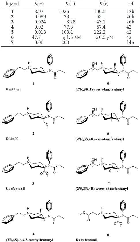
Some hints to the recognition elements that may confer high-affinity binding to the µ-receptor can be found in the SAR data reported on fentanyl. Substitu-tions to the piperidine ring have been shown to dra-matically affect ligand binding (Table 1).8 A simple methoxymethyl (as in R30490, 2)9 or acetate (as in carfentanil, 3)10substituent on the piperidine 4-axial position of 1 has been shown to increase the ligand binding affinity by 45- and 160-fold, respectively. As alluded to above, substitution of the N-phenethyl group effectively abolishes ligand binding in some cases.7SAR studies have further revealed that compounds which retain the ethyl spacer display high affinity to the µ-receptor. Fentanyls have also shown to display great variation in enantiospecific binding. With two chiral centers and four possible enantiomers, the most active cis-3R,4S stereoisomer of 3-methylfentanyl (4)11,12 ex-hibits 7000-fold increased in vivo potency compared to morphine.13Perhaps, the most significant results have come from studies of 2′-OH-3-methylfentanyl (Figure 1). Out of the eight optically active isomers, cis-(2′ R,3R,4S)-ohmefentanyl (5) belongs to the “super potent” class of µ-selective agonists.14 In contrast, the corresponding antipode cis-(2′R,3S,4R)-ohmefentanyl (6) is the least potent, weakly bound ligand in the ohmefentanyl series. * To whom correspondence should be addressed. Tel: 612-626-2601.
Fax: 612-626-4429. E-mail: [email protected].
381 J. Med. Chem. 2000, 43, 381 391
Among the four trans stereoisomers, the 2′S,3R,4R enantiomer 7 possesses an enhanced affinity for the µ-receptor as compared to the 2′R,3R,4R counterpart. Of the various N-phenethyl modifications that have been reported,15 remifentanil (8) is one of the most unique.16,17This highly selective compound contains a hydrolytically labile ester group in place of the phenyl ring and, while sterically smaller than 1-7, displays similar potencies.
Although SAR data provide insights into the phar-macophore features imparting specificity for the µ-re-ceptor binding, attempts to understand the structural basis for ligand recognition have been hindered by the difficulties in obtaining the crystal structure of the receptor or the ligand-bound complex. Fortunately, some
insights on the ligand binding domain have been gained through binding studies of fentanyl and related com-pounds using cloned opioid receptors. Chimeric con-structs of opioid receptors have shown transmembrane (TM) helices I-III, VI, and VII to be involved in sufentanil (9) binding.18More specifically, site-directed mutagenesis data suggest that an aspartate residue in TM-III may be a critical anchor point for the binding of fentanyl derivatives.19 This aspartate is highly con-served in opioid and related G-protein-coupled receptors and is thought to form a salt bridge with the protonated amino group of a variety of ligands. Although the binding data is limited, available pharmacological results20a,21strongly suggest fentanyls bind in the TM domain of theµ-opioid receptor.
Despite the importance of fentanyl and related com-pounds in pain management, a detailed investigation of the receptor-ligand interactions responsible for high-affinity binding and selectivity to theµ-receptor has not yet been reported. This is partly due to the inherent difficulties in modeling opioid receptors at a molecular level and also to the manifold conformations fentanyl may adopt in the condensed phase. Mosberg et al.22 recently proposed a docking arrangement for cis-meth-ylfentanyl to theµ-opioid receptor. However, the focus of that study was on receptor model building, so the ligand docking results are quite limited. The “manual docking” performed places the ligand (4) toward the top of the receptor cavity and consequently does not com-pletely explain available site-directed mutagenesis and chimeric data.18-21 In addition, the conformational variability of 4 was not fully considered, potentially biasing the results. Although a number of computational studies on the conformational preferences of fentanyl analogues have been reported,23-27no conclusive agree-ment has emerged with respect to the bioactive con-former for members of this ligand series.
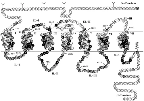
In this study, we combine SAR and ligand binding data with molecular docking experiments to understand the molecular determinants governing the recognition of fentanyls at theµ-receptor. Using molecular dynamics (MD) simulations in the condensed phase, the most populated conformations of 1-8 are identified and docked to the humanµ-receptor (Figure 2). A refined µ-opioid receptor model is also reported that takes advantage of the 6 Å projection map of bovine rhodop-sin28and other biophysical data obtained from rhodop-sin-likereceptors.Asinourearlierworkonarylacetamides,29b an automated docking protocol is applied to evaluate the binding site orientations in this receptor model. The results are compared with available site-directed mu-tagenesis, chimeric, and SAR data to identify key ligand-receptor interactions involved in the molecular recognition process. Finally, the results are applied to provide new insight to the design of experiments aimed at understanding the structural basis to the differential selectivities of ligands at theµ-,δ-, andκ-opioid recep-tors.
Computational Methods
µ-Receptor Model. The structural model of the human µ-opioid receptor has been derived from electron cryomicros-copy data30 and the CR coordinate template provided by
Baldwin.28 That template was generated from rhodopsin
[image:2.612.53.296.67.486.2]projection data in combination with a sequence analysis of 493 Table 1. Experimental Binding Affinity (nM) of 1-7 for theµ-,
δ-, andκ-Opioid Receptor
ligand Ki(µ) Ki(δ) Ki(κ) ref
1 3.97 1035 196.5 12b
2 0.089 23 63 26b
3 0.024 3.28 43.1 26b
4 0.02 77.3 57.4 42
5 0.013 103.4 122.2 42
6 47.7 >1.5µM >0.5µM 42
7 0.06 200 14e
Figure 1. Structures of fentanyl derivatives 1-8.
[image:2.612.111.234.619.681.2]GPCRs, including opioid receptors.28Substitution of the opioid
receptor sequence (SWISS-PROT accession number, P35372; entry name, OPRM•HUMAN) onto the template produced a residue conservation profile similar to that previously obtained by us using a smaller alignment of 201 GPCR sequences.31
The length of the helices was determined by considering the minimum length in the lipid bilayer, as defined by Baldwin, with subsequent addition of residues at the extracellular and intracellular limits based on sequence analysis of this regions. Coordinates for the TM helices were obtained by model building idealR-helices with the appropriate kinks added to helices V and VI at P246 and P297. Side-chain conformations were determined using the backbone-dependent rotamer li-brary of Dunbrack and Karplus.32These structures were then
aligned to the CRtemplate of Baldwin based on the orientation and vertical position of highly conserved residues across the rhodopsin family. After each addition to the TM bundle, the system was energy-minimized and equilibrated with 100 ps of restrained MD at 300 K to relieve steric contacts introduced in model building.
The model was further refined using a series of MD simulations in which positional constraints on the peptide backbone atoms were gradually reduced from 5 to 0.05 kcal/ Å2/mol. Hydrogen-bonding distance constraints (32 kcal/Å2/
mol) were also imposed between the carbonyl oxygen of residue (i) and the amide proton of residue (i+4) to maintain an R-helical conformation. Given the known problems of rhodop-sin templates to capture all relevant structural data derived from functional studies of GPCRs, additional hydrogen-bond-ing constraints were applied to several residues to account for potential interactions. In particular, the initial CRtemplate
places D116 and N334 more than 10 Å apart. Mutagenesis studies, however, have suggested these highly conserved residues may be part of a hydrogen-bonding network that ties together helices I (N88), II (D116), and VII (N334).33-35To
account for this effect, distance constraints were added be-tween N88, D116, and N334 during the refinement process. The entire process required approximately 700 ps of MD at 300 K. The resulting model was qualitatively evaluated for consistency with ligand binding data derived from site-directed mutagenesis data36,37and with our previous models used in
docking opiates, dynorphin A, and arylacetamides.29,31All key
elements of the receptor, including the highly conserved aspartate in TM-III, the cluster of aromatic residues within the receptor cavity, and the position of selectivity elements at the top of TM domains VI and VII, aligned favorably. The structural quality of the model was also verified using PROCHECK38which showed 98% of the residues in the most
favored helical region and the remaining 2% in the additionally allowed helical region. Side-chain χ1 and χ2 angles were
determined to be in their most favorable regions without stereochemical conflicts. A final energy minimization was performed on the model, which was subsequently used as the starting structure for ligand docking studies.
Conformational Analysis. The crystal structures of 1,39 2,404,415, and 742were used as starting ligand geometries,
while those of 3, 6, and 8 were model-built from closely related X-ray structures. On the basis of the reported pKavalues of
several fentanyl derivatives,16b,23the protonated form of 1-8 was considered to be the pharmacologically relevant species. The geometries of 1-8 were optimized using molecular orbital
[image:3.612.57.558.43.394.2]program package.43The partial atomic charges for the
energy-minimized structures (1-8) were obtained using the restrained
electrostatic potential (RESP) charge fitting formalism.44
Subsequent computations of 1-8 adopted the Cornell et al.
force field45(parm94.dat file) incorporated in AMBER 4.1 suite
of programs46 with appropriate additions (see Supporting
Information). The ester parameters for 3 and 8 were taken from the recently published work of Kollman and co-workers.47
The MD simulations on the in vacuo energy-minimized geometries of 1-8 were performed to obtain the conformational
preferences of the fentanyls in solution. Accordingly, 1-8 were
placed in a periodic box of TIP3P water molecules.48Freezing
the geometry of the ligand, the solvent molecules alone were energy-minimized until a rms convergence of 0.1 kcal/mol in energy was achieved. This was followed by a 5 ps equilibration of the solvent and energy minimization of the entire system (0.01 kcal/mol rms deviation in energy). A 2 ns MD simulation of the solvated ligands was then carried out at 1 atm of constant pressure. SHAKE was used to constrain bonds involving hydrogen, and the temperature of the entire system was maintained at 300 K using Berendsen algorithm49with
0.2 ps coupling constant. A 1 fs time step with an 8 Å nonbonded cutoff was also used, and the simulations were restarted every 250 ps.
Ligand Docking. Docking of the solution conformation of 1-8 to the model-built TM domain of the µ-receptor was performed using the automated docking procedure (DOCK 3.5 program package)50by following the protocols described in our
previous work29band the DOCK manual. The program
SPH-GEN was used to create spheres that fill the “putative ligand binding pocket” on the solvent-accessible molecular surface51
of theµ-receptor TM helices. After initial refinement of the spheres thus generated, a cluster of 43 spheres that serve as negative image descriptors was used for docking of 1-8. A
maximum of two to six bad (steric) contacts between the ligand atoms and the receptor residues were allowed while itentifying potential docking configurations (orientations) in theµ -recep-tor binding pocket. To find the most probable ligand alignment in the binding pocket, all predicted ligand orientations in the
µ-receptor TM domain were scored (Eff, force field) individually
based on the empirical evaluation of the van der Waals repulsion (Erep) and attraction (Eatt) and electrostatic (Ees)
energy contributions (Eff )Ees + Erep +Eatt). Site-directed
mutagenesis studies onµ-opiate ligands have shown that a D147A/D147N/D147E point mutation in TM-III of ratµ-opioid receptor led to a diminished binding affinity,19,52presumably
due to the loss of a salt bridge anchoring the receptor-ligand complex. Since the basic nitrogen in fentanyl analogues (Figure 1) is protonated under physiological conditions, ligand orienta-tions that had this proton proximal to the TM-III aspartate (D149 in humanµ-opioid receptor) were given preference in the selection process. Consequently, the force-field scores and pharmacological results were used together to identify the most probable docking orientation in theµ-receptor.
Refinement of the initial receptor-ligand complex was achieved by in vacuo energy minimization (0.001 kcal/mol rms deviation). A 5.0 kcal/mol positional constraint was applied to the receptor backbone atoms along with an 8 Å cutoff for nonbonded interaction. This energy-minimized complex was then subjected to an initial 250 ps MD simulation ()4) with the above constraints. The force constant was subsequently reduced to 3.5 kcal/mol for the next 250 ps simulation and to 2.0 kcal/mol for the remaining 0.5 ns MD simulation. A 1 fs time step was used and the nonbonded pair list updated every 25 fs. The temperature of the system was maintained at 300 K using the Berendsen algorithm with a 0.2 ps coupling constant. Since the simulations over the last 500 ps were quite stable, the transient structure obtained from the last simula-tion step (1 ns) was used in the final refinement of the receptor-ligand (1-8) complex. With a 2 kcal/mol force
constant applied for the receptor backbone atoms, the geom-etry optimization ()4r) was terminated when the projected decrease of 0.001 kcal/mol in energy was met. Unless otherwise specified, all the docking analyses discussed hereafter are
based on results of the last 0.5 ns MD simulation and the subsequent minimization step.
Results and Discussion
The relative populations of the solution structures of 1-8 are reported in Table 2. Consistent with X-ray structures,39-42spectroscopic data,53and prior compu-tational investigations,23-27the piperidine ring favored only a chair conformation throughout the MD simula-tions. In addition, the methoxymethyl (2) and acetate (3 and 8) substituents to the piperidine ring were restricted mostly in the trans form. No preferences were noted, however, in the rotation of the terminal methyl group of the amide in 1-8. In evaluating the remaining torsional degrees of freedom, ligand substitutions were found to have a significant impact on conformational sampling especially onθ3which defines the orientation of the N-phenylpropanamide moiety (refer to Table 2 for the definition ofθ1-θ3torsions). In particular, aθ3 conformational rigidity is observed for 4- and 3-axial piperidine substitutions (g-in 2-5, 8; g+in 6). On the other hand, an equatorial methyl substitution in 7 and the unsubstituted fentanyl (1) allow flexibility between the g+ and g- forms of θ3 and consequently are in dynamic equilibrium throughout the simulation. This equilibrium may contribute to the observed trans con-figuration of this torsion in the X-ray crystallographic structures of 1 (174.9°)39and 7 (170.8°).42Using a model compound (N-phenyl-N-isopropylacetamide), the energy difference between the trans and g+/-conformers was
determined to be quite low (0.08 kcal/mol) based on ab initio Hartree-Fock (HF/6-31G*) calculations. The X-ray structures reported may therefore represent an average of these two forms or may simply be the result of lattice packing forces in the crystal. While cis θ3 conformers have been identified in a previous compu-tational study,26this arrangement is not populated in solution due to steric crowding, especially in the pip-eridine 4-substituted fentanyls.
[image:4.612.318.559.81.228.2]A scatter plot of the remainingθ1andθ2 angles for the observed θ3 torsions in 1-8 is given in Figure 3. These angles essentially define the overall shape of the ligand and are therefore critical to identifying the most probable “bioactive” conformation of 1-8. An analysis
Table 2. Population (%) of [θ1,θ2,θ3] Torsion of Fentanyl Analogues 1-8 Obtained from the 2 ns MD Simulation of the
Solvated Ligandsa
[θ1,θ2] [g+,g+] [g+,t] [g+,g-] [t,g+] [t,t] [t,g-] [g-,g+] [g-,t] [g-,g-] θ3
1 1.4 28.7 23.4 32.5 10.5 3.5 g+/g
-2 20.8 11.6 21.5 46.2 0.1 g
-3 16.6 27.6 33.8 22.0 0.1 g
-4 48.4 6.4 9.5 34.9 0.9 g
-5 0.1 96.3 3.7 g
-6 40.6 7.7 35.0 16.8 0.1 g+
7 16.7 46.0 16.1 21.3 g+/g
-8 0.4 59.6 24.2 13.3 2.5 g
-aGauche+(g+∼60.0°), trans (t∼180.0°), gauche-(g-∼300.0°) forθ1torsion. Gauche+(g+∼40.0°), trans (t∼180.0°), gauche-(g -∼320.0°) for θ2 torsion. Gauche+ (g+ ∼150.0°), cis (c ∼30.0°),
of this plot indicates the trans or extendedθ2torsion is not populated in solution. Except for 1, 7, and 8, the [g-,g-,θ3] orientation is not sampled in the MD run and the [g-,g+,θ3] arrangements are scarcely populated. Discarding the conformers that are<5% populated in solution for 1-8, a maximum of 4 conformers are sampled in solution out of the 27 conformational pos-sibilities (3-fold rotation around eachθ). However, their relative populations vary across the series (Table 2) and reveal different conformational preferences for each ligand. For instance, the [t,g-,g-] X-ray structure40is also the highly populated (46%) solution structure for 2, while the [t,g-,g-] crystal structure orientation of cis-3-methylfentanyl41is the second most populated (35%) isomer observed for the solvated species. The most potent ohmefentanyl derivative, 5, exists in the [t,g+,g-] X-ray structure42 orientation and is conformationally rigid with 96% populated in solution. In contrast, the inactive enantiomer (6) is flexible with the [g+,g+,g+] and [t,g+,g+] isomers sampled around 35% and the [t,g-,g+] isomer populated by 17% (Table 2). Although the chirality (3R,4S) is the same, the solution conformer of 4 differs significantly in the preference forθ1andθ2 torsions as compared to 5. A striking observation of the MD simulations, however, is the strong preference for trans and gauche-θ1andθ3 torsions, respectively, for most of the active ligands, while the gauche+θ3torsion dominates the weakly bound ligand 6.
Table 3 lists the initial conformations of 1-8 used in automated docking as well as the resulting DOCK force-field scores. Although priority was given to the most highly populated solution structures in docking,
incon-sistencies were noted in the binding modes of 4 and 6 when compared with the remainder of the ligands in the series.54This was addressed by selecting the second most populated solution structure for these ligands. Automated docking of this revised set of conformations to the TM domain of theµ-opioid receptor resulted in several (∼50 to∼215) plausible docking orientations for each ligand. In the process of evaluating these docking configurations using the force-field scores, we found the best overall scored binding modes did not always correlate with the results from site-directed mutagenesis studies.52aExcept for 2, none of the best scored docking orientations had the protonated nitrogen of the piperi-dine ring close to D149 in TM-III.
The origin of this disparity can be traced to the components of the DOCK scores. In comparing the best scored orientations with those that contain the key D149 ligand contact, significant differences are noted in the repulsive component of the score (Table 3). For all ligands (except 2), the repulsive energy dominates the score of the D149-bound orientations. This problem is most likely due to uncertainties in the side-chain conformation of the model-built receptor but may also stem from limitations in the use of rigid docking protocols. Table 3 therefore lists the overall best scored docking orientation as well as the selected orientations that account for the key D149-ligand contact.
Finally, the sensitivity of the docking orientation and the conformation of the initial receptor-ligand complex were verified by performing additional MD simulations for fentanyl starting from a different ligand docking orientation (Eff(s))[-0.7,33.8,-40.4]) and for
carfen-Figure 3. Scatter plot of the [θ1,θ2] conformational population of the solvated ligands (1-8) observed during the 2 ns MD
[image:5.612.96.519.50.218.2]simulation. Anglesθ1(x-axis) andθ2(y-axis) within 0-120°∼gauche+, 120-240°∼trans, and 240-360°∼gauche-.
Table 3. Electrostatic (Ees) and van der Waals Repulsive (Erep) and Attractive (Eatt) Contributions (kcal/mol) for the Best (Eff(b)) and
Selected (Eff(s)) Docking Configuration of Various Fentanyl Analogues in theµ-Receptor TM Domaina
Eff(b) Eff(s)
no.
starting conformer
for docking [θ1,θ2,θ3] Ees Erep Eatt Ees Erep Eatt
populated conformer
in complex [θ1,θ2,θ3] %
1 [t,g-,g+] -3.7 13.6 -33.9 0.3 31.8 -39.5 [g-,g-,g-] 74.6b
2 [t,g-,g-] -3.8 15.6 -36.0 -3.5 15.0 -34.9 [t,g-,g-] 85.6
3 [t,g+,g-] -3.2 12.3 -32.7 -1.7 47.9 -44.8 [t,g-,g-] 80.6
4 [t,g-,g-] -2.4 12.7 -34.6 -10.2 61.8 -60.9 [t,g-,g-] 97.2
5 [t,g+,g-] -4.8 15.1 -34.9 -3.0 36.3 -48.7 [t,g-,g-] 81.6
6 [t,g+,g+] -0.9 22.8 -47.4 -7.7 89.8 -69.5 [g-,g+,g+] 90.0
7 [t,g-,g+] -4.0 18.6 -39.6 -8.4 92.6 -75.5 [t,g-,g-] 99.4
8 [t,g+,g-] -3.2 10.2 -29.4 -10.5 214.0 -77.4 [t,g+,g-] 99.2
[image:5.612.57.559.273.373.2]tanil starting with a different ligand conformation ([t,g-,g-]). Irrespective of the starting receptor-ligand geometry, the MD simulations of both the complexes resulted in final configurations that were similar to the ones reported in Table 3.
The MD trajectories of the receptor-ligand complexes indicate the fentanyl analogues align diagonally across the receptor cavity, spanning TM-II, -III, -VI, and -VII. A depiction of the ligand binding mode and superposi-tion of the bound conformasuperposi-tions of 1-8 is given in Figure 4. This binding site model is supported by ligand binding studies toµ/κandµ/δchimera that have shown TM-I-III, -VI, and -VII are essential to sufentanil binding.18The proposed docking orientation places the
N-phenylpropanamide group toward the extracellular side of the cavity (between TM-III and -VI), while the N-phenethyl group slides through TM-III and -VII toward the intracellular end of the cavity. A detailed examination of the binding modes for 1-8, however, reveals some differences in the orientation of these functional groups across the series. In the case of the N-phenethyl group, differences are noted in the binding of 1, 6, and 8 as compared to that of 2-5 and 7. More specifically, the phenethyl group of 2-5 and 7 is projected toward TM-II while the same moiety is directed toward TM-III in 1, 6, and 8 (Figure 4). As a result, the receptor-ligand complexes of 2-5 and 7 gain additional stabilization from residues in TM-II and -VII. These subtle differences in the ligand conformations may be a contributing factor in reducing the affinity of 5 (by 590-fold) to aκ/µchimeric receptor in which TM-I and -II are taken fromκand TM-III-VII are taken from µ.21The binding affinity of 6, on the other hand, was effected much less (40-fold), lending support to the hypothesis. Similarly, the proximity of the phenethyl group of 1 to TM-III, and especially to N150 (which appears to sterically hinder binding), is corroborated by the 20-fold increase observed in fentanyl binding upon
[image:6.612.105.512.48.301.2]a N150A point mutation.20aA listing of residues proxi-mal to the N-phenethyl group in 1-8 can be found in Table 4.
The position of the N-phenylpropanamide group is of particular interest. This group is directed toward the top of TM-VI which contains key recognition elements in the binding of several opioid ligands. In particular, position VI:23 (K305; Figure 2) has been identified in the binding and selectivity of the rigidµ-opiates such as norBNI55and GNTI56(E297 inκ) and theδ-selective piperazine derivative SNC8057(W284 inδ). A compari-son of the binding modes of 1-8 in this domain shows the N-phenylpropanamide group of 1-7 adopts a similar arrangement between TM-III and -VI. However, the fentanyl skeleton is simply too small to span the receptor cavity (i.e. D149-K305) and consequently cannot extend up to the similarly placed µ-specific residue, K305. This tends to rule out this position in conferring selectivity of 1-8 and suggests different elements may drive the molecular recognition process at theµ-receptor.
An analysis of the residues flanking the remaining functional groups also reveals some ligand-specific differences. Perhaps the most provocative involves the Figure 4. Key residues surrounding cis-3-methylfentanyl (4) in theµ-receptor binding pocket (left). A superposition of 1-8 in
[image:6.612.316.557.368.442.2]theµ-receptor binding pocket is shown on the right.
Table 4. Selected Residues within 5 Å Surrounding the
Various Functional Moieties of 1-8 in the TM Domain of the
µ-Receptor Binding Pocket (µ-receptor-specific residues are underlined)
helix no. residue no. in theµ-receptor
TM-II (T120, L123)a, T122b
TM-III (N152b, M153)a, (I146, D149b)c, Y150d, S156a TM-V (F239, F243)d
TM-VI I298c, H299d, (V302, K305)e
TM-VII (N330, S331b, N334, P335)a, (G327, Y328b)c, (W320, C323, I324, L326)e
key D149 salt link. This interaction has been shown to be a key anchor to recognition and binding of a number of opioid ligands, including peptides, rigid opiates, and nontypical opioids such as U50,488.19,58The MD simu-lations of the receptor-ligand complexes, however, indicate this salt link is lost in one case. Although each ligand-receptor complex was started with this contact intact, the orientation of 6 changed within the first 100 ps breaking this critical link. This appears to be due to the g+θ3torsion of 6 that orients the 3-methyl group much closer to M153 in the initial complex (note that the same torsion is g-in 5 and 7; Table 3). Similarly, the N-phenyl group encounters steric clashes with I298 and I324 as does the N-phenethyl group with A119 and Y328. Hindered by repulsive interactions with the receptor, the ligand realigns during the simulation disrupting the salt bridge. As a result, the 2′-OH group of 6 is close to hydrogen-bonding distance with the carboxylate group of D149. This may, in part, explain the reduced binding affinity of 6 compared to other members in the ohmefentanyl series.14
Ligand-specific differences are also noted in the receptor interactions with substituents on the piperidine ring (2, 3). Although docked close to H299 in TM-VI, the polar substituents of this ring are not positioned appropriately to take advantage of donor-acceptor interactions with the imidazole side chain of this residue, suggesting that H299 is not directly involved in fentanyl binding. This general hypothesis is sup-ported by ligand binding studies that indicate sufentanil binding remains unaffected by H297N or H297Q point mutations in the rat µ-opioid receptor.59 The axial 3-methyl substitutions in 4 and 5 are ideally placed to benefit from hydrophobic interactions with residues in TM-III and -VII, while the equatorial methyl group of 7 points to a domain between TM-III and -V.
Although such differences in hydrophobic interactions with the receptor may be an important factor contribut-ing to the differential bindcontribut-ing of 5 and 7, these ligands also contain a 2′-hydroxyl group that may attenuate affinity. In fact, the hydroxyl group of 5 and the hydroxyl group of S331 come in close contact during some stages of the MD simulation, providing an ad-ditional anchor point to receptor-ligand association. A similar interaction is possible for the hydroxyl group of T122 and the 2′-OH of 5 and 7. It is important to point out, however, that an alternative docking arrangement has been proposed for ohmefentanyl. Tang et al. hy-pothesized that this hydroxyl may form a hydrogen bond with Y150.60While Xu et al.61have reported a reduction in binding affinity of 5 and 6 to a Y150F mutant µ-receptor (Y148 in rat sequence), the effect is marginal (4-7-fold) which casts some doubt over the interpreta-tion. Similar reductions in affinity have been noted for other residues in the cavity of opioid receptors that are not directly involved in ligand binding. Nevertheless, the alternative docking arrangement for the 2′-OH cannot be ruled out at this point in time.
The docking mode(s) proposed here is also consistent with SAR data reported for a number of fentanyl derivatives. As seen in Figure 4, positionsRto the basic nitrogen in fentanyl dock between TM-III and -VII in a fairly restricted binding domain. Substitutions to theR position may therefore encounter steric repulsions with
the receptor side chains, thereby disrupting the salt link with D149. This may explain the dramatic loss in activity observed for 2,3-, 2,5-, and 3,5-dimethylpiperi-dine ring derivatives of fentanyl.62Similarly, the prox-imity of the 3-methyl substitution in 4 and 5 to G327 (TM-VII) is consistent with the diminished binding affinities observed for 3-allyl- and 3-propylpiperidine substitutions.63 In general, SAR studies suggest rela-tively small, planar fragments as favored terminal groups to the ethyl spacer. Thus, constrained deriva-tives,64 which include benzo-fuzed heteropentacycles and other bicyclic derivatives, show a marked reduction in binding affinity (ca. 300-fold) when compared to 1.65 While cyclization increases steric bulk in this region of the ligand, it also restricts the θ1 and θ2 angles that define critical recognition elements. The results reported here suggest the most strongly bound ligands prefer an extended or transθ1. Cyclization may, in effect, lock the structure in a less than optimal conformation, leading to an overall decrease in binding affinity. These same arguments could be extended to explain the decreased affinity of cyclized derivatives of the N-phenylpropana-mide and piperidine rings.66The N-phenyl ring of 1-8 is close to a number of side-chain residues in TM-III. Cyclization may therefore produce steric hindrance and crowding of the ligand in this region. The position of the phenyl ring in this local binding domain is further supported by SAR data on ortho/para/meta substitu-tions.6bWhereas the ortho positions of the N-phenyl ring lie close to residue side chains in TM-III, the meta and para positions project between TM-III and -VI. This is consistent with the reduction in binding affinity re-ported for fentanyl derivatives that contain bulky groups ortho to the N-phenyl bond.6
31 for a detailed discussion of opiate docking). While site-directed mutagenesis studies have implicated sev-eral of these aromatic residues in binding opiates, it is important to point out that similar results have not surfaced to link fentanyl-based ligands to this binding domain in theµ-receptor. This lends some support to the relative disposition of the aromatic rings shown in Figure 5.
Conclusions
This study has presented a binding site model for a series of fentanyl derivatives using a combination of solution-phase conformational analysis and automated docking techniques. One of the more interesting results of the work regards the potential relationship observed between ligand conformation and binding affinity. The results of the solution-phase dynamics calculations suggest that the most potent fentanyl derivatives pos-sess an extended conformation for the phenethyl group withθ1 ) trans andθ2 ) gauche. This suggests that minor differences in the conformations of substituted fentanyls may have a significant impact on ligand binding. Given the close structural relationship of 1-7, this outcome was unexpected at the outset of our study. Support for this conclusion can be found in the reduced affinity of 6 (which adopts a more compact form) and the diminished activity of cyclized derivatives that lock the torsions in less than optimal values.64,65This may also explain the differences in binding modes reported by Pogozheva et al.22with that reported here for 4 (cis-3-methylfentanyl). The conformation selected for dock-ing in that study possessedθ1andθ2torsion angles of 282° and 201°, respectively. This structure, however, was not populated during our solution-phase simula-tions. This is true of all fentanyl derivatives with a trans θ2 which are predicted to be higher in energy when compared with the corresponding gauche forms. In fairness to Pogozheva et al.,22it is important to point out that the focus of their study was on receptor structure, and not directed to a detailed analysis of fentanyl and fentanyl derivatives. Nevertheless, it is clear that conformer selection has a significant impact on the predicted binding mode. Taken overall, this
indicates great care must be taken in evaluating the conformational properties of all ligand derivatives, regardless of the close structural relationships displayed across a given series.
The automated ligand docking results have also led to the development of a unique binding site model for fentanyl that explains available pharmacological data as well as SAR data for a number of fentanyl deriva-tives. To date, most models of opioid recognition place the ligand in a main cavity of the receptor formed by TM-III-VII. This cavity is essentially bounded by the conserved Asp in TM-III (D149) and a cluster of aromatic residues in TM-V and -VI. The docking mode proposed here, however, places the N-phenethyl group of fentanyl deep in a crevice between TM-II and -III with the N-phenylpropanamide moiety projected diagonally toward TM-VI and -VII. This also aligns the cationic amine of fentanyl with D149 but leaves the aromatic pocket of the receptor unoccupied. While the model is generally supported by ligand binding data to µ/κ chimeras,18the most convincing evidence can be found in Figure 5. A comparison of the binding modes of cis-3-methylfentanyl and N-phenethylnormorphine (10) indicates the phenethyl groups project into the same binding domain in theµ-receptor, despite clear differ-ences in the overall binding modes of the ligands. (Note the phenolic moiety of the opiate is bound in the aforementioned aromatic pocket of the cavity.) These results suggests that the µ-receptor may contain an important recognition site relatively deep into the receptor cavity between TM-II and -III. The significance of such a conclusion is further heightened by the finding that changes in TM-II may be linked with receptor activation and high/low-affinity binding of agonists. A single mutation of D116 (D114 in rat) has been shown to constitutively deactivate opioid receptors, leading to a decrease in agonist affinity.68The interaction of the
N-phenethyl ring of fentanyl (and the opiate) with TM-II may therefore play some role in receptor activation. Although this is highly speculative, steric interactions between the retinal chromophore and TM-III of rhodop-sin have been shown to trigger signal transduction.69 The results of this study have further provided insight to potential targets for site-directed mutagenesis studies and the design of novel fentanyl derivatives. Such experiments not only are critical to evaluating the binding site model proposed but also are of great importance to understanding the structural basis to selectivity for this ligand class. For example, mutation of S331 may help define the orientation of the phenethyl ring within the receptor cavity. This residue is a potential hydrogen-bonding partner with the 2′-OH of the ohmefentanyl derivatives. The comparison with the opiate is especially enlightening and shows additional steric bulk can be accommodated in the 4-axial position of the piperidine ring. Although 4-phenyl/heteroaryl substitutions have been shown to improve analgesic potency at theµ-receptor,70the effect of incorporating a phenolic group in this position has not been tested. It may be of particular interest to examine the effect of mutating H299 on the binding of such derivatives as this residue has been linked with opiate binding59(but not with fentanyl). The overlap noted in the N-phenethyl groups of the opiate and 4 is also suggestive.
Substitu-Figure 5. Superposition of cis-3-methylfentanyl (4; dashed)
tion of this position in opiates is known to convert agonists to antagonists and vice versa (e.g. oxymor-phone/naloxone/naltrexone). Given the results of Figure 5, it may therefore be of value to examine the effect of substituting the N-phenethyl group of (ohme)fentanyl with an allyl (as in naloxone) or cyclopropylmethyl (as in naltrexone).2c Such experiments may lead to the characterization of a hitherto unknown fentanyl-based antagonist that may be of extreme value as a pharma-cological tool and for the development of analgesics with modified properties.
Finally, it is important to emphasize that ligand binding studies have not yet determined key selectivity sites for nonpeptide ligands in theµ-receptor. In sharp contrast, site-directed mutagenesis studies have located specific residues near the extracellular boundary of TM-VI and -TM-VII that are involved in ligand selectivity in both theδ- and κ-receptors.55,57 Albeit indirectly, this suggests the recognition process at theµ-receptor may be unique. In reviewing the results of this study and those of others, it is clear that the selectivity of fentanyl cannot be explained by close proximity of a single residue (as inκorδ) but appears to be more dependent on a number ofµ-specific contacts within the receptor. The structural basis to fentanyl binding may therefore be related to subtle differences in the steric makeup or conformation of the opioid receptors which may or may not be related to the high/low-affinity binding states. This may also cause fentanyl to adopt different binding modes in the µ-,δ-, and κ-receptors. While this adds another dimension to the design and interpretation of structure-based experiments (including mutational stud-ies, ligand design, and molecular modeling), it may explain the difficulties encountered in developing mod-els of recognition for theµ-receptor. The results of this study strongly suggest the binding domain surrounding the N-phenethyl group may be an excellent starting point to explore this phenomenon. Given the recent success reported in modulating the selectivity of opiates based onκ- andδ-models of recognition,56the resolution of problems in determining µ-receptor binding and selectivity may ultimately lead to the rational design of ligands for all three opioid subtypes.
Acknowledgment. The authors thank NIH/NIDA for financial support to D.M.F., P.S.P., and M.G.P.
Supporting Information Available: Force-field
param-eters and experimental pharmacological data used in this study along with the in vacuo geometry-optimized coordinates (PDB format) and RESP partial atomic charges of 1-8 and 10 and also the putative ligand binding pocket in theµ -recep-tor TM domain and the binding orientations of 1-8 and 10
obtained from 1 ns MD simulation of the receptor-agonist complex. This material is available free of charge via the Internet at http://pubs.acs.org.
References
(1) Janssen, P. A. J. A Review of the Chemical Features Associated with Strong Morphine-like Activity. Br. J. Anaesth. 1962, 34, 260-268.
(2) (a) Helsley, G. C.; Lunsford, C. D.; Welstead, W. J., Jr.; Boswell, R. F.; Funderburk, W. H.; Johnson, D. N. Synthesis and Analgesic Activity of Some 1-substituted 3-pyrroldinylanilides and dihydrobenzoxazinones. J. Med. Chem. 1969, 12, 583-586. (b) van Bever, W. F. M.; Niemegeers, C. J. E.; Janssen, P. A. J. Synthetic Analgesics. Synthesis and Pharmacology of the Dias-tereoisomers of N-[3-Methyl-1-(2-phenethyl)-4-piperidyl]-N-phe-nylpropanamide and
N-[3-Methyl-1-(1-methyl-2-phenylethyl)-4-piperidyl]-N-phenylpropanamide. J. Med. Chem. 1974, 17, 1047
-1051. (c) Finney, Z. G.; Riley, T. N. 4-Anilidopiperidine Analgesics. 3. 1-Substituted 4-(Propananilido)perhydroazepines as Ring-Expanded Analogues. J. Med. Chem. 1980, 23, 895-899. (d) Borne, R. F.; Law, S.-Y.; Kapeghian, J. C.; Masten, L. W. Evaluation of 2-Azabicyclo[2.2.2]octane Analogues of 4-Anili-dopiperidine Analgesics. J. Pharm. Sci. 1980, 69, 1104-1106. (e) Maryanoff, B. E.; Simon, E. J.; Gioannini, T.; Gorissen, H. Potential Affinity Labels for the Opiate Receptor Based on Fentanyl and Related Compounds. J. Med. Chem. 1982, 25, 913-919. (f) Fernandez, M. J.; Huertas, R. M.; Galvez, E.; Orjales, A.; Berisa, A.; Labeaga, L.; Gago, F.; Fonseca, I.; Sanz-Aparicio, J.; Cano, F. H.; Albert, A.; Fayor, J. Synthesis, and Structural, Conformational and Pharmacolgical Studies of New Fentanyl Derivatives of the Norgranatane System. J. Chem. Soc., Perkin Trans. 2 1992, 687-695.
(3) (a) Martin, W. R. Pharmacology of Opioids. Pharmacol. Rev.
1983, 35, 283-323. (b) Millan, M. J. Multiple Opioid systems and Pain. Pain 1986, 27, 303-347. (c) Lenz, G. R.; Evans, S. M.; Walters, D. E.; Hopfinger, A. J. Opiates; Academic Press: New York, 1986. (d) Jaffe, J. H.; Martin, W. R. Opioid Analgesics and Antagonists. In The Pharmacological Basis of Therapeutics, 8th ed.; Gilman, A. G., Rall, T. W., Nies, A. S., Taylor, P., Eds.; Pergamon Press: New York, 1990; pp 485-521. (e) Raynor, K.; Kong, H.; Chen, Y.; Yasuda, K.; Yu, L.; Bell, G. I.; Reisine, T. Pharmacological Characterization of the Cloned κ-, δ-, and µ-opioid Receptors. Mol. Pharmacol. 1994, 45, 330-334. (f) Satoh, M.; Minami, M. Molecular Pharmacology of the Opioid Receptors. Pharmacol. Ther. 1995, 68, 343-364. (g) Kieffer, B. L. Recent Advances in Molecular Recognition and Signal Trans-duction of Active Peptides: Receptors for Opioid Peptides. Cell. Mol. Neurobiol. 1995, 15, 615-635. (h) Dhawan, B. N.; Cesselin, F.; Raghubir, R.; Reisine, T.; Bradley, P. B.; Portoghese, P. S.; Hamon, M. International Union of Pharmacology. XII. Clas-sification of Opioid Receptors. Pharmacol. Rev. 1996, 48, 567 -592.
(4) (a) Colapet, J. A.; Diamantidis, G.; Spencer, H. K.; Spaulding, T. C.; Rudo, F. G. Synthesis and Pharmacological Evaluation of 4,4-Disubstituted Piperidines. J. Med. Chem. 1989, 32, 968
-974. (b) France, C. P.; Winger, G.; Madzihradsky, F.; Seggel, M. G.; Rice, K. C.; Woods, J. H. Mirfentanil: Pharmacological Profile of a Novel Fentanyl Derivative with Opioid and Nonopioid Effects. J. Pharmacol. Exp. Ther. 1991, 258, 502-510. (c) France, C. P.; Gerak, L. R.; Flynn, D.; Winger, G. D.; Medzihradsky, F.; Bagley, J. R.; Brockunier, L. L.; Woods, J. H. Behavioral Effects and Receptor Binding Affinities of Fentanyl Derivatives in Rhesus Monkeys. J. Pharmacol. Exp. Ther. 1995, 274, 17-28. (5) Strand, F. L. Neuropeptides: Regulators of Physiological Pro-cesses; MIT Press: Cambridge, 1999; Chapter 14 and references therein.
(6) (a) Lobbezoo, M. W.; Soudijn, W.; van Wijngaarden, I. Structure and Receptor Interactions of Morphinomimetics. Part I. Hydroxy and Methoxy Derivatives of Fentanyl and some Morphine Analogues. Eur. J. Med. Chem-Chim. Ther. 1980, 15, 357
-361. (b) Lobbezoo, M. W.; Soudijn, W.; van Wijngaarden, I. Opiate Receptor Interaction of Compounds Derived from or Structurally Related to Fentanyl. J. Med. Chem. 1981, 24, 777 -782.
(7) (a) Casy, A. F.; Hassan, M. M. A.; Simmonds, A. B.; Staniforth, D. Structure-activity Relations in Analgesics Based on 4-anili-nopiperidine. J. Pharm. Pharmacol. 1969, 21, 434-440. (b) Maguire, P.; Tsai, N.; Kamal, J.; Cometta-Morini, C.; Upton, C.; Loew, G. Pharmacological Profiles of Fentanyl Analogues atµ, δandκOpiate Receptors. Eur. J. Pharmacol. 1992, 213, 219
-225.
(8) Feldman, P. L.; James, M. K.; Brackeen, M. F.; Bilotta, J. M.; Schuster, S. V.; Lahey, A. P.; Lutz, M. W.; Johnson, M. R.; Leighton, H. F. Design, Synthesis, and Pharmacological Evalu-ation of Ultrashort- to Long-Acting Opioid Analgetics. J. Med. Chem. 1991, 34, 2202-2208 and references therein.
(9) van Bever, W. F. M.; Niemegeers, C. J. E.; Schellekens, K. H. L.; Janssen, P. A. J. N-4-Substituted 1-(2-Arylethyl)-4-piperidi-nyl-N-phenylpropanamides, a Novel Series of Extremely Potent Analgesics with Unusually High Safety Margin. Arzneim-Forsch.
1976, 26, 1548-1551.
(10) van Daele, P. G. H.; DeBruyn, M. F. L.; Boey, J. M.; Sanczuk, S.; Agten, J. T. M.; Janssen, P. A. J. Synthetic Analgesics: N-(1-[2-Arylethyl]-4-substituted 4-piperidinyl) N-Arylalkanamides. Arzneim-Forsch. 1976, 26, 1521-1526.
(11) Wang, Z.-X.; Zhu, Y.-C.; Chen, Z.-J.; Ji, R.-Y. Stereoisomers of 3-Methylfentanyl: Synthesis, Absolute Conformation and An-algesic Activity. Acta Pharm. Sin. 1993, 28, 905-910. (12) (a) Band, L.; Xu, H.; Bykov, V.; Greig, N.; Kim, C.-H.; Newman,
Jacobson, A. E.; Rice, K. C.; Rothman, R. B. (+ )-cis-3-Methylfen-tanyl and its Analogues Bind Pseudoirreversibly to the Mu Opioid Binding Site: Evidence for Pseudoallosteric Modulation. Neuropharmacology 1991, 30, 455-462.
(13) Jin, W.-Q.; Xu, H.; Zhu, Y.-C.; Fang, S.-N.; Xia, X.-L.; Huang, Z.-M.; Ge, B.-L.; Chi, Z.-Q. Studies on Synthesis and Relationship Between Analgesic Activity and Receptor Affinity for 3-Meth-ylfentanyl Derivatives. Sci. Sin. 1981, 24, 710-720.
(14) (a) Xu, H.; Jie, C.; Chi, Z.-Q. Ohmefentanyl-A New Agonist forµ-opiate Receptor. Sci. Sin. 1985, 28, 504-511. (b) Brine, G. A.; Stark, P. A.; Carroll, F. I.; Xu, H.; Rothman, R. B. Enanti-omers of (( )-cis-N-[1-(2-Hydroxy-2-phenylethyl)-3-methyl-4-pi-peridyl]-N-phenylpropanamide Influence of the Hydroxyl Group. Med. Chem. Res. 1992, 2, 34-40. (c) Ni, Q.; Xu, H.; Partilla, J. S.; Stark, P. A.; Carroll, F. I.; Brine, G. A.; Rothman, R. B. Stereochemical Requirements for Pseudoirreversible Inhibition of Mu Receptor Binding by the 3-Methylfentanyl Congeners, RTI-46144 and its Enantiomers: Evidence for Different Binding Domains. Synapse 1993, 15, 296-306. (d) Wang, Z.-X.; Zhu, Y.-C.; Chen, X.-J.; Ji, R.-Y. Ohmefentanyl Enantiomers and its Analgesic Activity. Chin. Sci. Bull. 1994, 9, 2004-2008. (e) Wang, Z.-X.; Zhu, Y.-C.; Jin, W.-Q.; Chen, X.-J.; Chen, J.; Ji, R.-Y.; Chi, Z.-Q. Stereoisomers of N-[1(2-Hydroxy-2-phenylethyl)-3-methyl-4-piperidyl]-N-phenylpropanamides: Synthesis, Ster-eochemistry, Analgesic Activity, and Opioid Receptor Binding Characteristics. J. Med. Chem. 1995, 38, 3652-3659. (f) Lu, Y.-F.; Xu, H.; Partilla, J. S.; Brine, G. A.; Carroll, F. I.; Rice, K. C.; Sadee, W.; Rothman, R. B. Opioid Peptide Receptor Studies. 5. Further Evidence that the Methylfentanyl Congener RTI-4614-4 and its Four Stereoisomers Bind to Different Domains of the Ratµ-opioid Receptor. Analgesia 1996, 2, 291-297. (g) Xu, H.; Lu, Y.-F.; Partilla, J. S.; Brine, G. A.; Carroll, F. I.; Rice, K. C.; Lai, J.; Porreca, F.; Rothman, R. B. Opioid Peptide Receptor Studies. 6. The 3-Methylfentanyl Congeners RTI-4614-4 and its Enantiomers Differ in Efficacy, Potency, and Intrinsic Efficacy as Measured by Stimulation of [35S]GTP-γ-S Binding Using Clonedµ-opioid Receptors. Analgesia 1997, 3, 35-42. (15) (a) Niemegeers, C. J. E.; Schellekens, K. H. L.; van Bever, W. F.
M.; Janssen, P. A. J. Sufentanil, a very Potent and Extremely Safe Intravenous Morphine-like Compound in Mice, Rats, and Dogs. Arzneiml-Forsch. 1976, 26, 1551-1556. (b) Monk, J. P.; Beresford, R.; Ward, A. Sufentanil: A Review of its Pharmaco-logical Properties and Therapeutic Use. Drugs 1988, 36, 286
-313.
(16) (a) James, M. K.; Feldman, P. L.; Schuster, S. V.; Bilotta, J. M.; Brackeen, M. F.; Leighton, H. J. Opioid Receptor Activity of GI 87084B, A Novel Ultra-short Acting Analgesic in Isolated Tissues. J. Pharmacol. Exp. Ther. 1991, 259, 712-718. (b) Glass, P. S. A. Remifentanil: A New Opioid. J. Clin. Anes. 1995, 7, 558-563. (c) Scholz, J.; Steinfath, M. Ist Remifentanil ein Ideales Opioid fu¨ r das ana¨sthesiologische Management im 21. Jahr-hundert? Ana¨sthesiol Intensivmed. Notfallmed. Schmerzther.
1996, 31, 592-607.
(17) James, M. K.; Vuong, A.; Grizzle, M. K.; Shuster, S. V.; Shaffer, J. E.; Hemodynamic Effects of GI 87084B, An Ultra-short Acting Mu-opioid Analgesic in Anesthetized Dogs. J. Pharmacol. Exp. Ther. 1992, 263, 84-91.
(18) Zhu, J.; Xue, J.-C.; Law, P.-Y.; Claude, P. A.; Luo, L.-Y.; Yin, J.-L.; Chen, C.-G.; Liu-Chen, L.-Y. The Region in theµOpioid Receptor Conferring Selectivity for Sufentanil over theδ Recep-tor is Different from that over theκReceptor. FEBS Lett. 1996, 384, 198-202.
(19) Heerding, J.; Raynor, K.; Kong, H.; Yu, L.; Reisine, T. Mutagen-esis Reveals that Agonists and Peptide Antagonists Bind in Fundamentally Distinct Manners to the Rat Mu Receptor than do Nonpeptide Antagonists. Reg. Pept. 1994, 54, 119-120. (20) (a) Mansour, A.; Taylor, L. P.; Fine, J. L.; Thompson, R. C.;
Hoversten, M. T.; Mosberg, H. I.; Watson, S. J.; Akil, H. Key Residues Defining theµ-opioid Receptor Binding Pocket: A Site-directed Mutagenesis Study. J. Neurochem. 1997, 68, 344-353. (b) Bot, G.; Blake, A. D.; Li, S.-X.; Resine, T. Fentanyl and its Analogues Desensitize the Cloned Mu Opioid Receptor. J. Pharmacol. Exp. Ther. 1998, 285, 1207-1218.
(21) Lu, Y.-F.; Xu, H.; Liu-Chen, L.-Y.; Chen, C.-G.; Partilla, J. S.; Brine, G. A.; Carroll, F. I.; Rice, K. C.; Lai, J.; Porreca, F.; Sadee, W.; Rothman, R. B. Opioid Peptide Receptor Studies. 7. The Methylfentanyl Congener RTI-4614-4 and its Four Enantiomers Bind to Different Domains of the ratµOpioid Receptor. Synapse
1998, 28, 117-124.
(22) Pogozheva, I. D.; Lomize, A. L.; Mosberg, H. I. Opioid Receptor Three-dimensional Structures from Distance Geometry Calcula-tions with Hydrogen Bonding Constraints. Biophys. J. 1998, 75, 612-634.
(23) Tollenaere, J. P.; Moereels, H.; van Loon, M. On Conformation Analysis, Molecular Graphics, Fentanyl and its Derivatives. Prog. Drug Res. 1986, 30, 91-126.
(24) Brandt, W.; Barth, A.; Ho¨ltje, H.-D. A New Consistent Model Explaining Structure (Conformation)-activity Relationships of Opiates withµ-selectivity. Drug Des. Discov. 1993, 10, 257-283. (25) Martins, J.; Andrews, P. Conformation-activity Relationships of Opiate Analgesics. J. Comput.-Aided Mol. Des. 1987, 1, 53-72. (26) (a) Cometta-Morini, C.; Loew, G. H. Development of a Confor-mational Search Strategy for Flexible Ligands: A Study of the Potentµ-selective Opioid Analgesic Fentanyl. J. Comput.-Aided Mol. Des. 1991, 5, 335-356 and references therein. (b) Cometta-Morini, C.; Maguire, P. A.; Loew, G. H. Molecular Determinants ofµReceptor Recognition for the Fentanyl Class of Compounds. Mol. Pharmacol. 1992, 41, 185-196.
(27) Rong, S.-B.; Zhu, Y.-C.; Jiang, H.-L.; Wang, Q.-M.; Zhao, S.-R.; Chen, K.-X.; Ji, R.-Y. Interaction Models of 3-Methylfentanyl Derivatives withµOpioid Receptors. Acta Pharmacol. Sin. 1997, 18, 128-132.
(28) Baldwin, J. M.; Schertler, G. F. X.; Unger, V. M. An Alpha-carbon Template for the Transmembrane Helices in the Rhodopsin Family of G-protein-coupled Receptors. J. Mol. Biol. 1997, 272, 144-164.
(29) (a) Paterlini, G.; Portoghese, P. S.; Ferguson, D. M. Molecular Simulation of Dynorphin A(1-10) Binding to Extracellular Loop 2 of theκ-opioid Receptor. A Model for Receptor Activitation. J. Med. Chem. 1997, 40, 3254-3262. (b) Subramanian, G.; Pater-lini, M. G.; Larson, D. L.; Portoghese, P. S.; Ferguson, D. M. Conformational Analysis and Automated Receptor Docking of Selective Arylacetamide-Basedκ-opioid Agonists. J. Med. Chem.
1998, 41, 4777-4789.
(30) Unger, V. M.; Hargrave, P. A.; Baldwin, J. M.; Schertler, G. F. X. Arrangement of Rhodopsin Transmembrane Alpha Helices Obtained by Electron Cryo-microscopy. Nature 1997, 389, 203
-206.
(31) Metzger, T. G.; Paterlini, M. G.; Portoghese, P. S.; Ferguson, D. M. Application of the Message-Address Concept of the Docking of Naltrexone and Selective Naltrexone-Derived Opioid Antago-nists into Opioid Receptor Models. Neurochem. Res. 1996, 21, 1287-1294.
(32) Dunbrack, R. L., Jr.; Karplus, M. Backbone-dependent Rotamer Library for Proteins. Application to Side-chain Prediction. J. Mol. Biol. 1993, 230, 543-574.
(33) Xu, W.; Ozdener, F.; Li, J.-G.; Chen, C.; de Riel, J. K.; Weinstein, H.; Liu-Chen, L.-Y. Functional Role of the Spatial Proximity of Asp114(2.50) in TMH 2 and Asn332(7.49) in TMH 7 of theµ Opioid Receptor. FEBS Lett. 1999, 447, 318-324.
(34) Zhou, W.; Flanaga, C., Ballesteros, J. A.; Konvicka, K.; Davidson, J. A.; Weinstein, H.; Millar, R. P.; Sealfon, S. C. A Reciprocal Mutation Supports Helix 2 and Helix 7 Proximity in the Gonadotropin-releasing Hormone Receptor. Mol. Pharmacol.
1994, 45, 165-170.
(35) Javitch, J. A.; Fu, D.; Chen, J. Residues in the Fifth Membrane-spanning Segment of the Dopamine D2 Receptor Exposed in the Binding-site Crevice. Biochemistry 1995, 34, 16433-16439. (36) Perlman, J. H.; Colson, A. O.; Wang, W.; Bence, K.; Osman, R.;
Gershengorn, M. C. Interaction Between Conserved Residues in Transmembrane Helices 1, 2, and 7 of the Thyrotropin-releasing Hormone Receptor. J. Biol. Chem. 1997, 272, 11937
-11942.
(37) Thirstrup, K.; Elling, C. E.; Hjorth, S. A.; Schwartz, T. W. Construction of a High Affinity Zinc Switch in the κ-opioid Receptor. J. Biol. Chem. 1996, 271, 7875-7878.
(38) Laskowski, R. A.; MacArthur, M. W.; Moss, D. S.; Thorton, J. M. PROCHECK: A Program to Check the Stereochemical Quality of Protein Structures. J. Appl. Crystallogr. 1993, 26, 283-291.
(39) Peeters, O. M.; Blaton, N. M.; De Ranter, C. J.; van Herk, A. M.; Goubitz, K. Crystal and Molecular Structure of N-[1-(2-phenylethyl)-4-piperidinylium]-N-phenylpropanamide (fentanyl) citrate-toluene Solvate. J. Cryst. Mol. Struct. 1979, 9, 153-161. (40) Koch, M. H. J.; De Ranter, C. J.; Rolies, M.; Dideberg, O. Acta
Crystallogr. 1976, B32, 2529.
(41) Flippen-Anderson, J. L.; George, C.; Bertha, C. M.; Rice, K. C. X-ray Crystal Structures of Potent Opioid Receptor Ligands: Etonitazene, cis-(+)-3-Methylfentanyl, Etorphine, Diprenor-phine, and Buprenorphine. Heterocycles 1994, 39, 751-766. (42) Brine, G. A.; Stark, P. A.; Liu, Y.; Carroll, F. I.; Singh, P.; Xu,
H.; Rothman, R. B. Enantiomers of Diastereomeric cis-N-[1-(2- Hydroxy-2-phenylethyl)-3-methyl-4-piperidyl]-N-phenylpropana-mides: Synthesis, X-ray Analysis, and Biological Activities. J. Med. Chem. 1995, 38, 1547-1557.
J. L.; Replogle, E. S.; Gomperts, R.; Martin, R. L.; Fox, D. J.; Binkley, J. S.; Defrees, D. J.; Baker, J.; Stewart, J. J. P.; Head-Gordon, M.; Gonzalez, C.; Pople, J. A. Gaussian94, revision E2; Gaussian Inc.: Pittsburgh, PA, 1995.
(44) Cieplak, P.; Cornell, W. D.; Bayly, C.; Kollman, P. A. Application of the Multimolecule and Multiconformational RESP Methodol-ogy to Biopolymers: Charge Derivation for DNA, RNA, and Proteins. J. Comput. Chem. 1995, 16, 1357-1377.
(45) Cornell, W. D.; Cieplak, P.; Bayly, C. I.; Gould, I. R.; Merz, K. M., Jr.; Ferguson, D. M.; Spellmeyer, D. C.; Fox, T.; Caldwell, J. W.; Kollman, P. A. A Second Generation Force Field for the Simulation of Proteins, Nucleic Acids, and Organic Molecules. J. Am. Chem. Soc. 1995, 117, 5179-5197.
(46) (a) Pearlman, D. A.; Case, D. A.; Caldwell, J. W.; Ross, W. S.; Cheatham, T. E., III; Debolt, S.; Ferguson, D. M.; Seibel, G. L.; Kollman, P. A. AMBER, a Package of Computer Programs for Applying Molecular Mechanics, Normal-mode Analysis, Molec-ular Dynamics, and Free Energy Calculations to Simulate the Structural and Energetic Properties of Molecules. Comput. Phys. Commun. 1995, 91, 1-41. (b) Pearlman, D. A.; Case, D. A.; Caldwell, J. W.; Ross, W. S.; Cheatham, T. E., III; Ferguson, D. M.; Seibel, G. L.; Singh, U. C.; Weiner, P. K.; Kollman, P. A. AMBER, version 4.1; Department of Pharmaceutical Chemistry, University of California: San Fransisco, CA, 1995.
(47) Fox, T.; Scanlan, T. S.; Kollman, P. A. Ligand Binding in the Catalytic Antibody 17E8. A Free Energy Perturbation Calcula-tion Study. J. Am. Chem. Soc. 1997, 119, 11571-11577. (48) Jorgenson, W. L.; Chandrasekhar, J.; Madhura, J. D.; Impey,
R. W.; Klein, M. L. Comparison of Simple Potential Functions for Simulating Liquid Water. J. Chem. Phys. 1983, 79, 926
-935.
(49) Berendsen, H. J. C.; Postma, J. P. M.; van Gunsteren, W. F.; DiNola, A.; Haak, J. R. Molecular Dynamics with Coupling to an External Bath. J. Chem. Phys. 1984, 81, 3684-3690. (50) (a) Meng, E. C.; Shoichet, B. K.; Kuntz, I. D. Automated Docking
with Grid-based Energy Evaluation. J. Comput. Chem. 1992, 13, 505-524. (b) Meng, E. C.; Gschwend, D. A.; Blaney, J. M.; Kuntz, I. D. Orientational Sampling and Rigid-body Minimiza-tion in Molecular Docking. Proteins: Struct. Funct. Genet. 1993, 17, 266-278. (c) Connolly, M.; Gschwend, D. A.; Good, A. C.; Oshiro, C.; Kuntz, I. D. DOCK, version 3.5; Department of Pharmaceutical Chemistry, University of California: San Fran-cisco, CA, 1995.
(51) (a) Connolly, M. L. Solvent-accessible Surfaces of Proteins and Nucleic Acids. Science 1983, 221, 709-713. (b) Connolly, M. L. Analytical Molecular Surface Calculation. J. Appl. Crystallogr.
1983, 16, 548-558.
(52) (a) Surratt, C. K.; Johnson, P. S.; Moriwaki, A.; Seidleck, B. K.; Blaschak, C. J.; Wang, J. B.; Uhl, G. R.µOpiate Receptor. J. Biol. Chem. 1994, 269, 20548-20553. (b) Befort, K.; Tabbara, L.; Bausch, S.; Chavkin, C.; Evans, C.; Kieffer, B. The Conserved Aspartate Residue in the Third Putative Transmembrane Do-main of theµ-opioid Receptor is not the Anionic Counterpart for Cationic Opiate Binding but is a Constituent of the Receptor Binding Site. Mol. Pharmacol. 1996, 49, 216-223.
(53) (a) Castiglione-Morelli, M. A.; Lelj, F.; Pastore, A.; Salvadori, S.; Tancredi, T.; Tomatis, R.; Trivellone, E. A 500-MHz Proton Nuclear Magnetic Resonance Study ofµOpioid Peptides in a Simulated Receptor Environment. J. Med. Chem. 1987, 30, 2067-2073. (b) Brine, G. A.; Boldt, K. G.; Huang, P.-T.; Sawyer, D. K.; Carroll, F. I. Carbon-13 Nuclear Magnetic Resonance Spectra of Fentanyl Analogues. J. Heterocycl. Chem. 1989, 26, 677-686.
(54) Attempts to identify a probable docking arrangement for the highly sampled [g+,g+,g-] and [g+,g+,g+] solution conformations for 4 and 6 resulted in initial docking arrangements where the ligands occupied the larger pocket between TM-III and -V-VII helices and are not consistent with experimental point mutation results and the binding modes of other members in this series. (55) Hjorth, S. A.; Thirstrup, K.; Grandy, D. K.; Schwartz, T. W. Analysis of Selective Binding Epitopes for theκ-opioid Receptor Antagonist Nor-binaltorphimine. Mol. Pharmacol. 1995, 47, 1089-1094.
(56) (a) Jones, R. M.; Hjorth, S. A.; Schwartz, T. W.; Portoghese, P. S. Mutational Evidence for a CommonκAntagonist Binding Pocket in the Wild-typeκand Mutantµ[K303E] Opioid Recep-tors. J. Med. Chem. 1998, 41, 4911-4914. (b) Stevens, W. C., Jr.; Jones, R. M.; Subramanian, G.; Metzger, T. G.; Ferguson, D. M.; Portoghese, P. S. Potent and Selective Indolomorphinan Antagonists of theκ-Opioid Receptor. J. Med. Chem., manuscript in preparation.
(57) Valiquette, M.; Vu, H. K.; Yue, S. Y.; Wahlestedt, C.; Walker, P. Involvement of Trp-284, Val-296, and Val-297 of the Human δ-opioid Receptor Binding ofδ-selective Ligands. J. Biol. Chem.
1996, 271, 18789-18796.
(58) Kong, H.; Raynor, K.; Reisine, T. Amino Acids in the Cloned Mouse Kappa Receptor That are Necessary for High Affinity Agonist Binding but not Antagonist Binding. Reg. Pept. 1994, 54, 155-156.
(59) Spivak, C. E.; Beglan, C. L.; Seidleck, B. K.; Hirshbein, L. D.; Blaschak, C. J.; Uhl, G. R.; Surratt, C. K. Naloxone Activation ofµ-opioid Receptors Mutated at a Histidine Residue Lining the Opioid Binding Cavity. Mol. Pharmacol. 1997, 52, 983-992. (60) Tang, Y.; Chen, K. X.; Jiang, H. L.; Wang, Z. X.; Ji, R. Y.; Chi,
Z. Q. Molecular Modeling ofµOpioid Receptor and its Interaction with Ohmefentanyl. Acta Pharmacol. Sin. 1996, 17, 156-160. (61) Xu, H.; Lu, Y.-F.; Partilla, J. S.; Zheng, Q.-X.; Wang, J.-B.; Brine, G. A.; Carroll, I.; Rice, K. C.; Chen, K.-X.; Chi, Z.-Q.; Rothman, R. B. Opioid Peptide Receptor Studies, 11: Involvement of Tyr148, Trp318 and His319 of the Ratµ-Opioid Receptor in Binding ofµ-Selective Ligands. Synapse 1999, 32, 23-28. (62) Riley, T. N.; Hale, D. B.; Wilson, M. C. 4-Anilidopiperidine
Analgesics I: Synthesis and Analgesic Activity of Certain Ring-methylated 1-substituted 4-propananilidopiperidines. J. Pharm. Sci. 1973, 62, 983-986.
(63) Casy, A. F.; Ogungbamila, F. O. 3-Allyl and 3-Propyl Analogues of Fentanyl. Eur. J. Med. Chem.-Chim. Ther. 1983, 18, 56
-60.
(64) Maryanoff, B. E.; McComsey, D. F.; Taylor, R. J., Jr.; Gardocki, J. F. Synthesis and Stereochemistry of 7-Phenyl-2-propionanili-dobenzo[a]quinolizidine Derivatives. Structural Probes for Fen-tanyl Analgesics. J. Med. Chem. 1981, 24, 79-88.
(65) Bagley, J. R.; Thomas, S. A.; Rudo, F. G.; Spencer, H. K.; Doorley, B. M.; Ossipov, M. H.; Jerussi, T. P.; Benvenga, M. J.; Spaulding, T. New 1-(Heterocyclylalkyl)-4-propionanilido-4-piperidinyl Meth-yl Ester and MethMeth-ylene MethMeth-yl Ether Analgesics. J. Med. Chem.
1991, 34, 827-841.
(66) (a) Janssen, P. A. J.; van der Eycken, C. A. M. The Chemical Anatomy of Potent Morphine like Analgesics. In Drugs Affecting the CNS; Burger, A., Ed.; Dekker: New York, 1968; Vol. 2, pp 25-60. (b) Klein, W.; Back, W.; Mutschler, E. Uber die Synthese und Pharmakalogische Wirkung Cyclischer Analoga des Fen-tanyl. Arch. Pharm. 1975, 308, 910-914. (c) Berger, J. G.; Davidson, F.; Langford, G. E. Synthesis of Some Conformation-ally Restricted Analogues of Fentanyl. J. Med. Chem. 1977, 20, 600-602. (d) Borne, R. F.; Fifer, E. K.; Waters, I. W. Confor-mationally Restrained Fentanyl Analogues. 2. Synthesis and Analgetic Evaluation of Perhydro-1,6-naphthyridin-2-ones. J. Med. Chem. 1984, 27, 1271-1275.
(67) (a) Clark, R. L.; Pessolano, A. A.; Weijlard, J.; Pfister, K. N-substituted Epoxymorphinans. J. Am. Chem. Soc. 1953, 75, 4963-4967. (b) Winter, C. A.; Orahovats, P. D.; Lehman, E. G. Analgesic Activity and Morphine Antagonism of Compounds Related to Nalorphine. Arch. Int. Pharmacodyn. 1957, CX, 186 -202.
(68) Kong, H.; Raynor, K.; Yasuda, K.; Moe, S. T.; Portoghese, P. S.; Bell, G. I.; Reisine, T. A Single Residue, Aspartic Acid 95, in theδOpioid Receptor Specifies Selective High Affinity Agonist Binding. J. Biol. Chem. 1993, 268, 23055-23058.
(69) Shieh, T.; Han, M.; Sakmar, T. P.; Smith, S. O. The Steric Trigger in Rhodopsin Activation. J. Mol. Biol. 1997, 269, 373-384. (70) (a) Bagley, J. R.; Wynn, R. L.; Rudo, F. G.; Doorley, B. M.;
Spencer, H. K.; Spaulding, T. New 4-(Heteroanilido)piperidines, Structurally Related to the Pure Opioid Agonist Fentanyl, with Agonist and/or Antagonist Properties. J. Med. Chem. 1989, 32, 663-671. (b) Kudzma, L. V.; Severnak, S. A.; Benvenga, M. J.; Ezell, E. F.; Ossipov, M. H.; Knight, V. V.; Rudo, F. G.; Spencer, H. K.; Spaulding, T. C. 4-Phenyl- and 4-Heteroaryl-4-anilidopi-peridines. A Novel Class of Analgesic and Anesthetic Agents. J. Med. Chem. 1989, 32, 2534-2542. (c) France, C. P.; Ahn, S. C.; Brockunier, L. L.; Bagley, J. R.; Brandt, M. R.; Winsauer, P. J.; Moerschbaecher, J. M. Behavioral Effects and Binding Affinities of the Fentanyl Derivative OHM3507. Pharmacol. Biochem. Behav. 1998, 59, 295-303.
![Table 2. Population (%) of [θ1,θ2,θ3] Torsion of FentanylAnalogues 1-8 Obtained from the 2 ns MD Simulation of theSolvated Ligandsa](https://thumb-us.123doks.com/thumbv2/123dok_us/1485707.688470/4.612.318.559.81.228/table-population-torsion-fentanylanalogues-obtained-simulation-thesolvated-ligandsa.webp)
![Figure 3. Scatter plot of the [θ1,θ2] conformational population of the solvated ligands (1-8) observed during the 2 ns MDsimulation](https://thumb-us.123doks.com/thumbv2/123dok_us/1485707.688470/5.612.96.519.50.218/figure-scatter-conformational-population-solvated-ligands-observed-mdsimulation.webp)


