l
KEYWORDS
Cell culture, 3-D, epigenetics, environmental cues.
PAGES
39 – 48
REFERENCES
Vol. 4 No. 1 (2017)
ARTICLE HISTORY
Submitted: May 30, 2017 Accepted: July 04 2017 Published: November 26 2017
CORRESPONDING AUTHOR Tiziana A. L. Brevini
Laboratory of Biomedical Embryology, Centre for Stem Cell Research, Università degli Studi di Milano
Via Celoria 10, Milan, 20133, Milan, Italy
mail: [email protected] fax +39-02-50317980
JOURNAL HOME PAGE
Review
“Bridging the gap between cell
culture and live tissue”
Stefan Przyborski1, Fulvio Gandolfi2, 4, Tiziana A. L. Brevini2 , 3, *
1 Department of Biosciences, Durham University, Durham, DH1 3LE, United Kingdom.
2 Unistem, Centre for Stem Cell Research, Università degli Studi di Milano, Milan, Italy.
3 Department of Health, Animal Science and Food Safety, Università degli Studi di Milano, Milan, Italy.
4 Department of Agricultural and Environmental Sciences-Production, Landscape, Agroenergy, Università degli Studi di Milano, Milan, Italy.
Abstract
Traditional in vitro two-dimensional (2-D) culture systems only partly imitate the physiological and biochemical features of cells in their original tissue. In vivo, in organs and tissues, cells are surrounded by a three-dimensional (3-D) organization of supporting matrix and neighbouring cells, and a gradient of chemical and mechanical signals. Furthermore, the presence of blood flow and mechanical movement provides a dynamic environment (Jong et al., 2011). In contrast, traditional in vitro culture, carried out on 2-D plastic or glass substrates, typically provides a static environment, which, however is the base of the present understanding of many biological processes, tissue homeostasis as well as disease. It is clear that this is not an exact representation of what is happening in vivo and the microenvironment provided by in vitro cell culture models are significantly different and can cause deviations in cell response and behaviour from those distinctive of in vivo tissues.
1
Introduction
Traditional in vitro two-dimensional (2-D) culture systems only partly imitate the physiological and biochemical features of cells in their original tissue. In vivo, in organs and tissues, cells are surrounded by a three-dimensional (3-D) organization of supporting matrix and neighbouring cells, and a gradient of chemical and mechanical signals. Furthermore, the presence of blood flow and mechanical movement provides a dynamic environment (Jong et al., 2011). In contrast, traditional in vitro culture, carried out on 2-D plastic or glass substrates, typically provides a static environment, which, however is the base of the present understanding of many biological processes, tissue homeostasis as well as disease.
It is clear that this is not an exact representation of what is happening in vivo and the microenvironment provided by in vitro cell culture models are significantly different and can cause deviations in cell response and behaviour from those distinctive of in vivo tissues.
In order to translate the present basic knowledge in cell control, cell repair and regeneration from the laboratory bench to the clinical application, we need a better understanding of the cell and tissue interactions. This implies a detailed comprehension of the natural tissue environment, with its organization and local signals, in order to more closely mimic what happens in vivo, developing more physiological models for efficient in vitro systems. In particular, it is imperative to understand the role of the environmental cues which can be mainly divided into those of a chemical and mechanical nature.
2
Environmental cues of chemical nature
2.1
Exosomes and Non-coding RNA
Exosomes are secreted vesicles that include membrane particles, microvesicles, ectosomes, exosome-like vesicles or apoptotic bodies (Ostrowski et al., 2010), often found in physiological fluids such as normal urine (Pisitkun et al., 2004), plasma (Caby et al., 2005) and bronchial lavage fluid (Admyre et al., 2003). They are secreted by several types of cell, such as dendritic cells (Zitvogel et al., 1998), mast cells (Raposo et al., 1997), T cells (Peters et al., 1989), platelets (Heijnen et al, 1999), Schwann cells (Fevrier and Raposo, 2004), tumor cells (Andre et al., 2001), endometrial cells (Greening et al., 2016) and embryos (Wydooghe et al., 2015). They contain evolutionarily conserved set of proteins but also have unique tissue/cell type-specific proteins that reflect their cellular source (Mathivanan et al., 2010).
2.2
Chemical gradient and macromolecular crowding
Gradients of distinct molecules play key roles in a variety of processes, affecting the commitment of cells as well as their role in vivo (Keenan and Folch, 2008) and a main need is to create in vitro methods that allow cells to be exposed to chemical gradients that may be tuned, quantified and controlled in such a way to mimic the gradients that are present in vivo (Benny et al., 2016).
It is obvious that a gradient across 3-D space more closely recreates the in vivo milieu and may be more advantageous to help elucidate biological processes are modulated by biomolecular gradients.
An alternative strategy is based on the principles of macromolecular crowding (MMC), a biophysical phenomenon that directs the intra- and extra-cellular milieu in multicellular organisms and increases thermodynamic activities and biological processes (Zimmerman and Harrison, 1987). MMC uses the principles of volume occupancy, where macromolecules take volumes larger than their ‘real’ volume owing to their high hydrodynamic radius, thereby reducing the space for other macromolecules belonging to the same system (Ellis, 2001; Lareu et al., 2007).
Several reports have recently shown the impact of these aspects. For instance, the addition of neutral or negatively charged molecules to the culture media was shown to increase collagen type I deposition in in vitro culture of human fibroblasts (Lareu et al., 2007). This has been explained with the observation that in vivo cells are entrapped in highly crowded extracellular space, where the conversion of the de novo synthesised procollagen to collagen takes place rapidly, whereas in the diluted culture environment the conversion of procollagen to collagen is very slow (Lareu et al., 2007). Although the full applicability of this approach is under evaluation, a promising application of MMC for cell based therapies is addressed to the combination with cell sheet technology (L’Heureux et al., 2007; Peck et al., 2012) to accelerate the production of cell sheets rich in extracellular matrix (English et al., 2012).
2.3
Epigenetic modifications
Cell differentiation processes are regulated by the expression of different sets of genes responsible for a distinct phenotype, under the control of regulatory mechanisms that include DNA methylation and histone modifications.
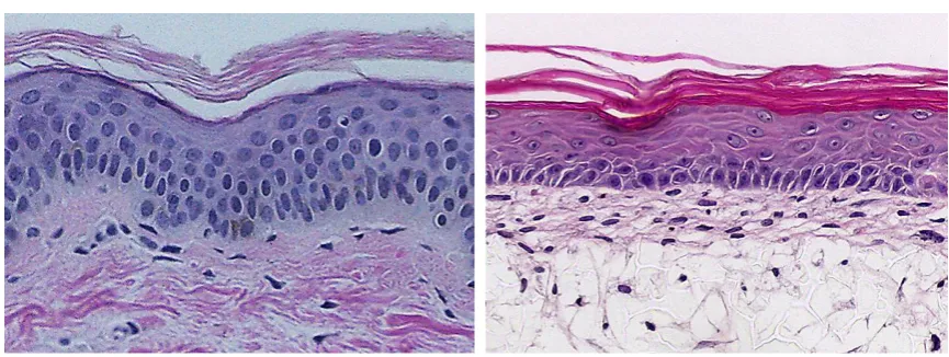
Figure 1: Fibroblasts obtained from biopsies cultured on gelatin coated culture dishes (left) and exposed to epigenetic modification (right). Striking changes in morphology are evident and are accompanied by a decrease in the global DNA methylation.
3
Environmental cues of mechanical nature
In vivo, cells are surrounded by a complex 3-D organization of neighbouring cells and ECM, which interact to provide chemical as well as mechanical stimuli. Integrin and surrounding matrix are not only in charge of ensuring physical attachment, but also convey chemical and mechanical signals from the outer environment (Janmey and McCulloch, 2007), suggesting that the dimension where cells are grown is a key point for the determination of cell fate. Until recently, cell culture has been performed in monolayers, that, although providing useful biological information, lack the ability to reproduce the morphology and 3-D architecture of the original tissue. The benefits of 3-D cell cultures are widely appreciated and more cell-based technologies are now becoming available that enable researchers to preserve the native 3-D structure of cells in vitro, offering many advantages over conventional monolayer culture. First of all, the mechanisms that underline the process of tissue formation, such as migration, proliferation, adhesion, differentiation and apoptosis can be better investigated. In addition, the 3-D environment enables cells to form cell-cell and cell-matrix interaction that may otherwise be precluded in monolayer culture (Baker and Chen, 2012). Deconstructing the elements contributing to the 3-D microenvironment and the associated processes will aid us in better understanding the underlying mechanisms that guide cell growth in vivo.
3.1
Technologies for 3-D cell culture
has strengths and weaknesses, and researchers must select the technology most appropriate for their needs to address the scientific questions that they are interested in.
Aggregate-based technologies promote bringing cells together to create 3-D tissue-like
masses or spheroids, often called ‘micro-tissues’ by exploiting the biophysical properties
acting on the media in which they are grown. Cells grown as 3-D aggregates secrete their own extra-cellular matrix (ECM) and self organise creating multiple cell-cell interactions. Different methods have been developed to generate this type of culture for routine use. The traditional hanging drop approach involves the culture of cells in a drop of medium suspended from the lid of a culture dish. These suspension cultures are adequate for cells that can proliferate in a non-adhesive environment where aggregation is favoured. An alternative strategy involves using attachment-resistant surfaces such as coatings with hydrophilic polymers (Jo and Park, 2000) or micro-patterned surfaces (Yoshii et al., 2011). Both methods inhibit cell adherence and forces cells to float in the medium, stimulating them to coalesce and form spheroids. Regardless of how cell aggregates are formed, these methods make it possible to scale down experiments and work in smaller volumes and are therefore amenable for higher throughput applications. Co-culture of different cells types is also possible, establishing signal rich environments to study the effect of paracrine signalling in real tissue (Torisawa et al., 2009).
Hydrogels differ from solid scaffolds in terms of the strength of physical support. Hydrogels are loose scaffolds consisting of cross-linked natural or synthetic materials within an aqueous environment for cell encapsulation. These highly absorbent matrices are better suited to modelling soft tissues basic of their tissue-like flexibility and viscoelasticity (Tibbit and Anseth, 2009). Hydrogels can be derived from a variety of sources that in turn affect their compatibility and properties. For example, animal-derived hydrogels mainly use collagen, which is the most abundant protein in the ECM. Matrigel® is an example popular commercially available hydrogel composed of tumour extract derived from mouse sarcoma cells. It is known to contain growth factors, a rich protein mix including collagen IV, laminin and entactin and other undefined constituents (Vukicevic et al., 1992). Matrigel® can promote cellular functions that would otherwise be unseen by providing a 3-D microenvironment and the necessary endogenous factors (Benton et al., 2014). The use of defined synthetic hydrogels overcomes some of the issues of animal derived materials such as batch variation and unknown constituents.
have risen as a more consistent alternative. Inert and non-degradable materials such as synthetic polymers can be carefully tweaked to capture the cellular niche, creating scaffolds suitable for cell culture (Bokari et al. 2007; Knight et al. 2011). The lack of biological activity and natural cell adhesion sites can be overcome by coating these substrates with ECM proteins such as laminin and fibronectin (Knight and Przyborski, 2014). Despite providing physical support in the form of 3-D spaces where cells can proliferate, these voids have poor mass transfer since these cultures are static systems. For these reasons, scaffolds are usually engineered as thin membranes (e.g. 200μm) that permit sufficient exchange of nutrients and waste products. This in turn enriches the physiological accuracy of these models allowing researchers to study in vivo phenomena in a controlled in vitro setting.
Advances in technology have led to new opportunities for growing cells in culture and the creation of 3-D tissue-like constructs. This is primarily as a consequence of inter-disciplinary research between cell biology and the biophysical sciences, introducing new materials and methods of manufacture to create platforms tailored to support 3-D cell growth in vitro. The success of these technologies will depend on their adoption, validation and application. The creation of tissue-like constructs in a reliable and reproducible manner according to clearly defined protocols is essential. Moreover, in-depth characterisation of the anatomy and physiology in vitro models alongside their native counterparts will be critical to convincing the scientific community and encourage users to adopt these new approaches (Figure 2).
Figure 2: This figure showcases the potential of 3-D cell culture technology and how it can be
There is no doubt that 3-D cell culture is a rapidly growing and important field of science and requires interdisciplinary research to be innovative and develop. These advances will continue to enable biomedical researchers to recreate the structure and function of human tissues in the lab for research, screening and safety assessment. This technology combined with human stem cell science will open new opportunities for tissue engineering in the lab where renewable sources of human cells can be generated to create robust and reproducible 3-D models of human tissues. Furthermore, to reproduce the conditions in vivo requires many other factors such as oxygen control, perfusion, growth factors, cytokines, hormones, mechanical stiffness, etc. Today we are applying technology to improve current practice, to make incremental advances over existing models, to enable greater insight into biological processes. As technology advances, we will further improve our cell culture models, edging closer to in vivo conditions, but researchers must always remember that it is a model they are studying in the lab and not real tissue. 3-D cell culture takes a big step towards achieving the goal of recreating the growth conditions cells experience in vivo.
Acknowledgments: The authors are members of the COST Actions CA16119. TALB
participates to COST Action CM1406. The authors thank Dr. Elena FM Manzoni for her help in the preparation of this manuscript.
References
Admyre, C., Grunewald, J., Thyberg, J., Gripenbäck, S., Tornling, G., Eklund, A., Scheynius, A., Gabrielsson, S., 2003. Exosomes with major histocompatibility complex class II and co-stimulatory molecules are present in human BAL fluid. Eur Respir J. 22(4):578-83.
Andre, F., Andersen, M., Wolfers, J., Lozier, A., Raposo, G., Serra, V., Ruegg, C., Flament, C., Angevin, E., Amigorena, S., Zitvogel, L., 2001. Exosomes in cancer immunotherapy: preclinical data. Adv Exp Med Biol. 495:349-54.
Baker, M.B., Chen, C.S., 2012. Deconstructing the third dimension – how 3D culture microenvironments alter cellular cues. J Cell Sci. 125(13): 3015–3024.
Benny, P., Badowski, C., Lane, E.B., Raghunath, M., 2016. Improving 2D and 3D Skin In Vitro Models Using Macromolecular Crowding. J Vis Exp. (114).
Benton, G., Arnaoutova, I., George, J., Kleinman, H.K., Koblinski, J., 2014. Matrigel: From discovery and ECM mimicry to assays and models for cancer research. Adv Drug Deliv Rev. 79-80:3-18.
Bokari, M., Carnachan, R., Przyborski, S.A., Cameron, N.R., 2007. Effect of synthesis parameters on emulsion-templated porous polymer formation and evaluation for 3D cell culture scaffolds. J Mater Chem. 17, 4088-4094.
granulosa cells exposed to 5-Azacytidine and addressed toward muscular differentiation. Stem Cell Rev. 10(5):633-42.
Caby, M.P., Lankar, D., Vincendeau-Scherrer, C., Raposo, G., Bonnerot, C., Exosomal-like vesicles are present in human blood plasma. Int Immunol. 17(7):879-87.
Chandrakantan V., Yeola, A., Kwan, J.C., Oliver, R.A., Qiao, Q., Kang, Y.C., Zarzour, P., Beck, D., Boelen, L., Unnikrishnan, A., Villanueva, J.E., Nunez, A.C., Knezevic, K., Palu, C., Nasrallah, R., Carnell, M., Macmillan, A., Whan, R., Yu, Y., Hardy, Philip, Grey, S.T., Gladbach, A., Delerue, F., Ittner, L., Mobbs, R., Walkley, C.R., Purton, L.E., Ward, R.L., Wong, J.W.H., Hesson, L.B., Walsh, W., Pimanda, J.E., 2016. PDGF-AbB and 5-Azacytidine induce conversion of somatic cells into tissue-regenerative multipotent stem cells. Proc Natl Acad Sci U S A. 113: 2306-2315.
Ellis, R.J., 2001. Macromolecular crowding: an important but neglected aspect of the intracellular environment. Curr Opin Struct Biol. 11(1):114-9.
English, A., Azeem, A., Gaspar, D.A., Keane, K., Kumar, P., Keeney, M., Rooney, N., Pandit, A., Zeugolis, D.I., 2012. Preferential cell response to anisotropic electro-spun fibrous scaffolds under tension-free conditions. J Mater Sci Mater Med. 23(1):137-48.
Fevrier, B., Raposo, G., 2004. Exosomes: endosomal-derived vesicles shipping extracellular messages, Curr Opin Cell Biol. 16(4):415-21.
Gerecht, S., Burdick, J.A., Ferreira, L.S., Townsend, S.A., Langer, R., Vunjak-Novakovic, G., 2007. Hyaluronic acid hydrogel for controlled self-renewal and differentiation of human embryonic stem cells. Proc Natl Acad Sci U S A. 104, 11298–11303.
Greening, D.W., Nguyen, H.P., Elgass, K., Simpson, R.J., Salamonsen L.A., 2016. Human Endometrial Exosomes Contain Hormone-Specific Cargo Modulating Trophoblast Adhesive Capacity: Insights into Endometrial-Embryo Interactions. Biol Reprod. 94(2):38.
Heijnen, H.F., Schiel, A.E., Fijnheer, R., Geuze, H.J., Sixma, J.J., 1999. Activated platelets release two types of membrane vesicles: microvesicles by surface shedding and exosomes derived from exocytosis of multivesicular bodies and alpha-granules. Blood. 94(11):3791-9.
Janmey, P.A., McCulloch, C.A., 2007. Cell mechanics: integrating cell responses to mechanical stimuli. Annu Rev Biomed Eng. 9:1-34.
Jo, S., Park, K., 2000. Surface modification using silanated poly(ethylene glycol)s. Biomaterials. 21, 605-615.
Jong, H. S., Yu, J., Luo, D., Shuler, M.L., March, J.C., 2011. Microscale 3-D hydrogel scaffold for biomimetic gastrointestinal (GI) tract model. Lab Chip. 11, 389-392.
Keenan, T. M., Folch, A., 2008. Biomolecular gradients in cell culture systems. Lab Chip. 8(1):34-57
Knight, E., Murray, B., Charnachan, R., Przyborski, S., 2011. Alvetex®: polystyrene scaffold technology for routine three dimensional cell culture. Methods Mol Biol. 695, 323-40.
L’Heureux, N., McAllister, T.N., de la Fuente, L.M., 2007. Tissue-engineered blood vessel for
adult arterial revascularization”, N Engl J Med. 357(14):1451-3.
Lareu, R.R., Subramhanya. K.H., Peng, Y., Benny, P., Chen, C., Wang, Z., Rajagopalan, R., Raghunath, M., 2007. Collagen matrix deposition is dramatically enhanced in vitro when crowded with charged macromolecules: the biological relevance of the excluded volume effect. FEBS Lett. 581(14):2709-14.
Manzoni, E.F., Pennarossa, G., deEguileor, M., Tettamanti, G., Gandolfi, F., Brevini, T.A.L., 2016. 5-azacytidine affects TET2 and histone transcription and reshapes morphology of human skin fibroblasts. Sci Rep. 6:37017.
Mathivanan, S., Lim, J.W., Tauro, B.J., Ji, H., Moritz, R.L., Simpson, R.J., 2010. Proteomics analysis of A33 immunoaffinity-purified exosomes released from the human colon tumor cell line LIM1215 reveals a tissue-specific protein signature. Mol Cell Proteomics. 9(2):197-208.
Mikos, A. G., Sarakinos, G., Leite, S.M., Vacanti, J.P., Langer, R., 1993. Laminated three-dimensional biodegradable foams for use in tissue engineering. Biomaterials. 14, 323-330.
Ostrowski, M.,, Carmo, N.B., Krumeich, S., Fanget, I., Raposo, G., Savina, A., Moita, C.F., Schauer, K., Hume, A.N., Freitas, R.P., Goud, B., Benaroch, P., Hacohen, N., Fukuda, M., Desnos, C., Seabra, M.C., Darchen, F., Amigorena, S., Moita, L.F., Thery, C., 2010. Rab27a and Rab27b control different steps of the exosome secretion pathway. Nat Cell Biol. 12(1):19-30; sup pp 1-13.
Peck, M., Gebhart, D., Dusserre, N., McAllister, T.N., L'Heureux, N., 2012. The evolution of vascular tissue engineering and current state of the art. Cells Tissues Organs. 195(1-2):144-58
Pennarossa, G., Maffei, S., Campagnol, M., Tarantini, L., Gandolfi, F., Brevini, T.A.L., 2013. Brief demethylation step allows the conversion of adult human skin fibroblasts into insulin secreting cells. Proc Natl Acad Sci U S A. 110: 8948-8953
Peters, P.J., Geuze, H.J., Van der Donk, H.A., Slot, J.W., Griffith, J.M., Stam, N.J., Clevers, H.C., Borst, J., 1989. Molecules relevant for T cell-target cell interaction are present in cytolytic granules of human T lymphocytes. Eur J Immunol. 19(8):1469-75.
Pisitkun, T., Shen, R.F., Knepper, M.A., 2004. Identification and proteomic profiling of exosomes in human urine. Proc Natl Acad Sci U S A. 101(36):13368-73.
Raposo, G., Tenza, D., Mecheri, S., Peronet, R., Bonnerot, C., Desaymard, C., 1997. Accumulation of major histocompatibility complex class II molecules in mast cell secretory granules and their release upon degranulation. Mol Biol Cell. 8(12):2631-45.
Taylor, S.M., Jones, P.A., 1979. Multiple new phenotypes induced in 10T1/2 and 3T3 cells treated with 5-azacitydine. Cell. 17:771-9
Torisawa, Y.-S., Mosadegh, B., Luker, G.D., Morell, M., O’shea, K.S., Takayama, S., 2009. Microfluidic Hydrodynamic Cellular Patterning For Systematic Formation Of Co-Culture Spheroids. Integr Biol. 1, 649-645.
Vukicevic, S., Kleinman, H.K., Luyten, F.P., Roberts, A.B., Roche, N.S., Reddi, A.H., 1992. Identification of multiple active growth factors in basement membrane matrigel suggests caution in interpretation of cellular activity related to extracellular matrix components. Exp Cell Res. 202, 1-8.
Wydooghe, E., Vandaele, L., Heras, S., De Sutter, P., Deforce, D., Peelman, L., De Schauwer, C., Van Soom, A., 2015. Autocrine embryotropins revisited: how do embryos communicate with each other in vitro when cultured in groups?. Biol Rev Camb Philos Soc. 92 (1), 505-520.
Yoshii, Y., Waki, A., Yoshida, K., Kakezuka, A., Kobayashi, M., Namiki, H., Kuroda, Y., Kiyono, Y., Yoshii, H., Furukawa, T., Asai, T., Okazawa, H., Gelovani, J.G., Fujibayashi, Y., 2011. The use of nanoimprinted scaffolds as 3D culture models to facilitate spontaneous tumor cell migration and well-regulated spheroid formation. Biomaterials. 32, 6052–6058.
Zimmerman, S.B., Harrison, B., 1987. Macromolecular crowding increases binding of DNA polymerase to DNA: an adaptive effect. Proc Natl Acad Sci U S A. 84(7):1871-5.