Z
UZANNAD
RULIS−K
AWA1, 2, A
RTURB
EDNARKIEWICZ3, G
ABRIELAB
UGLA1,
W
IESŁAWS
TRĘK3, W
ŁODZIMIERZD
OROSZKIEWICZ1Bactericidal Effects of the Fotolon (Chlorin e6)
on Gram−Negative and Gram−Positive Strains Isolated
from Wound Infections
Bakteriobójczy wpływ Fotolonu (chlorinu e6) na Gram−ujemne
i Gram−dodatnie szczepy izolowane z zakażonych ran
1 Institute of Genetics and Microbiology, University of Wroclaw, Poland 2 Korczak Children’s Hospital in Wroclaw, Poland
3 Institute of Low Temperature and Structure Research, Polish Academy of Sciences, Wroclaw, Poland Adv Clin Exp Med 2006, 15, 2, 279–283
ISSN 1230−025X
ORIGINAL PAPERS
Abstract
Background. Surgical site infections and diabetes foot infections are the very serious therapeutic problem. Because of the alarming bacterial drug resistant level and difficulty in wound infections treatment, an alternative or supportive bactericidal cure have to be developed. One of them is Photo Dynamic Inactivation (PDI) of bacter− ial strains.
Objectives. Test and discussion of the PDI efficiency using the Fotolon (chlorin e6) against Gram−positive and Gram−negative standard and wild type strains.
Material and Methods. Eight clinical isolates of Pseudomonas sp. and one clinical strain ofAcinetobacter bau− maniiwere tested. As references the authors used control strains from American Type Culture Collection: Gram− positive: Staphylococcus aureus ATCC 29213, and Gram−negative: Klebsiella pneumoniae ATCC 700601,
Escherichia coliATCC 25922 and Pseudomonas aeruginosaATCC 27853. PDI procedure were carried out using photosensitive compound – the Fotolon (chlorin e6) and laser light.
Results.For S. aureusstrain fivefold decrease of colony forming units was obtained. For other control bacteria only twice decrease level was observed. The P. putidaand P. stutzeriisolates were as sensitive as Gram−positive
S. aureus, what is very interesting phenomenon. The remaining Gram−negative strains showed 0.5–2 log of cfu decrease. The authors found that high concentration of the Fotolon in short incubation time gives smaller bacteri− cidal effect than lower concentration in long incubation period.
Conclusions.Both Gram−positive and Gram−negative types of bacteria were sensitive to PDI with Fotolon, how− ever different PDI susceptibility of clinical isolates were observed (Adv Clin Exp Med 2006, 15, 2, 279–283).
Key words:photodynamic inactivation, Fotolon (chlorin e6), Gram−positive bacteria, Gram−negative bacteria.
Streszczenie
Wprowadzenie.Zakażenia ran pooperacyjnych oraz infekcje związane ze „stopą cukrzycową” są bardzo poważ− nym problemem terapeutycznym. Ze względu na alarmujący wzrost oporności drobnoustrojów na antybiotyki oraz trudności w leczeniu zakażeń ran istnieje konieczność rozwoju nowych alternatywnych lub wspomagających me− tod leczniczych. Jedną z nich może być fotodynamiczna inaktywacja (PDI) szczepów bakteryjnych.
Cel.Przebadanie skuteczności PDI w stosunku do Gram−dodatnich i Gram−ujemnych szczepów bakteryjnych, za− równo standardowych, jak i dzikich.
Materiały i metody.Przetestowano sześć klinicznych szczepów Pseudomonassp. i jeden kliniczny izolat Acine− tobacter baumanii. Jako kontrole użyto szczepy z kolekcji ATCC (American Type Culture Collection): Gram−do− datni Staphylococcus aureusATCC 29213 oraz Gram−ujemne: Klebsiella pneumoniaeATCC 700601, Escherichia coliATCC 25922 iPseudomonas aeruginosaATCC 27853. Procedurę fotodynamicznej inaktywacji przeprowa− dzono za pomocą Fotolonu (chlorinu e6) i światła laserowego.
The wound infections, especially surgical site infections and diabetes foot infections are the very serious therapeutic problem. They account for 14–16% of the estimated 2 million nosocomial infections affecting hospitalized patients in the United States [1]. The World Health Organization demonstrates a prevalence of these infections varying from 5 to 34% of total nosocomial infec− tions [2].
The most common bacteria isolated from the wound infections belong to [3–5]: Gram−positive:
Staphylococcus aureus, Staphylococcus coagu− lase−negative, Streptococcus sp., Enterococcus
sp.; Gram−negative: Enterobacteriaceae (Escheri− chia coli,Enterobacter sp., Klebsiella sp., Proteus
sp.) and Pseudomonadales (Pseudomonas aeru− ginosa, Acinetobactersp.).
The bacteria causing the hospital−acquired in− fections are in most cases multidrug resistant [4–6]. From the Gram−positive bacteria there are isolated methicillin resistant Staphylococcus aureus (MRSA) [5–7] and vancomycin resistant enterococci (VRE) [5, 6, 8]. The Pseudomo− nadales are natural resistant to many antibiotics and chemotherapeutics and nosocomial isolates could be unsusceptible to almost all active antibi− otics [9–12]. Because of this alarming resistance level and difficulty in wound infections treatment, an alternative or supportive bactericidal cure have to be developed. One of them is Photo Dynamic Inactivation (PDI) of bacterial strains.
The photodynamic therapy of bacterial infec− tions starts with topical administration of PS. Owing to the fact, the time needed to bind large PS molecules (molecular weight ~18 500) to the bac− teria cells is relatively short (up to 30 minutes) in comparison with eukaryotic cells, the PDI show good selectivity for bacteria [13, 14].
Next the PS is excited with light of proper wavelength and proper fluency, to initiate cyto− toxic damage to the surrounding living structures by highly reactive species generation (e.g. single oxygen, hydroxyl radicals, etc.).
The PDI itself is regarded as efficient and non− recovering antimicrobial therapeutic procedure against Gram−positive bacteria. Moreover, its activity is independent of the antibiotic sensitivity spectrum of the treated pathogen [15, 16].
The photodynamic activity of photosensitizer (PS) used in PDT (photodynamic therapy) is depen− dent on their hydrophobic or hydrophilic properties accountable to their side chains on the tetra−pyrrole ring, as well as to the structure and chemical com− position of the cellular targets like proteins or lipids. Whereas traditional photosensitizes (non−charged) have shown to be effective in Gram−positive bacte− ria inactivation, structural features (outer mem− brane) occurring in the Gram−negative bacteria cre− ates chemical and physical barrier which does not allow to photo−inactivate these bacteria.
There are three approaches to extend antimicro− bial PDI effectiveness towards Gram−negative bac− teria, e.g. 1) destabilization of the outer membrane structure making it permeable to photosensitizes molecules [17–20], 2) application of the positively charged phthalocyanine photosensitizers [21–24], which electro−statically bind to negatively charged outer membrane of the bacteria, 3) the use of specif− ic antibody−linked PS to increase the binding selec− tivity to Gram−negative bacteria covers [25, 26].
The first approach combines PDI treatment with small peptide polymyxin−B application. Also other compounds like Tris/EDTA together with non− cationic photosensitiser were used to increase effec− tiveness. The second approach utilizes poly−L−lysine (non−bactericidial polymer) and chlorin e6 conju− gates with different length of the lysine chain. The PS modified towards polycationic character binds to anionic sites of lipopolysaccharide (LPS) – the com− ponent of the outer membrane. This binding is regarded to weaken the intermolecular interaction of the LPS, disorganize its structure leading to increased permeability for drugs to reach the cyto− plasmic membrane.
In the study the authors have tested and dis− cussed the Photo Dynamic Inactivation (PDI) effi− ciency of the Fotolon against Gram−positive and Gram−negative standard and wild bacteria strains.
Material and Methods
Microorganisms
In the study the authors have used the clinical strains isolated from wound infections of the
ły tak samo wrażliwe na PDI jak Gram−dodatni S. aureus, co jest bardzo interesującym zjawiskiem. Pozostałe szczepy Gram−ujemne wykazywały spadek cfu w granicach 0,5–2 log. Stwierdzono, że wysokie stężenie Fotolo− nu zastosowane przy krótkim czasie inkubacji charakteryzowało się mniejszym efektem bakteriobójczym niż ni− skie stężenie aplikowane przez długi czas.
Wnioski.Zarówno bakterie Gram−dodatnie, jak i Gram−ujemne były wrażliwe na PDI z zastosowaniem Fotolonu, szczepy kliniczne jednak wykazywały zróżnicowaną wrażliwość (Adv Clin Exp Med 2006, 15, 2, 279–283).
patients hospitalized in surgical wards in Wroclaw. The bacteria belonging to Pseudomonadales were used as the appropriate experimental strains. These were: Pseudomonas aeruginosa – 6 strains,
Pseudomonas stutzeri – 1 strain, Pseudomonas putida – 1 strain, Acinetobacter baumanii – 1 strain. As references the authors used control strains from American Type Culture Collection: Gram−positive: Staphylococcus aureus ATCC 29213, and Gram−negative: Klebsiella pneumoni− aeATCC 700601, Escherichia coli ATCC 25922 and Pseudomonas aeruginosaATCC 27853.
Photosensitizer
The authors used the Fotolon containing chlo− rin e6 (Ce6) and PVP polyvinylpyrrolidone dis− solved in 0.9% NaCl solution (PF). The ratio of mass fractions of chlorin e6 and PVP is 1 : 1. The PVP polymer does not influence spectral proper− ties of Fotolon in comparison to pure chlorin e6, however Ce6 builds into the hydrophobic part of the PVP, leading to lack of photosensitizer agglomeration [27]. Photo bleaching of the Fotolon was experimentally examined and it was determined, that in PF solution it looses 80% of its activity with 30 J/cm2 light dose delivered.
Moreover it was determined from absorption spec− tra that due to polymerization of e6 molecules, PF solution of Fotolon looses its activity after several days from the day of preparation, leading to decrease in effective photo activity of PS concen− tration. Therefore the authors used PS solution prepared just before the experiments.
Light Source
As an excitation source, pigtailed laser diode was used (LaserSecura, Poland) emitting at 650 nm matching exactly Q absorption band’s maxi− mum of chlorin e6 (Fotolon) in phosphate buffer (0.9% NaCl). The plate was fixed about 4 cm above the laser fiber tip and power meter (Coherent, Field Master) equipped with measuring head (Coherent, LM−10) shielded with 0.5 cm2
diaphragm was used to obtain 200 mW/cm2light
flux. The surface of the plastic plate reflected around 12% of light, however the 96−well plates were covered with the same kind of plastic reflect− ing 12% of the laser light back to the well. Thus 200 mW/cm2light flux is assumed to be preserved
during all experiments. The total light dose deliv− ered was then 120 J/cm2, which is in excess
enough to photo activating all e6 molecules. In
fact, 2.5−minute lasting treatment procedure could have been used to obtain the same results.
Experimental procedure
Two kinds of experiments were carried out: 1) 50 µg/ml chlorin e6 dose with long (120 min) incubation time with bacterial culture before illu− mination, and 2) 500 µg/ml chlorin e6 dose with shorter (30 min) incubation time with bacterial culture before illumination. Bacteria were incubat− ed in the dark at room temperature. The volume of 100 µl of bacterial culture in concentration 106
cells/ml diluted in sterile phosphate buffer (PBS) was placed into the wells of a flat bottom 96−well microtitration plate in triplicate and illuminated. To determine colony forming units (cfu) the aliquots 10 µL were taken from each well, serially diluted, placed on Mueller Hinton Agar (MHA) and incubated for 24 h at 37°C in the dark. The controls were bacteria untreated with PS or light, bacteria exposed to the light in the absence of PS and bacteria treated with PS but not illuminated. The cfu of the last two control groups, exhibited no changes of cfu in comparison to no PS – no light control group.
Results
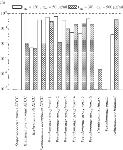
The reduction of colony forming units of the reference and clinical strains is presented on Figure 1. The presented data are normalized to the respective (no PS, no light) control group cfu, to easily compare different strains.
In control group the authors have obtained fivefold decrease of colony forming units of S. aureus. It is well known that chlorin e6 has a good activity on Gram−positive bacteria. The next three control strains were more susceptible to the higher concentration of photosensitizer, and only about twice decrease level was observed.
The clinical strains behavior could not be pre− dicted from Gram−positive or Gram−negative char− acter of the bacteria. The Pseudomonas putidaand
Pseudomonas stutzeriisolates were as sensitive as Gram−positive S. aureus, what is very interesting. The other strains showed various susceptibility to PDI of the Fotolon. It held within 0.5–2 orders decrease of cfu.
The mechanisms of Pseudomonas aeruginosa
resistance to carbapenems are well known [9–12]. These are 1) drug inactivation through out of enzymes activity (carbapenemases), 2) target alter− nation – is not detected, 3) prevention of drug
influx correlated with loss of membrane protein called OprD porin and 4) active extrusion of drug from the cell by efflux pump system.
Studied Pseudomonas aeruginosastrains No 4 and No 5 exhibited the reduced number of porin OprD. The MIC (minimum inhibitory concentration) of imipenem for these strains makes 16–32 µg/ml (low level resistance). This means that the en− trance of the drug into the cell is limited.
In studied mutant isolates the authors have small number of this channel, so when the chlorin e6 use this porin for influx, one will see that lower concentration of photosensitizes but longer incu− bation time will be more effective than higher con− centration acting during short time as this was the result of conducted experiment. That allows the authors to suppose that OprD is most probably responsible for influx of chlorin e6 into the
Pseudomonas aeruginosacell.
Both types of bacteria Gram−positive and Gram−negative were sensitive to PDI with Foto− lon, however different PDI susceptibility of clini− cal isolates was observed. Larger clinical strain collections should then be verified for therapeuti− cal application of Fotolon in PDI. Moreover, due to non−specific PDI action against bacterial and patient cells, possibly short times of incubation the bacteria with the PS compound should be applied. Thus modifications to the chlorin e6 compound or other photosensitiser are expected to increase the antimicrobial efficiency. Nevertheless it was pos− tulated that OprD was probably responsible for influx of chlorin e6 into the Pseudomonas aerugi− nosa cell. Since the photodynamic antimicrobial inactivation mechanism is not precisely known, the last conclusion seems to be extremely impor− tant for further PDI efficiency enhancement.
References
[1] Emori TG, Gaynes RP:An overview of nosocomial infections, including the role of microbiology laboratory. Clin Microb Rev 1993, 4, 428–442.
[2] Mayon−White RT, Ducel G, Kereselidze T:An international survey of the prevalence of hospital−aquired infec− tion. J Hosp Infect 1988, 2, 11 Suppl A: 43–48.
[3] NNIS System: National Nosocomial Infections Surveillance (NNIS) report, data summary from October 1986–April 1996, issued May 1996. A report from the National Nosocomial Infections Surveillance (NNIS) System. Am J Infect Control 1996, 5, 380–388.
[4] Revathi G, Puri J, Jain BK:Bacteriology of burns. Burns 1998, 4, 347–359.
[5] Siami G, Christou N, Eiseman I, Tack KJ and the Clinafloxacin Severe Skin And Soft Tissue Infections Study Group:Clinafloxacin versus Piperacillin−Tazobactam in Treatment of Patients with Severe Skin and Soft Tissue Infections. Antimicrob. Agents Chemother 2001, 45, 525–531.
[6] Carmeli Y, Castro J, Eliopoulos GM, Samore MH: Clinical Isolation and Resistance Patterns of and
Superinfection with 10 Nosocomial Pathogens after Treatment with Ceftriaxone versus Ampicillin−Sulbactam. Antimicrob. Agents Chemother 2001, 45, 275–279.
[7] Bell JM, Turnidge JD and SENTRY APAC Participants: High Prevalence of Oxacillin−Resistant Staphy−
lococcus aureusIsolates from Hospitalized Patients in Asia−Pacific and South Africa: Results from SENTRY Antimicrobial Surveillance Program, 1998–1999. Antimicrob. Agents Chemother 2002, 46, 879–881.
[8] Harbarth S, Cosgrove S, Carmeli Y: Effects of Antibiotics on Nosocomial Epidemiology of Vancomycin−
Resistant Enterococci. Antimicrob. Agents Chemother 2002, 46, 1619–1628.
Fig. 1. Cfu parameter for all strains examined within the two kinds of experiments. The data are normalized to the cfu of respective control group: tinc– incubation
time; ce6– chlorin e6; cfu – colony forming units
Ryc. 1. Wskaźnik cfu dla wszystkich badanych szcze− pów w dwóch rodzajach eksperymentów. Dane zostaly znormalizowane w stosunku do cfu odpowiedniej gru− py kontrolnej: tinc– czas inkubacji; ce6– chlorin e6;
cfu – jednostki tworzące kolonie
inc
t = 120', c = 50 µg/ml cfu
Staphylococcus aur
eus
Klebsiella pneumoniae
Escherichia coli
Pseudomonas aeruginosa
Pseudomonas aeruginosa
A
TCC
A
TCC
A
TCC
A
TCC
1
Pseudomonas aeruginosa Pseudomonas aeruginosa Pseudomonas aeruginosa Pseudomonas aeruginosa Pseudomonas aeruginosa
2 3 4 5 6
Pseudomonas stutzeri Pseudomonas putida
Acinetobacter baumani
e6
10−5 10−4 10−3 10−2 10−1 100
inc
[9] Livermore DM, Woodford N:Carbapenemases: a problem in waiting? Curr Opin Microbiol 2000, 3, 489–495.
[10] Livermore DM:Of Pseudomonas, porins, pumps and carbapenems. J Antimicrob Chemother 2001, 47, 247–250.
[11] Quinn JP, Dudek EJ, DiVincenzo CA, Lucks DA, Lerner SA:Emergence of resistance to imipenem during
therapy for Pseudomonas aeruginosainfections. J Infect Dis 1986, 154, 289–294.
[12] Senda K, Arakawa Y, Nakashima K, Ito H, Ichiyama S, Shimokata K, Kato N, Ohta M:Multifocal outbreaks of metallo−—lactamase−producing Pseudomonas aeruginosaresistant to broad−spectrum β−lactams, including car− bapenems. Antimicrob Agents Chemother 1996, 40, 349–353.
[13] Hamblin MR, Zahra T, Contag CH, McManus AT, Hasan T:Optical monitoring and treatment of potentially
lethal wound infections in vivo.J Infect Dis 2003, 187, 1717–1725.
[14] Soukos NS, Wilson M, Burns T, Speight PM:Photodynamic effects of toluidine blue on human oral ker−
atinocytes and fibroblasts and Streptococcus sanguisevaluated in vitro. Lasers Surg Med 1996, 18, 253–259.
[15] Embleton ML, Sean PN, Cookson BD, Wilson M: Selective lethal photosensitization of methicillin−resistant
Staphylococcus aureususing an IgG−tin (IV) chlorin e6 conjugate. J Antimicrob Chemother 2002, 50, 857–864.
[16] Soncin M, Fabris C, Busetti A, Dei D, Nistri D, Roncucci G, Jori G:Approaches to selectivity in the Zn(II)− phthalocyanine−photosensitized inactivation of wild−type and antibiotic−resistant Staphylococcus aureus.
Photochem Photobiol Sci 2002, 10, 815–919.
[17] Ehrenberg B, Malik Z, Nitzan Y: Fluorescence spectral changes of hematoporphyrin derivative upon binding to lipid vesicles Staphylococcus aureusand Escherichia colicells. Photochem Photobiol 1985, 41, 429–435.
[18] Malik Z, Ladan H, Nitzan Y: Photodynamic inactivation of Gram−negative bacteria Problems and possible solu− tions. J Photobiol Photochem B: Biol 1992, 1, 262–266.
[19] Bertoloni G, Rossi F, Valduga G, Jori G, van Lier J:Photosensitiseing activity of water− and lipid−soluable phthalocyanines on Escherichia coli.FEMS Microbiol Letters 1990, 59, 149–55.
[20] Nitzan Y, Malik Z, Ehrenberg B:Photosensitization of microbial cells. In Photobiology: the Science and its Applications. Eds.: Riklis E. Plenum Press, New York, 815–820.
[21] Merchat M, Bertoloni G, Giacomoni P, Villanueva A, Jori G:Meso−substituted cationic porphyrins as efficient photosensitizers of Gram−positive and Gram−negative bacteria. J Photochem Photobiol 1996, 32, 153–157.
[22] Hamblin MR, O’Donnell DA, Murthy N, Rajagopalan K, Michaud N, Sherwood ME, HasanT:Polycationic
photosensitiser conjugates: effects of chain length and Gram classification on the photodynamic inactivation of bacteria. J Antimicrob Chemother 2002, 49, 941–951.
[23] Minnock A, Vernon DI, Schofield J, Griffiths J, Parish JH, Brown ST:Photoinactivation of bacteria. Use of a cationic water−soluble zinc phthalocyanine to photoinactivate both Gram−negative and Gram−positive bacteria. J Photochem Photobiol B: Biol 1996, 32, 159–164.
[24] Vaara M:Agents that increase the permeability of the outer membrane. Microbiol Rev 1992, 56, 395–411.
[25] Berthiaume F, Reiken SR, Toner M, Tompkins RG, Yarmush ML:Antibody−targeted photolysis of bacteria in vivo.Bio−Technology 1994, 12, 703–706.
[26] Friedberg JS, Tompkins RG, Rakestraw SL, Warren SW, Fischman AJ, Yarmush ML:Antibody−targeted
photolysis. Bacteriocidal effects of Sn (IV) chlorin e6−dextran−monoclonal antibody conjugates. Ann NY Acad Sci 1991, 618, 383–393.
[27] Parkhots MV, Knyukshto VN, Isakov GA, Petrov PT, Lepeshkevich SV, Khairullina AY, Dzhagarov BA:
Spectral−luminescent studies of the “photolon” photosensitizer in model media and in blood of oncological patients. J App Spectr 2003, 70, 921–926.
Address for correspondence:
Zuzanna Drulis−Kawa
Institute of Genetic and Microbiology, University of Wroclaw Przybyszewskiego 63/77
51−148 Wroclaw Poland
Tel./fax: 0048 071 325 21 51 e−mail: [email protected]
Conflict of interest: None declared
Received: 8.07.2005 Revised: 12.08.2005 Accepted: 29.09.2005
Praca wpłynęła do Redakcji: 8.07.2005 r. Po recenzji: 12.08.2005 r.