This is an open access journal, and articles are distributed under the terms of the Creative Commons Attribution-Non Commercial-ShareAlike 4.0 License, which allows others to remix, tweak, and build upon the work non-commercially, as long as appropriate credit is given and the new creations are licensed under the identical terms.
© 2017 Journal of Advanced Pharmacy Education & Research | Published by SPER Publication
464
Prevalence of Methicillin-Resistant Staphylococcus Aureus
Colonization in Children Admitted to Motahari Hospital of
Urmia
Ebrahim Sadeghi
1, Amir Nasimfar
1*, Maliheh Haghir Madadi
21Department of pediatric, Urmia university of medical sciences. Urmia, IR Iran, 2Urmia university of medical sciences. Urmia, IR Iran.
Correspondence: Amir Nasimfar, Department of pediatric, Urmia university of medical sciences. Urmia, IR Iran, E_mail:Amir [email protected]
ABSTRACT
Background and Objective: Staphylococcus aureus is an important bacteria associated with a range of infectious diseases in children and adults. Over-prescription of antibiotics effective in this bacterium in recent years has caused a special type of this species, called methicillin-resistant staphylococcus aureus (MRSA). It is considered as major problem to health systems around the world. The research was conducted to evaluate the prevalence of MRSA colonization in patients admitted to the infectious ward of educational-medical center of Shahid Motahari Hospital of Urmia. Methodology: This research is a cross-sectional analytical study. Samples were prepared from the anterior part of the nose and they were cultured on Mannitol-Celtic-Agar culture medium (Tian Biotech, India) after placing them in a Stuart transport medium. Samples were examined by microbiologist after incubation. Yellow grown colonies indicated the mannitol fermentation and they were selected for catalase and coagulase testing. Clones confirmed by these tests were isolated as staphylococcus aureus to measure the susceptibility to methicillin. SPSS version 21 software was used for statistical analysis. Results: The results of this research revealed that staphylococcus is a resistant and growing bacterium and it is still found in treatment centers. Antibiotic use has a significant relationship with positive culture results of MSSA (methicillin-susceptible staphylococcus aureus) and MRSA (methicillin-resistant staphylococcus aureus). Screening the children is required in terms of type of antibiotic use. Conclusion: Most of children admitted to the medical centers are often carriers of Staphylococcus aureus bacterium. Thus, screening the children in terms of Staphylococcus aureus is necessary by care centers in community and educational centers (schools, kindergartens).
Keywords: MRSA،MSSA, staphylococcus aureus, infectious disease, pediatrics
Introduction
Staphylococcus aureus bacterium is an environmental organism found everywhere and its colonization is often seen in the nose and skin. Staphylococcus aureus bacterium is considered as one of the main causes of infectious diseases in hospitals around the world. It is transmitted by resistant strains of this bacterium (MRSA) to common antibiotics, such as penicillins and cephalosporins [1]. Hospital spread of these microorganisms is
higher in specific wards such as surgery, ICU, nursing homes and burn wards [2]. Staphylococcus aureus diagnosis in the
laboratory is feasible through production of specific golden colonies, β-hemolysis production in blood agar and the similar gram-positive appearance of grape cluster in hot dyeing [3].
MRSA genome (methicillin-resistant staphylococcus aureus) can be usually distinguished from MSSA (methicillin- susceptible staphylococcus aureus) with the presence of the mecA gene or its homologous MecC [4]. Genome sequence analysis to diagnose the presence of mecA gene predicts the resistant phenotype with high accuracy [5, 6]. An important sign
of infection with this bacterium is the formation of an abscess, which might be associated with involvement of the lymph nodes. Invasive infections might involve lungs, liver, bones, joints, kidneys, endocardium and foreign objects, considered as life threatening [7].
Methicillin-resistant staphylococcus aureus (MRSA) was first recognized in 1959 and in 1989 in the United States. Since that time, its prevalence has been reported throughout of world. In the mid-1970s, gentamicin-resistant and methicillin-resistant staphylococcus and more than 20 other antimicrobials were reported. In the same decade, methicillin-resistant staphylococcus aureus was introduced as an important hospital pathogen [8]. Given the results of previous studies, the
Access this article online
Website: www.japer.in E-ISSN: 2249-3379
How to cite this article:Ebrahim Sadeghi, Amir Nasimfar, Maliheh Haghir Madadi. Prevalence of Methicillin-Resistant Staphylococcus Aureus Colonization in Children Admitted to Motahari Hospital of Urmia. J Adv Pharm Edu Res 2017;7(4):464-468.
Journal of Advanced Pharmacy Education & Research | Oct-Dec 2017 | Vol 7 | Issue4 465
prevalence of colonization with MRSA in admitted patients is more than 70% in Asian countries such as China, Korea and Taiwan, more than 50% in North America, 20% in Europe, and about 50% in Iran [9, 10]. Based on the results of these
studies, it seems that there are major geographical and racial differences in the prevalence of colonization with MRSA. It reflects the needs to conduct further studies in this area [11, 12].
Some studies have indicated that the prevalence of MRSA in infants and children is higher and the clinical outcomes associated with it are worse. In their studies, Wu et al. reported Pyogenic liver abscess, which its cause is staphylococcus aureus. It is considered a rare disease in children [13-26]. In addition, it has been shown that MRSA
infections have increased in children in recent years [14, 26-32].
The frequency of MRSA colonization was examined in the pediatric infectious ward of educational-medical center of Shahid Motahari Hospital in Urmia.
Methodology
This research was a cross-sectional study conducted on 200 children (115 male children (57.5%) and 85 female children (42.5%) admitted to Shahid Motahari Hospital in Urmia. Demographic information including age, gender and other variables including family size, parent job, history and type of disease, cause of current hospitalization, history and duration of previous hospitalization, history of receiving antibiotic in the last 6 months and its type, history of previous surgery, history of attending in kindergarten or elementary school and the history of contact with health care providers were recorded through asking question from parents. The samples were prepared by sterile cotton swabs from the anterior part of the nose admission time and before hospitalization, and before discharge from the hospital. Then, they were transferred to Microbiology Department of Shahid Motahari Hospital in Urmia after placing them in the Stuart transport medium, and they were cultured on Mannitol-Salt-Agar culture medium (Tian Biotech, India). Samples were examined by microbiologist after incubation for 24-48 hours at 35 ° C. Yellow grown colonies indicate mannitol fermentation and they were selected for catalase and coagulase testing [13].
Colonies confirmed as staphylococcus aureus were isolated for measuring the methicillin susceptibility. For this purpose, Mannitol-Salt-Agar culture medium (Tian Biotech, India) and a 1-microgram Oxacillin antibody medicine disc [14] were used.
For all staphylococcus aureus collected, a microfibial suspension of 0.5% McFarland was prepared from 18 to 24-hour colonies. Then, a sterile swab was inserted into a bacterial suspension and after withdrawing its excessive water, it was cultured on the whole surface of the Muller-Hinton-Agar medium. After 15 minutes, 1-microgram oxacillin disc was placed in the plate. Then, the plates were placed at 35 ° C for 18-24 hours. Non-growth zone diameter was measured by mm ruler and the results were interpreted using the CLSI guideline. Non-growth zone less than or equal to 10 mm was considered as an oxacillin-resistant strain. It should be noted that this disc was used instead of methicillin disc due to its unavailability. SPSS version 21 software was used for statistical analysis. Quantitative data were presented in mean, standard deviation, minimum and maximum, and qualitative data were presented as frequency and percentage .
Results
In this research, 200 children (115 male (57.5%) and 85 female (42.5%)) were divided into three age groups, including 1-5 years group, 5-10 years group, and 11-15 years group. Out of these 200 children, 99 (49.5%) were placed in 1-5 years group, 73 (36.5%) were placed in the -10 years group, and 28 (14%) were placed in the 10-14 years group. The minimum age for these children was 2 months and the maximum age of them was 14 years. Out of 99 children in the 1-5 years age group, 57 (57.5%) were female and 42 (42.5%) were female. Out of 73 children in the 5-10 years age group, 41 (56.1%) were male and 32 (% 43.9) were female, and out of 28 children in 10-15 age group, 17 (60.7%) were female, and 11 (39.3%) were female (Table 1).
Table 1: Distribution of absolute and relative frequency of age groups in children studied
Age group gender Total
male female
1-5 years )57.5 %(57 )42.5 %(42 99
5-10 years )56.1 %(41 )43.9 %(32 73
10-15 years )60.7 %(17 )39.3 %(11 28
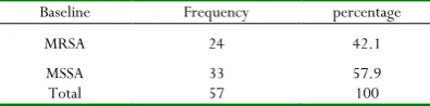
Out of 200 patients studied, 57 (28.5%) patients were positive in terms of Staphylococcus aureus at baseline and 143 (71.5%) were negative. Out of 57 children who were positive in terms of Staphylococcus aureus at baseline, 34 children (59.6%) were female and 23 (40.4%) were female, which 33 children (57.9%) were in the 1-5 years age group, 15 children (% 26.3) were in the of 5-10 years age group, and 9 children (15.8%) were in the 10-15 years age group. In addition, out of these 57 children, 24 children (42.1%) were methicillin-resistant (MRSA), and 33 children (57.9%) were methicillin- susceptible (MSSA) (Table 2).
Table 2: Distribution of absolute and relative frequency of MRSA and MSSA in positive children
at baseline
Baseline Frequency percentage
MRSA 24 42.1
MSSA 33 57.9
Total 57 100
Out of 33 MSSA children at baseline, 19 (57.57%) were male and 14 (42.43%) were girls. Out of these 19 male children, 12 children (63.2%) had non risk factor, 2 children (10.5%) had two risk factors, and 5 children (26.3%) had one risk factor. Out of these 14 children, 9 children (64.3%) had no risk factor, 2 children (14.3%) had 2 risk factors, (and 3 children (21.4%0 had one risk factor. Based on Chi-square test, no relationship was found between gender of patients and risk factors for MSSA (P = 0.9).
466 Journal of Advanced Pharmacy Education & Research | Oct-Dec 2017 | Vol 7 | Issue4
Table 3: Distribution of absolute and relative frequency of risk factor based on the gender of MRSA children
baseline Risk factor Total
No risk factor Two risk factors One risk factor
male )26.7 %(4 )46.7 %(7 )26.7 %(4 15
female )33.3 %(3 )44.4 %(4 )22.2 %(2 9
total )29.2 %(7 )45.8 %(11 )25 %(6 24
Moreover, based on the results obtained and based on Chi-square statistical test, no significant relationship was found between the gender of the patients and the result of the culture (P = 0.4). Out of 33 MSSA patients who had positive test results at baseline, 18 people (54.5%) were in the age group of 1-5 years, 10 children (30.3%) were in the age group of 5-10 years, and 5 children (15.2%) were in the age group of 10-15 years, and out of 24 patients with MRSA at baseline, 15 patients (62.5%) were in the age group of 1-5 years, 6 patients (25%) were in the age group of 5-10 years, and 3 patients (12.5%) were in the age group of 10-15 years. Based on Chi-square test, no significant relationship was found between the gender of the patients and the positive result of the sample (P = 0.8).
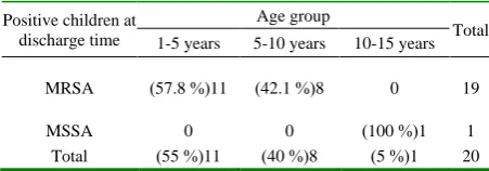
Out of 33 MSSA patients, 21 patients (63.6%) had no risk factor, 4 patients (12.1%) had two risk factors, and 8 patients (24.2%) had one risk factor, and out of 24 MRSA patients, 7 patients (29.2%) had no risk factor, 11 patients (45.8%) had two risk factors, and 6 patients (25%) had one risk factor. Based on Chi-square test, there is a significant relationship between risk factor and MSSA and MRSA in children (P = 0.03). The patients were also studied the discharge time. Out of 57 children who were positive in terms of staphylococcus bacteria, 20 patients (35.1%) were positive at the discharge time, but the test results of 37 patients (64.9%) were negative. Out of these 20 patients with positive staphylococcus bacterium at the discharge time, 19 patients had MRSA (95%) and 1 patient (5%) had MSSA. Out of 19 MRSA children at discharge time, 11 patients (57.8%) were in the age group of 5-10 years and 8 patients (42.1%) were in the age group of 10-15 years. One patient with MSSA at the discharge time was in the age group of 10 -15 years. The results showed no relationship between the age group of patients and the result of culture at the discharge time (P = 0.4) (Table 4).
Table 4: Distribution of absolute and relative frequency of MRSA and MSSA at discharge time
Positive children at discharge time
Age group
Total
1-5 years 5-10 years 10-15 years
MRSA )57.8 %(11 )42.1 %(8 0 19
MSSA 0 0 )100 %(1 1
Total )55 %(11 )40 %(8 )5 %(1 20
Out of 143 children who were negative at the baseline in terms of staphylococcus bacterium, the results of 31 (21.6%) of them were positive at the discharge time, that 29 (93.5%) of them had MRSA and 2 of them (6.5%) had MSSA (31%). These 31 patients were studied based on age group. The results showed that 20 patients (68.9%) were in the age group of 1-5 years, 4 patients (13.7%) were in the age group of 5-10 years, and 5 patients (17.2%) were in the age group of 10-15 years, and 2 MSSA patients were in the age group of 10-15 years. Chi-
square statistical test showed a significant relationship between age group and culture result (P = 0.04).
Out of 19 MRSA children at the time of discharge, 12 were male, which 2 of them (16.6%) had no risk factor, 7 of them (58.3%) had two risk factors, and 3 of them (25%) had one risk factor. Out of 7 female patients, 5 patients (71.4%) had two risk factors, and 2 patients (28.6%) had one risk factor. Based on the Chi-square statistical test, no significant relationship was found between gender and risk factor (P = 0.25).
Table 5: Distribution of absolute and relative frequency of gender of discharged MRSA children based on risk factor
Discharge time Risk factor Total
No risk factor Two risk factors One risk factor
female )16.6 %(2 )58.3 %(7 )25 %(3 12
Male 0 )71.4 %(5 )28.6 %(2 7
total )10.5 %(2 )63.2 %(12 )26.3 %(5 19
Out of 20 patients who had positive results at the time of discharge, 9 patients (45%) had no risk factor, 10 patients (50%) had one risk factors, and 1 patient (5%) had two risk factors, and out of 37 patients who had negative test result at discharge time, 19 patients (51.4%) had no risk factor, 5 patients (13.5%) had one risk factor, and 13 patients (35.1%) had two risk factors. Based on Chi-square test, significant relationship was found between risk factors and positive culture result of patients (P = 0.003). One patient with MSSA was reported at discharge time, that this patient had one risk factor, and out of 19 patients who had MRSA at discharge time, 9 patients (47.4%) had no risk factor, and 10 patients (52.6%) had two risk factors. Based on Chi-square test, a significant relationship was found between risk factor and MRSA and MSSA of patients at discharge time (P = 0.001).
Discussion
Methicillin-resistant staphylococcus aureus (MRSA) is an important disease agent, leading to various infectious diseases
[15]. Healthcare-associated MRSA (HA-MRSA) usually causes
pneumonia and sepsis and MRSA in the community (CA-MRSA), leading to soft tissue infections (SSTIs) [16]. In this
research, 57 (28.5%) patients had colonization culture of Staphylococcus aureus at the baseline, that 33 of them (57.9%) were susceptible and 24 (42.1%) were methicillin-resistant. Out of 57 patients carried Staphylococcus aureus at the baseline, after care and treatment, 20 patients remained positive for Staphylococcus aureus at discharge time, that 5% of them were diagnosed to MSSA and 95% were diagnosed methicillin-resistant (MRSA). These findings were in line with those of research conducted by Naderi Nasan et al. [12]. In the
research conducted by Naderi Nasan et al. [12], out of 600
patients hospitalized, 239 patients (39.8%) had staphylococcus colonization, that 141 of them (59%) were methicillin-resistant and.2% had colonization at discharge time and 92.3% were methicillin-resistant.
Journal of Advanced Pharmacy Education & Research | Oct-Dec 2017 | Vol 7 | Issue4 467
attending in kindergarten). Thus, the hospitalized children were probably carriers of methicillin-resistant species, which this number increased over time in the infectious ward. Hence, the spread of Staphylococcus aureus in infectious wards can be prevented by screening the patients in terms of risk factors involved in the positive culture results. In this research, 42.1% of children had MRSA at the baseline, which these results are in line with those of research conducted by Moreno et al. [17]. In
this research, out of 143 children who were negative at the base line in terms of staphylococcus bacterium, 21.6% had positive culture of staphylococcus at the time of discharge. Thus, during the hospitalization, children acquired various types of staphylococcus phenotypes from health care providers and children who were infected before hospitalization and were under medical cases in the infection centers during. The type of staphylococcus phenotype should be examined in medical and health centers. In addition, the likelihood of being infected by health care providers should also be examined.
In a research conducted by Creech et al. [18], out of 500
children studied, 182 cases (36.4%) had staphylococcus aureus in their nose, which 46 (9.2%) cases were methicillin-resistant. It is in line with result of our research in terms of children with staphylococcus aureus at baseline (28.5%), while our research showed higher percentage in terms of the presence of methicillin-resistant bacterium, compared to the study conducted by Creech (42.1%). Our study revealed that the gender and age of the studied children were not related had no relationship with MSSA and MRSA in hospitalized children, which this result is consistent with that of the research conducted by Rijal [19].
In this research, methicillin-resistant children had two risk factors, which these risk factors included the history of using antibiotic in the last 6 months or attending in kindergarten and elementary school. Our research revealed a significant relationship between the risk factor of MSSA and MRSA without considering the gender and age group of children. In the research conducted by Calfee in 2017, it was reported that MRSA was diagnosed in most cases at the hospitalization time, and there were various MRSA species in both community-acquired samples and hospital samples [20-26]. Given the results
of this research, screening the children in terms of staphylococcus is required by health centers in community and educational centers (schools, kindergartens).
Conclusion
Staphylococcus as a resistant and growing bacterium is found still in health centers and most children admitted to treatment centers are often the carriers of this bacterium in the community. The children infected to this bacterium during their hospitalization transmit the infection to critical wards such as surgery, PICU and NICU, leading to adverse consequences for children. Antibiotic use has a significant relationship with the positive culture results of MSSA and MRSA in children. Thus, the children should be screened in terms of antibiotic use.
Recommendations
Staphylococcus aureus as an environmental organism can be found everywhere and given the resistance to more than 11 antimicrobial agents, more extensive studies are needed in this regard. The risk factors involved in prevalence of this bacterium in children have not been accurately identified. Determining the prevalence of staphylococcus in the medical
centers requires more extensive studies. We hope that other researchers to examine the risk factors involved in this regard in the form of case and control studies.
References
1. Ledda A, Price JR, Cole K2, Llewelyn MJ, Kearns AM, Crook DW, Paul J, Didelot X. Re-emergence of methicillin susceptibility in a resistant lineage of Staphylococcus aureus. J Antimicrob Chemother. 2017 May 1;72(5):1285-1288. doi: 10.1093/jac/dkw570. 2. Shahin R, Johnson IL, Jamieson F, McGeer A, Tolkin J,
Ford-Jones EL. Methicillin-resistant Staphylococcus aureus carriage in a child care center following a case of disease. Toronto Child Care Center Study Group. Arch Pediatr Adolesc Med. 1999. 153(8), 864-868.
3. Deresinski S. Methicillin-resistant Staphylococcus aureus: an evolutionary, epidemiologic, and therapeutic odyssey. Clin Infect Dis. 2005. 40(4), 562-573.
4. Wyllie D, Paul J, Crook D. Waves of trouble: MRSA strain dynamics and assessment of the impact of infection
control. J Antimicrob Chemother. 2011
Dec;66(12):2685-8. doi: 10.1093/jac/dkr392.
5. Gordon NC, Price JR, Cole K, Everitt R, Morgan M, Finney J, Kearns AM, Pichon B, Young B, Wilson DJ, Llewelyn MJ, Paul J, Peto TE, Crook DW, Walker AS, Golubchik T. Prediction of Staphylococcus aureus antimicrobial resistance by whole-genome sequencing. J Clin Microbiol. 2014 Apr;52(4):1182-91. doi: 10.1128/JCM.03117-13.
6. Bradley P, Gordon NC, Walker TM, Dunn L, Heys S, Huang B, Earle S, Pankhurst LJ, Anson L, de Cesare M, Piazza P, Votintseva AA, Golubchik T, et al. Rapid antibiotic-resistance predictions from genome sequence data for Staphylococcus aureus and Mycobacterium tuberculosis. Nat Commun. 2016 Apr 20; 7:11465. doi: 10.1038/ncomms11465.
7. Haley RW, Hightower AW, Khabbaz RF, Thornsberry C, Martone WJ, Allen JR, et al. The emergence of methicillin-resistant Staphylococcus aureus infections in United States hospitals. Possible role of the house staff-patient transfer circuit. Ann Intern Med. 1982; 97(3), 297-308.
8. Mandell GL, Bennett JE, Dolin R. Mandell, Douglas, and Bennett's principles and practice of infectious diseases. 7th Edition Churchill Livingstone/Elsevier, Philadelphia, PA, 18th September 2009.
9. Wang JT, Chen YC, Yang TL, Chang SC. Molecular epidemiology and antimicrobial susceptibility of methicillin-resistant Staphylococcus aureus in Taiwan. Diagn Microbiol Infect Dis. 2002; 42(3), 199-203. 10. Abbas Farahani, Parviz Mohajeri, Babak Gholamine,
468 Journal of Advanced Pharmacy Education & Research | Oct-Dec 2017 | Vol 7 | Issue4
11. Shokri R, Salouti M, Sorouri Zanjani R, Heidari Z. Frequency of meticillin resistant Staphylococcus aureus strains isolated from clinical samples in Mousavi Hospital, Zanjan, and recognition mec A gene using PCR. Journal of Microbial World. 2014; 7(1), 58-65.
12. Naderi-Nasab M, Heydari A, Booryazadeh V, Zarifian A, Meshkat Z, Afzalaghaei M. Investigating Colonization of Staphylococcus aureus among Patients Admitted to the Infectious Diseases Ward of Imam Hospital in Mashhad. Journal of Medical Bacteriology. 2014. 3(3), 32-39. 13. Wu X, Ye YZ, Wang CQ, Wang AM, He LY, Yu H. A
case report of hepatic abscesses with soft tissue infection caused by methicillin resistant Staphylococcus aureus in a young child. Medicine (Baltimore). 2017 Dec;96(50): e9260. doi: 10.1097/MD.0000000000009260.
14. Aires de Sousa M, Crisostomo MI, Sanches IS, Wu JS, Fuzhong J, Tomasz A, et al. Frequent recovery of a single clonal type of multidrug-resistant Staphylococcus aureus from patients in two hospitals in Taiwan and China. J Clin Microbiol. 2003 41(1), 159-163.
15. Ochoa TJ, Mohr J, Wanger A, Murphy JR, Heresi GP. Community-associated methicillin-resistant Staphylococcus aureus in pediatric patients. Emerg Infect Dis. 2005; 11(6), 966-968.
16. Marcinak JF, Frank AL. Treatment of community-acquired methicillin-resistant Staphylococcus aureus in children. Curr Opin Infect Dis. 2003; 16(3), 265-269. 17. Rashidpanah M, Abolghasemi J, Toosi MN, Salehi M.
Investigating the different stages in the progress of cirrhosis using the Markov model. Govaresh. Volume 21, Issue 3, Autumn 2016, Pages 157-166.
18. Shamszadeh S, Sheikh-Al-Eslamian SM, Hasani E, Abrandabadi AN, Panahandeh N. Color stability of the bulk-fill composite resins with different thickness in response to coffee/water immersion. International Journal of Dentistry. AccessVolume 2016, 2016, Article number 7186140.
19. Brown DF, Edwards DI, Hawkey PM, Morrison D, Ridgway GL, Towner KJ, et al. Guidelines for the laboratory diagnosis and susceptibility testing of methicillin-resistant Staphylococcus aureus (MRSA). J Antimicrob Chemother. 2005; 56(6), 1000-1018. 20. Wallet F, Roussel-Delvallez M, Courcol RJ. Choice of a
routine method for detecting methicillin-resistance in staphylococci. J Antimicrob Chemother. 1996; 37(5), 901-909.
21. Wong VW, Cheung YS, Wong J, et al. A community-acquired methicillin-resistant Staphylococcus aureus liver abscess. Hong Kong Med J 2010; 16:227–9.
22. Cherian J, Singh R, Varma M, et al. Community-acquired methicillin-resistant pyogenic liver abscess: a case report. J Investig Med High Impact Case Rep 2016; 4:2324709616660576.
23. Moreno F, Crisp C, Jorgensen JH, Patterson JE. Methicillin-resistant Staphylococcus aureus as a
community organism. Clin Infect Dis. 1995; 21(5), 1308-1312.
24. Creech CB, Saye E, McKenna BD, Johnson BG, Jimenez N, Talbot TR, et al. One-year surveillance of methicillin-resistant Staphylococcus aureus nasal colonization and skin and soft tissue infections in collegiate athletes. Arch Pediatr Adolesc Med. 2010; 164(7), 615-620.
25. Rijal KR, Pahari N, Shrestha BK, Nepal AK, Paudel B, Mahato P, et al. Prevalence of methicillin resistant Staphylococcus aureus in school children of Pokhara. Nepal Med Coll J. 2008; 10(3), 192-195.