R E S E A R C H A R T I C L E
Open Access
Intra-observer and inter-observer
repeatability of ocular surface interferometer
in measuring lipid layer thickness
Yang Zhao
1, Carin Lay San Tan
2and Louis Tong
2,3,4,5,6*Abstract
Background:Tear lipid morphology is important for normal tear function. Recently, there have been clinical studies using interferometry to assess lipid layer thickness (LLT). The aim of the study is to examine the repeatability of a commercially available interferometer.
Methods:Two observers measured LLT in twenty Asian subjects (20 eyes) using an interferometer (LipiView® ocular surface interferometer, TearScience Inc, Morrisville, NC). Dry eye symptoms, tear break up time (TBUT) and corneal fluorescein staining were also prospectively evaluated.
Results:Data for 20 participants are presented for either right or left eye (randomly selected). The mean LLT ± standard deviation of these participants was 53.53 ± 14.59 nm. When a single observer repeated the imaging on the same day, the coefficient of repeatability was 16 nm and the 95 % limits of agreement were between−11 nm and 18 nm. When a different observer repeated the scan, the coefficient of repeatability was 13 nm and limits of agreement were−9 nm and 16 nm. LLT was not significantly associated with TBUT, presence of any corneal staining in any corneal zones, or symptomatic status.
Conclusion:With the repeatability of measurements being known, the significance of LLT changes measured by this interferometer may be better interpreted. In this small Asian study, the LLT was lower than previously reported studies.
Keywords:Imaging, Human, Clinical study, Cornea, Lipid, Tear
Background
Dry eye is a common condition that carries significant pa-tient morbidity and healthcare cost [1, 2]. For many years, symptomatic dry eye has been qualitatively evaluated and cannot be externally graded in research trials. While there are routine quantitative tests, such as the tear break up time (TBUT) and Schirmer’s test, these tests are highly variable in their measurements [3]. Recently, advances have been made in developing more objective and reliable tests, which employ modalities such as optical coherence tomography [4–7], tear osmolarity measurement [8] and interferometry [9, 10].
Disturbance to the preocular tear film is a key feature of dry eye [11]. The preocular tear film, about 3 μm thick [12], provides vital nutrients to the corneal epithe-lium [13, 14], and serves as a barrier against the external environment [13, 15]. Being the first refractive interface for incident light, the tear film also plays an important role in ensuring good visual quality [16]. The tear lipid layer, measuring 20–180 nm in thickness [9, 14, 17–19], is the outermost layer of the tear film, superficial to the aqueous layer and the mucin layer.
The lipid layer has traditionally been thought to contrib-ute to tear film stability [20–22]. Since the lipid layer serves as a barrier for the underlying aqueous tear to es-cape, it may reduce tear evaporation [23–25]. Blinking of the eyelids also plays an important role in the normal function and physiology of the ocular surface, including the reconstitution of the tear film [14, 26–29]. During each blink, the tear lipid layer dynamically changes in * Correspondence:[email protected]
2
Singapore National Eye Center, 11 Third Hospital Avenue, 168751 Singapore, Singapore
3
Ocular Surface Research Group, Singapore Eye Research Institute, 168751 Singapore, Singapore
Full list of author information is available at the end of the article
morphology [14, 27, 30]. Therefore, apart from the func-tion of tear stability, the lipid layer thickness (LLT) is also a measure of firstly the regularity of the surface [31], sec-ondly the evenness/dynamics of tear spreading [18, 32], and lastly the amount of underlying aqueous [22, 32].
Measurement of the LLT is potentially important in diseases of the ocular surface. The tear lipids are pro-duced by the meibomian glands and a common ocular surface disease is meibomian gland dysfunction (MGD), defined as a chronic eyelid condition with occlusion of terminal meibomian gland ducts and qualitative and quantitative changes of the expressed meibum [33]. In hypersecretory MGD, LLT may be increased whereas in hyposecretory MGD, it may be reduced [34].
In addition, LLT is correlated to the number of ex-pressible meibomian glands [35] and meibomian gland loss [19]. Measurement of LLT therefore leads to a greater understanding of diseases that affect lipid expres-sion and aid in their assessment, such as in the diagnosis of MGD [36–39]. Some studies have also shown an in-crease in LLT after treatment of MGD [40, 41], suggest-ing that it may be used as a monitorsuggest-ing tool after commencement of treatment.
Despite the potential applications of LLT, it is challen-ging to directly quantify. The development of interfero-metric methods has made LLT assessment more feasible. Interferometry has received major scientific attention re-cently, partly related to technological advancement in imaging and publication of treatment trials [40, 41].
In interferometry, when white light is projected over the cornea, a color interference pattern is produced due to specular reflection at the lipid-aqueous interface [18]. By correlating interference color with LLT [18, 42], a re-cently released interferometer (LipiView® ocular surface interferometer, TearScience Inc, Morrisville, NC) can objectively quantify the LLT [9, 10]. Being the first com-mercial interferometer to do so, it can measure LLT in interferometric color unit, which is equivalent to nano-meter. This is potentially more useful than evaluating LLT in ordinal grades [20, 43] and may be better for lon-gitudinal evaluation of patients.
Repeatability of measurements is crucial in ensuring the reliability of results, but there is no existing data on the re-peatability of this interferometer. There were also no stud-ies on the repeatability of LLT in repeat scans. To address these issues, we aim to investigate the inter-observer and intra-observer repeatability of the LipiView® ocular surface interferometer in the measurement of LLT.
Methods
Participants
The SingHealth Centralised Institutional Review Board approved this study and it adhered to the tenets of the Declaration of Helsinki. This study was registered under
the clinicaltrials.gov database (NCT01933165). 20 partic-ipants (20 eyes) were recruited from the public via pos-ter recruitment and verbal announcement.
The inclusion criterion was the absence of prior dry eye diagnosis. Exclusion criteria were: eye surgery done within the past 3 months, and active ocular surface con-ditions such as infection or pterygium that may affect tear film stability. As dry eye is a heterogenous condi-tion, one expects in clinical studies that groups of pa-tients with varying disease severity and tear parameters are included. We do not expect that the studied interfer-ometer will only be used for a specific type of dry eye patients. For this reason, the participant selection cri-teria were not excessively restrictive and aimed to in-clude a variety of normal and mild dry eye cases.
Potential participants were screened for eligibility and written informed consent was sought for each participant by the investigators. Biodata, history of past contact lens wear, and history of ophthalmic surgery were documented.
Symptom score
A dry eye questionnaire used in our previous study [44] was administered to each participant prior to the meas-urement of the lipid layer thickness. Participant was con-sidered as symptomatic if any of the symptoms was reported as“often”or“all the time”.
Interferometric assessment of lipid layer thickness Each eye was assessed thrice by each of the two investiga-tors (ZY and CTLS) using an interferometer (LipiView® ocular surface interferometer, TearScience Inc, Morrisville, NC). Both investigators were trained and validated for the use of the device. Between every measurement, there was a 5-min interval for participant to rest, during which time the participant removed his head from the chin rest. All measurements for each participant were performed on the same visit, in the same room with relatively unchanged conditions, namely room humidity, temperature and am-bient lighting (clinic lighting).
For each measurement, the participant was instructed to rest his head on the chin-rest and to blink freely during imaging. The measurement area was digitally set over the cornea, about 1 mm above the inferior tear meniscus and manually focused with interface controls. The interferom-eter was run for its maximum recording duration and the recorded video was automatically analysed for LLT in nanometers based on recorded interferometric color units. The output LLTs were copied to the data-recording sheet and later collated for further analysis.
layer. The ocular surface was stained with fluorescein by introducing a wetted Fluoret® (1 mg Fluorescein Sodium Ophthalmic Strip, Bausch & Lomb, Rochester, NY) into the inferior fornix and the participant was instructed to blink afterward. Then, tear break-up time (TBUT) was measured once by recording the time taken for any dry spot to form over the tear film from the moment of eye opening [44]. A shorter TBUT indicates a less stable tear film and is associated with dry eyes. Afterwards, punc-tate staining or erosions of the corneal epithelium were documented and graded according to the Cornea and Contact Lens Research Unit (CCLRU) scheme as pub-lished [45]. Briefly, each of the five corneal zones was scored between 0 (no staining/scarring) to 4 (severe staining). The presence of clinically relevant staining in each corneal zone was taken as a CCLRU staining grade of 1 or greater.
Visual acuity screening and comfort post imaging
Participants were screened for their best corrected spec-tacle visual acuity using a Snellen chart. Participants were asked about any ocular or periocular symptoms after the assessment.
Statistical analysis
Data was tested for normality using the skewness and kurtosis test, Kolmogorov-Smirnov test, histogram and q-q plot. Coefficient of repeatability was calculated as 2 times the standard deviation of the differences [46]. Bland-Altman plots [46] were also plotted to assess both intra-observer and inter-observer repeatability and out-liers were identified visually with scatter plots and box-plots. Linear regression was used for univariate and multivariate analysis of LLT. Correlation of LLT with TBUT was measured using Spearman’s rank correlation coefficient. Statistically significant difference was based on alpha of 0.05. All analyses were performed with SPSS, version 21 (SPSS Inc, Chicago, IL).
Results
Characteristics of participants
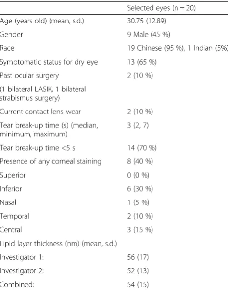
20 volunteers (20 eyes) were recruited for the study. We present the eye data from a randomized side (using a random number generator) for each patient. Table 1 shows the study sample’s charateristics, namely biodata, clinical history and parameters.
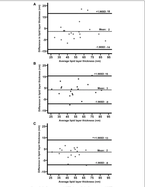
Intra-observer and Inter-observer repeatability of LLT In terms of intra-observer repeatability, the Bland-Altman plot showed a coefficient of repeatability of 16 nm and limits of agreement (95 % CI of differences) between−14 nm and 18 nm (Fig. 1a).
For inter-observer repeatability, when a single scan of one observer was compared to that of the other
observer, the coefficient of repeatability was 13 nm. In the Bland-Altman plot (Fig. 1b), the limits of agreement were between−9 nm and 16 nm. When the averaged trip-licate measurement of one observer was compared to that of the other, the coefficient of repeatability was 11 nm. Its corresponding limits of agreement (Fig. 1c) were −9 nm and 13 nm.
There was no significant systematic error in the intra-observer and inter-intra-observer comparisons, with a mean difference of 2 nm and 3 nm respectively. In all the ana-lyses above, the differences were not associated with the magnitude of the means. 1–2 outliers were removed from the Bland-Altman plots before analysis.
Possible associations and correlation with clinical parameters
The median TBUT (minimum, maximum) was 3 (2, 7) s and 70 % of the participants had a TBUT of less than 5 s (Table 1). TBUT was not significantly correlated with LLT (p = 0.874, Spearman’s r = 0.038).
Among the participants, 40 % had corneal fluorescein staining in at least one of the 5 corneal zones (Table 1). LLT was not significantly associated with the presence of any corneal staining (p = 0.325). In addition, LLT was not significantly associated with age, gender, history of current contact lens wear, history of ocular surgery and
Table 1Descriptive characteristics of participants
Selected eyes (n = 20)
Age (years old) (mean, s.d.) 30.75 (12.89)
Gender 9 Male (45 %)
Race 19 Chinese (95 %), 1 Indian (5%)
Symptomatic status for dry eye 13 (65 %)
Past ocular surgery 2 (10 %)
(1 bilateral LASIK, 1 bilateral strabismus surgery)
Current contact lens wear 2 (10 %)
Tear break-up time (s) (median, minimum, maximum)
3 (2, 7)
Tear break-up time <5 s 14 (70 %)
Presence of any corneal staining 8 (40 %)
Superior 0 (0 %)
Inferior 6 (30 %)
Nasal 1 (5 %)
Temporal 2 (10 %)
Central 3 (15 %)
Lipid layer thickness (nm) (mean, s.d.)
Investigator 1: 56 (17)
Investigator 2: 52 (13)
symptomatic status for dry eyes on univariate and multi-variate analysis (p > 0.05).
Assessment after scanning
Amongst the participants, 60 % had visual acuity of 6/ 9 or better, while 15 % were from 6/9−1 to 6/12 and 25 % were from 6/12−1 to 6/18. None of the partici-pants who went through interferometry complained about increased discomfort.
Discussion
For any given reading of the same subject, LLT was found not to differ from the mean by more than 16 nm. The limits of agreement for inter-observer and intra-observer measurements were similar. This suggests that the repeat-ability of measurements was independent of its observer. The single-scan inter-observer agreement and the aver-age of triple-scan inter-observer LLT measurements were also similar. There was no systematic difference between different measurements (whether intra-observer or inter-observer) as mean differences were not signifi-cantly different from zero. In this study, none of the tested clinical factors were associated with LLT. Despite the high number of scan acquisitions, no participant complained of discomfort.
Prior study on the repeatability of quantitative LLT mea-surements using interferometry involved a new spectral interferometer that has been developed by Fogt and King-Smith [12, 30, 47]. However, this interferometer is not commercially available. Moreover, although the study re-ported a good correlation coefficient (Spearman’s r = 0.835) [17], it must be noted that correlation alone is not an ap-propriate measure of repeatability [46].
Repeatability was also assessed in the pre-production model of the LipiView® interferometer. The pre-production model measured LLT by asking two observers to deter-mine the LLT based on subjective appreciation of the inter-ference colors and these measurements were found not to defer by more than 30 nm [9]. Compared to its pre-production model, the commercially released interferom-eter employed software processing and analysis of the recorded interference colors to calculate LLT [10]. As such, repeatability of measurements may be improved by the objective nature of software analysis.
To put our repeatability finding in perspective, mean LLT (SD) was 76 (25) nm in dry eye patients in a study by Finis et al. [16], and 65.0 (19.1) nm and 54.2 (17.9) nm respectively in healthy controls and MGD patients
in another study by Eom et al. [6]. In our Asian study population, the average LLT of 54 (15) nm appeared to be lower than these two studies. The difference may be due to greater heterogeneity in our study sample.
In terms of correlations between LLT and TBUT or fluorescein staining, this study showed no significant correlations, and was generally similar with these two prior studies [6] [16]. In Finis et al’s study, a weak correl-ation between LLT and Ocular Surface Disease Index (OSDI) (r =−0.13) was found, whilst we did not find any difference in LLT between symptomatic and non-symptomatic participants. In Eom et al’s study, correl-ation between LLT and TBUT was only found in the MGD group (r = 0.415). The primary objective of our study was to investigate repeatability and not to examine associations with clinical parameters of dry eye or mei-bomian gland dysfunction. Inter-ethnic differences, if any, will require future studies to elicit.
The strength of our study is that it was conducted in a very controlled condition with trained observers. Our study had the following limitations. Firstly, during LLT measurement, actual room humidity and temperature were not actually measured. However, the evaluation room was centrally air-conditioned and Singapore does not have any seasonal variations in climate. Secondly, as the sample size was small, differences in repeatability of measurements in different patient subgroups could not be assessed. Thirdly, the repeatability results could not be generalized to patients with specific characteristics which differ from this study’s sample.
In the future, technological advances may improve the repeatability of the instrument further. In software ana-lysis, perhaps the pattern (open meshwork, closed mesh-work, wave, colour fringe) can be considered in addition to the colour of the spectral reflection. Statistical model-ing with these additional variables may more precisely estimate LLT measurements. It may also be desirable for interferometry to be able to function over a wider area of the cornea or in a specific area of the cornea, or per-haps to measure LLT in non-blinking (stressed) condi-tions. A Kowa DR-1-based software has been developed to sample LLT over multiple corneal zones and interpret kinetic changes in tear lipid spreading [32, 37, 48].
Now that a certain degree of repeatability is established, interferometry may be a useful modality in monitoring changes of LLT in clinical trials. Single scan on each occa-sion is adequate for LLT measurement, and repeat mea-surements need not be performed by the same examiner. (See figure on previous page.)
Conclusion
With the repeatability of measurements being known, the significance of treatment-induced changes in the LLT measurements of this interferometer may be better interpreted. This is useful in clinical studies where a group of patients has undergone intervention related to the tear film. The size of the group will need to be deter-mined by the treatment effect that these future studies aim to detect.
Abbreviations
LLT:Lipid layer thickness; MGD: Meibomian gland dysfunction; TBUT: Tear break-up time; CCLRU: Cornea and Contact Lens Research Unit; SD/ s.d.: Standard deviation; 95 %CI: 95 % Confidence interval.
Competing interests
The authors declare that they have no competing interests.
Authors’contributions
ZY, CTLS and LT designed the study, drafted and revised the manuscript. ZY and CTLS recruited and assessed all the participants. ZY analyzed and interpreted the data.
Acknowledgements
Louis Tong was supported by the National Medical Research Council, Singapore (NMRC/CSA/045/2012) and the Biomedical Research Council, Singapore (BMRC (TCRP) 10/1/35/19/670 R828).
Grant support
This study was supported by the National Medical Research Council, Singapore (NMRC/CSA/045/2012), and the Biomedical Research Council, Singapore (BMRC (TCRP) 10/1/35/19/670 R828).
Author details
1Yong Loo Lin School of Medicine, National University of Singapore,
Singapore, Singapore.2Singapore National Eye Center, 11 Third Hospital Avenue, 168751 Singapore, Singapore.3Ocular Surface Research Group, Singapore Eye Research Institute, 168751 Singapore, Singapore.4Department of Cornea and External Eye Disease, Singapore National Eye Center, 11 Third Hospital Avenue, 168751 Singapore, Singapore.5Office of Clinical, Academic and Faculty Affairs, Duke-NUS Graduate Medical School, Singapore, Singapore.6Department of Ophthalmology, Yong Loo Lin School of Medicine, National University of Singapore, Singapore, Singapore.
Received: 9 June 2014 Accepted: 27 April 2015
References
1. The epidemiology of dry eye disease: report of the Epidemiology Subcommittee of the International Dry Eye WorkShop (2007). The ocular surface 2007, 5(2):93–107.
2. Pflugfelder SC. Prevalence, burden, and pharmacoeconomics of dry eye disease. Am J Manag Care. 2008;14(3 Suppl):S102–6.
3. Sullivan BD, Crews LA, Sonmez B, de la Paz MF, Comert E, Charoenrook V, et al. Clinical utility of objective tests for dry eye disease: variability over time and implications for clinical trials and disease management. Cornea. 2012;31(9):1000–8.
4. Kanellopoulos AJ, Asimellis G. In vivo 3-dimensional corneal epithelial thickness mapping as an indicator of dry eye: preliminary clinical assessment. Am J Ophthalmol. 2014;157(1):63–8. e62.
5. Wu S, Tao A, Jiang H, Xu Z, Perez V, Wang J. Vertical and horizontal corneal epithelial thickness profile using ultra-high resolution and long scan depth optical coherence tomography. PLoS One. 2014;9(5), e97962.
6. Cui X, Hong J, Wang F, Deng SX, Yang Y, Zhu X, et al. Assessment of corneal epithelial thickness in dry eye patients. Optom Vision Sci: Off Publ Am Acad Opto. 2014;91(12):1446–54.
7. Tittler EH, Bujak MC, Nguyen P, Zhang X, Li Y, Yiu SC, et al. Between-grader repeatability of tear meniscus measurements using Fourier-domain OCT in
patients with dry eye. Ophthalmic Surg Lasers Imaging : Off J Intl Soc Imaging Eye. 2011;42(5):423–7.
8. Versura P, Profazio V, Campos EC. Performance of tear osmolarity compared to previous diagnostic tests for dry eye diseases. Curr Eye Res. 2010;35(7):553–64. 9. Blackie CA, Solomon JD, Scaffidi RC, Greiner JV, Lemp MA, Korb DR. The
relationship between dry eye symptoms and lipid layer thickness. Cornea. 2009;28(7):789–94.
10. Korb DR, Grenon SM, Blackie C, Willis TR, Bacich S. Apparatuses and methods for determining tear film break-up time and/or for detecting lid margin contact and blink rates, particulary for diagnosing, measuring, and/ or analyzing dry eye conditions and symptoms, Google Patents. 2014. 11. The definition and classification of dry eye disease: report of the Definition
and Classification Subcommittee of the International Dry Eye WorkShop (2007). The ocular surface 2007,5(2):75–92.
12. King-Smith PE, Fink BA, Fogt N, Nichols KK, Hill RM, Wilson GS. The thickness of the human precorneal tear film: evidence from reflection spectra. Invest Ophthalmol Vis Sci. 2000;41(11):3348–59.
13. Mishima S. Some physiological aspects of the precorneal tear film. Arch Ophthalmol. 1965;73:233–41.
14. Bron AJ, Tiffany JM, Gouveia SM, Yokoi N, Voon LW. Functional aspects of the tear film lipid layer. Exp Eye Res. 2004;78(3):347–60.
15. Govindarajan B, Gipson IK. Membrane-tethered mucins have multiple functions on the ocular surface. Exp Eye Res. 2010;90(6):655–63. 16. Tutt R, Bradley A, Begley C, Thibos LN. Optical and visual impact of tear
break-up in human eyes. Invest Ophthalmol Vis Sci. 2000;41(13):4117–23. 17. King-Smith PE, Hinel EA, Nichols JJ. Application of a novel interferometric
method to investigate the relation between lipid layer thickness and tear film thinning. Invest Ophthalmol Vis Sci. 2010;51(5):2418–23.
18. Korb DR, Baron DF, Herman JP, Finnemore VM, Exford JM, Hermosa JL, et al. Tear film lipid layer thickness as a function of blinking. Cornea. 1994;13(4):354–9. 19. Eom Y, Lee JS, Kang SY, Kim HM, Song JS. Correlation between quantitative
measurements of tear film lipid layer thickness and meibomian gland loss in patients with obstructive meibomian gland dysfunction and normal controls. Am J Ophthalmol. 2013;155(6):1104–10. e1102.
20. Yokoi N, Takehisa Y, Kinoshita S. Correlation of tear lipid layer interference patterns with the diagnosis and severity of dry eye. Am J Ophthalmol. 1996;122(6):818–24.
21. Nichols JJ, Nichols KK, Puent B, Saracino M, Mitchell GL. Evaluation of tear film interference patterns and measures of tear break-up time. Optom Vision Sci: Off Publ Am Acad Opto. 2002;79(6):363–9.
22. Isreb MA, Greiner JV, Korb DR, Glonek T, Mody SS, Finnemore VM, et al. Correlation of lipid layer thickness measurements with fluorescein tear film break-up time and Schirmer’s test. Eye. 2003;17(1):79–83.
23. Mishima S, Maurice DM. The oily layer of the tear film and evaporation from the corneal surface. Exp Eye Res. 1961;1:39–45.
24. Iwata S, Lemp MA, Holly FJ, Dohlman CH. Evaporation rate of water from the precorneal tear film and cornea in the rabbit. Invest Ophthalmol. 1969;8(6):613–9.
25. Guillon M, Guillon JP. Hydrogel lens wettability during overnight wear. Ophthalmic & Physiological Optics: J Br Coll Ophthalmic Opticians. 1989;9(4):355–9.
26. Braun RJ, King-Smith PE. Begley CG, Li L. Dynamics and Function of the Tear Film in Relation to the Blink Cycle. Progress in retinal and eye research: Gewecke NR; 2014.
27. Yokoi N, Bron AJ, Georgiev GA. The precorneal tear film as a fluid shell: the effect of blinking and saccades on tear film distribution and dynamics. Ocul Surf. 2014;12(4):252–66.
28. Palakuru JR, Wang J, Aquavella JV. Effect of blinking on tear dynamics. Invest Ophthalmol Vis Sci. 2007;48(7):3032–7.
29. Harrison WW, Begley CG, Liu H, Chen M, Garcia M, Smith JA. Menisci and fullness of the blink in dry eye. Optom Vision Sci: Off Publ Am Acad Opto. 2008;85(8):706–14.
30. King-Smith PE, Fink BA, Nichols JJ, Nichols KK, Braun RJ, McFadden GB. The contribution of lipid layer movement to tear film thinning and breakup. Invest Ophthalmol Vis Sci. 2009;50(6):2747–56.
31. Kojima T, Ishida R, Dogru M, Goto E, Takano Y, Matsumoto Y, et al. A new noninvasive tear stability analysis system for the assessment of dry eyes. Invest Ophthalmol Vis Sci. 2004;45(5):1369–74.
33. Tomlinson A, Bron AJ, Korb DR, Amano S, Paugh JR, Pearce EI, et al. The international workshop on meibomian gland dysfunction: report of the diagnosis subcommittee. Invest Ophthalmol Vis Sci. 2011;52(4):2006–49. 34. Knop E, Knop N, Millar T, Obata H, Sullivan DA. The international workshop
on meibomian gland dysfunction: report of the subcommittee on anatomy, physiology, and pathophysiology of the Meibomian gland. Invest Ophthalmol Vis Sci. 2011;52(4):1938–78.
35. Finis D, Pischel N, Schrader S, Geerling G. Evaluation of lipid layer thickness measurement of the tear film as a diagnostic tool for Meibomian gland dysfunction. Cornea. 2013;32(12):1549–53.
36. Yokoi N, Mossa F, Tiffany JM, Bron AJ. Assessment of meibomian gland function in dry eye using meibometry. Arch Ophthalmol. 1999;117(6):723–9. 37. Goto E, Tseng SC. Differentiation of lipid tear deficiency dry eye by kinetic
analysis of tear interference images. Arch Ophthalmol. 2003;121(2):173–80. 38. Mitra M, Menon GJ, Casini A, Hamada S, Adams D, Ricketts C, et al. Tear film
lipid layer thickness and ocular comfort after meibomian therapy via latent heat with a novel device in normal subjects. Eye. 2005;19(6):657–60. 39. Spiteri A, Mitra M, Menon G, Casini A, Adams D, Ricketts C, et al. Tear lipid
layer thickness and ocular comfort with a novel device in dry eye patients with and without Sjogren’s syndrome. J Fr Ophtalmol. 2007;30(4):357–64. 40. Korb DR, Greiner JV. Increase in tear film lipid layer thickness following
treatment of meibomian gland dysfunction. Adv Exp Med Biol. 1994;350:293–8.
41. Olson MC, Korb DR, Greiner JV. Increase in tear film lipid layer thickness following treatment with warm compresses in patients with meibomian gland dysfunction. Eye Contact Lens. 2003;29(2):96–9.
42. McDonald JE. Surface phenomena of the tear film. Am J Ophthalmol. 1969;67(1):56–64.
43. Guillon M, Styles E, Guillon JP, Maissa C. Preocular tear film characteristics of nonwearers and soft contact lens wearers. Optom Vision Sci: Off Publ Am, Acad, Optom. 1997;74(5):273–9.
44. Tong L, Chaurasia SS, Mehta JS, Beuerman RW. Screening for meibomian gland disease: its relation to dry eye subtypes and symptoms in a tertiary referral clinic in singapore. Invest Ophthalmol Vis Sci. 2010;51(7):3449–54. 45. CCLRU. Grading Scales. Sydney, Australia: Cornea and Contact Lens
Research Unit, School of Optometry, University of New South Wales; 1996. 46. Bland JM, Altman DG. Statistical methods for assessing agreement between
two methods of clinical measurement. Lancet. 1986;1(8476):307–10. 47. Fogt N, King-Smith PE, Tuell G. Interferometric measurement of tear film
thickness by use of spectral oscillations. Invest Ophthalmol Vis Sci. 1998;15(1):268–75.
48. Goto E, Dogru M, Kojima T, Tsubota K. Computer-synthesis of an interference color chart of human tear lipid layer, by a colorimetric approach. Invest Ophthalmol Vis Sci. 2003;44(11):4693–7.
Submit your next manuscript to BioMed Central and take full advantage of:
• Convenient online submission
• Thorough peer review
• No space constraints or color figure charges
• Immediate publication on acceptance
• Inclusion in PubMed, CAS, Scopus and Google Scholar
• Research which is freely available for redistribution