I
IJJMMCCMM Original Article S
Suummmmeerr22001133,,VVooll22,,NNoo33
A Comparison between Antibacterial Activity of Propolis and
Aloe vera on Enterococcus faecalis (an In Vitro Study)
Maryam Ehsani1, Mahmood Amin Marashi2, Ebrahim Zabihi3,4∗, Maryam Issazadeh1, Soraya Khafri5
1. Dental Materials Research Center, School of Dentistry, Babol University of Medical Sciences, Babol, Iran. 2. Department of Bacteriology and Virology, School of Medicine, Alborz University of Medical Sciences, Karaj, Iran. 3. Cellular and Molecular Biology Research Center (CMBRC), Babol University of Medical Sciences, Babol, Iran. 4. Department of Pharmacology & Physiology, School of Medicine, Babol University of Medical Sciences, Babol, Iran. 5. Department of Social Medicine, School of Medicine, Babol University of Medical Sciences, Babol, Iran.
Removing the bacteria, including Enterococcus faecalis, from the root canal is one of the important aims in
endodontic treatment.We aimed to compare the antibacterial activity of Chlorhexidine with two natural drugs.
The antibacterial activities of three different propolis extracts (alcohol concentrations: 0, 15, 40%) and Aloe vera
gel on E. faecalis were compared using three methods: disk diffusion, microdilution and direct contact test. In
addition to the above bacterium, the Aloe vera gel effect on Staphylococcus aureus and Streptococcus mutans
was evaluated. Disk diffusion test revealed that propolis ethanolic extracts (the alcohol concentration of 15 and
40%) and Aloe vera gel have antibacterial activities but aqueous extract of propolis did not show any effect in
this test. The MICs for propolis ethanolic extracts, Aloe vera gel and aqueous extract of propolis (0% alcohol)
were 313 µg/ml, 750 µg/ml, 2250 µg/ml, and ≥ 500 µg/ml respectively, much higher than the Chlorhexidine
one. In direct contact test, contrary to Aloe vera, all three propolis extracts showed antibacterial effects on E.
faecalis. The Aloe vera gel also showed significant antibacterial effect on S.aureus and S.mutans. The
hydroalcoholic extracts of propolis and Aloe vera gel had antibacterial effects on E. faecalis, however, propolis
is more potent than Aloe vera. The antibacterial effect of Aloe vera on S. aureus and S. mutans is low (MIC ≥
2250 µg/ml). Appropriate concentrations of alcoholic extracts of propolis and some fractions of Aloe vera gel
might be good choices for disinfecting the root canal in endodontic treatments.
Key words: Chlorhexidine , root canal, antiseptic, S. aureus, S. mutans
∗
Corresponding author: Cellular and Molecular Biology Research Center, Babol University of Medical Sciences, Ganje-Afrooz Avenue, Babol, Iran. Email: [email protected] & [email protected]
ne of the main goals in endodontic treatments
is removing the bacteria from the root canal
system. Although chemo - mechanical preparation
of root canal is able to decrease the bacterial load,
the resistant microorganisms usually remain in the
canal space even after the instrumentation and
O
Submmited 28 July 2013; Accepted 17 Aug 2013
washing processes. The main reasons behind this
contamination are: the complex anatomy of pulp
system, existence of the secondary canals, and
ability of microorganisms to survive in harsh
conditions (1-2). E. faecalis is an anaerobic
gram-positive bacterium which is found in periapical
lesions. It is able to attack dentinal tubules and
easily copes with hard condition of root canal
which make it a resistant microorganisms (3). Some
studies on root treated teeth have shown that E.
faecalis bacteria are prevalent up to 77% in the
periradicular lesions. In fact, the involvement of
this bacterium in root canal treatment failure is
more likely than the primary endodontic lesions (4).
Sodium hypochlorite has been used as an intracanal
irrigant, however, due to its adverse effects
including damage to tissues and inducing
emph-ysema, its used has been restricted. Chlorhexidine
2% solution is used as an intracanal irrigant with
antibacterial properties and great ability to disinfect
the dentinal tubules against E. faecalis, however its
use has been restricted due to: discoloration of the
teeth and tongue, decreasing the sense of taste,
irritation of oral mucosa and mouth dryness.
Nowadays, due to its antibacterial properties,
calcium hydroxide is highly used as the intracanal
medication. But again, because of its high pH, this
subtance is so toxic to the tissues which can lead to
chronic inflammation and cell necrosis (5-6).
Because of the cytotoxicity induced by common
intracanal drugs, their inability to remove some
bacteria from the dentinal tubules, and the
microorganisms’ resistance phenomenon, looking
for new intracanal drugs especially among natural
resources are highly recommended (7).
Propolis is a dense yellow-brown resin-like
material which its solubility is low in water, but
high in ethanol (8). This material is made from
resin, bud and other parts of the plants by bees. It is
used for protecting the hive against the outside
pollutions and blocking the slots and cracks.
Propolis has antibacterial, antifungal, antiviral,
antiinflammation, antioxidant and anti-tumor
effects (8-9) and many applications for this
substance in dentistry has been recently reported
(7). Aloe vera, along with other 360 species,
belongs to liliaceae family. This plant can grow in
hot and dry weather due to its high capacity in
maintaining water. Aloe vera has antibacterial,
anti-fungal, antivirus, antiinflammation, and anti-tumor
properties which make it useful in broad
range of ailments including: arthritis, asthma,
gastrointestinal diseases, and skin problems (e.g.
psoriasis, burning and wounds).
In dentistry, Aloe vera has been used in
recurrent aphthous ulcers, alveolar osteitis, and
lichen planus lesions (10-12).
The aim of this study was to determine the
antibacterial potency of Aloe vera compared to
propolis and Chlorhexidine. Also, the effect
of ethanol concentration on antibacterial activity
of hydroalcoholic extracts of propolis was
investigated.
Materials and Methods
Propolis quality control assays
About 150 grams of propolis was freshly
collected from Amirkola’s (Mazadaran-Iran) honey
bees’ nests during the 2012 winter. Standard
microbiological and chemical assays were
performed on the sample by Suren Tak Toos Lab.
Co. (Mashhad-Iran).
Propolis hydroalcoholic extraction
Propolis was dispersed in absolute ethanol
(500 mg in 50 ml) at 37ºC using magnet stirring for
1.5 hours. The obtained opaque yellow liquid
passed through filter (Whatman#1) and centrifuged
at 22ºC for 10 minutes (800 g). The clear
supernatant was diluted with appropriate amounts
of sterile distilled water to give ethanol
concentration of either 15%. or 40%. To make
aqueous extract, propolis was dispersed in sterile
distilled water (500 mg in 50 ml) at 22ºC using
magnet stirring for 4 hours. The obtained opaque
liquid was filtered and centrifuged at 22ºC for 10
minutes (800g). These extracts were kept in the
fridge (less than 1 week) and by warming up to
37ºC any precipitate was dissolved before use.
Aloe vera physicochemical analyses
Aloe vera gel was kindly gifted by Barij
Essence (Kashan-Iran). Standard physicochemical
assays including carbohydrates content, dry
substance, ash weight, and capillary viscometry
were performed.
The test microorganisms
The sample of standard strains of E. faecalis
PTCC 1394, S. mutans ATCC 1601 and S. aureus
ATCC 25923, were obtained from the
Scientific-Industrial Research Center of Asre-Enghelab
(Tehran-Iran) and were inoculated in Brain Heart
Infusion (BHI) culture medium.
Disk diffusion test
The method of Kirby-Bauer disk-diffusion
was performed for this assay. Briefly sterile paper
disks (6.4 mm) were soaked in the test material
solutions for 10 minutes. Ethanol (15, 40%) and
distilled water were used as negative control. The
impregnated paper disks were placed on the surface
of blood agar culture plates previously inoculated
by the test microorganism (E. faecalis, S. mutans, S.
aureous). The inhibition zone was measured for
each test material.
Direct contact test
The test material solutions (500 µL each) were
dried on the bottom of a 24-well plate. Then 50 µ L
of the test bacterial suspension (1.5×10⁷ CFU/ml)
was poured into each well and left to dry in a
laminar airflow. After that, 500 µL of BHI was
added to each well and the plate was incubated at
37ºC. After 24 hours, the colony count of 5 µL of
each well’s solution was measured.
The microdilution test
Broth microdilution test was performed as
described in M27-A2 (CLSI) with minor
modi-fications. The test material solutions was firstly
diluted 50:50 in 2X BHI medium then serial
dilutions were made using (100 µL) 1X BHI in
each well, then 10 µL of microbial suspension
(1.5×10⁷ CFU/ml) was added. After 24 hours
incubating at 37ºC, the last well without opacity
was considered as minimum inhibitory
concen-tration (MIC). The well with lowest concenconcen-tration
of the tested material, which could not lead to
microbial growth (99.9% inhibition) after
inocu-lating the blood agar plate, was considered as the
minimum bactericidal concentration (MBC). Also
the microdilution test was performed on Aloe vera
using two additional microorganisms (S.aureus and
S.mutans).
Statistical analyses
The data are presented as mean±SD and
analyzed by ANOVA. In case of significance, the
multi fold Scheffe comparisons and t-test were used
for two by two comparisons. P < 0.05 was
consid-ered significant.
Results
The antibacterial activity of propolis
hydroalcoholic extracts (with 0, 15, 40% ethanol),
Aloe vera gel, and Chlorhexidine 2% on E. faecalis
bacteria are compared using three methods: disk
diffusion, direct contact and microdilution. In
regards to Aleo vera, disk diffusion and
micro-dilution tests, have been performed using
two additional bacteria (S. aureus, S. mutans) to
investigate more its antimicrobial spectrum.
Propolis and Aloe vera quality control assays
The results of some quality control tests on
propolis are shown in table 1. The physicochemical
analysis data of Aloe vera are shown in table 2.
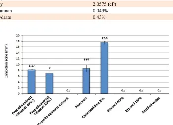
Disk diffusion test
Propolis hydroalcoholic extract (with 15 and
40% ethanol) and Aloe vera gel showed
antibacterial effect with no significant difference
among them. However, no inhibition zone was
observed with propolis aqueous extract (0%
ethanol). Chlorhexidine 2% produced significantly
higher inhibition zone compared to the other
Fig 1. Growth inhibition zone (mean ±SD) induced by different propolis hydroalcoholic extracts (with 0, 15, 40% ethanol), Aloe vera gel
and chlorohexidine 2% in the method of disk diffusion with E. faecalis.
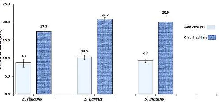
extracts (P< 0.001) (Fig. 1). The Aloe vera gel was
less effective than Chlorhexidine 2% not only
against E. faecalis but also against S. aureus and S.
mutans (Fig. 2).
Table 2. The physicochemical analysis of Aloe vera sample
Color Colorless
pH 4.45
Density 0.9739 (g/ml)
Dry weight 0.9 %
Ash weight 0.29%
Viscosity 2.0575 (cP)
Glucomannan 0.049%
Carbohydrate 0.43%
Table 1. The quality control assays on propolis sample and its extracts
Result (unit) Conducted assay
brown Sample color
26.2 (%) Total polyphenol content
Negative (cfu/g) E. coli growth
Negative (cfu/g)
Staphylococcus aureus growth
Negative (cfu/g) Pseudomonas Sp. growth
Negative (cfu/g) Aspergillus growth
55.8 (%) Dried mass
16.3 (%) Total carbohydrate content
0.5 (%) Total protein content
Positive Free amino acid (detected by TLC)
Positive Free sugars (4 and 5 carbon detected by TLC)
2.35 (%) Insoluble substances in 10% alcohol
2.87 (%) Reduced sugar
0.1 (%) Dry substance of saturated aqueous extract (0% ethanol)
0.5 (%) Dry substance of propolis hydroalcoholic extract (40% ethanol)
0.3 (%) Dry substance of propolis hydroalcoholic extract (15% ethanol)
Fig 2. A comparison between Aloe vera and chlorohexidine 2% antibacterial activity against 3 test microorganisms using disk diffusion test.
The label numbers are the mean of inhibition zone for three replicate disks.
Microdilution test
The MIC results for propolis hydroalcoholic
extracts, Aloe vera gel and Chlorhexidine 2%
solution have been presented below (Table 3).
The propolis aqueous extract (0% ethanol) did not
show any inhibition in microdilution test (MIC>
propolis solubility). Chlorhexidine showed the
lowest MIC (2 µg/ml) compared to the other tested
materials. In addition to E. faecalis, Aloe vera
showed antibacterial activity against two gram
positive cocci (S. aureus, S. mutans) in this test
(Table 3).
Direct contact test
The number of colonies of bacteria grown
after 24 hours is shown in fig. 3. The
hydroalcoholic extract of propolis with 40% alcohol
showed significant antibacterial effect against E.
faecalis (similar to Chlorhexidine 2% solution).
The aqueous extract of propolis showed a lesser
extent in this antibacterial effect. However, Aloe
vera showed no antibacterial effect in this method
and the resulting colonies were practically
uncountable same as the negative controls (because
of countless resulting colonies, the negative
controls are not depicted in this figure).
Discussion
In this study, we showed that Aloe vera gel
and propolis ethanolic extracts have antibacterial
activity against E. faecalis in in vitro. However,
both these naturally available substances showed
lower potency compared to Chlorhexidine in either
disk diffusion and microdilution assays (Table 3,
Fig. 1, 2). On the other hand, propolis ethanolic
extract showed high antibacterial activity against E.
faecalis comparable to that of Chlorhexidine in
direct contact test (Fig. 3) which signifies the
importance of solubility issue. Some gram positive
bacteria such as E. faecalis resist the cleaning and
shaping of root canal, and potentially can lead to
endodontic failure (13-15).
Aloe vera gel and propolis are two naturally
occurring substances which have been long used in
the treatment of inflammation and infectious
diseases of the mouth (13, 16-17). The
physicochemical assays conducted on both Aloe
vera and propolis samples confirm their standard
characteristics (Tables 1, 2). Since the solubility of
propolis components in alcohol is different, the
concentration of ethanol used for extraction is
critical. The dry weight of each propolis alcoholic
extract is correlated to its ethanol concentration
(Table 2). In this study, we used high speed
centrifugation following filtration to omit any
dispersed solid material off the extract. Colloidal
particles in the extract might exert direct
Fig. 3. The number of grown colonies of E. faecalis after 24 hour contact with propolis hydroalcoholic extracts, Aloe vera, and
Chlorohexidine in direct contact test.
antibacterial effects. The noticeable difference in
antibacterial activity results obtained by the three
test procedures, especially with propolis aqueous
extract, indicates that ethanol soluble constituents
of propolis are responsible for its antibacterial
effect (8, 18). These components show quite high
antibacterial activity in direct contact test against E.
faecalis (Fig. 3).
The anti-microbial effect of hydroalcoholic
extracts of propolis in disk diffusion was less than
that in microdilution, this issue might be aroused by
low diffusion ability of alcohol soluble components
in agar. On the other hand, since Aloe vera gel is
aqueous, no such a difference was observable
between its microdilution and its disk diffusion test
(Fig. 1, Table 3)
In direct contact test the microorganism
gets in touch with the surface of the dried material
directly, hence, there is no problem with
insolubility of antimicrobial components. For this
reason, the aqueous extract of propolis, which
contains the least amount of ethanol soluble
antimicrobial components, only shows its weak
antibacterial activity in direct contact test (Fig. 3).
Table 3. MIC and MBC in hydroalcoholic extract of propolis, Aleo vera and Chlorohexidine 2 % using the microdilution test on E. faecalis
MBC(µg/ml) MIC(µg/ml)
Groups
625 313
Propolos hydroalcoholic extract (40% ethanol)
1500 750
Propolos hydroalcoholic extract (15% ethanol)
NA NA
Propolis aqueous extract (0% ethanol)
4500 NA (1) 4500 (2) 2250
4500 (1) 2250 (2) Aloe vera
4 2
Chlorohexidine 2%
NA NA
Ethanol40 %
NA NA
Ethanol 15%
NA NA
Distilled water
NA: without antibacterial inhibitory effect (1) The test microorganism was S. aureous (2) The test microorganism was S. mutans.
These substances have low solubility in water
but they are highly soluble in ethanol. Some
components in propolis, which have been suggested
as its active agents, include flavonoids, phenolic
and aromatic compounds like caffeic acid (19). Our
results are in concordance with a study conducted
by Mattigatti et al. (2012) who investigated the
effects of propolis on three microorganisms (E.
faecalis, S. aureus and Candida albicans) using
agar diffusion test (20). They have shown (same to
our results) that Chlorhexidine along with
MTAD® (a mixture of tetracycline, citric acid and
a detergent) has superior activity against the tested
micro-organisms.
On the other hand, Aloe vera gel which
showed weak antibacterial activity in disk diffusion
and microdilution tests, failed to show any activity
in direct contact test (Fig. 3). This might be the
result of low concentration of its antibacterial
components compared to nutrient polysaccharides
which could prevent the microorganism to be fully
in touch with the Aloe vera active components.
Aloe vera’s pharmacotherapeutic and cosmetic
properties have been studied since long time ago
(16-17). However, studies about its antibacterial
effect on E. faecalis and its comparison to intra
canal drug like Chlorhexidine 2% has not yet been
done. The leaf of Aloe vera contains some
active substances like acemanan, anthraquinone,
anthracine, cinnamonic acid with anti
inflamma-tory/antimicrobial properties (17, 21).
As a comparison between Aloe vera and
propolis, the antimicrobial effect of Aloe vera gel in
microdilution was less than hydroalcoholic extracts
of propolis and its obtained MIC on all tested
microorganisms (E. feaclais, S. aureus and S.
mutans) was more than 2250 µg/ml. Recently,
conducted studies with other test organisms or
methods of antibacterial activity assyas, have
shown similar results in our study. In the study by
Anuj Bhardwaj et al. in 2012, the antimicrobial
effect of some natural extracts and Aloe vera with
Chlorhexidine 2% on E. faecalis was compared
which similar to the present study (22-23).
Conclusion
Aloe vera gel has mild antibacterial effect
against E. faecalis, S. aureus and S. mutans. It
seems that Aloe vera gel has low antibacterial
potency compared to propolis, hence its
subfractionation may be a good choice to make a
better antibacterial compound for root canal
treatments. On the other hand, the hydroalcoholic
extract of propolis could be a good anti-microbial
agent against E. faecalis especially following direct
contact to this germ. Both tested natural substances
have less antibacterial activity compared to
Chlorhexidine , however their potency could be
significantly increased by improvement in the
extraction techniques. This could potentially lead to
root canal antibacterials with fewer side effects.
Acknowledgement
This research project has been funded by a
research grant from Babol University of Medical
Sciences (Grant No:9133725). The authors wish to
thank: Barij Essence company (Kashan-Iran) and
Behsa Pharmaceutical Company (Tehran-Iran) for
honoring the Aloe vera gel and Chlorhexidine
solution respectively.
References
1. Gomes BP, Souza SF, Ferraz CC, et al. Effectiveness of 2%
Chlorhexidine gel and calcium hydroxide against Enterococcus
faecalis in bovine root dentine in vitro. Int Endod J
2003;36:267-75.
2. Victorino FR, Bramante CM, Zapata RO, et al. Removal
efficiency of propolis paste dressing from the root canal. J Appl
Oral Sci 2010;18:621-4.
3. Siqueira JF, Jr., Rocas IN. Polymerase chain reaction-based
analysis of microorganisms associated with failed endodontic
treatment. Oral Surg Oral Med Oral Pathol Oral Radiol Endod
2004;97:85-94.
4. Molander A, Reit C, Dahlen G, et al. Microbiological status of
root - filled teeth with apical periodontitis. Int Endod J
1998;31:1-7.
5. Pujar M, Makandar S. Herbal Usage In Endodontics- A
Review. Int J contem dentis 2011;2:34-7.
6. Kayaoglu G, Omurlu H, Akca G, et al. Antibacterial activity
of Propolis versus conventional endodontic disinfectants against
Enterococcus faecalis in infected dentinal tubules. J Endod
2011;37:376-81.
7. Sforcin JM, Bankova V. Propolis: is there a potential for the
development of new drugs? J Ethnopharmacol 2011;133:253-60.
8. Viuda-Martos M, Ruiz-Navajas Y, Fernandez-Lopez J, et al.
Functional properties of honey, propolis, and royal jelly. J Food
Sci 2008;73:R117-24.
9. Sforcin JM. Propolis and the immune system: a review. J
Ethnopharmacol 2007;113:1-14.
10. Cera LM, Heggers JP, Robson MC, et al. The therapeutic
efficacy of aloe vera ceramin thermal injuries. two case report. J
Am Animal Hosp Assoc 1980;16:768-72.
11. West DP, Zhu YF. Evaluation of aloe vera gel gloves in the
treatment of dry skin associated with occupational exposure. Am
J Infect Control 2003;31:40-2.
12. Babaee N, Zabihi E, Mohseni S, et al. Evaluation of the
therapeutic effects of Aloe vera gel on minor recurrent aphthous
stomatitis. Dent Res J (Isfahan) 2012;9:381-5.
13. Chavez de Paz LE, Molander A, Dahlen G. Gram-positive
rods prevailing in teeth with apical periodontitis undergoing root
canal treatment. Int Endod J 2004;37:579-87.
14. Chu FC, Leung WK, Tsang PC, et al. Identification of
cultivable microorganisms from root canals with apical
periodontitis following two-visit endodontic treatment with
antibiotics/steroid or calcium hydroxide dressings. J Endod
2006;32:17-23.
15. Chávez de Paz LE, Molander A, Dahlén G. Gram-positive
rods prevailing in teeth with apical periodontitis undergoing root
canal treatment. Int Endod J 2004;37:579-87.
16. Reynolds T, Dweck AC. Aloe vera leaf gel: a review update.
J Ethnopharmacol 1999;68:3-37.
17. Wynn RL. Aloe vera gel: update for dentistry. Gen Dent
2005;53:6-9.
18. Miguel MG, Antunes MD. Is propolis safe as an alternative
medicine? J Pharm Bioallied Sci 2011;3:479-95.
19. Salomao K, Dantas AP, Borba CM, et al. Chemical
composition and microbicidal activity of extracts from Brazilian
and Bulgarian propolis. Lett Appl Microbiol 2004;38:87-92.
20. Mattigatti S, Ratnakar P, Moturi S, et al. Antimicrobial effect
of conventional root canal medicaments vs propolis against
Enterococcus faecalis, Staphylococcus aureus and Candida
albicans. J Contemp Dent Pract 2012;13:305-9.
21. Surjushe A, Vasani R, Saple DG. Aloe vera: a short review.
Indian J Dermatol 2008;53:163-6.
22. Bhardwaj A, Ballal S, Velmurugan N. Comparative
evaluation of the antimicrobial activity of natural extracts of
Morinda citrifolia, papain and aloe vera (all in gel formulation),
2% Chlorhexidine gel and calcium hydroxide, against
Enterococcus faecalis: An in vitro study. J Conserv Dent
2012;15:293-7.
23. Kaithwath G, Kumar A, Pandey H, et al. Investigation of
comparative antimicrobial activity of aloe vera gel and juice.
Pharmacologyonline 2008;1:239-43.