I
IJJMMCCMM Original Article S
Suummmmeerr22001144,,VVooll33,,NNoo33
Association Study of rs3184504 C>T Polymorphism in Patients
With Coronary Artery Disease
Sarah Sadat Aghabozorg Afjeh1, Sayyed Mohammad Hossein Ghaderian1∗, Reza Mirfakhraie1, Mohammad Piryaei1, Hooshang Zaim Kohan2
1. Department of Medical Genetics, Shahid Beheshti University of Medical Sciences, Tehran, Iran.
2. Department of Medical Genetics, Tehran University of Medical Sciences, Tehran, Iran.
Cardiovascular disease has become the main factor of death and birth defects in the world and also in Iran. New
clinical studies have shown that early diagnosis of patients with coronary artery disease (CAD) can contribute to
effective prevention or therapeutic structures, which reduce mortality or the next chance of cardiovascular
events, and increase the quality of life. Most studies on CAD disease and its genetic risk factors so far, have been
done excluding the Iranian population. PubMed was used to search for all relevant studies published on or before
2013 and rs3184504 was selected for association study for CAD. A total of 200 subjects with 100 cases and 100
controls were ultimately included in the analysis. Blood samples were collected and after DNA extraction the
DNA analysis was performed by TaqMan Probe Real Time PCR to evaluate the association between candidate
variant with the disease and some blood biochemical factors. Our study demonstrated that there was not a direct
association between rs3184504 C>T variant with risk of CAD in Iranian population, whereas, there is a
significant association between this variant with increased blood LDL and diastolic blood pressure. Further
molecular analysis and other disease association studies are necessary in the Iranian population.
Key words: CAD, polymorphism, blood pressure, Iran
∗
Corresponding author: Department of Medical Genetics, Shahid Beheshti University of Medical Sciences, Tehran, Iran. Email: [email protected]
ardiovascular disease (CVD), including its
most severe complication myocardial
infarction (MI), has become the main factor of
death in the world and also in Iran (1-2).
More than 80% of sudden cardiac deaths in
the world are caused by atherosclerotic coronary
artery disease (CAD), and the remaining 20% of
cases are caused by other diseases including
cardiomyopathies, congenital heart disease, left
ventricular hypertrophy, aortic valve disease and
other cardiac disorders. Many risk factors are
involved in the familial aggregation of coronary
heart disease which can be separated to
environ-mental risk factors and genetic risk factors.
Epidemiological studies have been done to elu
Department of Medical Genetics, Shahid Beheshti
University of Medical Sciences, Tehran, Iran.
Sghaderian @ yahoo.co.uk cidate the risk factors of
C
Submmited 24 February 2014; Accepted 21 June 2014; Published 12 July 2014
cardiovascular disease. On the other hand, other
studies have focused on specific candidate genes
influencing quantitative variation in lifestyle and
behavioral phenotypes (3). Two separated
environ-mental factors which cause the disease are
common. These include smoking or other lifestyles
and factors related with the location individual lives
such as air pollution. Many studies investigating the
association of environmental factors such as
weight, body mass index (BMI), height, cholesterol
level and pulse rate with blood pressure phenotypes
have shown a significant association (4-5).
Macrovascular and microvascular pathology
are related to cardiovascular disease such as CAD
and stroke (6- 8). Studies on genetic determinants
of CVD have been performed with focus on
macrovascular disease traits and rare studies were
conducted on the genetic analysis of microvascular
disease phenotype (6, 8-9).
According to recent publications,
microvasc-ular disease plays an important role in the
pathogenesis of cardiovascular disease.
Noninvasive direct assessment of the human
microcirculation can be done through quantitative
measurement of retinal blood vessels caliber from
photographs (10).
Researches demonstrate that alteration in
retinal vascular caliber such as narrower arteriolar
and wider venular caliber, can be related to a
range of cardiovascular diseases and their
risk factors (11-12), such as hypertension (13),
diabetes mellitus (14-15), stroke (16), coronary
heart disease (17) and cerebral small vessel
disease (18-19).
An essential step in vascular remodeling and
regeneration events is the migration of endothelial
cells. A cascade of events which can lead to
vascular disorders (e.g., atherosclerosis, vein graft
arteriosclerosis and restenosis) are often initiated
through the loss of endothelial function and
integrity, after percutaneous revascularization and
transplant arteriosclerosis (20-21).
Endothelial cell (EC) migration is a
mechani-cally integrated molecular process which involves
dynamic and coordinated alteration in cell
attachment and cytoskeleton organization (22-23).
A series of steps are required in this process
including cellular extension and formation of
membrane protrusion, termed lamellipodia that
push the leading front and lastly, cell contraction
which allows forward progression. Attachment of
protrusions to the extracellular matrix (ECM) is
mediated by integrins that function as receptors for
cell-adhesion molecules.
As mentioned, there are also genetic risk
factors causing CVD. The new strategy of
genome-wide association (GWA) studies is starting to
demonstrate novel genetic factors which contribute
to disease risk. Many genetic variants that
predispose to CVD spanning from common
polymorphisms (minor allele frequency >1%) to
rare, highly deleterious mutations responsible for
Mendelian diseases which are usually identified by
linkage studies, alone or in combination, modulate
the risk of the disease. A different approach named
genetic association analysis is used in order to
identify the genes involved in complex diseases
(24). In the case of complex diseases such as MI ,
linkage analysis is compounded by genetic
heterogeneity of the disease and other factors such
as incomplete penetrance of genes causing the
disease and their interaction with environmental
factors. In addition, the high prevalence of the
disease-causing allele in the population, and late
onset of disease can be named as another instance
(25). Studies based on Meta-analysis technique
published on 2011 demonstrate that specific genetic
loci were introduced in association with CVD and
more specifically with MI. Alteration in expression
in some of them causes increasing risk of MI
(25-26). Recent GWAs studies show that different
SNPs on 12q24 locus (such as rs3184504 on Lnk)
are associated with platelet count, hemoglobin
concentration, hematocrit and blood pressure (27).
Among the studied SNPs, one case, the
non-synonymous SNP rs3184504 C>T, Trp>Arg
(p-value: 6E-06) in the exon 3 of SH2B3 gene had
significant correlation with MI in six different
populations (28). The named SNP, in addition to its
companionship with eosinophil, cooperates with
other increased blood parameters such as the total
amount of platelets, leukocytes and red blood cells.
Regarding the platelets and leukocytes the
cooperation was evaluated by CAD onset (28).
Genetic diversity can be related to the
regulation of blood pressure fluctuation through
structural change of encoded proteins and alteration
in gene expression (quantity of proteins) (29).
Regarding the SH2B3 gene, based on existing
studies, there is a significant correlation between
the non-synonymous SNP rs3184504 and the
increased diastolic blood pressure. Lnk [Src
homology 2-B3 (SH2B3)] and the closely related
proteins SH2B1 and SH2B2 belong to a subfamily
of SH2-containing proteins with adaptor functions.
These adaptor proteins are the regulators of growth
factor and cytokine receptor-mediated pathways.
The Lnk protein contains a pleckstrin homology
(PH) domain, a NH2-terminal proline-rich region, a
Src homology 2 (SH2) domain, and potential
tyrosine phosphorylation sites (20, 30-31). Lnk−/−
mice show an abnormal accumulation of erythroid
cells, B lymphocytes and megakaryocytes in
different hematopoietic chambers which
demonstrates a defect in lymphoid and myeloid
homeostasis (32-33). Protrusions attachment to
extracellular matrix is led by integrin that acts as
receptor for cell attachment molecules. Lnk down
regulates stem cell factors and c-kit receptor
signaling in B-cell precursors (32, 34-38),
endothelial progenitor and mast cells (39).
Deficiency of Lnk may cause increased signaling
by cytokine and thrombopoietin receptors such as
c-kit and c-mpl that are essential and critical for
growth of hematopoietic stem and progenitor cells
(20, 39). Another study performed in London
confirmed association between increased LDL and
rs3184504 C>T variant (40). The aim of this study
was to investigate the association between CAD,
increased LDL, blood pressure and rs3184504 C>T
variant.
Materials and Methods
Patients and controls
A total of 200 genetically unrelated Iranian
subjects, comprising 100 individuals with MI and
100 normal controls from Tehran, Babol, and
Mashhad cities were enrolled in the present study.
All cases were selected based on the World Health
Organization (WHO) criteria as described (41).
Inclusion criteria: male and female, age over
40 years, angiography and ventriculography
positive with at least one coronary artery with
stenosis of 50% or more,
systolic blood pressure of ≥ 130 mm Hg,
diastolic blood pressure of ≥ 85 mm Hg and/or
taking medication for hypertension, triglycerides of
at least 150 mg/dL (≥ 1.65 mmol/L), HDL-C less
than 40 mg/dL (b 1.04 mmol/L) for men and less
than 50 mg/dL (b 1.30 mmol/L) for women, fasting
plasma glucose of at least 100 mg/dL (≥ 5.50
mmol/L) and waist circumference greater than 90
cm for men and greater than 80 cm for women.
Exclusion criteria: pregnant and lactating
women, history of previous angiography, heart
failure and history of drug use for the disease,
systemic and kidney disease and history of drug use
for the disease and genetic diseases influencing
heart.
Normal controls were randomly selected from
the same geographic area who were not suffering
MI or had no family history of CAD &
inflammation of the organ, with blood pressure >
140/90 mm Hg, blood cholesterol levels > 220
mg/dl, obesity, BMI ≥ 30, type 2 diabetes ,FBS >
126 mg/dl, smoking, in the past five years. Those
with medical illnesses such as endocrinological
abnormalities, chronic liver and/or renal diseases
and those with glucose-lowering medication and
alcohol consumption were excluded from the study.
All participants completed a standardized
questio-nnaire including information such as name, age,
ethnicity, dietary habits, family history of MI,
stroke, diabetes mellitus and past medical history.
Measurements
For all the subjects, height, weight, waist
circumference, hip circumference and waist/hip
ratio were measured using standardized measures
and scales. The body mass index (BMI) was
calculated as kg/m2 and a BMI of 20–25 was considered normal. Systolic blood pressure (SBP)
and diastolic blood pressure (DBP) were measured
using a standardized procedure. A full fasting
serum lipid profile including total cholesterol
(TC), HDL, LDL and TG, and fasting blood
glucose (FBS) concentrations were evaluated
by standard enzymatic techniques. Serum
C-reactive protein (CRP) concentrations were
determined by polye-thylene glycol-enhanced
immunoturbidimetry.
DNA isolation and SNP selection and genotyping
Genomic DNA was extracted from whole
blood using a commercially available DNA
isolation kit (High pure PCR template preparation
kit, Roche company) and then was checked by
agarose gel-electrophoresis for quality control and
quantitated by NanoDrop ND-1000
spectrophoto-meter (Thermo Fisher Scientific, Wilmington, DE,
USA). The rs3184504 of SH2B3 gene with a minor
allele frequency (MAF) ≥ 0.05 were selected based
on previous studies and NCBI dbSNP database
(http://www.ncbi.nlm.nih. gov/). The typing of
single nucleotide polymorphisms (SNPs) were
determined by predesigned TaqMan SNP
geno-typing assays (Real time PCR, Light cycler 96,
Roche company, Germany). The reaction was
performed in 25 µL final volume with real-time
polymerase chain reaction using 96-well plates on
Real time PCR, Light cycler 96, (RocheCompany).
The polymerase chain reaction conditions were as
follows: initial denaturation at 95 °C for 10 min
then 40 cycles of denaturation at 92 °C for 15 s;
and annealing and elongation at 60 °C for 1 min.
Individual genotype identification was analyzed by
SDS software version 1.3 (Applied Biosystems).
The genotyping success rates were greater than
95% for all SNPs. For the genotyping quality
control, 10% of samples were randomly selected
and measured in duplicates and the concordance
rate was 100%. Nuclease-free water was used as
negative control.
Results
In the present study 100 males and females
were chosen among CAD patients. Patients were
diagnosed through WHO index (confirmation by
coronary angiography and left ventriculography
was done). Patients had shown at least one 50% or
more congested vessel. In addition, 100 people aged
40 or above, who had never had heart attack or
CAD in their families and risk factors such as high
blood pressure, smoking, diabetes, high cholesterol
level, obesity and organ inflammation with negative
coronary angiography and ventriculography were
chosen as control group.
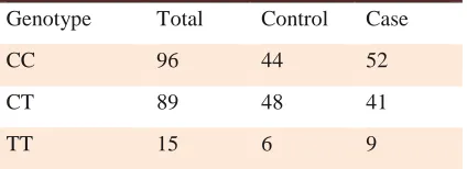
Data analysis show that there was not direct
significant association between C>T mutation in
rs3184504 SNP with coronary artery disease
(Table 1, 2)
The rate of biochemical factors such as
HDL-C, LDL, triglycerides as well as systolic and
diastolic blood pressure, and other factors like age,
weight, sex and waist circumference had significant
difference between case and control patients
(Table3).
Table 1. Genotypes frequencies in case and
control cohort.
Genotype Total Control Case
CC 96 44 52
CT 89 48 41
TT 15 6 9
Table 3. Association study of analyzed factors
with coronary artery disease.
Variants P-Value
Triglyceride 000
HDL-C 000
LDL 015
age 000
Weight 029
Systolic Blood Pressure 000 Diastolic Blood Pressure 000
Waist Circumference 008
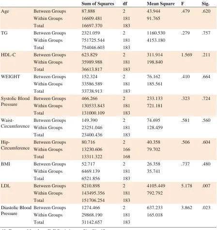
We also analyzed the association between
rs3184504 variant with biochemical factors such as
HDL-C, LDL, triglycerides, systolic and diastolic
blood pressure, and other factors like age, weight,
sex and some anthropometric measurements in
table 4. It was shown that there were not significant
associations between these factors, except for LDL
and diastolic blood pressure, and non-synonymous
SNP rs3184504(C>T) in case and control cohort
(table4). Regarding the SH2B3 gene and based on
the present study, there was a significant
association between the SNP and the increased
blood LDL and diastolic blood pressure (table 4).
Discussion
Many studies on CAD disease and its genetic
risk factors have been done so far excluding the
Iranian population. The locus which is proposed as
risk factor for development of CAD/MI includes a
candidate gene from previous studies which showed
that alleles frequencies may differ in population and
in genetic pool. Therefore, to determine the genetic
risk factors of the disease in any population, the
genetic pool of that population should be studied
first. Other factors such as personal characteristics,
biochemical factors and environment, can be
effective on the onset and developme
nt of the disease. Some of these factors can be
effective alone or in the association with other
factors or with a specific mutation.
In this research only one genetic factor has
been investigated. According to the results, there
was not a significant correlation between the
different alleles of the gene and CAD. However, a
significant correlation was observed between alleles
of this gene and increased LDL-C, diastolic and
systolic blood pressure which will be discussed
below.
One of the first studies on this SNP was done
by Carr et al. in 2009 on British population through
Realtime-Taqman method which demonstrated that
there is not a significant correlation between alleles
of C>T rs3184505 and vasculitis (the inflammation
of blood vessels) which can cause inflammation in
surface of blood vessels and therefore, reduction in
blood circulation and even blockage of vessels (42).
In the same year, another research by
Gudbjurtson et al. was performed on 12000
European and 5000 Asians which revealed that
rs3184504 had association with the number
of eosinophils in blood (27). Eosinophils are
Table 2. Association study between genotypes and risk of Coronary artery disease.
P-Value OR (95% CI)
Status=Control Status=Case
Genotype Model
0.42 1
44 (44.9%) 52 (51%)
CC
Codominant CT 41 (40.2%) 48 (49%) 1.38 (0.78-2.47) 0.79 (0.26-2.39) 6 (6.1%)
9 (8.8%) TT
0.39 1
44 (44.9%) 52 (51%)
CC Dominant
1.28 (0.73-2.23) 54 (55.1%)
50 (49%) CT-TT
0.47 1
92 (93.9%) 93 (91.2%)
CC-CT Recessive
0.67 (0.23-1.97) 6 (601%)
9 (8.8%) TT
0.21 1
50 (51%) 61 (59.8%)
CC-TT Overdominant
1.43 (0.82-2.50) 48 (49%)
41 (40.2%) CT
0.7 1.09 (0.70-1-70)
.... ....
....
Lod-Additive
SNP association with response Status (n=200, Crude analysis)
Table 4. Association of each polymorphic allele with different analyzed factors, using ANOVA test.
Sum of Squares df Mean Square F Sig.
Age Between Groups 87.888 2 43.944 .479 .620
Within Groups 16609.481 181 91.765
Total 16697.370 183
TG Between Groups 2321.059 2 1160.530 .279 .757
Within Groups 751725.544 181 4153.180
Total 754046.603 183
HDL-C Between Groups 623.829 2 311.914 1.569 .211
Within Groups 35989.988 181 198.840
Total 36613.817 183
WEIGHT Between Groups 152.324 2 76.162 .410 .664
Within Groups 33586.589 181 185.561
Total 33738.913 183
Systolic Blood Pressure
Between Groups 466.266 2 233.133 .323 .724
Within Groups 130533.843 181 721.181
Total 131000.109 183
Waist-Circumference
Between Groups 149.390 2 74.695 .581 .560
Within Groups 23251.046 181 128.459
Total 23400.436 183
Hip-Circumference
Between Groups 80.716 2 40.358 .506 .604
Within Groups 13230.606 166 79.702
Total 13311.322 168
BMI Between Groups 52.717 2 26.358 .737 .480
Within Groups 6469.139 181 35.741
Total 6521.856 183
LDL Between Groups 8210.898 2 4105.449 5.178 .007
Within Groups 143495.356 181 792.792
Total 151706.254 183
Diastolic Blood Pressure
Between Groups 1274.466 2 637.233 3.862 .023
Within Groups 29868.190 181 165.018
Total 31142.657 183
df : Degree of freedom; F: F Statistic test; Sig: Significancy
multifunctional leukocytes that have an important
role in the beginning of inflammation responses and
thus have an important role in inflammation
diseases such as heart attack (28). Hematopoietic stem cells (HSCs) exist in low quantity in adult’s bone marrow. However, the question, how
hemostasis of HSCs is preserved is not known
clearly yet. Findings demonstrate that in the
deficiency of HSCs adaptor, expression of BCL-X
in comparison to normal HSCs is increased from
the moment of thrombopoietin activation.
In addition, studies show that HSCs are
resistant to apoptosis because of high level of
expression in BCL-XI in patients with mutation in
SH2B3/LNK. As a result, the reduction of apoptosis
can lead to the aggregation of HCSCs which causes
several different diseases. Therefore, according to
studies, SH2B3 gene is introduced as a down
regulator for lymphocyte production and generally
a down regulator for hemostasis. Mutation in this
gene can be related to increased blood cells and
blood density (43).
Signal transduction of JAK-STAT by
intermediation of growth factors and cytokines has
an important role in biological trends such as
apoptosis, differentiation and proliferation (44).
Following the attachment of cytokines to the
receptors on cell surface, oligomerisation and
activation of JAk (a family of tyrosine kinase)
occurs (45). The family members of JAk are JAk1,
2, 3 and TYK. After activation of the cytoplasmic
region of the receptor, it is phosphorylated by JAk
and this action provides a region for STAT
adherence. STAT is also phosphorylated and
dimerised by JAK and immigrates to the cell
nucleus where gene transcription is regulated. The
SH2B3 gene has an important role by sticking to
JAK in this process (46).
In a research done by Natali et al. in 2000, it
was reported that 26% of the whole patients
suffering from heart disease have had high blood
pressure from which they have been suffering for
approximately 9 years as well. Only 12% of them
had recently been diagnosed with high blood
pressure (47). In addition, two other studies on
2009 and 2010 were performed based on case
control and meta-analysis methods in European and
Indian populations which expressed the correlation
between this SNP and high blood pressure (48).
Therefore, it appears that this SNP and several
other variants may contribute to the high blood
pressure. The metabolism of lipids in blood such as
triglyceride, increased cholesterol with low density
and decreased cholesterol with high density are
among the dangerous factors for coronary vessels.
It is not clear whether it reveals the improvement of
the coronary vessels (40).
Another study was done in London through
SNP array in which the relation of this variant and
the lipid blood factors such as LDL, HDL and apo
B was investigated. The results demonstrated a
significant correlation between this variant and
increased LDL (40). As mentioned, the present
study expresses the relationship and the role of this
variant in diastolic blood pressure and LDL level
increase.
According to several studies on CAD across
the world, by the evaluation of genes from
HAPMAP and GWAS data, there have been
different questions raised about each of the genetic
and environmental factors. One of the questions is
if a genetic factor alone or in association with
environment factors can lead to disease onset. This
question can be answered in cases that genetic and
environmental risk factors maps have been
previously traced for each population. Comparison
of these maps through complex and developed
software can lead to individual medicine. In case of
increased numbers of samples and the comparison
of their results related to gene mutation, a hope for
appropriate genetic map for each disease rises.
Increased samples and new techniques for the
diagnosis of CAD and SH2B3 gene in the future,
can be helpful for such studies through complete
sequencing of this gene and evaluation of
methylation.
Conflict of interest
The authors declared no conflicts of interest.
References
1. Musunuru K, Kathiresan S. Genetics of coronary artery
disease. Annu Rev Genomics Hum Genet 2010;11:91-108.
2. Donyavi T, Naieni KH, Nedjat S, et al. Socioeconomic status
and mortality after acute myocardial infarction: a study from
Iran. Int J Equity Health 2011;10:9.
3. Mitchell BD, Kammerer CM, Blangero J, et al. Genetic and
environmental contributions to cardiovascular risk factors in
Mexican Americans. The San Antonio Family Heart Study.
Circulation 1996;94:2159-70.
4. Badaruddoza, Kaur P. Familial Aggregation of Blood Pressure
With Respect to Anthropometric Variables Among the Lobana
(Nomadic Origin) Population in Punjab, India. Asia Pac J Public
Health 2012;24:104-16.
5. Harrap SB, Stebbing M, Hopper JL, et al. Familial patterns of
covariation for cardiovascular risk factors in adults: The
Victorian Family Heart Study. Am J Epidemiol 2000; 152:704-15.
6. Ikram MK, Sim X, Jensen RA, et al. Four novel Loci (19q13,
6q24, 12q24, and 5q14) influence the microcirculation in vivo.
PLoS Genet 2010;6:e1001184.
7. Camici PG, Crea F. Coronary microvascular dysfunction. N
Engl J Med 2007;356:830-40.
8. Watkins H, Farrall M. Genetic susceptibility to coronary
artery disease: from promise to progress. Nat Rev Genet
2006;7:163-73.
9. Wellcome Trust Case Control Consortium. Genome-wide
association study of 14,000 cases of seven common diseases and
3,000 shared controls. Nature 2007;447:661-78.
10. Hubbard LD, Brothers RJ, King WN, et al. Methods for
evaluation of retinal microvascular abnormalities associated with
hypertension/sclerosis in the Atherosclerosis Risk in
Communities Study. Ophthalmology 1999;106:2269-80.
11. Ikram MK, de Jong FJ, Vingerling JR, et al. Are retinal
arteriolar or venular diameters associated with markers for
cardiovascular disorders? The Rotterdam Study. Invest
Ophthalmol Vis Sci 2004;45:2129-34.
12. Wong TY, Mitchell P. Hypertensive retinopathy. N Engl J
Med 2004;351:2310-7.
13. Wong TY, Klein R, Sharrett AR, et al. Retinal arteriolar
diameter and risk for hypertension. Ann Intern Med
2004;140:248-55.
14. Wong TY, Klein R, Sharrett AR, et al. Retinal arteriolar
narrowing and risk of diabetes mellitus in middle-aged persons.
JAMA 2002;287:2528-33.
15. Qiu C, Cotch MF, Sigurdsson S, et al. Retinal and cerebral
microvascular signs and diabetes: the age, gene/environment
susceptibility-Reykjavik study. Diabetes 2008;57:1645-50.
16. McGeechan K, Liew G, Macaskill P, et al. Prediction of
incident stroke events based on retinal vessel caliber: a
systematic review and individual-participant meta-analysis. Am
J Epidemiol 2009;170:1323-32.
17. McGeechan K, Liew G, Macaskill P, et al. Meta-analysis:
retinal vessel caliber and risk for coronary heart disease. Ann
Intern Med 2009;151:404-13.
18. Ikram MK, De Jong FJ, Van Dijk EJ, et al. Retinal vessel
diameters and cerebral small vessel disease: the Rotterdam Scan
Study. Brain 2006;129:182-8.
19. Longstreth W, Jr., Larsen EK, Klein R, et al. Associations
between findings on cranial magnetic resonance imaging and
retinal photography in the elderly: the Cardiovascular Health
Study. Am J Epidemiol 2007;165:78-84.
20. Devalliere J, Chatelais M, Fitau J, et al. LNK (SH2B3) is a
key regulator of integrin signaling in endothelial cells and targets
alpha-parvin to control cell adhesion and migration. FASEB J
2012;26:2592-606.
21. Mitchell RN, Libby P. Vascular remodeling in transplant
vasculopathy. Circ Res 2007;100:967-78.
22. Lauffenburger DA, Horwitz AF. Cell migration: a physically
integrated molecular process. Cell 1996;84:359-69.
23. Sheetz MP, Felsenfeld D, Galbraith CG, et al. Cell migration
as a five-step cycle. Biochem Soc Symp 1999;65:233-43.
24. Cambien F, Tiret L. Genetics of Cardiovascular Diseases:
From Single Mutations to the Whole Genome. Circulation
2007;116:1714-24.
25. Winkelmann BR, Hager J. Genetic variation in coronary
heart disease and myocardial infarction: methodological
overview and clinical evidence. Pharmacogenomics
2000; 1:73-94.
26. Reilly MP, Li M, He J, et al. Identification of ADAMTS7 as
a novel locus for coronary atherosclerosis and association of
ABO with myocardial infarction in the presence of coronary
atherosclerosis: two genome-wide association studies. Lancet
2011;377:383-92.
27. Gudbjartsson DF, Bjornsdottir US, Halapi E, et al. Sequence
variants affecting eosinophil numbers associate with asthma and
myocardial infarction. Nat Genet 2009;41:342-7.
28. van der Harst P, Zhang W, Mateo Leach I, et al. Seventy-five
genetic loci influencing the human red blood cell. Nature
2012;492:369-75.
29. Levy D, Ehret GB, Rice K, et al. Genome-wide association
study of blood pressure and hypertension. Nat Genet
2009;41:677-87.
30. Huang X, Li Y, Tanaka K, et al. Cloning and
characterization of Lnk, a signal transduction protein that links
T-cell receptor activation signal to phospholipase C gamma 1,
Grb2, and phosphatidylinositol 3-kinase. Proc Natl Acad Sci U S
A 1995;92:11618-22.
31. Takaki S, Watts JD, Forbush KA, et al. Characterization of
Lnk. An adaptor protein expressed in lymphocytes. J Biol Chem
1997;272:14562-70.
32. Velazquez L, Cheng AM, Fleming HE, et al. Cytokine
signaling and hematopoietic homeostasis are disrupted in
Lnk-deficient mice. J Exp Med 2002;195:1599-611.
33. Devalliere J, Charreau B. The adaptor Lnk (SH2B3): an
emerging regulator in vascular cells and a link between immune
and inflammatory signaling. Biochem Pharmacol 2011;82:
1391-402.
34. Takaki S, Sauer K, Iritani BM, et al. Control of B cell
production by the adaptor protein lnk. Definition Of a conserved
family of signal-modulating proteins. Immunity 2000;
13:599-609.
35. Takaki S, Tezuka Y, Sauer K, et al. Impaired lymphopoiesis
and altered B cell subpopulations in mice overexpressing Lnk
adaptor protein. J Immunol 2003;170:703-10.
36. Seita J, Ema H, Ooehara J, et al. Lnk negatively regulates
self-renewal of hematopoietic stem cells by modifying
thrombopoietin-mediated signal transduction. Proc Natl Acad
Sci U S A 2007;104:2349-54.
37. Bersenev A, Wu C, Balcerek J, et al. Lnk controls mouse
hematopoietic stem cell self-renewal and quiescence through
direct interactions with JAK2. J Clin Invest 2008;118:2832-44.
38. Kwon SM, Suzuki T, Kawamoto A, et al. Pivotal role of lnk
adaptor protein in endothelial progenitor cell biology for
vascular regeneration. Circ Res 2009;104:969-77.
39. Tong W, Lodish HF. Lnk inhibits Tpo-mpl signaling
and Tpo-mediated megakaryocytopoiesis. J Exp Med
2004;200:569-80.
40. Talmud PJ, Drenos F, Shah S, et al. Gene-centric association
signals for lipids and apolipoproteins identified via the
HumanCVD BeadChip. Am J Hum Genet 2009;85:628-42.
41. World Health Organization. Prevention of cardiovascular
disease. Guidelines for assessment and management of
cardiovascular risk. Geneva, 2007.
42. Carr EJ, Niederer HA, Williams J, et al. Confirmation of the
genetic association of CTLA4 and PTPN22 with
ANCA-associated vasculitis. BMC Med Genet 2009;10:121.
43. Suzuki N, Yamazaki S, Ema H, et al. Homeostasis of
hematopoietic stem cells regulated by the myeloproliferative
disease associated-gene product Lnk/Sh2b3 via Bcl-xL. Exp
Hematol 2012;40:166-74 e3.
44. Dreesen O, Brivanlou AH. Signaling pathways in cancer and
embryonic stem cells. Stem Cell Rev 2007;3:7-17.
45. Valentino L, Pierre J. JAK/STAT signal transduction:
regulators and implication in hematological malignancies.
Biochem Pharmacol 2006;71:713-21.
46. Schindler C, Levy DE, Decker T. JAK-STAT signaling:
from interferons to cytokines. J Biol Chem 2007;282:20059-63.
47. Galvan AQ, Galetta F, Natali A, et al. Insulin resistance and
hyperinsulinemia: No independent relation to left ventricular
mass in humans. Circulation 2000;102:2233-8.
48. Lian J, Huang Y, Huang RS, et al. Meta-analyses of four
eosinophil related gene variants in coronary heart disease. J
Thromb Thrombolysis 2013;36:394-401.