ISSN: 2278-067X, Volume 1, Issue 9 (June 2012), PP.55-59
www.ijerd.com
Fast Evolution of Curve Based On Chan-Vese Model for Specific
Images
Madhu Anand
1, Vikram Singh
2, Sumit Kaushik
31,2
CSA department, Ch.Devilal University Sirsa (INDIA)
3
GNI, Mullana (India)
Abstract--In image processing, segmentation is an important intermediate step for object recognition. It is a fundamental task in image analysis responsible for partitioning an image into multiple sub-regions based on a desired feature. One such technique is through Curve Evolution and one of the applications of Geometric Curve Evolution is Active Contours.
In the research work being reported in this paper an attempt has been made to enhance Chan-Vese [1] Algorithm faster by taking different planes (red, green and blue) of an image. The plane in which the contour evolves fastest gets automatically selected and rest of the algorithm will work on that particular plane. Experimental results are presented on various domains like biomedical images, artificial images and painted images. Also, the contour evolves differently in different planes.
Key Terms- active contour, objects, object recognition, segmentation, standard deviation
I.
INTRODUCTION
Segmentation is an intermediate step in all high level object-recognition tasks. For example, if we want to locate the face of a particular person in an image of a crowd, we should first determine that which parts of the image correspond to human faces. Active Contours have been widely used as attractive image segmentation methods because they always produce sub-regions with continuous boundaries. For instance, starting with a curve around the object to be detected, the curve moves toward its interior normal and has to stop on the boundary of the object. An object in image processing is an identifiable portion of an image that can be interpreted as single unit.
The active contour model is more and more used in image segmentation because it relies on solid mathematical properties and its numerical implementation uses the level set method to track evolving contours. The motion of the curve is driven by the image itself. The driving force is obtained by defining a potential in the image that shall be small n ear objects contours.
If β is a grey level image, the external potential is defined at each point of the image and is of the type
g(x) = 1 (1+ǀDGσ*βǀ2)
where G is a gaussian with standard deviation s. The term appearing in the denominator is the gradient of a regularize d version of the image ( * denotes convolution.)
Active contours can be classified as parametric active contours and geometric active contours according to their representation and implementation. Geometric active contours were in use now a days, which provide solution to tackle the problem of topological changes required in curve evolution. In [5] a precise relationship between the geometric and parametric active contours has been developed which includes spatially-varying coefficients, both tension and rigidity, and non-conservative external forces. Geometric deformable models implemented using level set methods have advantages over parametric models due to their intrinsic behavior, parameterization independence, and ease of implementation.
Active contour models are also used for 2D and 3D biomedical images formulated using the level set method [6].These models can also be generalized to segmentation of images with more than two segments.
56
In [9] a graph cut based active contour without edges segmentation model has been discussed to track pedestrian in thermal images. The deformable model is based on the Mumford- Shah piecewise constant energy formulation.
An algorithm for automated segmentation of white matter in brain MRI images was proposed [11] which can be used to create connected representations of the gray matter in the cerebral cortex of the brain. Automating the postmortem identification of deceased individuals based on dental characteristics is receiving increased attention especially with the large number of victims encountered in mass disasters.
In [10] a novel formulation framework of the minimal surface problem, called Active Geometric Functions (AGF), is proposed to reach truly real-time performance in segmenting 4D ultrasound data.
II.
THE MODEL
The original idea of active contours was given by Kass, Witkin and Terzopoulos. We have chosen a model for active contours to detect objects in a given image, based on techniques of curve evolution, Mumford–Shah functional [3] for segmentation and level sets [2].
Chan-Vese approach involves geometric active contour model (based upon Mumford – Shah Functional ). The model begins with a contour in the image plane defining an initial segmentation and then contour is evolved according to evolution equation. The basis of Chan-Vese algorithm is a Fitting Energy Functional [1]. The goal of algorithm is to minimize this fitting energy for a given image and corresponding will define segmentation.
In general form it is written as follows:
F(ɸ) = µ(ʃǀ H(ɸ)ǀdx)ᵖ + ʋʃH(ɸ)dx Ω Ω +λ1ʃǀI- C1ǀ²H(ɸ)dx Ω +λ2ʃǀI-C2ǀ²(1-H(ɸ))dx Ω
μ, υ, λ1, λ2 and p are parameters selected to fit a particular class of images.
Here,
H=Heavy side function I is the image to be segmented Ω =domain of that image
C1 and C2 are averages of the image I in the regions where ɸ>=0 and ɸ<0 respectively.
In the above equation the first term can be thought as a penalty on the total length of the edge contour for a given segmentation. The second term is a penalty on the total area of the foreground region found by the segmentation. The third term is proportional to the variance of the image gray level in the in the foreground region. The fourth term does the same for background region. Usually, we take λ1=λ2=1 but if we set λ1=2, λ2=1 then our final segmentation will have a more uniform
foreground region at the expense of loss of uniformity in background.
The contour ultimately segments the image into foreground and background. Chan- Vese [1] algorithm evolves this contour via a level set method. The function (I,i,j) (the level set function where (i,j) are co-ordinates in the image and t is time). The segmentation is given by two regions{Φ>0} and {Φ<0}. Some PDE (Partial Differential Equation) is used to evolve level set function.
III.
RESULT AND ANALYSIS
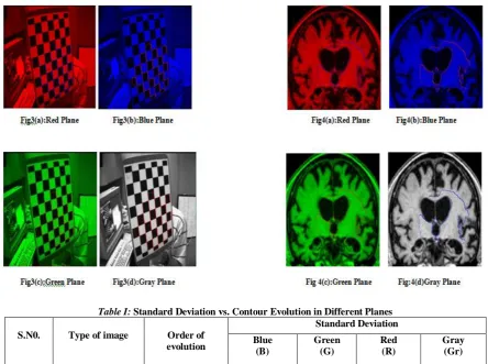
In the research work being reported in this paper, four planes red, green, gray and blue have been taken and standard deviation corresponding to each plane is calculated by keeping the number of iterations and size of each image fixed. It is found that the contour evolves faster in the plane having highest value of standard deviation. This is because the energy in different planes attracts the contour differently. The position of the initial curve can be anywhere in the plane, and it does not necessarily surround the objects to be detected.
It is shown that the images (fig 3 & fig 4) in which the red, blue and green components are present almost equally are having almost same values for standard deviation. In case of painted images (fig.1) the red plane is having highest value of standard deviation. The contour is evolving fast covering objects minutely as compare to blue and green planes.
58
Table I: Standard Deviation vs. Contour Evolution in Different Planes
S.N0. Type of image Order of evolution
Standard Deviation
Blue (B)
Green (G)
Red (R)
Gray (Gr)
1. Painted Image R>G>Gr>B 56.062 68.230 80.148 57.761
2. Astronomical Image
B>G>Gr>R 53.979 53.939 13.217 39.474
3. Biomedical Image B>G>R>Gr 71.460 71.457 71.425 71.398
4. Synthetic Image B=G=R=Gr 71.339 71.339 71.339 71.339
IV.
CONCLUSION & FUTURE SCOPE
Chan-Vese algorithm has been enhanced for specific application in image segmentation. It has also been shown that the proposed algorithm is effective on a wide variety of images in red, green and blue planes. Chan-vese presented a modified model for vector images [12] but its computation is expensive as different planes require λ1, λ2 and c1 and c2 (region averages) values to be computed separately.
For those types of images where there is not much difference in three component values (red, green, blue) proper plane can be selected for fast evolution without losing segmentation. In particular, the images where one or two components of color are negligible we can make use of standard deviation to select the plane which has fastest evolution. Thereby, reducing unnecessary computation like in astronomical image, red component is significantly small and thus ignorable. Hence saving much computation as compare to Chan–Vese model for vector images.
REFERENCES
[1]. Tony F. Chan, Luminita A. Vese ”Active contours without edges”, IEEE transactions on image processing, Vol.10, NO. 2, February 2001
[2]. Robert Crandall, “Image segmentation using Chan Vese algorithm”, ECE532 Project fall, 2009
[3]. Yingjie Zhang “Fast Segmentation for the Piecewise Smooth Mumford-Shah Functional”, International Journal of Signal Processing, Fall 2006
[4]. D. Mumford and J. Shah. Optimal approximations by piecewise smooth functions and associated variational problems. Comm. Pure Appl. Math., 42(5):577–685, 1989.
[5]. Chenyang Xu, Anthony Yezzi, Jr. and Jerry L. Prince, “On the Relationship between Parametric and Geometric Active Contours,” In Proc. of 34th Asilomar Conference on Signals, Systems, and Computers, pp. 483-489, October 2000.
[6]. Tony F.Chan Luminita A.Vese, “Active Contour and Segmentation Models Using Geometric PDE’s for Medical Imaging,” Geometric Methods in Bio-Medical Image Processing'', Series: Mathematics and Visualization, Springer, 2002, pp. 63-75.
[7]. Michael Kass, Andrew Witkin and D.Terzopoulos, “Snakes: Active Contour Models,” International Journal of Computer Vision, 321-331(1998).
[8]. Li Wang, Chunming Li, Quansen Sun, Deshen Xia, and Chiu-Yen Kao, “ Brain MR Image Segmentation Using Local and Global Intensity Fitting Active Contours/Surfaces,” Springer-Verlag Berlin Heidelberg 2008.
[9]. Noha El-Zehiry and Adel Elmaghraby, “A Graph Cut Based Active Contour without Edges with Relaxed Homogeneity Constraint,” International Conference on Pattern Recognition, 2008.
[10]. Qi Duan, Elsa D. Angelini, Shunichi Homma and Andrew F. Laine, “Real-Time Segmentation of 4D Ultrasound By Active Geometric Functions,” IEEE International Symposium on Biomedical Imaging, 2008.
[11]. Gowri Srinivasa, Vivek S. Oak, Siddharth J. Garg, Matthew C. Fickus and Jelena Kovacevic, “Voting-Based Active Contour Segmentation Of FMRI Images Of The Brain,” Proc. IEEE Conf.on Image Proc.,San Diego, USA Oct.2008.