A Framework for Evaluation of the
Higher-Risk Infant After a Brief
Resolved Unexplained Event
J. Lawrence Merritt, II, MD,aRicardo A. Quinonez, MD, FAAP, FHM,bJoshua L. Bonkowsky, MD, PhD, FAAP,c,d Wayne H. Franklin, MD, MPH, MMM, FAAP,eDavid A. Gremse, MD, FAAP,fBruce E. Herman, MD, FAAP,c Carole Jenny, MD, MBA, FAAP,aEliot S. Katz, MD, FAAP,gLeonard R. Krilov, MD, FAAP,hChuck Norlin, MD, FAAP,c Robert E. Sapién, MD, MMM, FAAP,iJoel S. Tieder, MD, MPH, FAAPa
abstract
In 2016, the American Academy of Pediatrics published a clinical practiceguideline that more specifically defined apparent life-threatening events as brief resolved unexplained events (BRUEs) and provided evidence-based recommendations for the evaluation of infants who meet lower-risk criteria for a subsequent event or serious underlying disorder. The clinical practice guideline did not provide recommendations for infants meeting higher-risk criteria, an important and common population of patients. Therefore, we propose a tiered approach for clinical evaluation and management of higher-risk infants who have experienced a BRUE. Because of a vast array of potential causes, the initial evaluation prioritizes the diagnosis of time-sensitive conditions for which delayed diagnosis or treatment could impact outcomes, such as child maltreatment, feeding problems, cardiac arrhythmias, infections, and congenital abnormalities. The secondary evaluation addresses problems that are less sensitive to delayed diagnosis or treatment, such as dysphagia, intermittent partial airway obstruction, and epilepsy. The authors recommend a tailored, family-centered, multidisciplinary approach to evaluation and management of all higher-risk infants with a BRUE, whether accomplished during hospital admission or through coordinated outpatient care. The proposed framework was developed by using available evidence and expert consensus.
The American Academy of Pediatrics (AAP) introduced a clinical practice guideline (CPG) defining the term“brief resolved unexplained event”(BRUE) to more precisely describe events for which the outdated term“apparent life-threating event”(ALTE) was previously used.1A BRUE is an event occurring in an infant younger than 1 year when the observer reports a sudden, brief, and now-resolved episode of 1 or more of the following: (1) cyanosis or pallor; (2) absent, decreased, or irregular breathing; (3) marked change in tone (hyper- or hypotonia); or an (4) altered
level of responsiveness.1BRUE is a diagnosis of exclusion and is used only when there is no explanation for the event after conducting an
appropriate history and physical.
The AAP CPG recommendations apply only to infants defined as“lower risk.” Lower-risk infants are defined as (1) age.60 days, (2) gestational age $32 weeks and postconceptional age $45 weeks, (3) no cardiopulmonary resuscitation required by trained medical provider, (4) BRUE duration
,1 minute, and (5)first BRUE (no repeat events, regardless of time
aDepartment of Pediatrics, University of Washington and
Seattle Children’s Hospital, Seattle, Washington;bSection of
Pediatric Hospital Medicine, Department of Pediatrics, Baylor College of Medicine, Houston, Texas;cDepartment of Pediatrics, School of Medicine, University of Utah, Salt Lake City, Utah;dBrain and Spine Center, Primary Children’s Hospital, Salt Lake City, Utah;eDepartment of Pediatrics,
Stritch School of Medicine, Loyola University, Maywood, Illinois;fDepartment of Pediatrics, University of South
Alabama, Mobile, Alabama;gDepartment of Medicine,
Harvard Medical School, Harvard University, Boston, Massachusetts;hDepartment of Pediatrics, New York
University Winthrop, Mineola, New York; andiDepartment of Emergency Medicine, Health Sciences Center, University of New Mexico, Albuquerque, New Mexico
Drs Merritt, Quinonez, Bonkowsky, Franklin, Gremse, Herman, Jenny, Katz, Krilov, Norlin, Sapién, and Tieder conceptualized and designed the study, drafted the initial manuscript, and approved thefinal manuscript as submitted.
The guidelines and recommendations in this article are not American Academy of Pediatrics policy, and publication herein does not imply endorsement.
DOI:https://doi.org/10.1542/peds.2018-4101 Accepted for publication May 24, 2019
Address correspondence to J. Lawrence Merritt II, MD, Biochemical Genetics, Seattle Children’s Hospital, MB.6.830, 4800 Sand Point Way NE, Seattle, WA 98105. E-mail: [email protected] PEDIATRICS (ISSN Numbers: Print, 0031-4005; Online, 1098-4275).
Copyright © 2019 by the American Academy of Pediatrics
interval) as well as having no concerning historical features (eg, sudden death in a relative) and no concerning physical examination findings (eg, unexplained bruising).1 There are currently no
recommendations for infants who do not meet these criteria and are considered higher risk, despite this situation being common and
challenging to manage. In this article, we propose a framework to guide the evaluation and management of infants meeting higher-risk criteria (Table 1). Because of a paucity of research on higher-risk infants, we do not provide recommendations in the format of a CPG. Rather, we offer an evidence-informed framework to guide initial medical decision-making, ongoing research, and improvements in managing this population.
METHODS
The authors included stakeholders across general pediatrics and multiple subspecialties, including child abuse, cardiology,
gastroenterology, hospital medicine, emergency medicine, pulmonology, neurology, infectious disease, and biochemical genetics. They reviewed articles identified in the original and updated systematic reviews of ALTE by Tieder et al.1,2Additional relevant subspecialty-focused articles related to the more-common diagnoses associated with such events (eg, child abuse, oropharyngeal
dysphagia, and cardiac arrhythmias) were reviewed, with emphasis on
those providing substantial evidence (levels A, B, or C).1From this expanded collection, authors identified and prioritized (considering accuracy, risk, and benefit) characteristics,findings, and evaluations on the basis of their ability to detect a serious underlying cause or lead to recurrent events (Fig 1). Through ongoing
communication and iterative drafts, consensus was developed among the authors, and conflicts were resolved through discussion. The proposed evaluations were then divided into 2 tiers. The goal of the“initial tier”is to detect problems that are (1) uncommon but could lead to serious adverse outcomes if not diagnosed or treated promptly (eg, medical child abuse [MCA] or pertussis) or (2) common and unlikely to lead to serious adverse outcomes but in which early diagnosis could prevent recurrent events and preclude unnecessary testing or caregiver concern (eg,“silent aspiration”or viral testing to identify infection). The goal of the“secondary tier”is to identify potential causes of recurrent events when clinical concerns regarding less-common characteristics, findings, and evaluations remain after completing the initial tier.
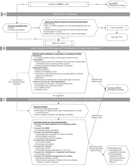
BRUE DIAGNOSIS AND RISK STRATIFICATION
In the initial medical assessment, a careful history and physical examination are needed to understand what occurred before, during, and after the event and to identify signs of potential causes. In the absence of an alternative explanation, a BRUE diagnosis can be made if the clinician characterizes the event as a sudden, brief, resolved episode involving 1 or more of the 4 BRUE characteristics. Once
a clinician has diagnosed a BRUE, it is important to obtain additional history and perform a thorough physical examination targeting
common and/or serious conditions that may present with a BRUE.1(We recommend reviewing the CPG’s Table 2 Historical Features To Be Considered in the Evaluation of a Potential BRUE and Table 3 Physical Examination Features To Be Considered in the Evaluation of a Potential BRUE.1) It is important to consider temporal relationships of the event to feedings, coughing, and sleeping. For example, dysphagia typically occurs with or shortly after feedings, whereas seizures can occur at any time.
It is important to note that if the history and physical examination reveal an explanation, then the patient did not, by definition, have a BRUE, and management specific to the diagnosis should be implemented according to that alternative
diagnosis. For example, events in infants with ongoing symptoms or abnormalfindings such as fever and respiratory symptoms do not meet BRUE criteria. If a cause is not found through history and physical examination, the infant’s risk for a serious underlying condition or adverse outcome should be determined and classified as lower or higher risk.1Subsequent testing and management to identify a cause should then be based on the risk determination. Risk criteria are summarized in Fig 1A and Table 1. Recommended management of infants at lower risk is detailed in the CPG1; in the remainder of this article, we discuss management of those at higher risk.
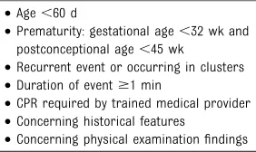
The evaluation and management of higher-risk infants is challenging because there are many potential causes of a BRUE and a dearth of evidence to support using clinical factors to quantify risk for adverse outcomes, such as recurrent events or diagnosis of a serious underlying cause. Potential causes are outlined in Table 2, with history and physical examinationfindings that may help guide clinicians in identifying TABLE 1Higher-Risk BRUE Criteria
•Age,60 d
•Prematurity: gestational age,32 wk and postconceptional age,45 wk
•Recurrent event or occurring in clusters
•Duration of event$1 min
•CPR required by trained medical provider
•Concerning historical features
•Concerning physical examinationfindings
FIGURE 1
specific concerning features listed in Table 3. As in lower-risk infants, nonspecific and indiscriminate testing, such as routine complete blood cell counts and chest radiographs, for all higher-risk infants is unlikely to reveal an
event’s cause, and it may increase harm by leading to false-positives and further unnecessary testing.10 Targeted evaluations based on specificfindings are discussed in greater detail in the following sections.
INITIAL EVALUATION AND MANAGEMENT
For higher-risk infants, initial tier evaluations (Fig 1B) may identify problems sensitive to delays in diagnosis and treatment.
Hospitalization may not be needed for many patients at this stage, particularly if close follow-up with a primary care clinician can be arranged and the services are available either in the emergency department (ED) or outpatient setting. These may include the following:
•continuous pulse oximetry monitoring for at least 4 hours; •consultation with social worker (or
other health care worker with similar experience and skills); •bedside feeding evaluation (by
a feeding therapist, if available); •electrocardiogram to be read by
a pediatric cardiologist or electrophysiologist;
•rapid viral respiratory panel testing;
•rapid pertussis polymerase chain reaction in endemic areas, during regional outbreaks, or in
underimmunized patients; •hematocrit;
•blood glucose, bicarbonate or venous blood gas, and lactate; and •if concerned for child
maltreatment, consultation with a child abuse expert, head imaging with computed tomography (CT) or MRI, and skeletal survey.
(Note that evidence supporting these recommendations is reviewed in subsequent sections.)
SECONDARY EVALUATION AND MANAGEMENT
If no explanation is identified through the initial tier of evaluations,
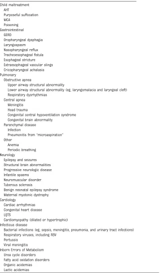
secondary tier evaluations may be tailored to specific individual concerns remaining from previously identified characteristics,findings, and evaluations or clinician and/or TABLE 2Potential Causes of BRUE in Higher-Risk Infants
Child maltreatment AHT
Purposeful suffocation MCA
Poisoning Gastrointestinal
GERD
Oropharyngeal dysphagia Laryngospasm
Nasopharyngeal reflux Tracheoesophagealfistula Esophageal stricture
Extraesophageal vascular slings Cricopharyngeal achalasia Pulmonary
Obstructive apnea
Upper airway structural abnormality
Lower airway structural abnormality (eg, laryngomalacia and laryngeal cleft) Respiratory dysrhythmias
Central apnea Meningitis Head trauma
Congenital central hypoventilation syndrome Congenital brain abnormality
Parenchymal disease Infection
Pneumonitis from“microaspiration” Other
Anemia
Periodic breathing Neurology
Epilepsy and seizures Structural brain abnormalities Progressive neurologic disease Infantile spasms
Neuromuscular disorder Tuberous sclerosis
Benign neonatal epilepsy syndrome Maternal myotonic dystrophy Cardiology
Cardiac arrhythmias Congenital heart disease LQTS
Cardiomyopathy (dilated or hypertrophic) Infectious disease
Bacterial infections (eg, sepsis, meningitis, pneumonia, and urinary tract infections) Respiratory viruses, including RSV
Pertussis Viral meningitis Inborn Errors of Metabolism
Urea cycle disorders Fatty acid oxidation disorders Organic acidemias
caregiver concerns in the history and physical examination. Depending on family preference and other circumstances, it is reasonable to perform the secondary tier of evaluations after discharge (Fig 1C). It is also reasonable to admit the infant to the hospital for a period of observation, continuous prolonged oximetry, and clinical swallow evaluation or feeding specialist consultation to better assess recurrent events and complete evaluations that could not be otherwise arranged in the ambulatory setting. Secondary tier evaluations can include any of the following consultations:
•gastroenterology; •otolaryngology;
•pulmonary or sleep expert; •child abuse expert;
•neurology; •cardiology; and •biochemical genetics.
Secondary tier evaluations that may be considered in combination with specialty consultation are as follows: •videofluoroscopic swallowing
study (VFSS) for "silent"
oropharyngeal dysphagia not seen in bedside evaluation;
•continuous prolonged oximetry to characterize recurring events; •comprehensive polysomnography
(PSG) to characterize and quantify central versus obstructive apnea; •prolonged ($12–24 hours) EEG;
and
•blood sodium, potassium, chloride, blood urea nitrogen, creatinine, calcium, and ammonia for metabolic disturbance.
If the BRUEs continue to occur, the following additional evaluations rarelyfind a cause of the BRUE but may be appropriate in certain circumstances in combination with specialty consultation. These could include upper gastrointestinal series (UGI), esophageal multichannel
intraluminal impedance–pH monitoring (MII-pH),
esophagogastroduodenoscopy with biopsy, arterial blood gas, chest radiograph, brain MRI or CT, echocardiogram, urine organic acids, plasma amino acids, or plasma acylcarnitines (Supplemental Information).
HOSPITAL ADMISSION
There is little evidence to guide which higher-risk infants with a BRUE are most likely to benefit from
hospitalization. Discussion of risks and benefits in a family-centered manner that incorporates shared decision-making and caregiver risk tolerance is advised,11,12using discussion tools when available (eg, Brief Resolved Unexplained Event: What Parents and Caregivers Need to Know, https://patiented.solutions. aap.org/handout.aspx?gbosid= 239090). The few prospective studies offering clinical decision rules to guide hospital admission used ALTE criteria and are thus difficult to apply to a more-specific BRUE
population.13–15If a decision to admit is made, it is important to establish and communicate the goals with caregivers up front because it is unlikely for events to recur during the inpatient stay or for the admission to reveal a diagnosis of a serious underlying condition. The availability of specialized testing and access to subspecialists and a primary care clinician should be considered in the decision for admission.
OUTPATIENT CONSIDERATIONS
It is possible for primary care clinicians to coordinate outpatient evaluation for higher-risk infants (including testing, consultants, or social work). Primary care clinicians may have an established relationship with the patient and family that could allow more efficient evaluation, more reliable follow-up, and optimal parent engagement for shared
decision-making and limit unnecessary anxiety and risk from a hospital setting.16It is recommended that primary care clinicians be aware of the indications for further evaluation in both lower-and higher-risk infants with a BRUE because they can efficiently arrange further monitoring, evaluation, consultation, and/or hospitalization as may be clinically appropriate.
Higher-risk infants with a BRUE who are evaluated and initially managed in the hospital or ED need effective follow-up by a primary care clinician. Timely and complete communication of thefindings and plan of care from the emergency or hospital setting are recommended.
SUBSPECIALTY EVALUATION
Child Maltreatment
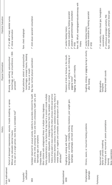
The most time-sensitive diagnoses presenting as a BRUE are abusive head trauma (AHT), inflicted suffocation, intentional poisoning, and MCA involving exaggerated, fabricated, or inflicted injury. The risk factors and physical examination findings for child abuse can be subtle and easily missed during a BRUE presentation, so a high index of suspicion and thorough evaluation is warranted (Tables 2 and 3).17,18It is important to remember that bruising is highly concerning in infants that are not mobile or when the bruises occur on the face, trunk, and ears.
Children who suffer AHT may present with a BRUE with higher-risk criteria, likely from central apneic episodes. Although likely symptomatic after the initial trauma, when care is delayed, children with AHT can appear normal. They also may not have external signs of trauma.19,20 One-third of infants and toddlers who presented for medical evaluation after an episode of AHT had been seen previously by a medical
complications, 27.8% were reinjured, and 2.8% died. Typically, AHT occurs when the child is in the care of a single person, more likely
a nonrelated man.6Other risk factors include a caretaker history of criminal offenses, previous social service involvement, and a history of domestic violence.3In a physical examination, subtle signs of trauma should be looked for.3–5It is recommended to obtain neuroimaging if there is anemia (hemoglobin,11.2 g/dL), seizures, or central apnea that could be from cranial bleeding.3,4Retinal
examination may be helpful to diagnose child abuse when there are concerns on neuroimaging.
The definition of MCA is a child receiving unnecessary harmful or potentially harmful medical care at the instigation of the caretaker, most often a parent.22When evaluating a higher-risk infant after a BRUE, features of concern for MCA include recurrent episodes and those only occurring in the presence of a particular person; presenting symptoms that are dramatically described, although the child looks perfectly normal; caretaker giving a complex history of multiple complaints or diagnoses involving multiple systems; or the caretaker having a complex personal medical history or other children with undiagnosed illness or multiple illness diagnoses. Diagnosis of MCA requires a careful review of all available medical records. Assessment for purposeful
suffocation should include looking for often-subtlefindings such as facial petechiae, scleral or subconjunctival hemorrhages, oronasal trauma, or bleeding from the nose or mouth. If there are signs of pulmonary hemorrhage, a chest radiograph may be indicated.5,23,24
If child maltreatment of any form is suspected, it is recommended to consult a pediatrician and social worker specializing in child abuse.
Gastrointestinal
Nearly every infant has some feeding difficulties or gastroesophageal reflux (GER) symptoms, so it can be difficult to know whether these problems contributed to a BRUE. Common gastrointestinal abnormalities in higher-risk infants include laryngospasm or pulmonary aspiration from oropharyngeal dysphagia or gastroesophageal reflux disease (GERD). Obtaining a careful feeding history is critical (Table 3).
Oropharyngeal dysphagia is dysfunctional suck, swallow, and breathe coordination and most common when infants arefirst learning to feed. Oropharyngeal dysphagia can be associated with aspiration, laryngeal penetration, or nasopharyngeal reflux.25Symptoms include choking, gagging, color change with feeds, taking
.30 minutes per feed, or pooling of feeds in the mouth.26Observation of feeding by a trained observer, typically an occupational or speech therapist, who specializes in feeding disorders can usually identify problems. If further evaluation is needed after bedside feeding observation and consideration by a pediatric gastroenterologist, to diagnose "silent" aspiration, VFSS may be considered a part of secondary diagnostic testing.
A history suggestive of obstructive apnea (respiratory effort but little air movement) that occurs during or shortly after feeding, especially when supine or with positional changes after feeding, may be associated with ascending gastric reflux–related laryngospasm. For infants with recurrent BRUE and unclear GER symptoms, it is reasonable to initiate a trial of conservative GER treatment (eg, upright position after feeds, smaller-volume feeds). If these fail, further diagnostic testing for
a gastrointestinal cause may helpfind a cause for the BRUE. A consultation with a pediatric gastroenterologist
can determine the usefulness of other diagnostic tests, such as MII-pH, listed in Table 3.
Pulmonary
A BRUE may be caused by an obstructive abnormality of the upper or lower airway, central apnea, or parenchymal lung disease. Obstructive pathology includes anatomic airway anomalies such as congenital malformations,
laryngomalacia, and tracheoesophagealfistulas. Obstructive sleep apnea (OSA) can also be seen in infants with low tone, problems with secretion
management, or certain hypotonia, GER, and craniofacial abnormalities. Central apnea can be caused by acquired problems, such as infection (eg, meningitis), trauma (eg, subdural hematoma), or congenital problems (eg, TORCH infections [toxoplasmosis, other (syphilis), rubella,
cytomegalovirus, herpes simplex virus], congenital central hypoventilation syndrome, brain malformations). Apnea of prematurity may also be an important cause to consider in premature infants before 44 weeks’postconceptional age. The most common pulmonary
parenchymal concerns include infections and aspiration of formula or gastric contents.27Respiratory symptoms may be nonspecific and may reflect other organ system disturbances. Concerning event characteristics should be used to help characterize the events and narrow the possible causes (Table 3).
in the higher-risk infant. Prolonged continuous oximetry of 24 hours was shown to be helpful in detecting hypoxemic events in infants with multiple ALTEs or a postconceptional age of,48 weeks.27Although the findings may not be generalizable to BRUE patients, this level of
monitoring could be a secondary consideration in a hospital admission but must be weighed against the risk of false-positives and alarm fatigue.13,27 Further research is necessary to determine the optimal duration of oximetry monitoring in high-risk infants presenting with BRUE.
For recurrent BRUEs, testing hemoglobin concentration and venous blood gas can be considered to evaluate for anemia and identify metabolic or respiratory acidosis.28 PSG may be indicated in select patients with prematurity, noisy respirations, or recurrent and/or severe BRUE in whom airway obstruction is suspected.
Neurology
Major categories of neurologic disorders that should be considered foremost in the evaluation of the higher-risk infant include epilepsy and seizures; structural brain abnormalities, such as hydrocephalus or an intracranial venous
malformation; neuromuscular disorders, such as spinal muscular atrophy; or progressive and/or degenerative neurologic disease. Concerning event characteristics should be reviewed (Table 3). For many neurologic disorders, there is evidence that obtaining a neurologic consultation and detailed neurologic examination and eventual neurologic follow-up can be of long-term use for some patients.18Features of concern that may indicate the need for neurologic consultation include family history, descriptions of the events, or observations of the parents. In addition, detailed physical and neurologic examinationfindings can
indicate the underlying neurologic disorder.
If the event was paroxysmal and/or recurrent, prolonged EEG monitoring ($12–24 hours) can be considered for secondary evaluation. More importantly, a prolonged EEG with video may record events that have been of concern to parents, thus confirming or allaying parental concerns about seizures versus nonepileptic events.
Cardiology
Possible cardiac causes in higher-risk infants include cardiac arrhythmias and congenital heart disease. Concerning characteristics include a positive family history of sudden unexplained death in a first- or second-degree relative before the age of 35, history of an ALTE or BRUE in a sibling, long QT syndrome (LQTS), or arrhythmia.
For patients with these family history risk factors, the initial evaluation includes an electrocardiogram reviewed by a pediatric cardiologist or a pediatric electrophysiologist, if available. Patients with a prolonged QTc or abnormal T waves should be referred to a pediatric cardiologist for consultation. If there are concerns found on history or physical examination of a higher-risk infant, consultation with a pediatric
cardiologist may be helpful and could include ruling out congenital heart disease with an echocardiogram (Supplemental Information).10,29
Infectious Disease
A spectrum of viral and bacterial infections can cause a BRUE, including sepsis, meningitis, pneumonia, urinary tract infection, pertussis, and respiratory infections. Although these infections typically present with fever or other symptoms, they can be absent in younger infants with immature neurologic and immunologic systems. Birth history, including gestational age,36 weeks, complicated neonatal
course, and previous receipt of antibiotics, especially in the infant
,2 months at presentation, may suggest a higher risk for infection.30 Invasive bacterial infection is unlikely to present as a BRUE because an infant who appears ill at presentation would not be considered a BRUE, and evaluation for a serious bacterial infection (eg, complete blood count, urinalysis, cerebrospinalfluid analysis, bacterial cultures) may be warranted.
In initial evaluations for higher-risk infants, rapid viral respiratory testing should be considered. In studies of children diagnosed with an ALTE who were tested for respiratory syncytial virus (RSV), 9% to 82% were found to be RSV-positive. The vast majority of these cases were in children
,2 months of age at the time of the event or those who had apnea before the onset of other respiratory symptoms.10,31,32History of exposure to older children or adults with upper respiratory infections and/or child care attendance in the several days before the event or seasonality (eg, winter months in most of the United States for RSV) may lead to focused evaluation.
Testing for pertussis may be initially considered in lower-risk and higher-risk infants when there is increased risk for pertussis exposure present in underimmunized families and communities.1The immunization history for the patient should be considered.
Inborn Errors of Metabolism
events of varying degrees of severity were also reported.34,39Newborn screening results should be verified because a false-negative“normal” newborn screen may provide a false sense of security.41
Measurement of glucose, bicarbonate, and lactic acid could be considered in higher-risk infants, although these are not necessarily specific for an IEM. Previous hypoglycemia may be
“masked”by caregiver treatment before presentation in the emergency setting. Secondary diagnostic testing in the higher-risk infants may include electrolytes, serum calcium,
ammonia, and blood gas. These disorders are unlikely to“ self-resolve”and so, therefore, would not meet the definition of a BRUE. Genetic testing for IEMs or any genetic disorder is not indicated in the evaluation of any BRUE in the acute care setting. If the initial tier of tests is concerning, consultation with a geneticist, genetic counselor, or subspecialist with experience in pretest and posttest genetic counseling is recommended.
DISCUSSION AND FUTURE DIRECTIONS
Infants presenting with a BRUE who are higher risk are challenging to manage because of the diverse and extensive diagnostic possibilities, rare but potential presence of a serious underlying disorder, and paucity of evidence to guide evaluation. There is a need to better understand the risk of subsequent events and a serious underlying diagnosis and to
determine if certain patient or event characteristics can be predictors and help guide diagnosis and
management. It is important to realize that diagnostic pursuits can be harmful, particularly in the setting of nonspecific signs and symptoms.42
The authors advise an individually tailored, family-centered,
multidisciplinary approach to caring for all children with a BRUE who are higher risk. The literature provides support for the differentiation of lower- and higher-risk groups as well as recommendations to limit medical interventions and testing in the lower-risk group. There is some evidence to guide the management of higher-risk infants who may be more likely to have a serious underlying condition.
Our review and recommendations have several constraints. The limited available literature on BRUEs resulted in an implicit design and selection bias in the expert review. Although we were unable to perform a systematic review specific to higher-risk infants, the literature cited in the CPG for lower-risk infants served as an evidence base. Further research is clearly necessary, including determining outcomes, characterizing relative prevalence of different causes of BRUE, developing algorithms for hospitalization versus outpatient care, and improved biomarkers for various clinical scenarios.
The tiered pathway approach presented can provide clinicians with a method of evaluating clinical and laboratory features often found in higher-risk infants presenting with a BRUE and can help prioritize the choices for testing, consultation, and follow-up. Although the pathway described in this article is largely based on literature in which authors describe the evaluation and outcomes of ALTEs that may not always be applicable to BRUEs, it may serve as a bridge to future investigation. Many studies do not provide sufficient information to properly stratify the level of risk in their population nor do they report comprehensive or longitudinal data. Additional research clearly is necessary to investigate the
predictive value of specific testing for an infant at higher risk after a BRUE. The clarity provided by defining lower- and higher-risk infants can lead to more-specific clinical studies to provide high-quality evidence. In turn, electronic medical coding and billing systems must also be modified to recognize and use these
definitions. Until research correlates presenting symptoms and testing in multivariate analyses with long-term outcomes to more clearly stratify risk aspects of the BRUE, it will still challenge the clinician.
ACKNOWLEDGMENTS
We are grateful to Michael B.H. Smith, MB, FRCPCH, FAAP, for his review of the article.
ABBREVIATIONS
AAP: American Academy of Pediatrics
AHT: abusive head trauma ALTE: apparent
life-threatening event BRUE: brief resolved
unexplained event CPG: clinical practice guideline CT: computed tomography ED: emergency department GER: gastroesophageal reflux GERD: gastroesophageal reflux
disease
IEM: inborn error of metabolism LQTS: long QT syndrome MCA: medical child abuse MII-pH: esophageal multichannel
intraluminal impedance–pH monitoring
OSA: obstructive sleep apnea PSG: comprehensive
polysomnography
RSV: respiratory syncytial virus UGI: upper gastrointestinal series VFSS: videofluoroscopic
FINANCIAL DISCLOSURE:Dr Merritt II received grant support from the National Institutes of Health; O’Malley Family Foundation; Horizon Pharma; SanofiGenzyme; Aeglea Biotherapeutics, Inc; Moderna Therapeutics; Ultragenyx Pharmaceutical; Kaleido Biosciences; Shire Pharmaceuticals; and BioMarin Pharmaceutical and honoraria from Horizon Pharma, SanofiGenzyme, and Asklepios Biopharmaceutical for activities outside the submitted work. Dr Bonkowsky reports grant support from the National Institutes of Health and European Leukodystrophy Association and consulting for Bluebird Bio, Calico, and Neurogene for activities outside the submitted work. Dr Krilov reports clinical research support from AstraZeneca (Medimmune) for activities outside the submitted work. Dr Sapién reports grant support from the Health Resources and Services Administration for activities outside the submitted work. The other authors have indicated they have nofinancial relationships relevant to this article to disclose.
FUNDING:No external funding.
POTENTIAL CONFLICT OF INTEREST:Drs Bonkowsky and Tieder report published research related to brief resolved unexplained events and apparent life-threatening events and are members of the Society of Hospital Medicine Apparent Life-Threatening Events Expert Panel; the other authors have indicated they have no potential conflicts of interest to disclose.
REFERENCES
1. Tieder JS, Bonkowsky JL, Etzel RA, et al; Subcommittee on Apparent Life Threatening Events. Brief resolved unexplained events (formerly apparent life-threatening events) and evaluation of lower-risk infants [published correction appears inPediatrics. 2016; 138(2):e20161487].Pediatrics. 2016; 137(5):e20160590
2. Tieder JS, Altman RL, Bonkowsky JL, et al. Management of apparent life-threatening events in infants: a systematic review.J Pediatr. 2013; 163(1):94–99.e1–6
3. Pierce MC, Kaczor K, Acker D, et al. History, injury, and psychosocial risk factor commonalities among cases of fatal and near-fatal physical child abuse.Child Abuse Negl. 2017;69: 263–277
4. Berger RP, Fromkin J, Herman B, et al. Validation of the pittsburgh infant brain injury score for abusive head trauma.
Pediatrics. 2016;138(1):e20153756
5. DeRidder CA, Berkowitz CD, Hicks RA, Laskey AL. Subconjunctival
hemorrhages in infants and children: a sign of nonaccidental trauma.Pediatr Emerg Care. 2013;29(2):222–226
6. Starling SP, Holden JR, Jenny C. Abusive head trauma: the relationship of perpetrators to their victims.
Pediatrics. 1995;95(2):259–262
7. Young A, Pierce MC, Kaczor K, et al. Are negative/unrealistic parent descriptors of infant attributes associated with physical abuse?Child Abuse Negl. 2018; 80:41–51
8. Kahn A, Rebuffat E, Sottiaux M, Dufour D, Cadranel S, Reiterer F. Lack of temporal relation between acid reflux
in the proximal oesophagus and cardiorespiratory events in sleeping infants.Eur J Pediatr. 1992;151(3): 208–212
9. Prachuapthunyachart S, Jarasvaraparn C, Gremse DA. Correlation of
Gastroesophageal reflux disease Assessment Symptom Questionnaire to impedance-pH measurements in children.SAGE Open Med. 2017;5: 2050312117745221
10. Brand DA, Altman RL, Purtill K, Edwards KS. Yield of diagnostic testing in infants who have had an apparent life-threatening event.Pediatrics. 2005; 115(4):885–893
11. Committee on Hospital Care and Institute for Patient- and Family-Centered Care. Patient- and family-centered care and the pediatrician’s role.Pediatrics. 2012;129(2):394–404
12. Kuo DZ, Houtrow AJ, Arango P, Kuhlthau KA, Simmons JM, Neff JM. Family-centered care: current applications and future directions in pediatric health care.Matern Child Health J. 2012;16(2): 297–305
13. Claudius I, Keens T. Do all infants with apparent life-threatening events need to be admitted?Pediatrics. 2007;119(4): 679–683
14. Mittal MK, Sun G, Baren JM. A clinical decision rule to identify infants with apparent life-threatening event who can be safely discharged from the emergency department.Pediatr Emerg Care. 2012;28(7):599–605
15. Kaji AH, Claudius I, Santillanes G, et al. Apparent life-threatening event: multicenter prospective cohort study to develop a clinical decision rule for
admission to the hospital.Ann Emerg Med. 2013;61(4):379–387.e4
16. Green M. Vulnerable child syndrome and its variants.Pediatr Rev. 1986;8(3): 75–80
17. Guenther E, Powers A, Srivastava R, Bonkowsky JL. Abusive head trauma in children presenting with an apparent life-threatening event.J Pediatr. 2010; 157(5):821–825
18. Bonkowsky JL, Guenther E, Filloux FM, Srivastava R. Death, child abuse, and adverse neurological outcome of infants after an apparent life-threatening event.Pediatrics. 2008; 122(1):125–131
19. Starling SP, Patel S, Burke BL, Sirotnak AP, Stronks S, Rosquist P. Analysis of perpetrator admissions to inflicted traumatic brain injury in children.Arch Pediatr Adolesc Med. 2004;158(5): 454–458
20. Maguire S, Pickerd N, Farewell D, Mann M, Tempest V, Kemp AM. Which clinical features distinguish inflicted from non-inflicted brain injury? A systematic review.Arch Dis Child. 2009;94(11): 860–867
21. Jenny C, Hymel KP, Ritzen A, Reinert SE, Hay TC. Analysis of missed cases of abusive head trauma.JAMA. 1999; 281(7):621–626
22. Roesler TA, Jenny C, American Academy of Pediatrics.Medical Child Abuse: Beyond Munchausen Syndrome by Proxy. Elk Grove Village, IL: American Academy of Pediatrics; 2009
23. Krous HF, Chadwick AE, Haas EA, Stanley C. Pulmonary intra-alveolar
hemorrhage in SIDS and suffocation.
24. McIntosh N, Mok JY, Margerison A, et al. The epidemiology of oro-nasal haemorrhage and suffocation in infants admitted to hospital in Scotland over 10 years.Arch Dis Child. 2010;95(10): 810–816
25. Duncan DR, Amirault J, Mitchell PD, Larson K, Rosen RL. Oropharyngeal dysphagia is strongly correlated with apparent life-threatening events.
J Pediatr Gastroenterol Nutr. 2017; 65(2):168–172
26. Gremse DA, Lytle JM, Sacks AI, Balistreri WF. Characterization of failure to imbibe in infants.Clin Pediatr (Phila). 1998; 37(5):305–309
27. Al-Kindy HA, Gelinas JF, Hatzakis G, Cote A. Risk factors for extreme events in infants hospitalized for apparent life-threatening events.J Pediatr. 2009; 154(3):332–337, 337.e1–2
28. Pitetti RD, Lovallo A, Hickey R. Prevalence of anemia in children presenting with apparent life-threatening events.Acad Emerg Med. 2005;12(10):926–931
29. Hoki R, Bonkowsky JL, Minich LL, Srivastava R, Pinto NM. Cardiac testing and outcomes in infants after an apparent life-threatening event.Arch Dis Child. 2012;97(12):1034–1038
30. Claudius I, Mittal MK, Murray R, Condie T, Santillanes G. Should infants presenting with an apparent life-threatening event undergo evaluation for serious bacterial infections and
respiratory pathogens?J Pediatr. 2014; 164(5):1231–1233.e1
31. Altman RL, Li KI, Brand DA. Infections and apparent life-threatening events.
Clin Pediatr (Phila). 2008;47(4):372–378
32. Church NR, Anas NG, Hall CB, Brooks JG. Respiratory syncytial virus-related apnea in infants. Demographics and outcome.Am J Dis Child. 1984;138(3): 247–250
33. McGovern MC, Smith MB. Causes of apparent life threatening events in infants: a systematic review.Arch Dis Child. 2004;89(11):1043–1048
34. Davies F, Gupta R. Apparent life threatening events in infants presenting to an emergency
department.Emerg Med J. 2002;19(1): 11–16
35. Kahn A; European Society for the Study and Prevention of Infant Death. Recommended clinical evaluation of infants with an apparent life-threatening event. Consensus document of the European Society for the Study and Prevention of Infant Death, 2003.Eur J Pediatr. 2004;163(2): 108–115
36. Weiss K, Fattal-Valevski A, Reif S. How to evaluate the child presenting with an apparent life-threatening event?Isr Med Assoc J. 2010;12(3):154– 157
37. Veereman-Wauters G, Bochner A, Van Caillie-Bertrand M. Gastroesophageal
reflux in infants with a history of near-miss sudden infant death.J Pediatr Gastroenterol Nutr. 1991;12(3):319– 323
38. Kiechl-Kohlendorfer U, Hof D, Peglow UP, Traweger-Ravanelli B, Kiechl S. Epidemiology of apparent life
threatening events.Arch Dis Child. 2005; 90(3):297–300
39. Arens R, Gozal D, Williams JC, Ward SL, Keens TG. Recurrent apparent life-threatening events during infancy: a manifestation of inborn errors of metabolism.J Pediatr. 1993;123(3): 415–418
40. Penzien JM, Molz G, Wiesmann UN, Colombo JP, Bühlmann R, Wermuth B. Medium-chain acyl-CoA
dehydrogenase deficiency does not correlate with apparent life-threatening events and the sudden infant death syndrome: results from phenylpropionate loading tests and DNA analysis.Eur J Pediatr. 1994; 153(5):352–357
41. Ficicioglu C, Coughlin CR II, Bennett MJ, Yudkoff M. Very long-chain acyl-CoA dehydrogenase deficiency in a patient with normal newborn screening by tandem mass spectrometry.J Pediatr. 2010;156(3):492–494
DOI: 10.1542/peds.2018-4101 originally published online July 26, 2019;
2019;144;
Pediatrics
R. Krilov, Chuck Norlin, Robert E. Sapién and Joel S. Tieder
Franklin, David A. Gremse, Bruce E. Herman, Carole Jenny, Eliot S. Katz, Leonard
J. Lawrence Merritt II, Ricardo A. Quinonez, Joshua L. Bonkowsky, Wayne H.
Unexplained Event
A Framework for Evaluation of the Higher-Risk Infant After a Brief Resolved
Services
Updated Information &
http://pediatrics.aappublications.org/content/144/2/e20184101
including high resolution figures, can be found at:
References
http://pediatrics.aappublications.org/content/144/2/e20184101#BIBL
This article cites 41 articles, 15 of which you can access for free at:
Subspecialty Collections
ine_sub
http://www.aappublications.org/cgi/collection/evidence-based_medic Evidence-Based Medicine
sub
http://www.aappublications.org/cgi/collection/quality_improvement_ Quality Improvement
e_management_sub
http://www.aappublications.org/cgi/collection/administration:practic Administration/Practice Management
following collection(s):
This article, along with others on similar topics, appears in the
Permissions & Licensing
http://www.aappublications.org/site/misc/Permissions.xhtml
in its entirety can be found online at:
Information about reproducing this article in parts (figures, tables) or
Reprints
http://www.aappublications.org/site/misc/reprints.xhtml
DOI: 10.1542/peds.2018-4101 originally published online July 26, 2019;
2019;144;
Pediatrics
R. Krilov, Chuck Norlin, Robert E. Sapién and Joel S. Tieder
Franklin, David A. Gremse, Bruce E. Herman, Carole Jenny, Eliot S. Katz, Leonard
J. Lawrence Merritt II, Ricardo A. Quinonez, Joshua L. Bonkowsky, Wayne H.
Unexplained Event
A Framework for Evaluation of the Higher-Risk Infant After a Brief Resolved
http://pediatrics.aappublications.org/content/144/2/e20184101
located on the World Wide Web at:
The online version of this article, along with updated information and services, is
http://pediatrics.aappublications.org/content/suppl/2019/07/24/peds.2018-4101.DCSupplemental
Data Supplement at:
by the American Academy of Pediatrics. All rights reserved. Print ISSN: 1073-0397.