Synthesis and Biological
Evaluation of Andrographolide
Derivatives as Potential
Anti-Inflammatory Agents
INTRODUCTION
Inflammation is increasingly being recognized as a risk factor and significant mechanistic contributor inseparably linked to the development of serious diseases, such as ulcerative colitis (UC), which is a chronic non-specific inflammatory bowel disease (IBD)1. Oxidative stress is an important player
in the pathogenesis of inflammatory. The activity of reactive oxygen and nitrogen species (ROS/NS) has been found to correlate with pathological changes observed in the inflammatory disease2,3. The method to reduce
or prevent the cumulative damage of ROS/NS-induced injury has been researched for many years. The currently available medications are limited, such as amino salicylates, steroids and immunosuppressants. Although these drugs have curative effects in clinic, their side effects are troubled, such as drug dependency and cytotoxicity4. Therefore, the efficient drugs and other
therapies are urgently needed in clinic.
Andrographolide (Andro) is the major bioactive ingredient in Andrographis paniculata. Modern pharmacological research reveals that Andro has a broad range of beneficial pharmacological effects, includ-ing anti-inflammation, anti-cancer, antibacterial, antiviral, etc.5 Andro
had a good effect on anti-inflammatory by inhibiting the NF-κB con-nected with DNA and reducing the expression of inflammation protein6.
Therefore, 14-Alpha-Lipoic acylandrographolide (AL-1) was designed and synthesized in our lab. Pharmacological research showed that AL-1 reduced reactive oxygen species (ROS) and nitric oxide (NO) generation induced by inflammation7. AL-1 was a novel andrographolide derivative,
with conjugating andrographolide and alpha lipoic acid (Scheme 1)8.
AL-1 inhibited the inflammatory response via lowering the level of inflammatory cytokines and myeloperoxidase (MPO) activity, which suggested that it is promoted as one of the most promising drugs in treatment for inflammation9. However, because of the instability and
Haibing Duan, Mei Jing, Zhenshen Li, Zaijun Zhang, Yuqiang Wang, Lipeng Xu*,
Pei Yu*
Institute of New Drug Research and Guangzhou Key Laboratory of Innovative Chemical Drug Research in Cardio-cerebrovascular Diseases, Jinan
University College of Pharmacy, Guangzhou, 510632, China
n Address reprint requests to: *Dr. Lipeng Xu, Pei Yu, Institute of New Drug Research and Guangzhou Key Laboratory of Innovative Chemical Drug Research in Cardio-cerebrovascular Diseases, Jinan University College of Phar-macy, Guangzhou, 510632, China E-mail: [email protected]; [email protected]
n Article citation: Duan H, Jing M, Li Z, Zhang Z, Wang Y, Xu L, Yu P. Synthesis and biological evaluation of andrographolide derivatives as potential anti- inflammatory agents. J Pharm Biomed Sci
2017;07(4):94–99.
Available at www.jpbms.info Statement of originality of work: The manuscript has been read and approved by all the authors, the requirements for authorship have been met, and that each author believes that the manuscript represents honest and original work.
Source of funding: National Natural Science Foundation of China (81673496 and 81001683) and the Science and Technology Planning Project of Guangdong Province, China (2015B020211011).
Competing interest / Conflict of interest: The author(s) have no competing interests for financial support, publication of this research, patents and royalties through this collaborative research. All authors were equally involved in discussed research work. There is no financial conflict with the subject matter discussed in the manuscript.
Disclaimer: Any views expressed in this paper are those of the authors and do not reflect the official policy or position of the Department of Defense.
NLM Title J Pharm Biomed Sci CODEN JPBSCT
2230-7885 ISSN No
DOI https://doi.org/10.20936/JPBMS/170405
ORIGINAL ARTICLE
ABSTRACT
Background Persistent inflammation might induce lipid metabolism disorders, which affected a significant proportion of the population worldwide, such as serious infections, cardiovascular diseases and autoimmune diseases. 14-Alpha-Lipoic acylandrographolide (AL-1) was previously synthesized and shown good activity to treat inflammatory diseases in our lab. However, AL-1 has a poor solubility in water and hygroscopicity, which brings problems in drug delivery.
Aim To enhance the water solubility of AL-1 and its anti-inflammatory activity.
Method By modifying its structure, we designed and synthesized the andrographolide derivative hydrochloric salt AL-2, which contains two glycine groups compared with AL-1. Then, the anti-inflammatory activity of AL-2 was evaluated in vitro.
Results and Conclusions Both AL-1 and AL-2 could inhibit the release of NO in inflam-matory cells. In addition, the water-solubility of AL-2 was improved as well as its cytotox-icity was reduced. Therefore, these results suggested that AL-2 may be a potential drug candidate in the treatment of inflammation.
J Pharm Biomed Sci JPBSCT
2230-7885
https://doi.org/10.20936/JPBMS/170405
poor water-solubility of AL-1, the modification of AL-1 should be performed.
Water solubility is extremely important physical chemistry property of organic small molecule drugs. Good water solubility will contribute to the improve-ment of efficacy and pharmacokinetic properties10.
Normally, drugs of lower solubility will bring a series of problems, for example, low solubility can affect metabolism in the body and it is difficult for poorly water-soluble drugs to be made into oral or intrave-nous agents11. To improve water solubility, structure
modification is a straight and effective method, includ-ing salt formation, polar group introduction, liposolu-bility reduction. Salt formation is the most commonly employed method for modifying aqueous solubility. It is widely applied in three aspects when the drug have been into salts12. First of all, salt can improve the drug
into medicinal properties. It can change the solubility of the drug, improve drug compliance and improve the stability of the drug, and so on. Second, salt can optimize preparation and purification process. It can remove the impurities in drugs which cannot be into salt. Finally, salt, a property of good medicine, can obtain patent protection and extend the patent protec-tion of the prototype drug. The solubility of drugs can affect the pharmacokinetic properties, chemical stabil-ity, and the choice of dosage form13.
To improve its druggability, the andrographolide derivative hydrochloric salt AL-2 was designed and syn-thesized, with the two hydroxyl groups of AL-1 conju-gated with two glycine groups. Finally, the protecting groups were cleaved by using dry hydrogen chloride gas in diethyl ether solvent and the hydrochloric salt AL-2 was obtained (Scheme 1). Through the determination of water-soluble and pharmacological experiments, micro-culture tetrazolium (MTT) assay was used to determine cell activity. The comparative results of AL-1 with AL-2 showed that AL-2’s solubility was improved in water and its cytotoxicity was reduced. Therefore, AL-2 may
be a potential new drug candidate in the treatment of inflammation.
MATERIALS AND METHODS
Chemistry
In the experiment, raw material AL-1 was synthesized in our lab having 97% purity by high performance liquid chromatography (HPLC) analysis. Boc-Glycine was purchased from Qiude Biological Chemical Company (Shanghai, China). Other chemicals were purchased from the Tianjin Fuyu Chemical Factory (Tianjin, China). 1H NMR and 13C NMR spectra were
recorded on a Bruker AV 300/400 spectrometer at 300/400 MHz. Mass spectra were recorded on an HP 1100 LC/MSD spectrometer (HP, Palo Alto, USA). High-resolution mass spectra were obtained on an SYNAPT G2 Mass Spectrometry (Weters, USA). The solubility was measured by HPLC (Agilent, USA). The melting point was measured by XT3A micro-melting point apparatus. Chromatographic purification was performed with silica gel (200–300 mesh) and seen under UV light at 254.
14-Alpha-Lipoic acyl andro grapholide (AL-1)
Compound AL-1 was synthesized in our laboratory. Yellow solid, 1H NMR (400 MHz, CDCl3) δ 6.98(t, J = 6.7 Hz, 1H), 5.91 (d, J = 5.8 Hz, 1H), 4.85 (s, 1H), 4.58 – 4.42 (m, 2H), 4.25 – 4.06 (m, 2H), 3.50 (ddd, J = 27.3, 14.8, 7.1 Hz, 2H), 3.29 (d, J = 6.8 Hz, 1H), 3.12 (tdd, J = 34.9, 21.0, 15.3 Hz, 4H), 2.52 – 2.26 (m, 6H), 2.01 – 1.74 (m, 7H), 1.74 – 1.58 (m, 5H), 1.35 – 1.10 (m, 7H), 0.65 (s, 3H).13C NMR (400 MHz, CDCl3) δ 174.42, 170.52,
29.57, 26.75,26.02, 25.11, 24.16, 16.59. MS (ESI) m/z 539.3 [M + H]+.
14-Alpha-Lipoic acylandrographolide-13,
19-di-tert-butoxycarbonyl-glycine (AL-2a)
To compound AL-1 (500 mg, 0.9 mmol) in 30 mL anhydrous CH2Cl2 at 0°C was added N-T-butoxy-carbonyl-glycine (Boc-Gly, 0.8 g, 4.5 mmol) and 1-(3-Dimethylaminopropyl)-3-ethylcarbodiimide hydrochloride(EDCI, 0.9 g, 4.5 mmol), then the cat-alyst 4-Dimethylamino-pyridine (DMAP, 20 mg) was added to the solution. The reaction was allowed to continue for 5 h. Later, the mixture was wished with saturated sodium chloride solution and extracted with dichloromethane (3 × 100 mL). The combined organic layers were dried over anhydrous NaSO4 and concentrated in vacuo. The residue was purified by column chromatography using ethyl acetate and petro-leum (1:1) as eluent to afford AL-2a as a white solid (650 mg, 82% yield), 1H NMR (300 MHz, CDCl3) δ 7.00 (dt, J = 10.6, 5.3 Hz, 1H), 6.15 (t, J = 7.5 Hz, 1H), 4.89 (s, 2H), 4.49 (d, J = 20.0 Hz, 3H), 3.87 (dd, J = 14.4, 5.8 Hz, 5H), 3.54 (t, J = 5.3 Hz, 1H), 2.53 – 2.28 (m, 5H), 2.09 – 1.99 (m, 2H), 1.92 – 1.78 (m, 4H), 1.49 – 1.37 (m, 19H), 1.31 – 1.22 (m, 5H), 1.02 (d, J = 3.7 Hz, 6H), 0.89 (t, J = 6.6 Hz, 4H), 0.74 (dd, J = 16.2, 6.5 Hz, 4H).13C NMR (300 MHz, CDCl3) δ 170.59, 170.12, 167.95, 156.17, 155.52, 146.08, 145.77, 144.75, 125.67, 124.69, 108.77, 105.29, 80.55, 80.09, 64.98, 64.46, 60.50, 55.57, 55.04, 42.59, 41.92, 41.61, 38.80, 37.32, 36.61, 29.97, 29.00, 28.31, 28.16, 27.99, 26.00, 24.20, 23.51, 23.18, 22.35, 14.45, 13.90, 10.76. MS (ESI) m/z 853.39 [M + H]+.14-Alpha-Lipoic acylandrographolide-13,
19-di-glycine hydrochloric salt (AL-2)
To compound AL-2a (500 mg, 0.58 mmol) in 15 mL ethyl acetate at room temperature was added. Then, anhydrous HCl gas was introduced into the solution and a white solid was precipitated. The solid was centrifuged and washed with ether to afford AL-2 hydrochloride as a white solid (285 mg, 75% yield), m.p.: 153-155 0C.1H
NMR (300 MHz, DMSO) δ 8.51 (d, J = 48.0 Hz, 4H), 7.33 – 7.13 (m, 1H), 6.82 (t, J = 6.2 Hz, 1H), 5.98 (d, J = 5.2 Hz, 1H), 4.87 (s, 1H), 4.66 – 4.58 (dd, J = 13.2, 8.3 Hz, 2H), 4.45 (d, J = 11.7 Hz, 1H), 4.35 (s, 1H), 4.26 (d, J = 11.2 Hz, 1H), 4.21 – 4.09 (m, 1H), 3.93 (d, J = 17.1 Hz, 1H), 3.82 (t, J = 12.6 Hz, 1H), 3.65 (s, 1H), 3.58 (dd, J = 13.2, 7.2 Hz, 1H), 3.14 (ddt, J = 13.5, 11.1, 6.8 Hz, 2H), 2.80 (s, 1H), 2.45 – 2.25 (m, 6H), 2.01 – 1.76 (m, 7H), 1.76 – 1.51 (m, 4H), 1.51 – 0.88 (m, 9H), 0.68 (s, 3H). 13C NMR (300 MHz,
DMSO) δ 172.95, 169.94, 169.90, 169.63, 169.32, 167.82, 151.78, 151.68, 149.72, 147.76, 124.92, 95.77, 85.04, 81.35, 78.63, 71.89, 68.10, 56.47,
45.45, 45.41, 44.37, 41.65, 38.57, 34.60, 34.40, 33.67, 28.64, 28.51, 27.78, 24.59, 22.43, 14.61. MS (ESI) m/z 653.29 [M + H]+.
Determination of solubility
A high performance liquid chromatography method was established to detect the solubility of AL-1 and AL-2. The Microsorb-MV (5 μM, 4.6 mm × 250 mm) column was adopted, the mobile phase of AL-1 and AL-2 were 0.05% KH2PO4 (PH 3.5)—CH3OH (25: 75) and 0.35% K2HPO4 (PH 10.0)—CH3OH (20: 80), respectively, at the flow rate of 1.0 mL min−1, and the
detection wavelength was 234 nm. First, the refer-ence substance solution of AL-1 (0.05, 0.1, 0.6, 0.8, 1, 2 mg/L) was made of methanol, while the refer-ence substance solution of AL-2 (0.03, 0.05, 0.1, 0.5, 3, 5 g/L) was made of purified water, we measured the peak area with HPLC and get the linear regres-sion equation, which put the peak area as the ordinate and the concentration as the abscissa. Then, we can calculate the drug solubility in the saturated solution according to the standard curve14,15.
PHARMACOLOGY
Cell culture
The mouse RAW 264.7 was grown in Dulbecco’s mod-ified Eagle’s medium supplemented with 10% fetal bovine serum and 100 units/mL penicillin/streptomy-cin sulfate. The cells were incubated in a humidified 5% CO2 atmosphere at 37°C. LPS from E. coli was used as the stimuli for all the experiments at a final concentration of 1 μg/mL.
Microculture tetrazolium (MTT) assay for
cell viability
The RAW 264.7 cells were plated at a density of 2 × 10⁴ cells per well in 96-well plates per 100 μL
of medium. For determination of cell viability, the 3-(4,5-dimethyl thiazol-2-yl)-2,5-diphenylthiazolium bromide (MTT) assay was performed. Fifty microliters of MTT (Sigma-Aldrich) was added to each well, and then cells were cultured for another 4 hours at 37ºC under a 5% CO2 atmosphere. The supernatant was dis-carded, and 150 mL of dimethyl sulfoxide was added to each well to dissolve the formazan formed. The opti-cal density was measured using a plate reader at 490 nm. The optical density of the formazan formed by the untreated cells was defined as 100%.
ROS production assay
The intracellular formation of ROS was quantified by flu-orescence with DCF-DA. The RAW 264.7 cells (2 × 10⁴
plates for 24h. The cells were then treated with different concentrations of each test compound and incubated for 24 h. RAW 264.7 cells were loaded with 20 μM DCF-DA in Hank’s buffered salt solution (HBSS) and incubated for 30 min in the dark. After washing out the excess probe, the fluorescence was measured at 480/20 nm exci-tation and 520/20 nm emission in a fluorescence multi detection reader (Synergy HT Multidetection Microplate Reader; BioTek, VT).
NO production assay
The RAW 264.7 cells were plated at 2 × 10⁴ cells/
well in 96 well plates, and then incubated with or without LPS (1 μg/mL) in the absence or presence of AL-1 or AL-2 for 24 h. NO levels in culture media were determined using the Griess reaction assay and presumed to reflect NO levels. Briefly, 100 μL of cell culture medium was mixed with 100 μL of Griess reagent (equal volumes of 1% (w/v) sulfanilamide in 5% (v/v) phosphoric acid, and 0.1% (w/v) naphthy-lethylenediamine dihydrochloride), incubated at room temperature for 10 min. The absorbance was measured at 540 nm using a microplate reader (PowerWaveXS). Fresh culture media were used as blanks in all experi-ments. NO levels in the samples were read off a stan-dard sodium nitrite curve.
RESULTS AND DISCUSSION
Chemistry
The synthesis of AL-2 was described in Scheme 1. Compound AL-1 was synthesized by using the method reported by our group8. It was converted to the
inter-mediate compound AL-2a through esterification with Boc-Glycine in the presence of EDCI and DMAP. Then, it was treated with anhydrous HCl to remove protec-tion groups (Boc) and afford AL-2 as hydrochloride salt. Glycine was selected to add on the AL-1, because it was the simple stamino acid and a kind of non-essential amino acid for human.
SOLUBILITY
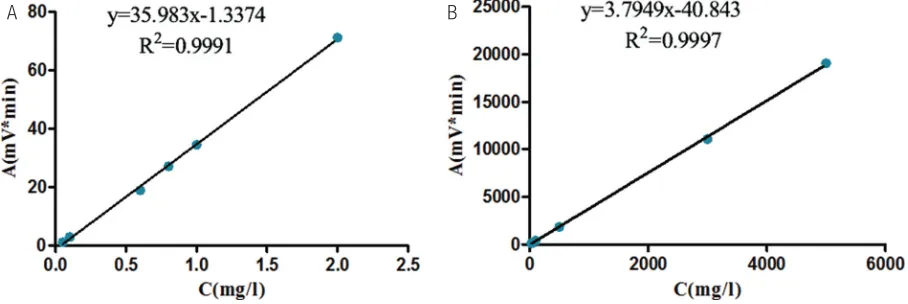
Due to the poor solubility, the standard solution of AL-1 was prepared in methanol. While AL-2’s standard solution was prepared in distilled water. The result was shown in Fig. 1. Typical equations of the standard curves of AL-1 and AL-2 were y = 35.983x-1.3374 and y = 3.7949x-40.843, respectively, with good cor-relation coefficient (R2 = 0.9991 and 0.9997,
respec-tively) during the appropriate concentration range. According to the standard curve, the water solubil-ity of AL-1 was 0.0705 mg/L, the water solubilsolubil-ity of AL-2 was 4.76 g/L. Therefore, the solubility in water of AL-2 was greatly increased without stronger hygroscopicity.
PHARMACOLOGY
Effects on cell viability in LPS-induced
RAW 264.7 cells
The effects of AL-1 and AL-2 on the viability of RAW 264.7 cells were determined by a colorimetric MTT assay after 24 h treatments. The data were expressed as percent cell viability compared to control. AL-2 did not cause any cytotoxicity at 10 μM in RAW 264.7 cells. As shown in Fig. 2 both AL-1 and AL-2 had anti-inflam-matory activity. Although the anti-inflamanti-inflam-matory cell activity of AL-2 was a little weakness than AL-1’s, AL-2’s cytotoxicity was greatly reduced, especially on concen-tration of 10 μM, the reason might be that salt could reduce drug stimulation to reduce the toxicity.
Effects on the production of ROS in
LPS-induced RAW 264.7 cells
In general, results showed that the generation of intra-cellular ROS decreased significantly in RAW 264.7 cells, which were, respectively, treated with AL-1 and AL-2 (Fig. 3). AL-1 and AL-2, respectively, compared with the model group. The elevated ROS levels were decreased by 22.82%, 42.85% and 44.12% after pretreatment with
Fig. 1 (A) The standard curve of AL-1’s solubility; (B) the standard curve of AL-2’s solubility.
Fig. 2 Effects of AL-1 and AL-2 on cell viability in LPS-induced RAW 264.7 cells. Cells were treated with the indicated concentration of AL-1, AL-2 and LPS for 24 h. Cell viabilities were assessed using MTT assay. Each value represents means ± SD of six independent experiments. **P < 0.01 indicates differences from the unstimulated control group. #P < 0.05 and ##P < 0.01 indicates differences from
the LPS-treated group.
Fig. 3 Effects of AL-1 and AL-2 on LPS-induced ROS production in RAW 264.7 cells. The level of intracellular ROS was measured with DCF-DA. The formation of ROS in the cells was evaluated by the arbitrary fluorescence unit and described as fold induction test via vehicle. Each value represents mean ± SD of six inde-pendent experiments. **P < 0.01 indicates differences from the unstimulated control group. #P < 0.05 and ##P < 0.01 indicates
differences from the LPS-treated group.
Fig. 4 Effect of AL-1 and AL-2 on the NO production in LPS-induced RAW 264.7 cells. The cells were treated with LPS only or with AL-1 and AL-2 ranging from 0.01 to 1 μM for 24 h. The culture media were collected, and the nitric oxide concentration was measured by the Griess reaction. Each value represents mean ± SD of triplicate experi-ments. **P < 0.01 indicates differences from the unstimulated control group. ##P < 0.01 indicates differences from the LPS-treated group. AL-1 at doses of 0.01, 0.1 and 1 μM, while the elevated
ROS levels were decreased by 16.08%, 16.42% and 19.94% after pretreatment with AL-2 at doses of 0.01, 0.1, and 1 μM. These results demonstrated that AL-2 could significantly reduce the level of ROS.
Effects on the production of NO in LPS-induced
RAW 264.7 cells
decreased significantly in RAW 264.7 cells, which were, respectively, treated with AL-1 and AL-2. The results showed that AL-1 and AL-2 were similar in reducing the production of NO. The reason might be that salt only changes the physical properties of AL-1.
CONCLUSION
In conclusion, a new andrographolide derivatives AL-2 was designed and synthesized. Both AL-1 and AL-2 had an active effect on inflammatory cells. Among these, two compounds, AL-2 had better solubility in water and lower cytotoxicity. AL-2 may be valuable for the treatment of inflammation. To explore the mechanism of action for this novel compound AL-2, we design the further experiments and the results will be reported in due course.
ACKNOWLEDGEMENT
This work was supported in part by grants from the National Natural Science Foundation of China (81673496 and 81001683) and the Science and Technology Planning Project of Guangdong Province, China (2015B020211011).
REFERENCES
1. Kawalec P, Pilc A. An indirect comparison of infliximab versus adalimumab or golimumab for active ulcerative colitis. Arch Med Sci. 2016;12:1097–1109.
2. Ramadass SK, Jabaris SL, Perumal RK, HairulIslam VI, Gopinath A, Madhan B. Type I collagen and its daughter peptides for tar-geting mucosal healing in ulcerative colitis: A new treatment strategy. Eur J Pharm Sci. 2016;91:216–24.
3. Lindblom R, Higgins G, Coughlan M, de Haan JB. Targeting mitochondria and reactive oxygen species-driven patho-genesis in diabetic nephropathy. Rev Diabet Stud. 2015;12: 134–56.
4. Romero-Castro A, Gutierrez-Sanchez M, Correa-Basurto J, Rosales Hernández MC, Padilla M, Mendieta-Wejebe JE. Pharmacokinetics in wistar rats of 5-[(4-carboxybutanoyl) amino]-2-hydroxybenzoic acid: a novel synthetic derivative of
5-aminosalicylic acid (5-ASA) with possible anti-inflammatory activity. PLoS One. 2016;11:e0159889.
5. Wang Z, Yu P, Zhang G, Xu L, Wang D, Wang L, et al. Design, synthesis and antibacterial activity of novel andrographolide derivatives. Bioorg Med Chem. 2010;18:4269–74.
6. Zhu YY, Yu G, Zhang Y, Xu Z, Wang YQ, Yan GR, et al. A novel andrographolide derivative AL-1 exerts its cytotoxicity on K562 cells through a ROS-dependent mechanism. Proteomics. 2013;13:169–78.
7. Li Y, Yan H, Zhang Z, Zhang G, Sun Y, Yu P, et al. Andro-grapholide derivative AL-1 improves insulin resistance through down-regulation of NF-κB signalling pathway. Br J Pharmacol. 2015;172:3151–8.
8. Jiang X, Yu P, Jiang J, Zhang Z, Wang Z, Yang Z, et al. Synthesis and evaluation of antibacterial activities of andrographolide analogues, Eur J Med Chem. 2009;44:2936–43.
9. Yang Y, Yan H, Jing M, Zhang Z, Zhang G, Sun Y, et al. Andrographolide derivative AL-1 ameliorates TNBS-induced
colitis in mice: involvement of NF-κB, and PPAR-γ signaling
pathways. Sci Rep. 2016;6:29716.
10. Muenster U, Mueck W, van der Mey D, Schlemmer KH, Greschat-Schade S, Haerter M, et al. Predicting biopharmaceutical per-formance of oral drug candidates - Extending the volume to dissolve applied dose concept. Eur J Pharm Biopharm. 2016;102:191–201.
11. Tilborg A, Norberg B, Wouters J. Pharmaceutical salts and cocrystals involving amino acids: a brief structural overview of the state-of-art. Eur J Med Chem. 2014;74:411–26.
12. Yao H, Li S, Yu P, Tang X, Jiang J, Wang Y. Reaction character-istics of andrographolide and its analogue AL-1 with GSH, as a simple chemical simulation of NF-κB inhibition, Molecules. 2012;17:728–39.
13. Fini A, Cavallari C, Bassini G, Ospitali F, Morigi R. Diclofenac salts, part 7: are the pharmaceutical salts with aliphatic amines stable?. J Pharm Sci. 2012;101:3157–68.
14. Xu R, Wang J, Du C, Han S, Meng L, Zhao H. Solubility deter-mination and thermodynamic dissolution functions of 1,3-diphenylguanidine in nine organic solvents at evaluated temperatures. The Journal of Chemical Thermodynamics. 2016;99:86–95.
15. Cong Y, Wang J, Du C, Han S, Zhao H. Solubility and solution thermodynamics of 2-methyl-6-nitroaniline in ten organic solvents at elevated temperatures. The Journal of Chemical Thermodynamics. 2016;101:180-9.