Original Research Article
Correlation between pulmonary function tests and inflammatory
markers in familial mediterranean fever patients
Tayyibe Saler
1, Murat Coşkun
2, Süleyman Ahbab
3, Benan Çağlayan
4, Zuhal Aydan Sağlam
5,
Betül Çavuşoğlu Türker
3*, Fatih Türker
6, Mehmet Bankir
7, Nurettin Ay
7, Şakir Özgür Keşkek
7INTRODUCTION
FMF is an autosomal recessive chronic inflammatory disease which is associated with recurrent attacks of fever, arthritis and poliserositis affecting commonly Armenians, Turks, Greeks and Arabic.1 Among several manifestations, pulmonary involvement prominently presents itself with chest attacks mostly due to serositis.2
As it has long been accepted, persistant inflammatory may lead to pulmonary amyloidosis.3 The close association between vasculitis and FMF also indicate the possible effect of vasculitis on lungs.3-5
Colchicine has been proven to be effective in most patients at preventing acute inflammatory manifestations depending on the frequency and severity of the episodes.
ABSTRACT
Background: Familial mediterranean fever (FMF) is a disease frequently seen in some races and characterized by recurrent attacks affecting various organs with chronic inflammation. As a result of chronic inflammation, microvascular changes may occur. In this study, we aimed to investigate the association between pulmonary function tests and inflammatory markers in FMF patients.
Methods: Seventy-five FMF patients followed up at the Department of Internal Medicine who did not have any comorbidity affecting lung functions and 64 healthy subjects were enrolled. Both groups underwent CO (carbon monoxide) diffusion analysis and pulmonary function tests. Inflammatory markers of the groups were recorded as well and compared with CO diffusion and pulmonary function tests.
Results: FVC%, FEV1, FEV1%, FEV1/FVC, PEF, FEF25-75% values of the patient group was significantly lower than those in the control group. When compared, DLCO%, DLCO/VA, DLCO/VA% were also significantly lower than controls. There was a significant correlation between pulmonary function tests and ESR, hsCRP and fibrinogen levels.
Conclusions: Inflammation in FMF patients may lead to damage on pulmonary tissue that causes an impairment in pulmonary functions.
Keywords: Familial mediterranean fever, Pulmonary function tests, Carbon monoxide diffusion analyses
Department of Internal Medicine, 1Adana SUAM, University of Health Sciences, Adana, 2Umraniye Training and Research Hospital, University of Health Sciences, Istanbul, 3Haseki Training and Research Hospital, University of Health Sciences, Istanbul, 6İstanbul Bilim University Medical Faculty, İstanbul, 7Numune Training and Research Hospital, University of Health Sciences, Adana, Turkey
4
Department of Pulmonology, Kartal Training and Research Hospital, University of Health Sciences, Istanbul, Turkey
5
Department of Family Medicine, Medeniyet University School of Medicine, Istanbul, Turkey
Received: 02 November 2018
Revised: 06 December 2018
Accepted: 08 December 2018
*Correspondence:
Dr. Betül Çavuşoğlu Türker,
E-mail: [email protected]
Copyright: © the author(s), publisher and licensee Medip Academy. This is an open-access article distributed under the terms of the Creative Commons Attribution Non-Commercial License, which permits unrestricted non-commercial use, distribution, and reproduction in any medium, provided the original work is properly cited.
Nevertheless it is reported that inflammation subsides but persists during symptom free periods despite regular colchicine treatment. It has been reported that chronic inflammation has harmful effects on vascular endothelium.6,7 To what extent the alveolar capillary zone effected remains unknown and is yet to be explained.
Based on these considerations, in this paper, we intended to determine the relationship between pulmonary function tests and inflammatory marker such as hsCRP, fibrinogen and ESR as an early indicator of endothelial damage and in FMF patients.
METHODS
This study was designed as a case control study. Patients with a confirmed diagnosis of FMF attending to Umraniye Training and Research Hospital, Department of Internal Medicine between 2012-2014 were invited to participate. The invitations were made consequently as the patients applied for their regular visits while they were at symptom-free period. Seventy-five patients accepted to participate during the reported time interval. The control group consisted of sixty-four healthy and volunteering relatives of the participants who had similar sociodemographic characteristics regarding age and gender. All of the participants were given information about the methodology and all gave written informed consent prior to the study.
According to the study design, all of the participants underwent physical examination. Body-mass indices of the participants were also recorded in order to exclude morbid. Participants with any comorbidity effecting pulmonary functions were excluded whereas any acute pulmonary disease like pneumonia was treated and participants were then invited again following complete recovery. The diagnosis of FMF patients were made according to Livneh criteria.8
Although different genetic mutations have been defined for FMF, genetic analysis is not required for diagnosis for every patient. Genetic analysis is required in patients with atypical clinical findings who do not have family history. Accordingly, we requested genetic analysis only for indetermined cases. Fourty-six patients had undergone genetic evaluation in order to support the diagnosis. Following the exclusion, all gave blood samples for erythrocyte sedimentation rate (ESR), high sensitive-C reactive protein (hsCRP) and leukocyte count. Serum samples were analyzed by the same technician for ESR, CRP and white blood cell count. ESR was measured using Westergren method with Sed Rate Screener 100 (SRS 100, Greiner Bio-one GmbH Kremsmünster, Austria). Hs-CRP was measured on Cobas Integra 400 Plus using a latex particle-enhanced immunoturbidimetric assay following the manufacturer's instructions (Roche Diagnostics, Indianapolis, IN). Plasma fibrinogen levels were determined using commercial kits on a STA compact autoanalyzer (STA, France). White blood cell
counts were determined using an automated equipment (Sysmex XE-2100; Japan).
Pulmonary function tests
Spirometric tests were carried out by the same team of technicians according to the recommended standard using a Jaeger Master Scobe (version 4.5). All respiratory parameters including force vital capacity (FVC), FVC%, force expiratory volume (FEV1), FEV1%, FEV1/FVC, force expiratory flow (FEF25-75), FEF25-75%, peak expiratory flow (PEF), PEF% were assessed. The best of at least three technically acceptable values for all of them were selected. Pulmonary diffusion capacity for carbon monoxide is defined as 1mL of a gas (a gas mixture containing 0.3% CO, 0.3% methane, 21% oxygen and nitrogen) diffused through the alveolocapillary membrane under 1 mmHg pressure in 1.9 Carbon monoxide has a high affinity for hemoglobin. The alveolocapillary membrane is the most important factor affecting the diffusion of carbon monoxide yet it is well known the diffusion capacity may be affected by a variety of factors including age, height, body surface area, smoking, hemoglobin levels, body posture and exercise, expiration and inspiration phases, high altitude, oxygen concentration, diurnal changes, menstruation, alcohol consumption, gender and ethnicity. So, obstructive lung diseases, parenchimal diseases, systemic diseases affecting lungs and cardiopulmonary diseases are all known to decrease DLCO levels.
In this study we measured lung volumes by a computerized system (SensorMedics Vmax 229; SensorMedics Corporation, Yorba Linda, CA, USA), following the standardized procedures (10) and DLCO and DLCO/VA using standardized single-breath method which were expressed as mean of two consecutive measurements.9 Spirometric volumes were expressed as mL and DLCO values were expressed as mL/mmHg/min. The pulmonary function results were expressed as percentages of predicted normal values. For the purpose of this study, the threshold of abnormality was identified as under 79% of the predicted value.
RESULTS
Fourty seven of FMF group (62.6%) and twenty eight of the control group (43.7%) were female. The mean age of FMF group was 31.4±9.2 years and the mean BMI was 25.7±4.3 kg/m2. Table 1 shows the demographic
characteristics and the comparison of demographic and biochemical characteristics of both groups. No significant difference was determined between the two groups with regard to gender, age, BMI, hsCRP, ESR, WBC (p>0.05). However, the fibrinogen levels of the patient group were significantly higher than controls (p=0.005) (Table 1).
Table 1: The comparison of demographic and biochemical characteristics of both groups.
Control group n=64
Study group
n=75 P value
Female n (%) 42 (65.6) 47 (62.6) 0.865
Age (years) 34.0±7.9 31.4±9.2 0.085
BMI (kg/m²) 27±2.9 25.7±4.3 0.054
hsCRP (mg/L) 0.47±0.45 0.82±1.7 0.095
ESR (mm/H) 11.1±6.6 12.6±11.2 0.096
WBC (1000/mm³) 7.9±1.9 7.4±1.9 0.067
Fibrinogen (mg/dL) 288.1±53.1 346.3±122..2 0.005
Table 2: The comparison of the pulmonary function and CO diffusion capacity tests of both groups.
Control group n=64
Study group
n=75 P value
DLCO 28.2±8.0 30.1± 16.2 0.747
DLCO% 101.6±28.3 91.2±29.9 0.016
DLCO/VA 5.2±0.6 4.7±0.8 0.003
DLCO/VA% 106.5±16.2 89.1±16.4 <0.001
FVC% 120.0±20.3 99.8±14.7 <0.001
Fev1 3.44±0.95 2.82±0.75 <0.001
Fev1% 110.5±20.3 97.5±14.3 <0.001
Fev1/Fvc 98.7±9.0 82.6±6.7 <0.001
PEF 7.0±2.0 6.4±1.8 0.040
PEF% 92.0±21.1 90.6±17.6 0.650
FEF25 75 3.4±2.9 3.0±1.0 0.144
FEF25 75% 87.7±23.8 79.1±19.1 0.019
Table 3: Multiple linear regression analyses (backward method) was performed with hsCRP as a dependent variable and with DLCO, DLCOVA, DLCOVA%, DLCO%, FEV1%, FEF25-75, FEF25-75%, FEV1, FEV/FVC,
PEF, PEF% as independent variables.
Independent variables Coefficient Standard error r partial T P value
(Constant) 2.9422
DLCO 0.04010 0.01216 0.3645 3.299 0.0015
DLCOVA% -0.03692 0.01176 -0.3491 -3.139 0.0025
DLCO% 0.04242 0.007361 0.5645 5.763 <0.0001
FEV1% -0.04011 0.01220 -0.3635 -3.287 0.0016
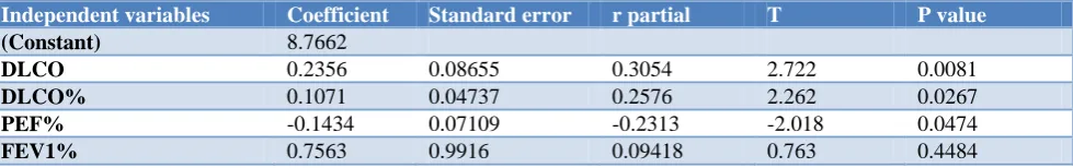
Table 4: Multiple linear regression analyses (backward method) was performed with ESR as a dependent variable and with DLCO, DLCOVA, DLCOVA%, DLCO%, FEV1%, FEF25-75, FEF25-75%, FEV1, FEV/FVC, PEF,
PEF% as independent variables.
Independent variables Coefficient Standard error r partial T P value
(Constant) 8.7662
DLCO 0.2356 0.08655 0.3054 2.722 0.0081
DLCO% 0.1071 0.04737 0.2576 2.262 0.0267
PEF% -0.1434 0.07109 -0.2313 -2.018 0.0474
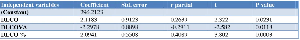
Table 5: Multiple linear regression analyses (backward method) was performed with fibrinogen as a dependent variable and with DLCO, DLCOVA, DLCO%, as independent variables.
Independent variables Coefficient Std. error r partial t P value
(Constant) 296.2123
DLCO 2.1183 0.9123 0.2639 2.322 0.0231
DLCOVA -2.2978 0.8898 -0.2911 -2.582 0.0118
DLCO % 2.0941 0.5508 0.4089 3.802 0.0003
When pulmonary function tests were compared, FVC%, FEV1, FEV1%, FEV1/FVC, PEF, FEF25 75% values of the FMF group were signicantly lower than the control group. Likewise when CO diffusion tests were compared, DLCO%, the DLCO/VA, DLCO/VA% values were significantly lower in the patient group than in the control group (Table 2). A significant correlation was determined between ESR, hsCRP, serum fibrinogen levels and pulmonary function tests (Tables 3-5).
DISCUSSION
The role of chronic inflammation in vascular damage has been acknowledged for many years.11,12 Inflammation also regulates the production of the acute phase proteins such as hsCRP, fibrinogen and serum amyloid A.13
The lung tissue is consisted of a large alveolar capillary area that allows gas exchange. Any disorder in the alveolar capillary membrane is expected to result with problems in the regulation of the gas exchange in the lungs.14 This study was conducted with the assumption that the dense microvascular structure of lungs in FMF patients may have been affected since they are consistently exposed to inflammation.
In this study we have observed that DLCO%, DLCO/VA, DLCO/VA% values of FMF patients were lower than those in the healthy group. We believe that the microvascular changes which might have been triggered by chronic inflammation, may have lead to impairment of CO diffusion tests in FMF patients. There are studies suggesting that chronic inflammation sets the grounds for atherosclerosis in many rheumatic diseases. In a study conducted by Çalışkan and colleagues, FMF patients were compared to healthy people in terms of coronary microvascular atherosclerosis and left ventriculary functions and it was concluded that the FMF group had significant microvascular impairment which was highly associated with CRP.15 The same author reported that coronary microvascular impairments were determined in inflammatory diseases, such as ankylozing spondylitis and inflammatory bowel diseases, which were again correlated with CRP.16-17 In our study, we detected a correlation between CO diffusion capacity impairments and inflammatory indicators such as hsCRP, fibrinogen and erythrocyte sedimantation rate. Each of FMF patients included in the study was under colchicine treatment which was supposed to suppress inflammation. Given
that no one suffered any attacks during study period, it was not possible to analyse the active period of the disease. In any case, we know that, even if the patients are clinically inactive, a certain amount of inflammation is present already.
In order to evaluate pulmonary functions, whole pulmonary function tests were run on the participants along with CO diffusion tests. The FMF group was observed to have significantly lower levels of FVC, FEV1, FEV1%, FEV1/FVC, PEF, FEF25-75% compared to the healthy population. During the tests were being processed, none of the patients had active disease. Therefore there was no presence of "serotisis" which might have acted as a worsener component for the respiratory functions. Hence the anomalies in the respiratory function were not caused by serotisis. Muscle involvement is an expected finding in FMF patients, even if rarely.18 In many diseases with muscle involvement, the respiratory muscles tend to be affected which lead to respiratory dysfunctions. Whereas this was not the case for any of our patients: none of them had such muscle involvement which may impair lung functions. There are few articles concerning the pulmonary functions in FMF patients. Some of which are simple reviews yet there's one single article comparing the pulmonary involvement and genetic variations in the FMF patients who had undergone genetic analysis.19 As far as we are concerned, there isn’t a case-control study conducted similar to ours, which analyses pulmonary functions in FMF patients. Thus we were unable to compare our results with the others.
In conclusion, this study shows that there is a significant decline in pulmonary function values of FMF patients such as DLCO%, DLCO/VA, DLCO/VA%, FVC%, FEV1/FVC and etc. This result may have several reasons. Microvascular changes in the endothelium of the alveolar capillary area that are affected by chronic inflammation, vasculitis or myopathy may have caused this. But given that our patients don't have any sign of myopathy or vasculitis, and that inflammatory signs are correlated with low values in respiratory function tests, we suggest that microvascular changes in physiopathology are very likely to be the main reason.
REFERENCES
1. Medlej-Hashim M, Loiselet J, Lefranc G, Mégarbané A. Familial Mediterranean Fever (FMF): from diagnosis to treatment. Santé. 2004;14(4):261– 6.
2. Brik R, Gershoni-Baruch R, Shinawi M, Barak L, Bentur L. Pulmonary manifestations and function tests in children genetically diagnosed with FMF. Pediatr Pulmonol. 2003;35(6):452-5.
3. Sahan C, Cengiz K. Pulmonary amyloidosis in familial Mediterranean fever Acta Clin Belg. 2006;61(3):147-51.
4. Lidar M, Pras M, Langevitz P, Livneh A. Thoracic and lung involvement in familial Mediterranean fever (FMF). Clin Chest Med. 2002;23(2):505-11. 5. Livneh A, Langevitz P, Pras M. Pulmonary
associations in familial Mediterranean fever. Curr Opin Pulm Med. 1999;5(5):326-31.
6. Ross R. Atherosclerosis: an inflammatory disease. N Engl J Med. 1999;340:115–26.
7. Koenig W, Sund M, Frohlich M, Fischer HG, Lowel H, Doring A, et al. C-Reactive protein, a sensitive marker of inflammation, predicts future risk of coronary heart disease in initially healthy middle-aged men: results from the MONICA (Monitoring Trends and Determinants in Cardiovascular Disease) Augsburg Cohort Study, 1984 to 1992. Circulation. 1999;99(2):237–42.
8. Livneh A, Langevitz P. Diagnostic and treatment concerns in familial Mediterranean fever. Baillieres Best Pract Res Clin Rheumatol. 2000;14:477-98. 9. American Thoracic Society. Single-breath carbon
monoxide diffusion capacity (transfer factor): recommendations for a standard technique. 1995 update. Am J Respir Crit Care Med. 1995;152:2185-8.
10. American Thoracic Society. Standardization of spirometry: 1994 update. Am J Respir Crit Care Med. 1995;152:1107-36.
11. Sinisalo J, Paronen J, Mattila KJ, Syrjälä M, Alfthan G, Palosuo T, et al. Relation of inflammation to
vascular function in patients with coronary heart disease. Atherosclerosis. 2000;149:403–11.
12. Libby P, Ridker PM, Maseri A. Inflammation and atherosclerosis. Circulation. 2002;105:1135–43. 13. Paffen E, DeMaat MP. C-reactive protein in
atherosclerosis: A causal factor? Cardiovasc Res. 2006;71(1):30-9.
14. Agostoni P, Bussotti M, Cattadori G, Margutti E, Contini M, Muratori M, et al. Gas diffusion and alveolar–capillary unit in chronic heart failure. Eur Heart J. 2006;27(21):2538-43.
15. Caliskan M, Gullu H, Yilmaz S, Erdogan D, Unler GK, Ciftci O, et al. Impaired coronary microvascular function in familial Mediterranean fever. Atherosclerosis. 2007;195(2):161-7.
16. Caliskan Z, Gokturk HS, Caliskan M, Gullu H, Ciftci O, Ozgur GT, et al. Impaired coronary microvascular and left ventricular diastolic function in patients with inflammatory bowel disease. Microvasc Res. 2015;97:25-30.
17. Caliskan M, Erdogan D, Gullu H, Yilmaz S, Gursoy Y, Yildirir A, et al. Impaired coronary microvascular and left ventricular diastolic functions in patients with ankylosing spondylitis. Atherosclerosis. 2008;196(1):306-12
18. Kavukcu S, Türkmen M, Soylu A, Kasap B, Tatli Güneş B. Skin and muscle involvement as presenting symptoms in four children with familial Mediterranean fever. Clin Rheumatol. 2009;28(7):857-60
19. Brik R, Gershoni-Baruch R, Shinawi M, Barak L, Bentur L. Pulmonary manifestations and function tests in children genetically diagnosed with FMF. Pediatr Pulmonol. 2003;35(6):452-5.