© Universiti Tun Hussein Onn Malaysia Publisher’s Office
IJIE
Journal homepage: http://penerbit.uthm.edu.my/ojs/index.php/ijie
The International
Journal of
Integrated
Engineering
ISSN : 2229-838X e-ISSN : 2600-7916
An Overview of Electrical Characterization Techniques for
Biological Cell
Abdul Hafiz Mat Sulaiman
1, Ida Laila Ahmad
2,3*, Ruzairi Abdul Rahim
2,4,
Jaysuman Pusppanathan
5, Dirman Hanafi
2, Elmy Johana Moahamad
2, Mohd
Fadzli Abdul Shaib
2, Leow Pei Ling
4, Yasmin Abdul Wahab
6, Suzanna Ridzuan
Aw
71 Institute of Microengineering and Nanoelectronics, Universiti Kebangsaan Malaysia, 43600 Bangi, Selangor,
MALAYSIA.
2 Instrumentation, Sensing and Tomography Research Group (InSet), Faculty of Electrical and Electronic Engineering,
Universiti Tun Hussein Onn Malaysia, 86400 Batu Pahat, Johor, MALAYSIA.
3 Microelectronics and Nanotechnology Shamsuddin Research Centre (MiNT-SRC), Universiti Tun Hussein Onn
Malaysia, 86400 Batu Pahat, Johor, MALAYSIA.
4 Process Tomography and Instrumentation (PROTOM-i) Research Group, Innovative Engineering Research Alliance
Universiti Teknologi Malaysia,81310 Skudai, Johor, MALAYSIA.
5 School of Biomedical Engineering and Health Sciences, Faculty of Engineering, Universiti Teknologi
Malaysia,81310 Skudai, Johor, MALAYSIA.
6 Department of Instrumentation and Control Engineering, Faculty of Electrical and Electronic Engineering, Universiti
Malaysia Pahang, 26600 Pekan, Pahang, MALAYSIA.
7 Faculty of Electrical & Automation Engineering Technology, Terengganu Advance Technical Institute University
College (TATiUC), Jalan Panchor, Telok Kalong, 24000 Kemaman, Terengganu, MALAYSIA.
*Corresponding Author
DOI: https://doi.org/10.30880/ijie.2019.11.06.018
Received 31 May 2018; Accepted 16 June 2019; Available online 12 September 2019
Abstract: In this paper, various electrical characterization techniques available for biological cell have been systematically reviewed. It covers both invasive and non-invasive approaches for population and single cell based studies. Examples of invasive technique consist of probing and patch clamp that measures the ionic current. However, depending on the applications, the non-invasive techniques are far more superior and popular. Some of the technique such as dielectric spectroscopy, electrorotation and dielectrophoresis measures the cell conductivity and dielectric constant. Furthermore, previous researchers proved that non-invasive technique may reduce the harmful effect on the cell due to electrical exposure. The review compares in terms of working principles, sample applications, advantages and limitations of each technique.
1.
Introduction
Electrical measurement with respect to cell properties can be performed either using population based or single cell-based investigation. Population based studies has been conventionally used in clinical and research setting had started since 1925 when the property of blood cells in a suspension can be determined by measuring the sample capacitance [1, 2]. This shows the potential of applying electrical measurement to study the biological cell properties. Few decades after, the growth on population measurement techniques have revolutionized automated devices concerning biological material. However, population based measurement were inaccurate due to the heterogeneity of the cells. Each cell with different shape and size for example possess different properties [3]. Furthermore, they also react differently towards similar stimuli thus causing larger room of error during measurement.
Single cell measurement offers a more accurate approach while probing for each individual cell response when exposed to certain stimuli [4, 5]. Researchers have developed various techniques to realize single cell measurement at cheaper costs and more portable platform. In this review, the discussions will be limited to population techniques in order to provide in depth understanding towards the fundamental concept and underlying theories. Nonetheless, an introduction towards single cell electrical characterization will be introduced in section 3.0.
2.
Biological Cells Electrical Properties Measurement Techniques Structure
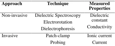
Several methods have been used in measuring electrical properties of single cell i.e. dielectric spectroscopy, electrorotation, dielectrophoresis, patch-clamp, and probing. Each of the techniques is measuring different single cell electrical properties and can be categorized by invasive method and non-invasive. Table 1 summarized the technique for single cell electrical properties measurement. Some of the techniques were measuring the same single cell electrical properties, i.e. dielectric constant and conductivity.
Table 1 - Available electrical measurement techniques Approach Technique Measured
Properties
Non-invasive Dielectric Spectroscopy Electrorotation Dielectrophoresis
Dielectric constant Conductivity
Invasive Patch-clamp Probing
Ionic current Current
2.1 Dielectric Spectroscopy
Dielectric spectroscopy also known as impedance spectroscopy is a technique where an impedance response of a biological suspension is being measured when applying an alternating current (AC) excitation signal. This technique uses a measurement cell of either two, three or four electrodes where the biological suspension is held [6]. When a
small AC voltage, is applied between the electrodes, the electrical current response, passing through the suspension is measured which in a form of frequency function to give the electrical properties of the particle or cell. The complex impedance of the response [6] can be described by equation (1) given as:
(1)
where is the real part or resistance and is the imaginary part or reactance of the complex impedance. For a spherical particle in a dilute suspension at a low volume fraction, the Maxwell mixture equation (please refer [7] for derivation) gives the steady state value of the equivalent complex permittivity mixture of suspending medium and particle is described by equation (2) given as:
(2)
where is the volume fraction or the ratio of the particle volume to the sensing area volume and is the Claus-Mossotti factor which can be described in equation (3) given as:
where and are the complex permittiviy of a particle and medium respectively. Complex permittivity is described in equation (4) given as:
(4)
where , is the permittivity and σ is the conductivity.
Complex impedance of the mixture of particle and suspending medium is then described by equation (5) given as:
(5)
where is the complex capacitance of the mixture. This technique is able to calculate single cell dielectric properties i.e. dielectric constant and conductivity without damaging the cells (non-invasive). Fig. 1 shows the dielectric spectroscopy technique.
Fig. 1 - Overview of Dielectric Spectroscopy technique
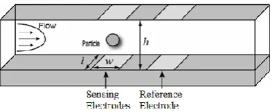
Dielectric spectroscopy has been numerously used by previous researchers with additional improvement on the techniques, for example simultaneous measurement on other properties, determination of mechanical properties [9-11], for medical diagnosis such as cancer detection [12,13]. By realizing some limitations on the current measurement technique, improvement was made in terms of precise positioning between particles of interest and placement of electrodes. Fig. 2 shows the improved measurement technique on dielectric spectroscopy. Even though the cells are now being tested individually, the measurement differences between with and without cells is too small and unobservable for a comparison [8].
Fig. 2 - Single cell dielectric spectroscopy integrated with flow cytometry
2.2 Electrorotation
Fig. 3 - Overview of electrorotation technique
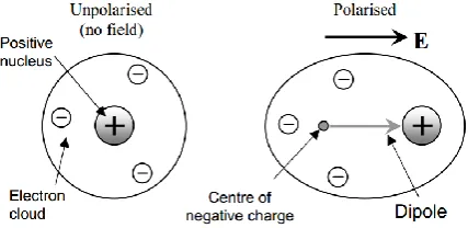
Fig. 4 - Polarization effect on a particle
The torque can be described by equation (6) given as:
(6)
where E is the electric field and is the imaginary part of the Claus-Mossotti factor. Proportionality between torque and the imaginary part of the Claus-Mossotti factor can be used to calculate dielectric properties of a cell. The torque is measured indirectly by analysing the rotation rate of the particle described by equation (7) given as:
(7)
where is the viscosity of the suspending medium and K is the scaling factor. The rotation rate of the particle is measured using a microscope and a stopwatch. The measured value is fitted to a mathematical expression of physical cell models for specific single cell dielectric properties [14].
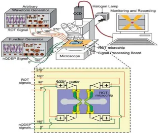
Electrorotation is able to measure cell membrane permittivity and cytoplasm conductivity [15]. However, there are several drawbacks with the technique. This technique relies on mathematical models for single cell. Many models have been proposed to calculate the electrorotational torque acting on a cell in a rotating electric field which then used to relate with cell membrane permittivity and cytoplasm conductivity [16-21]. Furthermore, ROT requires skilled operator for the measurement to be successfully performed. Positioning a micro scale particle in the middle of a rotating electric field is time consuming and labour intensive process. Hence, low throughput data are obtained. Alignment of particles is important due to the fact of irregular shape of biological samples. Spherical and non-spherical object will cause the induced dipole moment to be different due to the alignment with uniform electric field. This torque is always in parallel with one of the axes generated by the object shape.
Fig. 5 - Improvement on electrorotation (ROT) technique [22]
2.3 Dielectrophoresis
Similar with electrorotation, dielectrophoresis (DEP) utilize an AC electric field but the field is nonuniform and the particles move in a translational motion. Fig. 6 shows how a polarized particle reacts to nonuniform electric field intensity. Due to the electric field the particle moves according its polarization intensity. In short, particle with high polarization will move toward the high intensity electric field and vice versa.
Fig. 6 - Dielectrophoresis technique
In DEP , force acting on a particle can be approximated using equation (8) given as:
(8)
Where is the permittivity of the medium, r is the radius, is the real part of Claus-Mossotti fraction. Claus-Mossoti fraction equation which can be used determine particle movement direction.
Positive dielectrophoresis (pDEP) is defined for a particle that is more polarizable than the medium, and attracted to the high intensity electric field regions. On the contrary, negative dielectrophoresis
frequency. DEP cross over frequency is a transient frequency where pDEP change to nDEP. This frequency is can be written in term of the conductance and membrane capacitance of the cell by using the simplified shell model for a biological cell which can be described in equation (9) given as:
(9)
where is the crossover frequency, is the medium electrical conductivity, r is the radius, and are the capacitance and conductance of the membrane, respectively. Basically, drawbacks for DEP is similar with ROT i.e. labour intensive, time consuming process, and require a complete representation model of a which involve complex mathematical calculation. Combination of electrorotation spectrum and dielectrophoresis force can provide a unique and significant measurement on the single cell dielectrophoresis [24]. Beside characterizing single cell electrical properties, DEP has been used widely in single particle manipulation (sort, isolate, and trap) [25-27] and separation [28].
2.4 Patch-clamp
Patch-clamp is a technique where the cellular ion channel is being characterized. Ion channel plays a major role in the cell signaling, i.e. propagation and modulation of muscle and nerve cells. Fig. 7 shows the overview of the patch-clamp technique. In the conventional method, a cell membrane patch is being sucked into a micropipette to form a high electrical resistance. Then, the ionic current flow through ion channel is being measured.
Fig. 7 - Conventional Patch-clamp technique
This technique requires high precision which is a laborious task and low throughput rate. Skilled operator is required to move the tip of the micropipette over the single cell using micromanipulator and sucking the cell membrane without damaging the whole cell. This delicate process was done under an optical microscope. However, an improvement on the patch-clamp technique has been made by integrating with chip-based devices [29-39]. Fg. 8 shows an example of the improved version of the patch - clamp technique. The technique has been integrated with Micro-Electrical-Mechanical system for better cell immobilization and reduces labour works.
Fig. 8 - Patch-clamp in a Micro-Electrical-Mechanical system (MEMS) chip
long-term study. Practically, this technique has been mostly used in the study of electrophysiological cell properties, i.e. the characterization of the kinetics and steady-state effects of toxins.
3.
Single Cell Electrical Characterization using Probing Technique
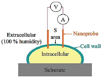
Probing is a new novel technique for single cell electrical measurement proposed by M. R. Ahmad et al. in 2009 [40]. Basically, this technique measure current flow passing through the single cell intracellular region, i.e. cytoplasm when a series of single pulse direct current (DC) voltage is applied through a pair of nanoprobe or also known as dual nanoprobe. Fig. 9 shows the overview of probing technique.
Fig. 9 - Single cell electrical measurements using dual nanoprobe
Single cell intracellular electrical measurement is performed by penetrating the cell wall and membrane layer of the cell to reach the cytoplasm part without cell bursting or exploding. This is achieved thanks to the size of the probe which is relatively smaller than the tested cells and a skilled operator for navigating the probe. The measurement is conducted inside an Environmental-Scanning Electron Microscopy (E-SEM). E-SEM is built for high resolution observation and capable to preserve the cell native state even when the cell is moving out of its buffer. Fig. 10 shows the manual Nano Manipulator under E-SEM system.
Fig. 10 - Manual Nano Manipulator under E-SEM(Environmental Scanning Microscopy)
Dual nanoprobe probing-based technique has been tested on W303 wild yeast cell. Interestingly, even though the approach is invasive the cell is able to recover from small wounds. Hence, the same cell can be used for a long term or multiple measurement study. The technique does not use complex single cell electrical model because only cytoplasm electrical properties is being measured. However, there are several drawbacks with the technique.
One of the applications for this technique is single cell viability detection which aims to tackle the disadvantages of conventional fluorescent-dye based method, i.e. qualitative and slow result processing. The dual nanoprobe technique is able to achieve instantaneous and quantitative results without using any chemical reagent. Since this technique is at an early stage of development, the application is limited. However, it has the potential to be improved for single cell electrical properties characterization, i.e. cytoplasm conductivity.
4.
Summary
The available methods for biological cell electrical characterization have been thoroughly presented. The results obtained from this investigation may lead to various correlations especially in disease diagnosis and disease progression [4]. All techniques have two common drawbacks which are slow throughput rate and labour intensive. In effort to overcome the mentioned problems, single cell electrical characterization has been introduced. This technique offer more accurate properties determination and can be conducted at much lower costs. Meanwhile, techniques based on electrokinetics, i.e. electorotation and dieletrophoresis, are commonly used in single cell manipulation [15]. The concept of the induced force on a particle when entering electric field without any physical contact makes it the preferred approach for single cell manipulation in preserving the cell physiology and viability. However, the technique suffers from limitations due to proper positioning and unwanted noise interference caused by the movement of tri-axial units towards the specimen placed on the cooling stage. In this regard, the integration of electrical characterization technique on a microfluidic platform may offer an excellent solution. Upon deployment of microfluidic technology, a more automated single cell electrical characterization procedures can be developed, hence better result can be obtained.
Acknowledgement
Authors wish to extend gratitude towards research collaborators who provide conducive research environment to inculcate further research effort. Special thanks to Microelectronics and Nanotechnology Shamsuddin Research Centre (MiNT-SRC), Universiti Tun Hussein Onn Malaysia for endless support. This research is funded by Universiti Tun Hussein Onn Malaysia Tier-1Grant (Vot No: H118).
References
[1] H. Fricke,. (1925). The Electric Capacity of Suspensions with Special Reference to Blood. The Journal of General Physiology, vol. 9, pp. 137-152.
[2] X. Mu, W. Zheng, J. Sun, W. Zhang, and X. Jiang. (2013). Microfluidics for Manipulating Cells. Small, vol. 9, pp. 9-21.
[3] M. R. Ahmad, M. Nakajima, S. Kojima, M. Homma, and T. Fukuda (2008). The Effects of Cell Sizes, Environmental Conditions, and Growth Phases on the Strength of Individual W303 Yeast Cells Inside ESEM. IEEE Transactions on NanoBioscience, vol. 7, pp. 185-193.
[4] K. Asami, T. Hanai, and N. Koizumi (1976). Dielectric properties of yeast cells. Journal of Membrane Biology, vol. 28, pp. 169-180.
[5] K. Asami (2002). Characterization of biological cells by dielectric spectroscopy. Journal of Non-Crystalline Solids, vol. 305, pp. 268-277.
[6] M. Hywel, S. Tao, H. David, G. Shady, and G. G. Nicolas (2007). Single cell dielectric spectroscopy. Journal of Physics D: Applied Physics, vol. 40, pp. 61-70.
[7] T. B. Jones. (1995). Electromechanics of Particles: Cambridge University Press.
[8] M. Thein, F. Asphahani, A. Cheng, R. Buckmaster, M. Zhang, and J. Xu (2010). Response characteristics of single-cell impedance sensors employed with surface-modified microelectrodes. Biosensors and Bioelectronics, vol. 25, pp. 1963-1969.
[9] H. Fricke (1924). A Mathematical Treatment of the Electrical Conductivity of Colloids and Cell Suspensions. The Journal of General Physiology vol. 6, pp. 375-384.
[10] T. Noll and M. Biselli (1998). Dielectric spectroscopy in the cultivation of suspended and immobilized hybridoma cells. Journal of Biotechnology, vol. 63, pp. 187-198.
[11] J. Chen, Y. Zheng, Q. Tan, Y. L. Zhang, J. Li, W. R. Geddie, et al. (2011). A microfluidic device for simultaneous electrical and mechanical measurements on single cells. Biomicrofluidics, vol. 5, p. 014113. [12] J. Chen, J. Li, and Y. Sun, (2012). Microfluidic Approaches for Cancer Cell Detection, Characterization, and
Separation. Lab on a Chip, vol. 12, pp. 1753-1767.
[14] R. Hölzel (1997). Electrorotation of Single Yeast Cells at Frequencies Between 100 Hz and 1.6 GHz. Biophysical Journal, vol. 73, pp. 1103–1109.
[15] C. Dalton, A. D. Goater, J. P. H. Burt, and H. V. Smith (2004). Analysis of parasites by electrorotation. Journal of Applied Microbiology, vol. 96, pp. 24-32.
[16] G. Fuhr and P. I. Kuzmin (1986). Behavior of Cells in Rotating Electric Fields with Account to Surface Charges and Cell Structures. Biophysical Journal, vol. 50, pp. 789-795.
[17] Y. Huang, R. Holzel, R. Pethig, and B. W. Xiao (1992). Differences in the AC electrodynamics of viable and non-viable yeast cells determined through combined dielectrophoresis and electrorotation studies. Physics in Medicine and Biology, vol. 37, p. 1499.
[18] J. Gimsa, T. Müller, T. Schnelle, and G. Fuhr (1996). Dielectric spectroscopy of single human erythrocytes at physiological ionic strength: dispersion of the cytoplasm. Biophysical Journal, vol. 71, pp. 495-506.
[19] J. P. Huang and K. W. Yu (2002). First-principles approach to electrorotation assay. Journal of Physics: Condensed Matter, vol. 14, p. 1213.
[20] M. Sancho, G. Martı́nez, and C. Martı́n (2003). Accurate dielectric modelling of shelled particles and cells. Journal of Electrostatics, vol. 57, pp. 143-156.
[21] P. R. C. Gascoyne, F. F. Becker, and X. B. Wang (1995). Numerical analysis of the influence of experimental conditions on the accuracy of dielectric parameters derived from electrorotation measurements. Bioelectrochemistry and Bioenergetics, vol. 36, pp. 115-125.
[22] S.-I. Han, Y.-D. Joo, and K.-H. Han (2013). An electrorotation technique for measuring the dielectric properties of cells with simultaneous use of negative quadrupolar dielectrophoresis and electrorotation. Analyst, vol. 138, pp. 1529-1537.
[23] N. G. Green and H. Morgan (1998). Dielectrophoresis of Submicrometer Latex Spheres. 1. Experimental Results," The Journal of Physical Chemistry B, vol. 103, pp. 41-50.
[24] F. F. Becker, X. B. Wang, Y. Huang, R. Pethig, J. Vykoukal, and P. R. Gascoyne (1995). Separation of human breast cancer cells from blood by differential dielectric affinity. Proceedings of the National Academy of Sciences, vol. 92, pp. 860-864.
[25] S. K. Mohanty, S. K. Ravula, K. L. Engisch, and A. B. Frazier (2003). A micro system using dielectrophoresis and electrical impedance spectroscopy for cell manipulation and analysis in 12th International Conference onTransducers, Solid-State Sensors, Actuators and Microsystems, pp. 1055-1058.
[26] R. Pethig (2010). Review Article---Dielectrophoresis: Status of the theory, technology, and applications. Biomicrofluidics, vol. 4, pp. 022811-35.
[27] Z. Zhu, O. Frey, D. S. Ottoz, F. Rudolf, and A. Hierlemann (2012). Microfluidic single-cell cultivation chip with controllable immobilization and selective release of yeast cells. Lab on a Chip, vol. 12, pp. 906-915. [28] P. R. C. Gascoyne and J. Vykoukal (2002). Particle separation by dielectrophoresis. Electrophoresis, vol. 23,
pp. 1973-1983.
[29] K. G. Klemic, J. F. Klemic, M. A. Reed, and F. J. Sigworth (2002). Micromolded PDMS planar electrode allows patch clamp electrical recordings from cells. Biosensors and Bioelectronics, vol. 17, pp. 597-604. [30] A. Stett, V. Bucher, C. Burkhardt, U. Weber, and W. Nisch (2003). Patch-clamping of primary cardiac cells
with micro-openings in polyimide films. Medical and Biological Engineering and Computing, vol. 41, pp. 233-240.
[31] S. Pandey, R. Mehrotra, S. Wykosky, and M. H. White (2004). Characterization of a MEMS BioChip for planar patch-clamp recording. Solid-State Electronics, vol. 48, pp. 2061-2066.
[32] R. Pantoja, J. M. Nagarah, D. M. Starace, N. A. Melosh, R. Blunck, F. Bezanilla, et al., (2004). Silicon chip-based patch-clamp electrodes integrated with PDMS microfluidics. Biosensors and Bioelectronics, vol. 20, pp. 509-517.
[33] C. Ionescu-Zanetti, R. M. Shaw, J. Seo, Y.-N. Jan, L. Y. Jan, and L. P. Lee (2005). Mammalian electrophysiology on a microfluidic platform. Proceedings of the National Academy of Sciences of the United States of America, vol. 102, pp. 9112-9117.
[34] C. Chen and A. Folch, (2006). A high-performance elastomeric patch clamp chip. Lab on a Chip, vol. 6, pp. 1338-1345.
[35] B. Matthews and J. W. Judy (2006). Design and fabrication of a micromachined planar patch-clamp substrate with integrated microfluidics for single-cell measurements. Journal of Microelectromechanical Systems, vol. 15, pp. 214-222.
[36] W.-L. Ong, J.-S. Kee, A. Ajay, N. Ranganathan, K.-C. Tang, and L. Yobas (2006). Buried microfluidic channel for integrated patch-clamping assay. Applied Physics Letters, vol. 89, p. 093902.
[37] T. Lehnert, D. M. T. Nguyen, L. Baldi, and M. A. M. Gijs (2007). Glass reflow on 3-dimensional micro-apertures for electrophysiological measurements on-chip. Microfluidics and Nanofluidics, vol. 3, pp. 109-117. [38] S. Li and L. Lin (2007). A single cell electrophysiological analysis device with embedded electrode. Sensors
[39] W.-L. Ong, K.-C. Tang, A. Agarwal, R. Nagarajan, L.-W. Luo, and L. Yobas (2007). Microfluidic integration of substantially round glass capillaries for lateral patch clamping on chip. Lab on a Chip, vol. 7, pp. 1357-1366.
[40] M. R. Ahmad, M. Nakajima, T. Fukuda, S. Kojima, and M. Homma (2009). Single cells electrical characterizations using nanoprobe via ESEM-nanomanipulator system in 9th IEEE Conference on Nanotechnology, pp. 589-592.
[41] A. H. M. Sulaiman and M. R. Ahmad (2012). Modeling and simulation of novel method of single cell viability detection via electrical measurement using dual nanoprobes in 2012 International Conference on Enabling Science and Nanotechnology (ESciNano), pp. 1-2.