214 | P a g e
SKIN LESION SEGMENTATION USING GENETIC
ALGORITHM
Mr. Sylvester M Gaikwad
1, Dr. Mrs. S . R. Chougule
21
M.E Electronics and Telecomunication
Bharati Vidyapeeth College of Engineering Shivaji University (India)
2
Dept. of Electronics and Telecomunication
Principal Bharati Vidyapeeth College of Engineering Shivaji University (India)
ABSTRACT
Image Segmentation for medical analysis play a very imortant role. Segmentation is the most important process
to assist in the visualisaton of the structure of importance in the medical images. Segmentation of medical
images is challenging due to poor image contrast and artifacts that result in missing or diffuse organ/tissue
boundaries. Skin lesion or cancer has been the most common and represents 50% of all new cancers detected
each year. If detected at an early stage, simple and economic treatment can cure it mostly. In this paper we
prresnt an image segmentation technique with the help of genetic algorithm were an accurate section of
inffected area is detected. This helps in better understanding the Lesion area and doing the treatment.
Index Terms: Skin Lesion, Image Segmentation, Genetic Algorithm, Region Growing
I.
INTRODUCTION
Medical image segmentation is the most important process to assist in the visualization of the structure of importance in medical images. A Large part of the modern medical data is expressed as images or other types of digital signals, such as X-Rays, MRI, computer tomography (CT), positron emission tomography, single-photon emission computed tomography, electrical impedance tomography, and ultrasound.
Skin lesion or cancer has been the most common and represents 50% of all new cancers detected each year. If detected at an early stage, simple and economic treatment can cure it mostly. Accurate skin lesion segmentation is critical in automated early diagnosis system.
One of the most challenging sub-fields of computer vision and image analysis is image segmentation. Image segmentation is a fundamental part of image analysis is extensively used in variety of image processing, video processing and computer vision applications. Its sole purpose is to reduce an image into useful information by identifying and isolating the objects of interest in the image. It is an essential first step in the imaging process , as the accuracy of any subsequent imaging task is dependent on the quality of the segmentation process. It is thus an integral part of any expert imaging and automatic object detection system.
II.
MEDICAL
IMAGE
SEGMENTATION
representing different anatomies. Segmentation of medical images is challenging due to the poor contrast and artifacts that result in the missing or diffuse organ or skin boundaries.
III.
SKIN
LESION
A Skin Lesion is a superficial growth or patch of skin that does not resemble the area surrounding it. Some skin lesion is being harmful for an individual as it may result in skin cancer. Recent events and change in global conditions have given rise to such kinds of lesion or commonly known as melanoma. For diagnosis of such melanoma image segmentation plays a vital role. Currently dermoscopy technology is being used for diagnosis of skin lesion. Dennoscopy has great possibility in the early diagnosis of this skin cancer, but their interpretation is time consuming.
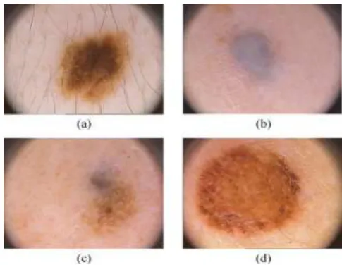
The Fig.1 shows the various patterns of dermoscopic images and the difficulties in getting a clear and proper image. Light falling on the skin undergoes reflection, refraction, diffraction and absorption. These phenomena are affected by the physical properties of the skin the entire image, or a set of contours extracted from the image. When applied to a stack of images, the output contours after image segmentation can be used to create three-dimensional reconstructions. There are mainly two approaches to segmentation namely edge segmentation and region segmentation. In segmentation stage the correctness of the subsequent steps is affected and hence it is the most important and difficult stage, because of lesion shapes, sizes, and colours along with different skin types and textures. Some lesions also have asymmetrical boundaries and sometimes there are smooth changeover between the lesion and the skin, dark hair covering the lesions and specular manifestations.
Fig. 1. Difficulties of Dermoscopic Images; (A) Presence Of Hair; (B) Smooth Transition
Between Lesion And Skin; (C) Multiple Colored Lesions; And (D) Specular Reflections.
IV.
GENETIC
ALGORITHM
TERMINOLOGY
A Genetic Algorithm is a class of adaptive stochastic optimization algorithms which involves the process of searching and optimizing. Optimization problems can be defined as an act, process, or methodology of making something such as a design, system, or decision; as fully perfect, functional, or effective as possible.
216 | P a g e The traditional methods are stuck at local optima or pre-mature convergence. They require the existence of derivatives of objective and constraint functions and also require mathematically well defined objective and constraint functions. Handling mixed variables is a very tedious process in traditional methods. The span of Genetic Algorithm includes the problem of real world being complex. Genetic Algorithm is useful in non linear multimodal, discrete optimization problems and non smooth search space. Multi objective optimization is possible in Genetic Algorithm. The Genetic Algorithm operators include selection (better individuals get higher chance), crossover (creates new individuals by combining parts from two individuals), mutation (creates new individual by making changes in a single individual), stopping criteria. Pignalberi used Genetic Algorithms to focus on range images, where a pixel is colored depending on the distance between the object and a sensor [10]. A genetic algorithm simulates Darwinian theory of evolution using highly parallel, mathematical algorithms that, transform a set (population) of solutions (typically strings of 1's and 0's) into a new population, using operators such as: reproduction, mutation and crossover.
4.1 Terms in Genetic Algorithm
Genetic algorithms try to imitate the Darwinian evolution process in computer programs. In evolutionary systems, populations evolve by selective pressures, mating between individuals, and alterations such as mutations. In genetic algorithms, genetic operators evolve solutions in the current population to create a new population, simulating similar effects. A genetic algorithm simulates Darwinian theory of evolution using highly parallel, mathematical algorithms that, transform a set (population) of solutions (typically strings of 1's and 0's) into a new population, using operators such as: reproduction, mutation and crossover.
4.2 Units
Biological GA terms
Chromosome String
Gene Feature or Character
Genomes Guesses solution ,collection of genes
5.1 Input Image
Here we use of a lesion affected skin .jpg image.An image is an array, or a matrix, of square pixels (picture elements) arranged in columns and rows.In a (8-bit) grey scale image each picture element has an assigned intensity that ranges from 0 to 255.
5.2 Initial Population
The initial population of the genetic algorithm is randomly generated i.e. programs (chromosomes) are formed by a randomly sequence of operators. The parameter values of operators are alo assigned randomly. It is made up of the primitive image analysis operators. For practical reasons, the size of each program is limited to a maximum depth. In our case we define the maximum depth of 15.
5.3 Genes
Genes are the building blocks of a chromosome. In genetic, these genes are basic low level image analysis operators also know as image primitive. We use simple notation to encode genes. The general layout of a gene can be seen as below
OPERATOR INPUT1 INPUT2 WEIGHT SE
The first field represents operator name , typically an image analysis function. The second and third fields represent the input planes to the operator. A gene can operate on one or two inputs, depending on the type of operator. The fourth field indicates a weight or parameter vaule (if needed) for the operator. Finally the last field refers to the type of morphological structuring element used by the operator.
5.4 Seed Region Growing
• Performs Region growing from a specified seed point.
• The region is iteratively grown by comparing all unallocated neighboring pixels to the region.
• The difference between a pixel's intensity value and the region's mean, % is used as a measure of similarity.
• The pixel with the smallest difference % measured this way is allocated to the respective region.
• This process stops when the intensity difference between region mean and % new pixel become larger than
the fitness function.
5.5 Fitness Function
Fitness is the measure of the optimality of a program present in the population. It reflects the accuracy of the segmentation algorithm. The sum of the absolute errors made by the program for pixels of all the images of training set can be transformed into a fitness function using some scaling techniques.
In our case, a segmented image consists of positive (object) and negative (non-object) pixels [18]. Ideally the segmentation of an image would result in an output image where positive pixels cover object pixels perfectly and nothing else while negative pixels cover non-object pixels perfectly and nothing else. Based on this ideal, we can view segmentation as a pixel-classification problem. Thus, the task of the segmentation program becomes assignment of the right class to every pixel in the image. As such, we can apply measure of classification accuracy to the problem of image segmentation.
218 | P a g e individual values in the best case scenario would be 1 and 0 in the worst case scenario. However, for the segmentation problem, achieving this is a challenging task, thus we define two more measures based on TPs, TNs, FPs and FNs called the False Positive Rate (FPR) and False Negative rate (FNR). FPR is the proportion of negative instances that were erroneously reported as being positive while FNR is the proportion of positive instances that were erroneously reported as negative.
For an ideal segmentation the values of FPR and FNR should be zero. For finding accuracy of a segmentation program we use pixel based accuracy formula based on FPR and FNR. This formula reflects the training and validation accuracy as follows.
Accuracy = k*(1-FPR)*(1-FNR) Where FPR- false positive rate FNR- false negative rate k – Constant
5.6 Selection
Selection of chromosome to undergo diversification. Tournament Selection scheme is used for diversification.
Size of the tournament window ᵞ is kept 10% of size of population. Quantity of parents selected is 50% of the size of population.
5.7 Crossover
Crossover is typically a two parent genetic operator. It works by exchanging the "genetic material" between two parent chromosomes. We have used a 1-point crossover for our GP. Two parents are chosen randomly from the parent pool. A random location is chosen in each of the parent chromosomes. The subsequences before and after this location in the parents are exchanged creating two offspring chromosomes.
5.8 Mutation
Mutation is a one parent genetic operator. It is applied to a single chromosome at a time and makes (small) changes in the genetic code of an individual.
We have used four mutations operators. They can be divided into two categories, Type A (inter-genomic - swap, insert and delete) and Type B (intra genomic - alter). 5.8.1 Type A Mutation
Type A mutation is typically inter-genomic. There are three inter-genomic mutations used - swap, insert and delete. The three inter-genomic mutations are as follows:
a) Swap: Two random locations inside a chromosome are chosen and the respective genes are swapped. b) Insert: A new gene is inserted in a randomly chosen position inside a parent chromosome.
c) Delete: A gene at a randomly chosen location gets removed from the chromosome. The remaining chromosome joins back together at the point of
the deletion.
5.8.2 Type B Mutation
Alter: Type B mutation is intra-genomic. Alter is a fitness based mutation. It is only performed if the fitness of the parent chromosome is above a minimum threshold
no mutation takes place. This operation essentially performs parameter tuning for the primitive image operators. The threshold was set at 70% accuracy, based on results of trial runs.
5.9 Elitism
Retains a certain percentage of the best available programs in the population pool. Here we save 1% of the chromosomes of population. Ensures that the best chromosomes always survive a generation.
VI.
RESULTS
By the help of genetic algorithm we were able to segment lesion accurately. This technique was very effective compared to other image segmentation technique.The results of image of skin lesion are as shown in following figs.
VII.
CONCLUSION
Combining the algorithms of Genetic Algorithm and region growth avoids the problems which arise while using a single method. The advantages of the Genetic Algorithm includes as being fast with simple binary operations and parallel searching scheme. The region growth procedure gets rid of the wrong target edge, making the target complete and more accurate. The region merging procedure helps in eradicating the problem of having many over segmented areas.
REFERENCES
[1] Angelina.S', L.Padma Suresh, S.H.Krishna Veni, IEEE 2012 “Image Segmentation Based On Genetic Algorithm for Region Growth and Region Merging”
[2] Lei Zhang, David Zhang (2011), "Automatic Image Segmentation by Dynamic Region Merging", IEEE Transactions, image process, volume 18, No.10.
[3] H. Ganster, P. Pinz, R.Rohnen, E.Wilding and H. Kittler(2001), "Automated Melanoma Recognition", IEEE Transactions, Medical Imaging. Volume 20, pages 233-239.
[4] Luis Garcia Ugarriza, Eli Saber (2009), "Automatic Image Segmentation by Dynamic Region Growth and Multiresolution Merging", IEEE Transactions, image process, volume 18, No. 10.
[5] Jacinto C Nascimento, Margarida Silveria (2009), "Comparison Of Segmentation Methods For Melanoma Diagnosis In Dermoscopy Images", IEEE Journal Of Selected Topic In Signal Processing, Volume 3, No.1.
220 | P a g e [7] Jie Wu, Skip Poehlman, Michael D. Noseworthy (2009), "Texture feature based automated seeded region
growing in abdominal MRI segmentation", J. Biomedical Science and Engineering, Scientific Research Publication, volume 2, pages 1-8.
[8] Mantas Paulinas, Andrius Usinskas (2007), "A survey of genetic algorithms applications for image enhancement and segmentation", ISSN, Information technology and control, volume 36, no.3.
[9] Melanie Mitchell, Payel Ghosh (2006), "Segmentation of Medical Images Using a Genetic Algorithm", GECCO, Image processing, volume 18.
[10] David 1. Crisp, Trevor C. Tao (2002), "Fast Region Merging Algorithms for Image Segmentation", ACCV2002: The 5th Asian Conference on Computer Vision, volume 38.
[11] G. Pignalberi, R. Cucchiara, L. Cinque, and S. Levialdi (2003), "Tuning range image segmentation by genetic algorithm", EURASIP Joumal on Applied Signal Processing, volume 8, pages 780-790.
[12] P. Zingaretti, G. Tascini, and L. Regini (2002), "Optimising the colour image segmentation", In VIII Convegno dell' Associazione Italiana per l'Intelligenza Artificiale, volume 1, pages 272-276.
[13] Elnomery A. Zanaty1,2 and Ahmed S. Ghiduk1,3 1College of Computers and IT, Taif University, Taif, Saudi Arabia “A Novel Approach Based on Genetic Algorithms and Region Growing for Magnetic Resonance Image (MRI) Segmentation”.
[14] L. Davis, Handbook of Genetic Algorithms. New York: Van Nostrand, 1991.
[15] Z. Michalewicz, Genetic Algorithms + Data Structures = Evolution Programs. New York: Springer-Verlag, 1992.
[16] Ning Situ1,Xiaojing Yuan2,Nizar Mullani3,George Zouridakis1“Automatic segmentation of skin lesion images using evolutionary strategy”.
[17] Margarida Silveira, Member, IEEE, Jacinto C. Nascimento, Member, IEEE, Jorge S. Marques, André R. S. Marçal, Member, IEEE, Teresa Mendonça, Member, IEEE, Syogo Yamauchi, Junji Maeda, Member, IEEE, and Jorge Rozeira “Comparison of Segmentation Methods for Melanoma Diagnosis in Dermoscopy Images”.