R E S E A R C H A R T I C L E
Open Access
Melatonin alleviates brain and peripheral
tissue edema in a neonatal rat model of
hypoxic-ischemic brain damage: the
involvement of edema related proteins
Li-Xiao Xu
1†, Yuan Lv
2†, Yan-Hong Li
1, Xin Ding
3, Ying Wang
3, Xing Han
3, Ming-Hua Liu
3, Bin Sun
3*and Xing Feng
3*Abstract
Background:Previous studies have indicated edema may be involved in the pathophysiology following hypoxic-ischemic encephalopathy (HIE), and melatonin may exhibit neuro-protection against brain insults. However, little is known regarding the mechanisms that involve the protective effects of melatonin in the brain and peripheral tissues after HIE. The present study aimed to examine the effects of melatonin on multiple organs, and the expression of edema related proteins in a neonatal rat model of hypoxic-ischemic brain damage (HIBD).
Methods:One hundred ninety-two neonatal rats were randomly divided into three subgroups that underwent a sham surgery or HIBD. After the HIBD or sham-injury, the rats received an intraperitoneal injection of melatonin or an equal volume vehicle, respectively. We investigated the effects of melatonin on brain, kidney, and colon edema via histological examination and the expression of edema related proteins, including AQP-4, ZO-1 and occludin, via qPCR and western blot.
Results:Our data indicated (1) Melatonin reduced the histological injury in the brain and peripheral organs induced by HIBD as assessed via H-E staining and transmission electron microscopy. (2) Melatonin alleviated the HIBD-induced cerebral edema characterized by increased brain water content. (3) HIBD induced significant changes of edema related proteins, such as AQP-4, ZO-1 and occludin, and these changes were partially reversed by melatonin treatment.
Conclusions:These findings provide substantial evidence that melatonin treatment has protective effects on the brain and peripheral organs after HIBD, and the edema related proteins, AQP4, ZO-1, and occludin, may indirectly contribute tothe mechanism of the edema protection by melatonin.
Keywords:Melatonin, Hypoxic-ischemic encephalopathy, Edema, Aquaporin-4, Zonula occludens-1, Occludin
Background
Hypoxic-ischemic encephalopathy (HIE) is a leading cause of mortality and morbidity. The most frequent eti-ologies of it are intrapartum or late antepartum brain hypoxia and ischemia [1, 2]. 40–60% of the HIE infants were die by 2 years of age or left severe disabilities—The
majority pathological mechanism of HIE is declined cerebral blood flow which results in less oxygen delivery to the brain disabilities [3] Despite the substantial re-search that has been conducted regarding HIE, the therapeutic interventions remain limited. Current HIE research is focused on the identification of the under-lying pathophysiology of perinatal HIE and the allevi-ation of these pathophysiological mechanisms, especially the reversible factors [4].
Neonatal asphyxia, or birth asphyxia is caused by deprivation of oxygen to a newborn baby that usually * Correspondence:[email protected];[email protected]
†Equal contributors
3Department of Neonatology, Children’s Hospital of Soochow University,
Suzhou 215006, China
Full list of author information is available at the end of the article
induce edema harm to the body, frequently to the brain to result in HIE [5]. Except brain injuries, severe neo-natal asphyxia is usually accompanied with multi-organ damages (such as in heart, lungs, liver, gut, kidneys) since the preferential blood supply to the brain will ag-gravate peripheral tissue ischemia [6, 7]. Therefore, the management of brain and peripheral tissue edema is critical for the prognosis of HIE patients. To date, a number of theories have been proposed to explain the development of edema in the brain and peripheral tis-sues [5, 8] Water channel aquaporins-4 (AQP-4) is specific membrane protein that have been widely investi-gated, which controls the influx and efflux of transmem-brane water [9]. As the constituent of the continuous intercellular barrier between epithelial cells, tight junc-tions (TJ) are required to separate tissue spaces and regulate the selective movement of solutes across the epithelium [10]. Because zonula occludens-1 (ZO-1) and occludin are identified within TJs, together they form a virtually impermeable barrier to fluid between closely as-sociated cells, which is very important in the mainten-ance of the mucosal and vascular endothelial barrier structure [11]. However, the dynamic changes in edema related proteins, such as AQP-4, ZO-1 and occludin, in the brain and peripheral tissues following HIE and their roles in edema development in central and peripheral tissues have rarely been reported. Early inhibition of edema related proteins in the brain and peripheral tis-sues may represent a new treatment protocol for edema following HIE.
In recent years, melatonin has been considered a promising neuro-protective drug for various acute and chronic brain injuries [12]. Melatonin is a naturally oc-curring hormone that is produced by the brain and was first identified to facilitate the regulation of the sleep-wake cycle [13]. Following ischemic brain injury/stroke, melatonin treatment has demonstrated a remarkable ability to reduce the infarct volume and/or inhibit neur-onal cell death in different mammalian species and ex-perimental models [12]. Moreover, it has been reported that melatonin decreased sensorimotor asymmetry and learning deficits, which thus protected pups from the long-term consequences of neonatal asphyxia [14]. In addition, histological analysis has also demonstrated melatonin treatment can increase the number of mor-phologically well preserved neurons in the CA1, CA2, and CA3 areas, as well as the dentate gyrus of the hippo-campus in a hypoxic-ischemic mode [12]. Melatonin can decrease the cellular damage induced by epilepsy in-volved activation of kainate-sensitive glutamate receptors [15]. However, to the best of our knowledge, little is known regarding the mechanisms that involve the neuro-protective effects of melatonin and its effects on periph-eral tissues in a hypoxic-ischemic encephalopathy model.
In the present study, we examined the effects of mela-tonin on brain, kidney and colon edema, and its roles in the expression of edema related proteins, including AQP-4, ZO-1 and occludin, in a neonatal rat model of hypoxic-ischemic brain damage (HIBD). We hypothe-sized that melatonin treatment would reduce the edema in both the brain and peripheral tissues that was induced by HIBD, at least in part, through the modulation of edema related proteins.
Methods
Animal preparation
192 Specific pathogen Free (SPF) Sprague Dawley (SD) rats at 7 days of age and a weight of 15–20 g, regardless of gender, were obtained from the SLRC Laboratory Animal Co. Ltd., China (License Key SCXK (Hu) 2012-0002). The neonatal rats were housed in groups of 10– 15 rats per cage in standard plastic cages, and they were freely fed by female rats. The animal room was main-tained on a 12-h dark-light cycle, and the temperature was maintained at 25±2°C. The protocols, which include all surgical procedures and animal usage, were approved by the Animal Care and Use Committee of Soochow University and conformed to the Guide for the Care and Use of Laboratory Animals by the National Institutes of Health.
HIBD model establishment
HIBD was induced in the present study using a modified Rice-Vannucci method [16]. In brief, 7-day-old postnatal pups were anesthetized with is oflurane (3% in a mixture of medical air and oxygen, 70:30 ratio). The left common carotid artery of each pup was identified, exposed, and permanently ligated with 5-0 surgical silk via a near-midline incision. The wound was closed, and the pups were allowed to recover from the anesthesia after the procedure, which lasted approximately 5 min per pup. Following recovery with their dams for 2 h, the pups were subsequently placed in a jar perfused with a hu-midified and pre-warmed gas mixture (8% oxygen bal-anced with nitrogen) for 2.5 h. A constant temperature of 37 °C was maintained throughout all procedures. Fol-lowing the hypoxia, the animals were returned to their dams, and the ambient temperature was maintained at 37 °C for 24 h. The sham animals underwent anesthesia, and the common carotid artery was exposed without ligation or hypoxia.
Experimental protocol
group (MT group). Immediately after the HIBD injury, the rats received an intraperitoneal injection of melatonin (10 mg/kg body weight) (Sigma, USA) in the MT group or an equal volume of vehicle in the Sham and Mod Groups.
Cerebral edema measurement
Cerebral edema was determined via brain water content (BWC) measurement using the wet weight—dry weight technique, and the results are expressed as a percentage of the water content [17].
Hematoxylin& eosin (H-E) staining of the brain, kidney and colon
The rats were sacrificed under deep anesthesia at 3, 6, 24 and 72 h after HIBD or sham-injury for H-E staining. The rats were perfused with heparinized saline and 4% neutral buffered formalin. The same cortical layer of left frontal cortex, renal cortex and colon were dissected. 5 μm thick paraffin-embedded sections were prepared for H-E staining.
Quantification of the swelling cell
For quantification analysis, the swelling cell in the H-E stained cortex, renal cortex and colon samples were de-fined as the cells which were abnormal enlargement of a body, transparent cytosol, light stained, and even balloon-like structures. The total number of the swelling glial cell, renal tubular epithelial cells, or colon epithelial cells were countered in 5 random fields under high mag-nification view. The swellingcells of cerebral glial, glom-erular epithelial, colon epithelial cells were quantified by dividing to the corresponding total cells.
Transmission electron microscopy (TEM) of the brain, kidney and colon
The rats were sacrificed under deep anesthesia at 3, 6, 24 and 72 h after HIBD or sham-injury for ultrastructure examination using TEM. Slices (approximately 1 mm thick) of the same cortical layer of left frontal cortex, renal cortex and colon were placed into cold 4% glutar-aldehyde solution immediately after harvest and main-tained at less than 4°C. The whole process of sampling required less than 3 min. Semi-thin sections were cut from these slices, rinsed overnight in 0.1 M phosphate buffer, post-fixed for 2 h in 1% osmium tetroxide, dehy-drated and then embedded in Araldite mixture. Ultra-thin sections stained in uranyl acetate and lead citrate were viewed in a Philips CM 120 electron microscope (Worcester, MA, USA).
Real time-quantitative polymerase chain reaction (RT-qPCR) for AQP4, ZO-1, and occludin
The rats in the three groups were sacrificed under deep anesthesia at 3, 6, 24 and 72 h after HIBD or
sham-injury for RT-qPCR [18]. The rats were trans-cardially perfused with 250 ml of cold heparinized 0.9% saline, and the left frontal cortex, renal cortex and colon were rapidly removed and stored in liquid nitrogen immediately until use. The AQP-4, ZO-1, and occluding mRNA levels were determined using RT-qPCR. The primer sequences and PCR conditions were as follows: AQP-4: forward, 5’- cggttcatggaaacct cact -3’; reverse, 5’- catgctggctccggtataat -3’; Zo-1: for-ward, 5’- gtatccgattgttgtgttcc -3’; reverse, 5’- tcacttg tagcaccatccgc -3’; occludin: forward, 5’- cacgttcgac caatgc -3’; reverse, 5’- cccgttccataggctc -3’; and β -actin: forward, cccatctatgagggttacgc -3’; reverse, 5’-tttaatgtcacgcacgatttc -3’. RT-qPCR analysis was per-formed using the iQ5™ Real-Time PCR System (Bio-Rad, USA), which applied real-time SYBR Green PCR technology. All samples were analyzed in triplicate. β -actin was selected as an acceptable endogenous refer-ence “housekeeping” gene. The relative change in the target cytokine mRNA expression was determined by the following equation: fold change = 2−[ΔΔCt], where the Ct value is the cycle number at which the fluorescence signal crosses the threshold.
Western blot
0.5–1 g of the mouse cortex, renal cortex, and colon were collected and lysed in 500 μl of homogenization buffer. Each 30μg of the extracted protein was separated on 10% SDS-PAGE gels and transferred onto a nitrocel-lulose membrane. Subsequently, the membrane was reacted with mouse monoclonal AQP-4 (all antibodies are from Abcam;Cambridge,MA),mouse monoclonal ZO-1, mouse monoclonal occluding, and mouse mono-clonal GAPDH. The probed membrane was then incu-bated with horseradish peroxidase-conjugated rabbit anti-mouse IgG. The probed proteins (AQP-4, ZO-1, oc-cluding, and GAPDH) were visualized by enhanced chemiluminescent.
Statistical analysis
SPSS 16.0 software was used for the statistical analyses (SPSS, Inc., Chicago, IL, USA). All data are presented as the mean ± SD. The data were subjected to a one-way analysis of variance followed by a Student-Newman-Keuls test. Statistical significance was determined as
P< 0.05.
Results
Cerebral edema measurement
the HIBD rats following melatonin treatment (P< 0.05; Fig. 1), which indicates an improvement in cerebral edema.
Edema estimation of the brain, kidney and colon by H-E staining and edema cell quantification
In the Sham group, the cellular morphologies were nor-mal in the brain (Fig. 2), kidney (Fig. 3) and colon (Fig. 4). In the Mod group, there was significant glial cell
swelling and karyopyknosis in combination with intersti-tial tissue edema, capillary contracture and blank zones around the capillary (Fig. 2). And a significant swelling of the renal tubular epithelial cells was identified, as well as interstitial tissue edema (Fig. 3). Moreover, significant edema and even ballooning degeneration of the renal tubular epithelial cells were identified (Fig. 4). In the MT group, there was an improvement in the edema com-pared with the Mod group (Figs. 2, 3 and 4). In addition, the corresponding edema cells were quantified (Fig. 2d, 3d, and 4d) which is consistent with the observation above.
TEM of the brain, kidney and colon
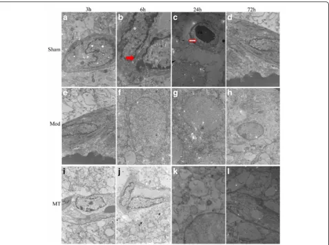
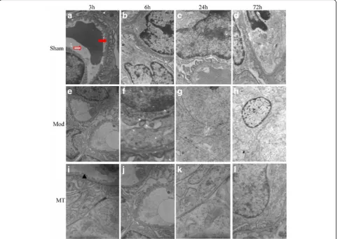
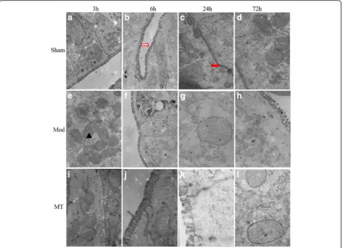
In the Sham group, the cellular morphologies were nor-mal in the brain (Fig. 5), kidney (Fig. 6) and colon (Fig. 7). In the Mod group, at 3 h after HIBD, there was a minimally dilated rough endoplasmic reticulum (ER) and edematous changes in the matrix and cristae of the mitochondria (Fig. 5). And the tubular epithelial cells were slightly swollen (Fig. 6). Moreover, the microvilli were sparse, and the rough ER was slightly dilated (Fig. 7). At 6 h after HIBD, there were more substantial edematous changes in the organelles and vacuolation of the mitochondria, as well as fusion in some intercellular junctions (Fig. 5). And the capillary lumen narrowed
Fig. 1Effects of melatonin treatment on the BWC (n= 6 per group). HIBD could result in a significant increase in the BWC in comparison with the one in the sham rats (P< 0.05), which was significantly attenuated following melatonin treatment (P< 0.05)
Fig. 3Renal cortex under a 400 × light microscope after H-E staining. Similar treatment as Fig. 2 induced significant swelling of the renal tubular epithelial cells and interstitial tissue edema, which was improved by melatonin treatment in H-Estained samples (aandb). However, there were few changes regarding the glomeruli in these groups. In addition, the swollen renal tubular epithelial cells were quantified in the total renal tubular epithelial cells in the indicated groups.↗represents cytotoxic edema, and▲represents vasogenic edema
while the epithelial hole increased (Fig. 6). Moreover, the edematous changes in the colon cells progressed, and the junctions between the cells became somewhat vague (Fig. 7). A remarkable pathologic change oc-curred at 24 h after HIBD, which was characterized by clearly decreased organelles and their extraordinary swelling. Many intercellular junctions had fused or disappeared (Fig. 5). And there was a vague mem-brane, which made it difficult to distinguish the three layers, and the foot process was randomly arranged and further fused (Fig. 6). In the colon (Fig. 7), even the sparse distributed microvilli swelled, and the epi-thelial cell organelles were vague. 72 h after the oper-ation, the situation had improved in the brain (Fig. 5), kidney(Fig. 6) and colon(Fig. 7). In the MT group, the time course of edema was similar com-pared with the Mod group, but the severity was re-duced (Figs. 5, 6 and 7).
AQP-4 expression in the cerebral cortex, renal cortex and colon
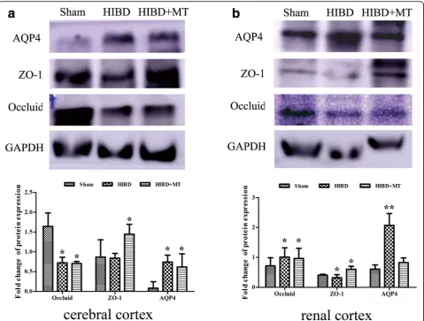
As shown in Fig. 8a, HIBD induced a significant increase in the AQP-4 mRNA expression in the cerebral cortex compared with the sham injury at 3, 6 and 24 h after in-jury (P< 0.05). Melatonin treatment significantly de-creased the AQP-4 mRNA expression in the cerebral cortex compared with the Mod Group at 3 and 24 h after the injury (P< 0.05). Furthermore, the data in Fig. 8b indicated that there was no significant difference in the AQP-4 mRNA expression in the renal cortex be-tween the three groups at any time-point (P> 0.05). In addition, the data in Fig. 8c indicated that HIBD induced a significant increase in the AQP-4 mRNA expression in the colon compared with the sham injury only at 24 h after the injury (P< 0.05). The mRNA changes in the cerebral cortex and renal cortex after 24 h HIBD treatment were verified as well by western blot (Fig. 11a
and b). AQP-1 protein expression of the cerebral cor-tex and renal corcor-tex were augmented significantly 24 h after HIBD and melatonin partially (cerebral cor-tex, Fig. 11a) or completely (renal corcor-tex, Fig. 11b) prevented this increase. The detection of the protein expression in the colon samples did not succeed (data did not show).
ZO-1 expression in the cerebral cortex, renal cortex and colon
As shown in Fig. 9a, HIBD induced a significant de-crease in the ZO-1 mRNA expression in the cerebral cortex compared with the sham injury at 6, 24 and 72 h after the injury (P< 0.05). There was no signifi-cant difference in the ZO-1 mRNA expression in the cerebral cortex between the Mod and MT groups at any time-point (P> 0.05). Furthermore, the data in Fig. 9b indicated that the ZO-1 mRNA expression in the renal cortex was significantly decreased compared
with the sham injury at 6 and 24 h after the injury (P< 0.05). Melatonin significantly increased the ZO-1 mRNA expression in the renal cortex compared with the Mod group at 24 h after the injury (P< 0.05). In addition, the data in Fig. 9c indicated that HIBD in-duced a significant decrease in the ZO-1 mRNA ex-pression in the colon compared with the sham injury at 6 and 24 h after the injury (P< 0.05), whereas melatonin treatment significantly increased the ZO-1 mRNA expression in the colon compared with the Mod group24 h after the injury (P< 0.05). The find-ings in the cerebral cortex and renal cortex at 24 h after injury were further confirmed by western blot (Fig. 11a and b). ZO-1 protein of the cerebral cortex has declined 24 h after HIBD and melatonin turn back this response significantly (Fig. 11a). Likewise, ZO-1 pro-tein expression of the renal cortex has slightly weakened 24 h after HIBD and this decrease was prohibited consid-erably by melatonin treatment (Fig. 11b).
Occludin expression in the cerebral cortex, renal cortex and colon
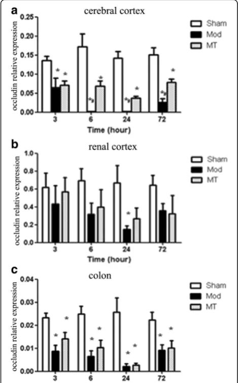
As shown in Fig. 10a, HIBD induced a significant de-crease in the occluding mRNA expression in the cerebral cortex compared with the sham injury at each time-point after the injury (P< 0.05). Melatonin treatment sig-nificantly increased the occluding mRNA expression in the cerebral cortex compared with the Mod group at 6, 24 and 72 h after the injury (P< 0.05). Moreover, the data in Fig. 10b indicated that the occluding mRNA expression in the renal cortex was significantly de-creased compared with the sham injury 24 h after the in-jury (P< 0.05). There was no significant difference in the occluding mRNA expression in the renal cortex between the Mod and MT groups at any time-point (P> 0.05). In addition, the data in Fig. 10c indicated that HIBD in-duced a significant decrease in the occluding mRNA ex-pression in the colon compared with the sham injury at each time-point after the injury (P< 0.05), whereas there
was no significant difference in the occluding mRNA ex-pression in the colon between the Mod and MT groups at any time-point (P> 0.05). In addition, these results in the cerebral cortex and renal cortex after 24 h HIBD were proven by western blot (Fig. 11a and b). Theocclu-din protein expression of the cerebral cortex and renal cortex have decreased significantly 24 h after HIBD and melatonin partially attenuated these changes.
Discussion
In the present study, we examined the effects of mela-tonin on brain, kidney and colon edema, and its roles in the expression of edema related proteins in a neonatal rat model of HIBD. The main findings are as follows: (1) Melatonin reduced the histological injuries of the brain and peripheral organs, which were induced by HIBD, as assessed via H-E staining and TEM. (2) Melatonin allevi-ated the HIBD-induced cerebral edema characterized by the decreased BWC. (3) HIBD induced significant
changes in the mRNA expression of edema related pro-teins, including AQP-4, ZO-1 and occludin, which were partially reversed by melatonin treatment.
AQP-4 is one of the most deeply lucubrated edema re-lated proteins in the central nervous system, especially regarding cytotoxic cerebral edema [19]. The expression of AQP-4 is positively related to edema at an early stage after HIBD insults in rats with a genetic knockout of AQP-4 [20], which is consistent with our findings that cerebral edema characterized by increased BWC is ac-companied by significantly increased AQP-4 expression in the cerebral cortex following HIBD insults. Moreover, we demonstrated that AQP-4 mRNA expression increased
as early as 3 h after HIBD injury and was accompanied by the histological signs of cerebral edema, such as mito-chondrial swelling, rough ER expansion, and capillary con-tracture surrounded by an electronic blank area, when examined by TEM, these alterations represented both cytotoxic edema and vasogenic edema. The present study demonstrated that AQP-4 mRNA expression reached its peak at 24 h after injury and subsequently decreased, which was accompanied by similar changes assessed via TEM.
Acute kidney injury typically presents with edema and necrosis, and the renal tubules exhibit an earlier and more severe manifestation [21]. In the present study, we
Fig. 8Real time-qPCR for AQP-4 in the cerebral cortex (a), renal cortex (b) and colon (c). HIBD induced a significant increase in the AQP-4 mRNA expression in the cerebral cortex and colon. Melatonin treatment significantly decreased the AQP-4 mRNA expression in comparison with the one without melatonin treatment in HIBD group. The data represent the mean ± SD
identified obvious edematous and necrotic changes to-gether with interstitial edema. However, AQP-4 mRNA expression was not significantly different after hypoxic and ischemic damage, which indicates that there may be no obvious correlation between AQP4 and renal tubular epithelial cell and interstitial tissue edema. Nevertheless, based on evidence regarding the organic distribution of AQPs and the existence of more than one subtype of AQPs positioned in the kidney [22] we speculated that tubular cell edemamay be affected by other AQP sub-types; thus
Additional research is required to verify this hypoth-esis. Neonates with HIE typically exhibit abnormal mani-festations in the digestive tract, such as gastrointestinal bleeding and feeding intolerance, and the most severe symptom is necrotizing enterocolitis. The ileum and
colon are more likely to be affected because of their spe-cial blood supply characteristics and molecular basis. Previous studies have demonstrated that AQP-4 is dis-tributed in the entire colon, mainly on the top of the villi epithelium and lumen [23]. However, little is known re-garding the relationship between AQP-4 and hypoxic-ischemic intestinal damage. In our experiment, we iden-tified colon epithelial cell edema using TEM, as well as an increased AQP-4 expression, which is consistent with the extent of edema in the colon. Thus, we could infer that AQP-4 expression may play a role in hypoxic-ischemic colon damage.
Tight junction proteins (TJPs) represent a series of proteins closely related to the blood-brain barrier (BBB) in the brain. In our experiment, we investigated two types of TJPs, including ZO-1 and occludin. The expres-sion of ZO-1 and occluding mRNA were decreased as early as 3 h after HIBD injury, which was accompanied by signs of vasogenic edema in the brain assessed with TEM. This finding further verifies that TJPs are nega-tively related to edema. Tight junctions are also very im-portant for the maintenance of epithelial homeostasis. Previous studies have indicated that TJPs are expressed on tubules [24]. Thus, the significant decrease in TJP ex-pression, especially ZO-1 exex-pression, may be related to the damage of tight junctions between tubules and renal interstitial, which tends to lead to vasogenic edema. In addition, the use of TEM in the present study demon-strated the existence of colon epithelial edema as early as 3 h after HIBD, and the edema peaked 24 h after in-jury, which was accompanied by significantly decreased ZO-1 and occluding mRNA expression in the colon. Thus, the damaged tight junction structure characterized by reduced TJP expression may be the reason for colon epithelial injury following HIBD.
To date, previous studies regarding endogenous melatonin and melatonin receptors have indicated that there is a significant decrease in both the release and function of melatonin following HIBD, which suggests melatonin may function in the pathogenesis of HIBD. Moreover, exogenous melatonin supplemen-tation can improve the stress state, exhibit beneficial effects in the maintenance of relative homeostasis, and improve the immunological functions of HIE ne-onates. Thus, it is necessary to further investigate the role of melatonin in the pathophysiological mecha-nisms of HIBD, such as edema in the central nervous system and peripheral organs. Our study provides timely and substantial evidence for the theoretical basis of melatonin application in the clinic.
Previous studies indicate that endogenous melatonin release decreased during the hypoxic-ischemic injury in rats, which may be involved in the pathogenesis of early HIBD [10, 12]. In addition, supplement of exogenous
melatonin after hypoxic-ischemic injury is beneficial to protect neonatal perinatal brain damage [14]. In a pro-spective study melatonin combining with hypothermia promoted neonatal HIE survival and health by brain magnetic resonance imaging (MRI) analysis [25]. In an-other preterm brain injury study, small dose (0.1 mg/kg) of melatonin demonstrated a protective activity on preg-nant ewe hyopoxic/ischemia injure model [26]. On the other hand, other study of melatonin treatment in an neonatal hypoxic-ischemic damage model did not show significant improvement in (1 h) early phase [27]. These results hint that different hypoxic/ischemia model and the method of melatonin employment may affect the re-sult, as well as more longer observation is necessary. Our results did demonstrate a significant anti-edema damage activity in longer time course observation. In addition, our result provided a possible mechanism as well that melatonin indirectly regulates the edema
related factors, AQP-4, ZO-1, and occludin, to partially protect hypoxic-ischemic injures.
Several mechanisms of melatonin may be involved in its effects. For example, based on its lipophilic and partly hydrophilic dissolubility, melatonin has a high diffusing capacity, which makes it easy to enter the central ner-vous system and act directly on cell membranes and nu-clei [28]. It was believed that metalonin acts for neuroprotection and ischemia harm protection by its anti-oxidative and anti-apoptosis properties [29–33]. Thus, regarding the edema protection property, mela-tonin could change the transcriptional activity of edema related molecules, such as AQP-4 and TJPs, which is im-portant for its improvement in brain edema. Further-more, the melatonin receptor belongs to a G-protein-coupled receptor family, and exogenous melatonin can combine with its receptors soon after HIBD, which fur-ther activates the downstream MAPK signal pathway,
especially the p38 MAPK and JNK/SAPK signal path-ways [34, 35], both of which are important in the regula-tion of HIBD pathogenesis, such as edema related proteins. Moreover, exogenous melatonin could increase endogenous melatonin release and melatonin receptor regulation via the positive feedback path, as well as en-large their effects via a cascade reaction. In addition, vas-cular endothelial growth factor (VEGF) has been reported to lead to the inactivation of TJPs, such as ZO-1, by decreasing their expression and increasing phosphoryl-ation; previous studies have indicated that melatonin can improve VEGF release [36, 37], which indirectly regulates edema related protein expression. Additional research re-garding the pathways involved in the regulation of edema related proteins for melatonin is necessary.
Conclusions
In conclusion, our data indicate that melatonin treat-ment has protective effects on the brain and peripheral organs after HIBD. The edema related proteins, AQP-4, ZO-1, and occludin, may participate in the mechanism of edema protection by melatonin.
Abbreviations
AQPs:Aquaporins; BBB: Blood-brain barrier; BWC: Brain water content; ER: Endoplasmic reticulum; HIBD: Hypoxic-ischemic brain damage; HIE: Hypoxic-ischemic encephalopathy; MT: Melatonin; qPCR: Real-time quantitative polymerase chain reaction; SD: Sprague Dawley; SPF: Specific pathogen free; TJ: Tight junctions; TJP: Tight junction proteins; VEGF: Vascular endothelial growth factor; ZO-1: Zonula occludens-1
Acknowledgements
Not applicable.
Funding
This work was supported by grants from National Natural Science Foundation (81471488, 81502157, 81271378, 81501291), Key medical subjects of Jiangsu province (XK201120), Jiangsu Province key research and development of special funds (BE2015644), Key Laboratory of Suzhou City (SZS201108), Science and Technology Project of Suzhou (SYSD2013105, SYS201446, SYS201441), Public health technology project of Suzhou (SS201536) and the Department of Pediatrics Clinical Center of Suzhou (Szzx201504).
Availability of data and materials
The data and analysis in this study could be reasonably acquired from the corresponding author.
Authors’contributions
XLX and LY participated in project design, all the experiment processes, data analysis, and manuscript writing. LYH provided technical support in histochemical staining analysis and helped revising the manuscript. DX designed the primer of PCR, completed the real-time PCR experiments and data collecting. HX and LMH helped accomplish HIBD model, participated in western blot experiments. WY was responsible for electronic microscopy analysis. SB and FX were primarily responsible for study design and protocol development. All authors have read and approved the manuscript.
Competing interests
The authors declare that they have no competing interests.
Consent for publication
Not applicable.
Ethics approval and consent to participate
The protocols, which include all surgical procedures and animal usage, were approved by the Animal Care and Use Committee of Soochow University [Licence No. SCXK(Hu) 2012-0002], and conformed to the Guide for the Care and Use of Laboratory Animals by the National Institutes of Health.
Publisher’s Note
Springer Nature remains neutral with regard to jurisdictional claims in published maps and institutional affiliations.
Author details
1Institute of Pediatric Research, Children’s Hospital of Soochow University,
Suzhou 215006, China.2Department of Neonatology, Northern Jiangsu People’s Hospital, Yangzhou 225001, China.3Department of Neonatology,
Children’s Hospital of Soochow University, Suzhou 215006, China.
Received: 30 October 2015 Accepted: 2 March 2017
References
1. Sarnat HB, Sarnat MS. Neonatal encephalopathy following fetal distress. A clinical and electroencephalographic study. Arch Neurol. 1976;33(10):696–705. 2. Volpe JJ. Neurology of the Newborn. 4. In: Hypoxic-ischemic encephalopathy:
Clinical aspects. Philadelphia: WB Saunders Co; 2000. p. 331–96.
3. Allen KA, Brandon DH. Hypoxic ischemic encephalopathy: pathophysiology and experimental treatments. Newborn Infant Nurs Rev. 2011;11(3):125–33. 4. Buonocore G, Perrone S, Turrisi G, Kramer BW, Balduini W. New
pharmacological approaches in infants with hypoxic-ischemic encephalopathy. Curr Pharm Des. 2012;18(21):3086–100.
5. Lupton BA, Hill A, Roland EH, Whitfield MF, Flodmark O. Brain swelling in the asphyxiated term newborn: pathogenesis and outcome. Pediatrics. 1988;82(2):139–46.
6. Wang X, Zhang J, Yang Y, Dong W, Wang F, Wang L, et al. Progesterone attenuates cerebral edema in neonatal rats with hypoxic-ischemic brain damage by inhibiting the expression of matrix metalloproteinase-9 and aquaporin-4. Exp Ther Med. 2013;6(1):263–7.
7. Barnett CP, Perlman M, Ekert PG. Clinicopathological correlations in postasphyxial organ damage: a donor organ perspective. Pediatrics. 1997;99(6):797–9.
8. Scallan J, Huxley VH, Korthuis RJ. Capillary fluid exchange: regulation, functions, and pathology[J]. Colloquium. 2010(1).
9. Liu K, Agre P. Aquaporin Water Channels. In: Lane WJLD, eidotr. Encyclopedia of Biological Chemistry. Waltham: Academic Press; 2013. p. 120-6.
10. Anderson JM, Van Itallie CM. Physiology and function of the tight junction. Cold Spring Harb Perspect Biol. 2009;1(2):a002584.
11. Günzel D, Yu AS. Claudins and the modulation of tight junction permeability. Physiol Rev. 2013;93(2):525–69.
12. Alonso-Alconada D, Alvarez A, Arteaga O, Martinez-Ibarguen A, Hilario E. Neuroprotective effect of melatonin: a novel therapy against perinatal hypoxia-ischemia. Int J Mol Sci. 2013;14(5):9379–95.
13. Brown GM. Light, melatonin and the sleep-wake cycle. J Psychiatry Neurosci. 1994;19(5):345–53.
14. Carloni S, Perrone S, Buonocore G, Longini M, Proietti F, Balduini W. Melatonin protects from the long-term consequences of a neonatal hypoxic-ischemic brain injury in rats. J Pineal Res. 2008;44(2):157–64. 15. Uz T, Giusti P, Franceschini D, Kharlamov A, Manev H. Protective effect of
melatonin against hippocampal DNA damage induced by intraperitoneal administration of kainate to rats. Neuroscience. 1996;73(3):631–6. 16. Chen W, Jadhav V, Tang J, Zhang JH. HIF-1alpha inhibition ameliorates
neonatal brain injury in a rat pup hypoxic-ischemic model. Neurobiol Dis. 2008;31(3):433–41.
17. Wang JW, Wang HD, Cong ZX, Zhang XS, Zhou XM, Zhang DD. Activation of metabotropic glutamate receptor 5 reduces the secondary brain injury after traumatic brain injury in rats. Biochem Biophys Res Commun. 2013;430(3):1016–21.
19. Wang H, Wang X, Guo Q. The correlation between DTI parameters and levels of AQP-4 in the early phases of cerebral edema after hypoxic-ischemic/reperfusion injury in piglets. Pediatr Radiol. 2012;42(8):992–9. 20. Da T, Verkman AS. Aquaporin-4 gene disruption in mice protects against
impaired retinal function and cell death after ischemia. Invest Ophthalmol Vis Sci. 2004;45(12):4477–83.
21. Basile DP, Anderson MD, Sutton TA. Pathophysiology of acute kidney injury. Compr Physiol. 2012;2(2):1303–53.
22. Wang W, Li C, Summer SN, Falk S, Wang W, Ljubanovic D, et al. Role of AQP1 in endotoxemia-induced acute kidney injury. Am J Physiol Renal Physiol. 2008;294(6):F1473–80.
23. Yde J, Keely S, Wu Q, et al. Characterization of AQPs in Mouse, Rat, and Human Colon and Their Selective Regulation by Bile Acids. Front Nutr. 2016;3:46.
24. Gonzalez-Mariscal L, Namorado MC, Martin D, Luna J, Alarcon L, Islas S, et al. Tight junction proteins ZO-1, ZO-2, and occludin along isolated renal tubules. Kidney Int. 2000;57(6):2386–402.
25. Aly H, Elmahdy H, El-Dib M, Rowisha M, Awny M, El-Gohary T, et al. Melatonin use for neuroprotection in perinatal asphyxia: a randomized controlled pilot study. J Perinatol. 2015;35(3):186–91.
26. Drury PP, Davidson JO, Bennet L, Booth LC, Tan S, Fraser M, et al. Partial neural protection with prophylactic low-dose melatonin after asphyxia in preterm fetal sheep. J Cereb Blood Flow Metab. 2014;34(1):126–35. 27. Berger HR, Morken TS, Vettukattil R, Brubakk AM, Sonnewald U, Wideroe M.
No improvement of neuronal metabolism in the reperfusion phase with melatonin treatment after hypoxic-ischemic brain injury in the neonatal rat. J Neurochem. 2015;136(2):339–50.
28. Hardeland R. Melatonin in plants and other phototrophs: advances and gaps concerning the diversity of functions. J Exp Bot. 2015;66(3):627–46. 29. Gilad E, Cuzzocrea S, Zingarelli B, Salzman AL, Szabo C. Melatonin is a
scavenger of peroxynitrite. Life Sci. 1997;60(10):L169–74.
30. Hardeland R, Tan DX, Reiter RJ. Kynuramines, metabolites of melatonin and other indoles: the resurrection of an almost forgotten class of biogenic amines. J Pineal Res. 2009;47(2):109–26.
31. Wakatsuki A, Okatani Y, Izumiya C, Ikenoue N. Melatonin protects against ischemia and reperfusion-induced oxidative lipid and DNA damage in fetal rat brain. J Pineal Res. 1999;26(3):147–52.
32. Tan DX, Manchester LC, Terron MP, Flores LJ, Reiter RJ. One molecule, many derivatives: a never-ending interaction of melatonin with reactive oxygen and nitrogen species? J Pineal Res. 2007;42(1):28–42.
33. Hassell KJ, Ezzati M, Alonso-Alconada D, Hausenloy DJ, Robertson NJ. New horizons for newborn brain protection: enhancing endogenous neuroprotection. Arch Dis Child Fetal Neonatal Ed. 2015;100(6):F541–52. 34. Xu L, Pathak PS, Fukumura D. Hypoxia-induced activation of p38
mitogen-activated protein kinase and phosphatidylinositol 3’-kinase signaling pathways contributes to expression of interleukin 8 in human ovarian carcinoma cells. Clin Cancer Res. 2004;10(2):701–7.
35. Ferreira GM, Martinez M, Camargo IC, Domeniconi RF, Martinez FE, Chuffa LG. Melatonin Attenuates Her-2, p38 MAPK, p-AKT, and mTOR Levels in Ovarian Carcinoma of Ethanol-Preferring Rats. J Cancer. 2014;5(9):728–35. 36. Fischer S, Wobben M, Marti HH, Renz D, Schaper W. Hypoxia-induced
hyperpermeability in brain microvessel endothelial cells involves VEGF-mediated changes in the expression of zonula occludens-1. Microvasc Res. 2002;63(1):70–80.
37. Kaur C, Sivakumar V, Ling EA. Melatonin protects periventricular white matter from damage due to hypoxia. J Pineal Res. 2010;48(3):185–93.
• We accept pre-submission inquiries
• Our selector tool helps you to find the most relevant journal
• We provide round the clock customer support
• Convenient online submission
• Thorough peer review
• Inclusion in PubMed and all major indexing services
• Maximum visibility for your research
Submit your manuscript at www.biomedcentral.com/submit