University of Pennsylvania
ScholarlyCommons
Publicly Accessible Penn Dissertations
2017
Multiscale Mechanical, Structural, And
Compositional Response Of Tendon To Static And
Dynamic Loading During Healing
Benjamin Freedman
University of Pennsylvania, [email protected]
Follow this and additional works at:https://repository.upenn.edu/edissertations Part of theBiomechanics Commons, and theBiomedical Commons
This paper is posted at ScholarlyCommons.https://repository.upenn.edu/edissertations/2289
For more information, please [email protected]. Recommended Citation
Freedman, Benjamin, "Multiscale Mechanical, Structural, And Compositional Response Of Tendon To Static And Dynamic Loading During Healing" (2017).Publicly Accessible Penn Dissertations. 2289.
Multiscale Mechanical, Structural, And Compositional Response Of
Tendon To Static And Dynamic Loading During Healing
Abstract
The extracellular matrix (ECM) is a major component of the biomechanical environment with which tendon cells (tenocytes) interact. Alterations to matrix structural and mechanical properties due to mechanical loading may promote normal tendon homeostasis or create pathological conditions. For example, fatigue loading of tendon elevates collagen fiber waviness (crimp), which correlates linearly with tissue laxity. The tendon ECM may also be altered following tendon injury. Aberrant tissue phenotypes caused by tendon ruptures are exemplified not only at transcript and protein levels, but also can extend to include disorganized collagen structure, inferior mechanical properties, and reduced in vivo limb function in animals. This
dissertation explores the interface between dynamic loading and tendon healing across multiple length scales using living tendon explants. This work begins to define the implications of macroscale mechanical loading on collagen structure and tenocyte response in uninjured and healing tendon, and provides a foundation for the development of new strategies to improve tendon healing. Ultimately, this work helps our understanding of tendon’s multiscale response to loading, provides a framework for the micromechanical environment that tenocytes interact in response to dynamic loading and healing, and lays important groundwork for benchmarks for tendon tissue engineering. The multiscale response to mechanical loading, which is a hallmark of clinical rehabilitation protocols, is necessary to determine the ramifications of various macroscale loading protocols. Additionally, these results provide benchmarks for the environments in which tendon cells may experience following cell delivery therapies. Several exciting future avenues of research are possible that would highly impact basic science research of tendon function and lead to potentially translatable approaches that could improve tendon injury onset and healing response. In conclusion, this dissertation provides a strong foundation on which future experimental and computational studies can build to fully elucidate the multiscale mechanisms that govern strain transfer in tendon.
Degree Type
Dissertation
Degree Name
Doctor of Philosophy (PhD)
Graduate Group
Bioengineering
First Advisor
Robert L. Mauck
Keywords
Biomechanics, Cell, ECM, Orthopaedics, Tendon, Tissue
Subject Categories
Biomechanics | Biomedical
MULTISCALE MECHANICAL, STRUCTURAL, AND COMPOSITIONAL RESPONSE OF TENDON TO STATIC AND DYNAMIC LOADING
DURING HEALING
Benjamin Ross Freedman
A DISSERTATION
in
Bioengineering
Presented to the Faculties of the University of Pennsylvania
in
Partial Fulfillment of the Requirements for the
Degree of Doctor of Philosophy
2017
Supervisor of Dissertation Graduate Group Chairperson
______________________ ________________________
Louis J. Soslowsky, Ph.D. Jason A. Burdick, Ph.D.
Fairhill Professor, Orthopaedic Surgery Professor, Bioengineering
Dissertation Committee
Robert L. Mauck, Ph.D. (Committee Chair)
Mary Black Ralston Professor, Orthopaedic Surgery, Bioengineering, University of Pennsylvania
Vivek B. Shenoy, Ph.D.
Professor, Materials Science, Mechanical Engineering and Applied Mechanics, Bioengineering, University of Pennsylvania
Rebecca Wells, M.D.
AssociateProfessor, Medicine, University of Pennsylvania
Lin Han, Ph.D.
ii
MULTISCALE MECHANICAL, STRUCTURAL, AND COMPOSITIONAL
RESPONSE OF TENDON TO STATIC AND DYNAMIC LOADING
DURING HEALING
COPYRIGHT
2017
iii
DEDICATION
I
would like to dedicate my dissertation to my parents, Sheryl and CurtFreedman, whom I would not be here without. They always encouraged me to pursue my
dreams and I feel very lucky to have been their son. I always looked up to my dad
growing up, as he was an extremely positive role model for me. My mom has been and
always will be the wind beneath my wings. Growing up, my mom was a constant source
of love and support, and was my number one fan. Her positive persona, funny
personality, creativity, and warmth are things I will always remember. My mom taught
me many things—most importantly—to not take life too seriously, to be happy with what
you have, and to never give up. She often quoted the poem, “It Couldn’t Be Done,” which
she would recite endlessly whenever we felt weary. The past six years have had their
highs and lows since her diagnosis and passing, and at times have seemed like the
challenge of a PhD just couldn’t be done. But I’m reminded that when things get tough,
…You just buckle right in with the trace of grin,
Just take off your coat and go to it.
Just start to sing, as you tackle the thing, that couldn’t be done,
iv
ACKNOWLEDGMENT
Words cannot describe how grateful I am for everyone that has supported me throughout my life—before graduate school, during my PhD, and in my next steps ahead
as a postdoctoral fellow. Earning an undergraduate degree in Biomedical Engineering is
not an easy one, let alone a graduate degree, and I’m so thankful for the incredible
individuals I have been able to lean on along the way for guidance, support, and
friendship.
I have been so fortunate to have landed in Dr. Lou Soslowsky’s laboratory. Lou
has been an absolutely stellar mentor, and has given me an extreme sense of freedom and
ability to ask interesting and important research questions. Lou has afforded me so many
opportunities to work on a diverse set of projects, mentor and train students, and pursue
opportunities within Penn that have made my graduate experience extremely rich and
fulfilling. I also wish to thank my thesis committee for all of their work in refining my
dissertation and providing key feedback. I am extremely grateful for Dr. Rob Mauck for
all of his keen insight and wisdom into my project and his help in developing and
focusing my initial ideas. I am also grateful for the opportunity to work closely with Dr.
Vivek Shenoy, Dr. Becky Wells, and Dr. Lin Han.
My project would not be possible without the support and enthusiasm of many
amazing scientists in our lab. In particular, I am most appreciative for the great
contributions Ashley Rodriguez, Stephanie Weiss, Dr. Cody Hillin, Ryan Leiphart, and
Joseph Newton, have made for this project. I am also thankful for the help of Biao Han
for teaching me AFM, Dr. Ehsan Ban for helping me with FE modeling, and Carrie
v
I will always remember the wonderful environment of the McKay Orthopaedic
Labs. There are so many individuals that have made long-lasting impacts on my career
and life. Dr. J Sarver was one of my first mentors in the lab and has been someone who
has given me substantial guidance and sparked my creativity and passion in tendon
biomechanics! I have also been fortunate to work with so many great graduate students
and postdocs, and am so glad to have gotten to be friends and colleagues with Dr. Mark
Buckley, Dr. Stephen Thomas, Dr. Katie Reuther, Dr. Sarah Rooney, Dr. Bri Connizzo,
(almost Dr.) Cori Riggin, Dr. Julianne Potocek, Dr. Joshua Gordon, Dr. Snehal Shetye,
Dr. Michael Hast, Jennica Tucker, Adam Pardes, and Zakary Beach. The group has been
extremely fun to work with both in and out of lab. I am also appreciative for my friends
and colleagues in the Mauck Lab who have supported me throughout this process,
especially discussions with Brian Cosgrove and Dr. Spence Sczcesny. Finally, a big
thank you (and hug) to Susan Dinella and Donna Seravello for everything that they have
done for me and for making our McKay Lab a family.
I am also thankful for the opportunities I was afforded outside of lab. I’m thankful
to the Graduate Association of Bioengineering (GABE) and our chapter of the BMES for
allowing me to be part of their e-board for four years. I am grateful for the support of Dr.
Jason Burdick and Dr. David Meaney to help develop many new activities for our
Bioengineering Department, and the tremendous help of Jen Gillard along the way. I
particularly enjoyed opportunities to collaborate with iPraxis and expand our outreach
efforts with them. I am also thankful to Dr. Tomas Isakowitz for allowing me to
participate in the Penn Center for Innovation Fellow’s Program. Finally, I am thankful to
vi
I would like to thank so many of my past mentors that have taken their time to
help and guide me. During my high school years at Longmeadow High, I was lucky to
have an immensely dedicated pre-calculus teacher, Mrs. Susan Peters, who spent extra
time tutoring me and shared so many creative teaching strategies that fostered my
interests in teaching. During my undergraduate years at the University of Rochester, Dr.
Amy Lerner played an instrumental role in my career development and start in the field
of biomechanics. Dr. Frances Sheehan at the National Institutes of Health has been truly
an amazing advocate and research mentor for me, an opportunity made possible by Dr.
Bob Lutz through the BESIP program. My undergraduate advisor, Dr. Scott Seidman,
played a large role in encouraging me to think big about what I wanted to do with my
career, and I am thankful for his frequent attention to my questions.
My family has always been a central source of love and support. Growing up, my
parents, Sheryl and Curt Freedman, were amazing role models and strived to nurture my
interests in science and provide me many opportunities in engineering at a young age. I
am also thankful for my brother, Alex, who has always been my there for me. I am
grateful to have gotten so close with my grandparents, Ruth and Herb Freedman, and
Ellie and Joel Glaser, and for their everlasting love and kindness in supporting me in my
career paths. The past six years have not been easy ones, and I so appreciative to have
had my amazing Aunt Brenda, Uncle Todd, and cousins nearby in Merion. They have
continuously opened their doors for me—treating me like their own son. Finally, I am
extremely thankful to my girlfriend, Jessica Liu, for all of her amazing support, enormous
vii
ABSTRACT
MULTISCALE MECHANICAL, STRUCTURAL, AND COMPOSITIONAL
RESPONSE OF TENDON TO STATIC AND DYNAMIC LOADING
DURING HEALING
Benjamin Ross Freedman
Dr. Louis J. Soslowsky
The extracellular matrix (ECM) is a major component of the biomechanical
environment with which tendon cells (tenocytes) interact. Alterations to matrix structural
and mechanical properties due to mechanical loading may promote normal tendon
homeostasis or create pathological conditions. For example, fatigue loading of tendon
elevates collagen fiber waviness (crimp), which correlates linearly with tissue laxity. The
tendon ECM may also be altered following tendon injury. Aberrant tissue phenotypes
caused by tendon ruptures are exemplified not only at transcript and protein levels, but
also can extend to include disorganized collagen structure, inferior mechanical properties,
and reduced in vivo limb function in animals. This dissertation explores the interface
between dynamic loading and tendon healing across multiple length scales using living
tendon explants. This work begins to define the implications of macroscale mechanical
loading on collagen structure and tenocyte response in uninjured and healing tendon, and
provides a foundation for the development of new strategies to improve tendon healing.
Ultimately, this work helps our understanding of tendon’s multiscale response to loading,
viii
response to dynamic loading and healing, and lays important groundwork for benchmarks
for tendon tissue engineering. The multiscale response to mechanical loading, which is a
hallmark of clinical rehabilitation protocols, is necessary to determine the ramifications
of various macroscale loading protocols. Additionally, these results provide benchmarks
for the environments in which tendon cells may experience following cell delivery
therapies. Several exciting future avenues of research are possible that would highly
impact basic science research of tendon function and lead to potentially translatable
approaches that could improve tendon injury onset and healing response. In conclusion,
this dissertation provides a strong foundation on which future experimental and
computational studies can build to fully elucidate the multiscale mechanisms that govern
ix
TABLE OF CONTENTS
DEDICATION... III ACKNOWLEDGMENT ... IV
ABSTRACT ... VII
LIST OF TABLES ... XIII
LIST OF ILLUSTRATIONS ... XIV
CHAPTER I: INTRODUCTION ... 1
1.1 Introduction ... 1
1.2 Background ... 1
1.2.1 Multiscale Tendon Mechanics and Structure ... 1
1.2.2 Tendon Healing ... 4
1.2.3 Role of the Extracellular Matrix and Mechanical Loading on Cell Behavior ... 6
1.3 Significance of Studies ... 7
1.3.1 Clinical Application ... 7
1.3.2 Structure-Function Relationships in Native Tendon ... 8
1.3.3 Assessment of Multiscale Properties on Living Tendons ... 9
1.3.4 Substrate Mechanics and Constitutive Models of Cell Mechanosensing ... 11
1.4 Specific Aims ... 13
1.5 Approach ... 15
1.5.1 Overall Strategy ... 15
1.5.2 Study Design and Animal Model ... 16
1.5.3 Hypothesized Outcomes... 16
1.5.4 Ex-vivo Assays ... 18
1.6 Chapter Overview ... 19
1.7 References ... 19
CHAPTER II: MACROSCALE MECHANICAL AND STRUCTURAL PROPERTIES OF TENDON ARE ALTERED DURING HIGH DYNAMIC LOADING AND TENDON HEALING ... 33
2.1 Introduction ... 33
x
2.2.1 Study Design ... 37
2.2.2 Sample Preparation ... 37
2.2.3 Fatigue Testing ... 38
2.2.4 Collagen Fiber Uncrimping, Alignment, and Birefringence ... 39
2.2.5 Data Analysis ... 40
2.2.6 Statistical Analysis ... 43
2.3 Results ... 44
2.3.1 Tissue-Level Mechanical Response During Fatigue Loading and Healing ... 44
2.4 Discussion ... 55
2.5 References ... 61
CHAPTER III: MULTISCALE MECHANICAL, STRUCTURAL, AND COMPOSITIONAL RESPONSE OF TENDON TO QUASI-STATIC LOADING DURING HEALING ... 71
3.1 Introduction ... 71
3.2 Methods ... 73
3.2.1 Study Design ... 73
3.2.2 Sample Preparation and Mechanical Testing ... 74
3.2.3 Cell Viability ... 76
3.2.4 Polarized Light Imaging ... 77
3.2.5 Fiber Recruitment Model ... 78
3.2.6 Immunofluorescence Staining ... 79
3.2.7 Confocal and Multiphoton Imaging ... 80
3.2.8 Atomic Force Microscopy (AFM) ... 82
3.2.9 F-Actin and Nuclear Shape Analysis ... 83
3.2.10 Statistical Analysis ... 83
3.3 Results ... 84
3.3.1 Macroscale Mechanics and Structure Are Inferior in Healing Tendon Across Applied Strain .... 84
3.3.2 Microscale Structural And Compositional Properties Are Inferior In Healing Tendon ... 86
3.3.3 Nanoscale Mechanical Properties are Inferior in Healing Tendon ... 92
3.4 Discussion ... 92
References ... 96
CHAPTER IV: MULTISCALE MECHANICAL, STRUCTURAL, AND COMPOSITIONAL RESPONSE OF TENDON TO DYNAMIC LOADING DURING HEALING ... 104
4.1 Introduction ... 104
4.2 Methods ... 106
4.2.1 Study Design ... 106
4.2.2 Sample Preparation and Mechanical Testing ... 107
xi
4.2.4 Polarized Light Imaging ... 110
4.2.5 Fiber Recruitment Model ... 111
4.2.6 Immunofluorescence Staining ... 112
4.2.7 Confocal and Multiphoton Imaging ... 112
4.2.8 F-Actin and Nuclear Shape Analysis ... 113
4.2.9 Statistical Analysis ... 114
4.3 Results ... 114
4.3.1 Macroscale Mechanics and Structure are Inferior Following High Magnitude Dynamic Loading ... 114
4.3.2 Microscale Structural and Compositional Properties are Inferior Following High Magnitude Dynamic Loading... 116
4.3.3 Effect of Dynamic Loading on Macroscale Tendon Properties in Healing Tendon ... 121
4.3.4 Effect of Dynamic Loading on Microscale Tendon Properties in Healing Tendon ... 124
4.4 Discussion ... 126
4.5 References ... 130
CHAPTER V: MODELING STRESS TRANSMISSION IN TENDON DURING HEALING AND DYNAMIC LOADING ... 138
5.1 Introduction ... 138
5.2 Methods ... 140
5.2.1 Background on the Constitutive Law ... 140
5.2.2 Model Inputs, Optimization, and Simulations... 142
5.2.3 Multiple Regression Analysis ... 144
5.2.4 Statistical Analysis ... 145
5.3 Results ... 146
5.3.1 Model Optimization ... 146
5.3.2 Tendon Cells in Uninjured Tissues Sense Further than in Healing and Fatigue Loaded Tissues ... 147
5.3.3 Matrix Alignment, Nuclear Organization, Cellularity, and F-Actin Predict Nuclear Deformation under Load ... 150
5.4 Discussion ... 152
5.5 References ... 155
CHAPTER VI: CONCLUSIONS AND FUTURE DIRECTIONS ... 160
6.1 Chapter Conclusions ... 160
6.1.1 Chapter II Conclusions ... 160
6.1.2 Chapter III Conclusions ... 161
6.1.3 Chapter IV Conclusions ... 162
6.1.4 Chapter V Conclusions ... 163
6.1.5 Overall Study Conclusions ... 164
xii
6.2.1 Additional Models and Perturbations of Tendon Loading ... 167
6.2.2 Additional Assessment of Multiscale Tendon Properties ... 170
6.2.3 Biological Adaptation and Remodeling Following Loading and Identification of Cell Types .. 172
6.2.4 Drug Delivery, Therapeutics, and Tissue Engineering ... 174
6.2.5 Improved Models of Stress Transmission ... 176
6.2.6 Development and Aging ... 177
6.2.7 Sex Differences ... 179
6.2.8 Genetic Knockout Studies ... 181
6.2.9 Clinical Translation: Combining In Vitro and In Vivo Animal Models to Design Studies in Humans ... 183
6.3 Final Conclusions ... 185
6.4 References ... 186
xiii
LIST OF TABLES
Chapter 2
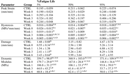
Table 2-1: Mechanical parameter comparisons throughout healing and fatigue life. ... 53
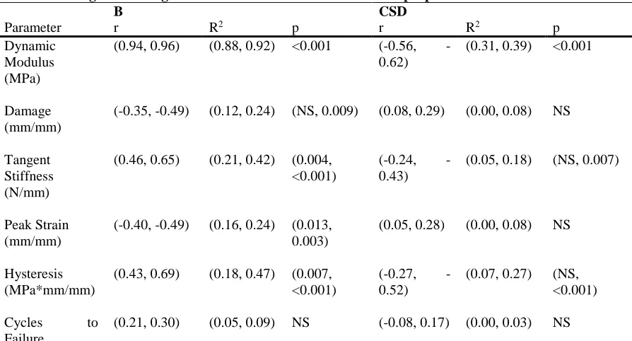
Table 2-2: Single linear regressions for structural and mechanical properties. ... 54
Table 2-3: Single and multiple linear regressions for structural and mechanical properties. ... 54
Chapter 5 Table 5-1. Model Inputs. ... 146
Table 5-2: Correlation to nAR from full dataset. ... 150
Table 5-3: Multiple regression modeling to predict nAR. ... 151
Table 5-4: Correlation coefficients for ∆nAR in uninjured and healing tendon (Aim 1). ... 151
xiv
LIST OF ILLUSTRATIONS
Chapter 1
Figure 1-1: Structure-function relationships in tendon during quasi-static and dynamic fatigue
loading. ... 3
Figure 1-2: Response of tendon to mechanical loading ... 7
Figure 1-3: Multiscale tendon properties. ... 10
Figure 1-4: Study Design. ... 17
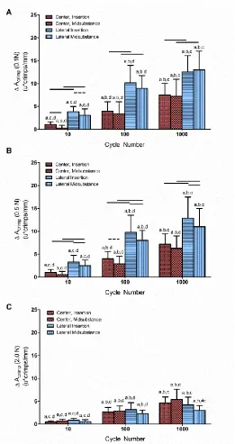
Chapter 2 Figure 2-1. Experimental setup of a plane polariscope. ... 36
Figure 2-2: Effect of fatigue loading on tendon mechanics. ... 36
Figure 2-3. Mechanical testing and image capture protocol. ... 45
Figure 2-4. Effect of fatigue loading on tendon mechanical properties. ... 46
Figure 2-5: Effect of Healing on Fatigue Mechanics. ... 46
Figure 2-6. Effect of fatigue loading on crimp amplitude. ... 50
Figure 2-7. Effect of fatigue loading on crimp frequency. ... 51
Figure 2-8: Effect of healing and fatigue on tendon structure. ... 51
Figure 2-9: Circular standard deviation (CSD) throughout fatigue life, healing, and at all loads. 52 Figure 2-10: Apparent birefringence (Bapp) throughout fatigue life, healing, and at all loads. ... 52
Figure 2-11: Normalized peak-to-peak intensity (Ip2p*) throughout fatigue life, healing, and at all loads. ... 52
Figure 2-12. Relationship between tendon laxity and crimp amplitude. ... 53
Chapter 3 Figure 3-1: Study Design ... 74
Figure 3-2: Quasi-static Mechanical Testing Protocol. ... 76
Figure 3-3: Experimental setup for live tendon testing ... 76
Figure 3-4: Polarized Light Imaging. ... 77
Figure 3-5: AFM Experimental Setup and Testing. ... 83
xv
Figure 3-7: Macroscale Mechanical Properties and Fiber Recruitment Model. ... 85
Figure 3-8: Macroscale Collagen Fiber Alignment. ... 85
Figure 3-9: Confocal and MP imaging. ... 87
Figure 3-10: Confocal and MP imaging. ... 88
Figure 3-11: Confocal and MP imaging. ... 88
Figure 3-12: Confocal and MP imaging. ... 88
Figure 3-13: Confocal and MP Imaging. ... 89
Figure 3-14: Local Matrix Alignment. ... 89
Figure 3-15: Cellularity and Actin Staining. ... 90
Figure 3-16: Nuclear Aspect Ratio. ... 90
Figure 3-17: Nuclear Aspect Ratio Histograms. ... 90
Figure 3-18: Nuclear Aspect Ratio Population Statistics. ... 91
Figure 3-19: Nuclear organization. ... 91
Figure 3-20: Correlations. ... 91
Chapter 4 Figure 4-1: Study Design. ... 106
Figure 4-2: Mechanical Testing Protocol... 109
Figure 4-3: Experimental setup for live tendon testing. ... 110
Figure 4-4: Macroscale Strain Stiffening. ... 115
Figure 4-5: Macroscale Dynamic Mechanical Properties. ... 115
Figure 4-6: Macroscale Collagen Alignment. ... 116
Figure 4-7: Macroscale Mechanical Recovery. ... 116
Figure 4-8: Multiphoton Imaging Comparing Effects of Strain Following Low Magnitude Short Duration Loading. ... 117
Figure 4-9: Multiphoton Imaging Comparing Effects of Strain Following Low Magnitude Long Duration Loading. ... 118
Figure 4-10: Multiphoton Imaging Comparing Effects of Strain Following High Magnitude Short Duration Loading. ... 118
Figure 4-11: Multiphoton Imaging Comparing Effects of Strain Following High Magnitude Long Duration Loading. ... 119
Figure 4-12: Multiphoton Imaging: Group Comparisons. ... 119
xvi
Figure 4-14: Nuclear Aspect Ratio. ... 120
Figure 4-15: Nuclear Organization. ... 120
Figure 4-16: Correlations. ... 121
Figure 4-17: Effect of Healing on Equilibrium Stress. ... 122
Figure 4-18: Effect of Healing on Dynamic Mechanical Properties. ... 123
Figure 4-19: Effect of Healing on Fatigue Properties. ... 123
Figure 4-20: Effect of Low Magnitude Long Duration Loading on Strain. ... 124
Chapter 5 Figure 5-1: Model fits for tendon healing. ... 146
Figure 5-2: Displacement transmission in uninjured and healing tendon.Error! Bookmark not defined. Figure 5-3: Normalized displacement transmission in uninjured and healing tendon. ... 149
1
CHAPTER I: INTRODUCTION 1.1 Introduction
Tendons function to transfer stresses and strains from muscle to bone during loading,
resulting in multiscale changes to its extracellular matrix (ECM). The extracellular matrix
(ECM) is a major component of the biomechanical environment with which tendon cells
(tenocytes) interact. Alterations to matrix structural and mechanical properties due to
mechanical loading may promote normal tendon homeostasis or create pathological
conditions. For example, fatigue loading of tendon elevates collagen fiber waviness
(crimp), which correlates linearly with tissue laxity (change in slack length). The tendon
ECM may also be altered following tendon injury. Aberrant tissue phenotypes caused by
tendon ruptures are exemplified not only at transcript and protein levels, but also can
extend to include disorganized collagen structure, inferior mechanical properties, and
reduced in vivo limb function in animals. This work explores the interface between
dynamic loading and tendon healing across multiple length scales using living tendon
explants. Overall, this work helps to define the implications of macroscale mechanical
loading on collagen structure and tenocyte response in uninjured and healing tendon, and
provides a foundation for the development of new strategies to improve tendon healing.
1.2 Background
1.2.1 Multiscale Tendon Mechanics and Structure
Multiscale strain transfer is the relationship of strain between structural
hierarchies of materials. Tendons allow for multiscale strain transfer due to their
2
loading, as disorganized ECM at the fascicle, fiber, and fibril levels becomes more
aligned and less wavy (i.e., crimped) (Figure 1-1A).3-6 Multiscale strain transfer ultimately affects cell microenvironments and therefore proliferation, differentiation, and
matrix production.7 Alterations to cell microenvironments due to mechanical loading may
provide a mechanism leading to normal tendon homeostasis8-13 or pathology.14-21
Previous studies have found that tissue and cellular level strains correlate in normal
uninjured tissues during quasi-static tensile loading in a variety of fiber-reinforced tissues
including tendons, menisci, and electrospun scaffolds.22 However, such studies have not
investigated the clinically important effects of dynamic loading and tendon healing,
which likely alter tendon multiscale mechanical, structural, and spatial distributions of
compositional proteins that affect how cells respond to their substrate, ECM loading, and
transfer stress over long distances.
Multiscale relationships during dynamic loading at high loads are important for
elucidating potential mechanical mechanisms governing fatigue-induced damage and
injury. When tendons are subjected to high dynamic loading, fatigue damage
accumulation occurs23 in concert with alterations in crimp properties.24 With fatigue
loading, tendon stiffness increases initially, reaches a maximum, and then gradually
decreases, and is coupled by a monotonic increase in peak cyclic strain (Figure 1-1B). This gradual decrease in stiffness is attributed to accumulated sub-rupture damage, which
ultimately leads to the dramatic increase in peak deformation and decrease in stiffness
prior to failure.25-27 Repeated subrupture loading results in collagen fibril kinking,28
which can affect cell morphology and matrix degradation.29 As these studies were
3
evaluating the specific response of tendon to load, thus removing confounding variables
present in whole tissue model systems. Many of the same mechanical and biological
mechanisms also occur in vivo, demonstrating the validity of these ex vivo models.
Figure 1-1: Structure-function relationships in tendon during quasi-static and dynamic fatigue loading. (A) During quasi-static tensile loading, a distinct nonlinearity or “toe” region exists that is attributed to collagen fiber uncrimping and re-alignment. (B) During fatigue testing, tendons follow a three-phase pattern of fatigue damage in both tangent stiffness and peak strain. Panels A and B were reproduced with permission from Connizzo, et al., 2013, and Freedman et al., 2013, respectively.
Multiscale tendon mechanics and structure can be evaluated using several
methods. Whole tendon mechanical properties are commonly determined from force and
displacement readings acquired with commercially available tensile testing systems. By
placing fiduciary markers on tissues to serve as optical extensometers, images can be
captured with external cameras to provide local assessment of tissue strain and stiffness.
Structural assessment during mechanical loading can be achieved with the integration of
a plane polariscope. Briefly, this device consists of two polarizing sheets on opposite
sides of the tendon. Recently, crimp was visualized with polarized light imaging during
4
load dependent crimp properties, to be inferred simultaneously during a mechanical test
without destructively altering the tendon. If the polarizers are rotated, measures of
collagen fiber orientation can be derived.31 Whole tissue can also be mounted under a
confocal microscope. Similar to fiduciary marker tracking of strain, microscale strains
can be assessed through strain field mapping of cells embedded within the collagen
matrix. If tendons are subject to sectioning, additional measures of multiscale mechanics
and structure can be determined. First, micro/nanoscale mechanics is achieved through
nanoindentation experiments using Atomic Force Microscopy (AFM).32 In this method, a
cantilever with a nano/micro-sized tip is placed on the surface of a tissue to derive local
mechanical measurements. Structurally, AFM can be used to assess nanoscale collagen
structural properties, such as fibril sliding.33 Multiphoton imaging can also be used to
study collagen structure through second harmonic generation. Additional nanometer
resolution imaging techniques including scanning electron microscopy (SEM) and
transmission electron microscopy (TEM), which are useful for looking at collagen fibrils
in both the longitudinal and transverse axes to evaluate the presence of fibril ends and
fibril diameters.34,35
1.2.2 Tendon Healing
Following injury, tendons are mechanically, structurally, and biologically
altered.36 Initially, the gross appearance of the tendon changes, becoming larger in cross
section,45, 46, 55 and translucent.37 In addition, a high density of inflammatory cells within
the repair site are present,36,38 expression of scleraxis (a tenocyte marker) is substantially
5
and biglycan have been shown to be upregulated.40 During this time, new fibrils are
deposited with altered diameter distributions39 and decreased longitudinal
organization.38,41 In addition, collagen-1,40 versican,40 decorin,40 matrix
metalloproteinases (MMPs),42,43 tissue inhibitory metalloproteinases (TIMPs),42 and
ADAMs42 become upregulated, indicating further tissue remodeling. With healing, cell
number and density gradually return to baseline values,36 but scarred portions may
remain non-recoverably disorganized with associated inferior mechanical properties.44
Examples of inferior quasi-static mechanical properties following injury include
decreased ultimate stress, maximum load, stiffness, elongation to failure, modulus, and
percent relaxation (a measure of tendon viscoelasticity).36,45,46 Together, these findings
suggest that healing results in altered tendon compositional properties that may likely
reflect the structural and mechanical quality of the newly deposited tissue.
Tendon homeostasis and healing ultimately depends on the ability of tenocytes to
remodel tissue. Due to the slow timescale of remodeling events in tendon,47 it is likely
that alterations in matrix properties (e.g., mechanics and structure) precede cell
responses. Such structural and mechanical changes alter the fibrous architecture of the
ECM, and may therefore influence the ability for resident tenocytes to recognize their
native “tendon” environment. For example, changes in tendon phenotype are common
with injury and healing. In particular, tendon stem cells are implicated to differentiate
towards chondrocyte, osteoblast, or adipocyte lineages, as altered matrix properties and
stresses on cells has been shown to affect cell fate.48-50 Therefore, to model the cellular
6
the tenocyte response to load and distances that cells can exert matrix stresses during
contraction.
1.2.3 Role of the Extracellular Matrix and Mechanical Loading on Cell Behavior
Tendons are fiber-reinforced musculoskeletal tissues that allow strain transfer
from the whole tissue level to subcellular levels,1,2 ultimately resulting in load-dependent
biochemical responses (Figure 1-2). For example, although dynamic moderate loading increased tensile strength,9 net collagen synthesis,8-12 tenogenic differentiation,13 and
cross-sectional area,9,51 excessive loading decreased tensile strength52,53 and collagen
organization,54,55 and increased net collagen degradation,56,57 aberrant differentiation,
13,58-62 cross-sectional area,63 vascularization,53,54,63 GAG production,64 and inflammatory
cytokines.53,65,66 However, mechanistic interpretation for these findings remains limited—
it is unknown what role mechanical loading played on the multiscale mechanical,
structural, and compositional response. Fiber orientation in the matrix is also critical for
various cell behaviors, including stem cell differentiation,67 cell alignment,68-70 and
migration.71 Quantifying these properties is necessary for understanding how mechanical
signals are transduced into molecular responses. For example, since alterations in nuclear
shape may affect cell phenotype,72,73 understanding the multiscale mechanisms to elicit
such changes is imperative. Knowledge of strain transfer is also important for defining
7
Figure 1-2: Response of tendon to mechanical loading. Low loading decreased tensile strength,14-16
collagen organization,14,15 and tenocyte markers;74 net collagen degradation and inflammatory cytokines
increased,16,74 and GAG content was unchanged.11 Moderate loading increased tensile strength,9 net
collagen synthesis,8-12 tenogenic differentiation,13 and cross-sectional area.9,51 Knowledge regarding
collagen organization, vascularization, GAG production, and inflammatory cytokines with moderate loading remains limited. Excessive loading decreased tensile strength52,53 and collagen organization,54,55
and increased net collagen degradation,56,57 aberrant differentiation,13,58-62 cross-sectional area,63
vascularization,53,54,63 GAG production,64 and inflammatory cytokines.53,65 Reproduced with permission
from Freedman, et al., 2015.
1.3 Significance of Studies
1.3.1 Clinical Application
Musculoskeletal injuries affect 28.6 million Americans per year and account for
$254 million in annual health care costs.75 This includes over 450,000 tendon, ligament,
or muscle related injuries each year.75 With increased participation in recreational
activities and sport, the incidence of acute and chronic tendon injuries is rising.76,77
Unfortunately, many treatments for tendon injuries remain controversial, with “weak” or
“inconclusive” evidence cited by a recent review from the American Academy of
8
relationships in an injured state have not been fully characterized, which may limit our
ability to design new targeted therapies. Specifically, multiscale alterations to the ECM
during macroscale mechanical loading may affect the local microenvironment of
tenocytes, and therefore provide a mechanism leading to normal tendon homeostasis8-13
or pathology.14-21 Therefore, since mechanical loading is a hallmark of clinical
rehabilitation protocols, fundamental, basic science studies are necessary to determine the
ramifications of various macroscale dynamic tissue loading protocols (magnitude and
duration) on the micro and nanoscale biomechanics in tendon during healing.
1.3.2 Structure-Function Relationships in Native Tendon
Tendon’s composition and structure are related to its mechanical function.
Non-linear mechanical properties of tendon are governed by changes in its architecture that
occur as a function of the applied stress and strain. For example, during mechanical
loading, a distinct nonlinearity or “toe” region is present that is often attributed to
collagen fiber re-alignment and uncrimping3-6 (Figure 1-1). Therefore, model systems which apply tensile loads to ex vivo tendon explants that preserve the native architecture
of the ECM, while also providing more controlled experimental conditions than are
possible in vivo, are important. Changes in tissue architecture may be critical for tissue
homeostasis and injury, particularly in response to dynamic loading. In injured tissues,
the formation of PG-rich inclusions has been implicated to attenuate strain transfer to
cells that may contribute to their ability to maintain homeostasis.22 Although tissue and
cellular level strains correlate in normal uninjured tissues during quasi-static tensile
9
loading and tendon healing. Additionally, although much research has investigated
structure-function relationships during quasi-static tensile loading (e.g., 54,79), evaluation
during dynamic loading has been limited.34,80 This loading environment is likely
generated in healing and injured tissues, and similar matrix disruption may be generated
in response to high magnitude dynamic loading. High dynamic loading and healing likely
alter tendon multiscale mechanical, structural, and spatial distributions of compositional
proteins that affect how cells respond to their substrate, ECM loading, and transfer stress
over long distances.
1.3.3 Assessment of Multiscale Properties on Living Tendons
Gross mechanical and biochemical changes may be due to aberrant strain transfer
from the cell to their ECM and between cells. Since cells interact with their substrate,
measures of fibril mechanics are also important. Substrate stiffness has been shown to
guide stem cell lineage specification in vitro and affect proliferation, motility,
contractility, and many other cell functions by changing both acute signaling and
transcriptional programs.48,81,82 Thus, specific measures of cell substrates (i.e., collagen
fibrils) in native and healing tendon are necessary to fully describe the mechanisms of
healing and response to dynamic loading. Altering tendon tensile forces can elicit
changes in integrin receptors,83 downstream ECM proteins,74 as well as cell-cell contacts
including N-cadherins and gap junctions84,85 (Figure 1-3). However, much of these findings remain disjointed since studies identified them in isolation, and the complete
multiscale mechanism remains unknown. Additionally, mechanisms of multiscale
10
understood. This is of particular importance since accumulation of proteoglycan
(PG)-rich inclusions or mucoid degeneration is common in injured tendon.22,57 Recent studies
showed that cells are more rounded within these regions and do not deform with applied
strain as much as cells embedded in normal fibrous matrices.22 This suggests that reduced
transfer may contribute to altered mechanosensing and potentially altered cellular
response.22
Figure 1-3: Multiscale tendon properties. Multiscale tendon response to load spanning from the subcellular (A) to whole tendon (B) levels. Macro-scale loading creates tension, compression, and shear loading on the ECM that is then transferred to the cell via transmembrane adhesion proteins termed integrins. Load is then transferred through the actin cytoskeleton via mechanosensitive pathways, such as RhoA, to the nucleus. It is noted that this illustration highlights only the basic subunits of these adhesion complexes, and that these components come together into higher order assemblies in vivo. Reproduced with permission from Freedman, et al., 2015.
This study will apply dynamic mechanical loading to uninjured and healing
tendon explants. We will use a sophisticated mechanical testing system to apply a variety
of dynamic mechanical loading protocols that vary both the magnitude and duration of
11
commonly studied static loading. To maintain tendon health during loading, tissues will
be immersed in a bath containing sterile DMEM supplemented with 5% fetal bovine
serum (FBS) at 37ºC, 20% O2, and 5% CO2. This innovative approach allows the
evaluation of multiscale mechanical and structural properties without any destructive
dissection to the tissue. Since the structure of tendons relates to mechanical strains
applied, following application of mechanical loading, tissues are flash frozen at set loads
and strains to maintain their native tissue architecture for subsequent assays. This
procedure provides advancement over traditional histological studies that do not control
for tissue prestresses or strains applied prior to fixation.
1.3.4 Substrate Mechanics and Constitutive Models of Cell Mechanosensing
Cells can sense and respond to mechanical signals over distances nearly 20 times
their cell radius (i.e., 250-1000μm).86,87 Traction forces on the underlying substrate are
generated during cell migration. These cell traction forces (CTF) are important for cell
migration, phenotype, and homeostasis. For example, cell traction force microscopy can
be shown to discriminate cell populations in tendon and are influenced by geometries that
tenocytes adhere 88. Recent modeling studies have suggested that the key mechanism
driving this process is the fibrous nature of extracellular matrices.1,89 Our collaborators
have developed a constitutive law accounting for mechanically driven collagen fiber
reorientation.89 Results show that tension-driven collagen-fiber alignment plays a crucial
role in force transmission.1 Small critical stretch for fiber alignment, large fiber stiffness,
and fiber strain-hardening behavior enable long-range interaction.1 Furthermore, the
12
larger than that for spherical cells with diagonal contraction.1 Building on this work, we
propose to develop a 3D model to elucidate the role of dynamic mechanical loading and
tendon healing on the ability for tenocytes to sense stress over long distances. In this
model, mechanical and cell morphological properties are used as model inputs. Use of
these properties at different strains following dynamic loading and healing provides
initial conditions for the changes in matrix architecture in response to loading.
This modeling framework is adapted to include experimentally derived measures
of tissue mechanics using AFM to evaluate the effects of dynamic loading and healing on
tenocyte stress transmission. Tenocyte stress transmission provides knowledge that may
have important ramifications for both cell-cell communication and load transfer within
the ECM in tendon. One potential mechanism for continued maintenance or failure to
heal following injury may be a lack of long range force transmission present in healing
tissue that may lack fibrous architecture. Therefore, this modeling framework aims to
build on previously existing models to develop a constitutive relationship that includes
several key features that are critical for clarifying the ability for cells to sense mechanical
perturbation and affect their surrounding matrix during contraction. These test conditions
are essential to specifically address the fundamental mechanisms governing the response
of tendon to dynamic loading and during healing.
To provide further insight into the relationships between multiscale mechanical,
structural, and compositional properties following dynamic loading in uninjured and
healing tendons, parameters from Aims 1 and 2 are used in single linear regressions to
determine the variables that show significant coefficients of determination in predicting
13
structure–function relationships in tendon.90-92 Significant variables are used in multiple
regression analyses to determine their predictability for tenocyte stress transmission
distances. Together, these models reveal the structure-function pathways of
mechanotransduction in normal and diseased tendons and elucidate the role of dynamic
mechanical loading in this process.
1.4 Specific Aims
This study uses a comprehensive approach, including novel methods for: (1)
application of mechanical loading on live tendons, (2) measurement of multiscale tendon
mechanics and structure during dynamic mechanical loading and healing, (3) assessment
of healing tissue composition using immunofluorescence (IF) and confocal imaging, and
(4) application of a constitutive model for fibrous matrices based on experimentally
derived parameters to predict tenocyte stress transmission and additional multiple
regression analyses.
Specific Aim 1 evaluates the multiscale mechanical, structural, and compositional
response of uninjured and healing tendon to quasi-static tensile loading. The first part of
this aim examines how macroscale mechanical loading affects macroscale structural
properties in nonliving uninjured control and healing tendons. Specifically, following
quasi-static loading, tissues are snap-frozen, embedded in OCT, and sectioned for
subsequent microscale assays.4,33,93 For structural assessment, we measure fascicle/fiber
alignment and crimp, and kinking using multiphoton confocal microscopy.
Specific Aim 2 evaluates the multiscale mechanical, structural, and compositional
14
Aim 1, the first part of this aim examines how macroscale mechanical loading affects
macroscale structural properties in nonliving uninjured control and healing tendons.
Subsequent parts of this aim will apply dynamic loading to living tendons to study how
macroscale mechanical loading is transduced to changes in cell and nuclear shape, and
the micro and nanoscale mechanical and structural properties that enable this mechanism.
Specifically, following various bouts of dynamic protocols, tissues are snap-frozen,
embedded in OCT, and sectioned for subsequent microscale assays.4,33,93 For structural
assessment, we will measure fascicle/fiber alignment and crimp, and kinking using
multiphoton confocal microscopy.
Specific Aim 3 applies a novel fibrous constitutive law for tendon matrices to
evaluate tenocyte stress transmission following low and high magnitude, long duration,
dynamic loading. We also relate tenocyte stress transmission to multiscale mechanical,
structural, and compositional properties determined from Aims 1 and 2 with multiple
regression. Our previous experimental and modeling studies in tendon incorporating
damage models have shown that empirical modeling techniques based on mechanical and
image based measures can predict mechanical properties in healing,94 aging,94 and
genetically altered95 tendons. Proteoglycans may be implicated in reductions in tendons
mechanical properties and collagen disorganization.95 However, the underlying
mechanisms for these processes have not been elucidated. As mentioned, mechanical
perturbation likely causes alterations to multiscale ECM biomechanics, which ultimately
affects cell behavior. Therefore, mechanical models that can directly measure the
complex interactions between tenocytes and their ECM are necessary. Mechanical,
15
constitutive law or multiple regression analyses. Since this study aims to directly measure
cell shape following various loading protocols, we apply realistic initial conditions for
cell strains prior to contraction.
1.5 Approach
1.5.1 Overall Strategy
This study determines the ramifications of various macroscale dynamic tissue
loading protocols on the micro and nanoscale biomechanics in tendon during healing.
These properties will encompass multiscale mechanics (tensile, compression) and
structure (alignment, kinking, crimp, composition), cell (shape, composition), and
subcellular (shape, composition) components. This comprehensive approach has
important implications for the fundamental mechanisms governing tendon homeostasis,
healing, and phenotype. A mouse model is used with several innovative methodologies
(mechanical testing with collagen fiber alignment, atomic force microscopy (AFM) to
assess fibril mechanics, confocal imaging to assess ECM, cell, and nuclear morphology
and composition, constitutive modeling to assess cell stress transmission, and multiple
regression analysis) to provide rigorous mechanical, structural, and compositional data on
the multiscale tendon response to dynamic loading during healing.Our overall hypothesis
is that dynamic loading results in distinct ECM and cell changes, not present with static
loading, which ultimately promote (e.g., via fiber recruitment) or reduce (e.g., via fiber
16 1.5.2 Study Design and Animal Model
This study employs a mouse patellar tendon injury model using female C57BL/6
mice at P150 (Figure 1-4). The patellar tendon model is advantageous since its tibia and
patellar insertions allow for gripping during mechanical testing, without any fine
dissection required of the patellar tendon. Additionally, the mouse patellar tendon allows
light transmission making it capable of polarized light imaging during mechanical
loading. Based on our previous rat Achilles mechanical data,96 we estimated the normal
variation in properties to determine our sample size. Given that rat Achilles stiffness
after 8 weeks of exercise was 60.2±9.0 N/mm, we can detect differences of 10 N/mm,
with 80% power using 10 animals per group.
An animal model is used to carefully control the injury and evaluate the
multiscale the tissue
properties. Such properties are incorporated into a mathematical model to predict
tenocyte stress transmission, for the first time, across a host of clinically relevant
paradigms. The ability for cells to sense and respond to their ECM over long distances,
we believe, is indicative of a return to a normal tissue phenotype.
1.5.3 Hypothesized Outcomes
Aim 1: We hypothesize that healing would reduce strain stiffening and fiber
recruitment compared to uninjured tendons. We use a comprehensive approach, including
application of mechanical loading on live tendons, measurement of multiscale tendon
mechanics and structure during healing, assessment of healing tissue composition using
17
Due to the slow timescale of remodeling events in tendon,47 it is likely that alterations in
matrix properties (e.g., mechanics and structure) precede cell responses. Such structural
and mechanical changes alter the fibrous architecture of the ECM, and may therefore
influence the ability for resident tenocytes to recognize their native “tendon”
environment.
Figure 1-4: Study Design. (A) Mice are randomized into three groups before quasi-static and dynamic loading are completed. The dynamic loading protocols vary the magnitude (low or high load) and duration (0, 10, or 1000 cycles) of loading.
Aim 2: We hypothesize that high magnitude long duration dynamic loading and
healing would reduce strain stiffening and fiber recruitment compared to uninjured
tendons and low magnitude long duration dynamic loading. Additionally, we hypothesize
that low magnitude dynamic loading will increase strain stiffening, stiffness, and
alignment, which ultimately increases tendon cell nAR, unlike high dynamic loading.
Aim 3: We hypothesize that higher magnitude cyclic loading and healing
decreases tenocyte stress transmission, as the ECM behaves more neo-Hookean than
fibrous. Through multiple regression analysis, we expect that in uninjured tendon,
18
However, in healing tendons, we hypothesize that modulus and crimp, contribute highest
to cell strain and stress transmission. We first determine the material parameters in the
model1 that provide the best fit for tendon experimental data and implementation of the
constitutive law in user material model in finite element software.
1.5.4 Ex-vivo Assays
Our mechanical loading approach allows for multiscale assessment of tendon
mechanical and structural properties in uninjured and healing tendons. At the macroscale,
we evaluate whole tissue dynamic mechanical properties and structural properties (matrix
alignment and crimp) during mechanical loading. We have successfully measured
collagen fiber alignment and crimp 97-100 using a custom apparatus with two precisely
controlled offset polarizing sheets on either side of a specimen imaged during tensile
testing, a linear backlight, and a digital camera. Briefly, as the tendon is loaded, a series
of polarized images are captured and analyzed to produce an alignment map.
Following loading perturbation, tissues are flash frozen at low and high strains to
provide load-dependent characterization of the effect of dynamic loading and healing.
Frozen tendons are then sectioned for micro and nano-level mechanical and structural
analysis, as well as confocal imaging of cell strains. Collagen fiber alignment is assessed
using polarized light microscopy. Fibril mechanics are assessed using atomic force
microscopy (AFM). Measurements using high resolution AFM provide nanometer
resolution of fibril and interfibrillar matrix compression modulus.
Tenocytes and nuclear strains are imaged in their native ECM environment at
19
using confocal microscopy will provide a mechanistic foundation for observed alterations
in multiscale mechanics and structure during healing and dynamic loading. Multiphoton
imaging provides high resolution characterization of collagen structure and allows for
simultaneous imaging of cell nuclei and the actin cytoskeleton. Together, these analyses
will present the first-ever quantification of multiscale ECM environment following
perturbation of dynamic loading in uninjured and healing tendons.
1.6 Chapter Overview
Chapter II will describe how macroscale mechanical and structural properties of
tendon are altered during fatigue loading and tendon healing. Chapter III will describe the multiscale mechanical, structural, and compositional response of tendon to quasi-static loading during healing. Chapter IV will describe the multiscale mechanical, structural, and compositional response of tendon to dynamic loading during healing. Chapter V will describe modeling stress transmission in tendon during healing and dynamic loading. Chapter VI will summarize the conclusions of the previous chapters and provide future directions for this area of research.
1.7 References
1. Wang H, Abhilash AS, Chen CS, Wells RG, Shenoy VB. Long-range force
transmission in fibrous matrices enabled by tension-driven alignment of fibers.
20
2. Arnoczky SP, Lavagnino M, Whallon JH, Hoonjan A. In situ cell nucleus
deformation in tendons under tensile load; a morphological analysis using
confocal laser microscopy. J Orthop Res. Jan 2002;20(1):29-35.
3. Franchi M, Fini M, Quaranta M, et al. Crimp morphology in relaxed and stretched
rat Achilles tendon. J Anat. Jan 2007;210(1):1-7.
4. Miller KS, Connizzo BK, Feeney E, Soslowsky LJ. Characterizing local collagen
fiber re-alignment and crimp behavior throughout mechanical testing in a mature
mouse supraspinatus tendon model. J Biomech. Aug 9 2012;45(12):2061-2065.
5. Diamant J, Keller A, Baer E, Litt M, Arridge RG. Collagen; ultrastructure and its
relation to mechanical properties as a function of ageing. Proc R Soc Lond B Biol
Sci. Mar 14 1972;180(1060):293-315.
6. Rigby BJ, Hirai N, Spikes JD, Eyring H. The Mechanical Properties of Rat Tail
Tendon. J Gen Physiol. Nov 1 1959;43(2):265-283.
7. Mammoto A, Mammoto T, Ingber DE. Mechanosensitive mechanisms in
transcriptional regulation. J Cell Sci. Jul 1 2012;125(Pt 13):3061-3073.
8. Mendias CL, Gumucio JP, Lynch EB. Mechanical loading and TGF-beta change
the expression of multiple miRNAs in tendon fibroblasts. J Appl Physiol (1985).
Jul 2012;113(1):56-62.
9. Heinemeier KM, Skovgaard D, Bayer ML, et al. Uphill running improves rat
Achilles tendon tissue mechanical properties and alters gene expression without
inducing pathological changes. J Appl Physiol (1985). Sep 1
21
10. Moerch L, Pingel J, Boesen M, Kjaer M, Langberg H. The effect of acute exercise
on collagen turnover in human tendons: influence of prior immobilization period.
Eur J Appl Physiol. Feb 2013;113(2):449-455.
11. Screen HR, Shelton JC, Bader DL, Lee DA. Cyclic tensile strain upregulates
collagen synthesis in isolated tendon fascicles. Biochem Biophys Res Commun.
Oct 21 2005;336(2):424-429.
12. Legerlotz K, Jones GC, Screen HR, Riley GP. Cyclic loading of tendon fascicles
using a novel fatigue loading system increases interleukin-6 expression by
tenocytes. Scand J Med Sci Sports. Feb 2013;23(1):31-37.
13. Zhang J, Wang JH. The effects of mechanical loading on tendons - an in vivo and
in vitro model study. PLoS One. 2013;8(8):e71740.
14. Ikoma K, Kido M, Nagae M, et al. Effects of stress-shielding on the dynamic
viscoelasticity and ordering of the collagen fibers in rabbit Achilles tendon. J
Orthop Res. Jul 6 2013.
15. Hannafin JA, Arnoczky SP, Hoonjan A, Torzilli PA. Effect of stress deprivation
and cyclic tensile loading on the material and morphologic properties of canine
flexor digitorum profundus tendon: an in vitro study. J Orthop Res. Nov
1995;13(6):907-914.
16. Uchida H, Tohyama H, Nagashima K, et al. Stress deprivation simultaneously
induces over-expression of interleukin-1beta, tumor necrosis factor-alpha, and
transforming growth factor-beta in fibroblasts and mechanical deterioration of the
22
17. Forde MS, Punnett L, Wegman DH. Prevalence of musculoskeletal disorders in
union ironworkers. J Occup Environ Hyg. Apr 2005;2(4):203-212.
18. Jobe FW, Kvitne RS, Giangarra CE. Shoulder pain in the overhand or throwing
athlete. The relationship of anterior instability and rotator cuff impingement.
Orthop Rev. Sep 1989;18(9):963-975.
19. McMaster WC, Troup J. A survey of interfering shoulder pain in United States
competitive swimmers. Am J Sports Med. Jan-Feb 1993;21(1):67-70.
20. Renstrom P, Johnson RJ. Overuse injuries in sports. A review. Sports Med.
Sep-Oct 1985;2(5):316-333.
21. Sommerich CM, McGlothlin JD, Marras WS. Occupational risk factors associated
with soft tissue disorders of the shoulder: a review of recent investigations in the
literature. Ergonomics. Jun 1993;36(6):697-717.
22. Han WM, Heo SJ, Driscoll TP, Smith LJ, Mauck RL, Elliott DM. Macro- to
microscale strain transfer in fibrous tissues is heterogeneous and tissue-specific.
Biophys J. Aug 6 2013;105(3):807-817.
23. Wang XT, Ker RF, Alexander RM. Fatigue rupture of wallaby tail tendons. J Exp
Biol. Mar 1995;198(Pt 3):847-852.
24. Freedman BR, Zuskov A, Sarver JJ, Buckley MR, Soslowsky LJ. Evaluating
changes in tendon crimp with fatigue loading as an ex vivo structural assessment
of tendon damage. J Orthop Res. Mar 13 2015.
25. Fung DT, Wang VM, Andarawis-Puri N, et al. Early response to tendon fatigue
damage accumulation in a novel in vivo model. J Biomech. Jan 19
23
26. Fung DT, Wang VM, Laudier DM, et al. Subrupture tendon fatigue damage. J
Orthop Res. Feb 2009;27(2):264-273.
27. Wren TA, Lindsey DP, Beaupre GS, Carter DR. Effects of creep and cyclic
loading on the mechanical properties and failure of human Achilles tendons. Ann
Biomed Eng. Jun 2003;31(6):710-717.
28. Veres SP, Harrison JM, Lee JM. Repeated subrupture overload causes
progression of nanoscaled discrete plasticity damage in tendon collagen fibrils. J
Orthop Res. May 2013;31(5):731-737.
29. Veres SP, Brennan-Pierce EP, Lee JM. Macrophage-like U937 cells recognize
collagen fibrils with strain-induced discrete plasticity damage. J Biomed Mater
Res A. Jan 2015;103(1):397-408.
30. Buckley MR, Sarver JJ, Freedman BR, Soslowsky LJ. The dynamics of collagen
uncrimping and lateral contraction in tendon and the effect of ionic concentration.
J Biomech. Sep 3 2013;46(13):2242-2249.
31. Lake SP, Miller KS, Elliott DM, Soslowsky LJ. Effect of fiber distribution and
realignment on the nonlinear and inhomogeneous mechanical properties of human
supraspinatus tendon under longitudinal tensile loading. J Orthop Res. Dec
2009;27(12):1596-1602.
32. Li Q, Doyran B, Gamer LW, et al. Biomechanical properties of murine meniscus
surface via AFM-based nanoindentation. J Biomech. Mar 11 2015.
33. Connizzo BK, Sarver JJ, Han L, Soslowsky LJ. In situ fibril stretch and sliding is
location-dependent in mouse supraspinatus tendons. J Biomech. Dec 18
24
34. Dunkman AA, Buckley MR, Mienaltowski MJ, et al. Decorin expression is
important for age-related changes in tendon structure and mechanical properties.
Matrix Biol. Jan 2013;32(1):3-13.
35. Provenzano PP, Vanderby R, Jr. Collagen fibril morphology and organization:
implications for force transmission in ligament and tendon. Matrix Biol. Mar
2006;25(2):71-84.
36. Chamberlain CS, Duenwald-Kuehl SE, Okotie G, Brounts SH, Baer GS,
Vanderby R. Temporal Healing in Rat Achilles Tendon: Ultrasound Correlations.
Ann Biomed Eng. Nov 13 2012.
37. Eliasson P, Andersson T, Aspenberg P. Rat Achilles tendon healing: mechanical
loading and gene expression. J Appl Physiol (1985). Aug 2009;107(2):399-407.
38. Ehrlich HP, Lambert PA, Saggers GC, Myers RL, Hauck RM. Dynamic changes
appearing in collagen fibers during intrinsic tendon repair. Ann Plast Surg. Feb
2005;54(2):201-206.
39. Maeda T, Sakabe T, Sunaga A, et al. Conversion of mechanical force into
TGF-beta-mediated biochemical signals. Curr Biol. Jun 7 2011;21(11):933-941.
40. Bring DK, Reno C, Renstrom P, Salo P, Hart DA, Ackermann PW. Joint
immobilization reduces the expression of sensory neuropeptide receptors and
impairs healing after tendon rupture in a rat model. J Orthop Res. Feb
2009;27(2):274-280.
41. Sasaki K, Yamamoto N, Kiyosawa T, Sekido M. The role of collagen
arrangement change during tendon healing demonstrated by scanning electron
25
42. Jones GC, Corps AN, Pennington CJ, et al. Expression profiling of
metalloproteinases and tissue inhibitors of metalloproteinases in normal and
degenerate human achilles tendon. Arthritis Rheum. Mar 2006;54(3):832-842.
43. Karousou E, Ronga M, Vigetti D, Passi A, Maffulli N. Collagens, proteoglycans,
MMP-2, MMP-9 and TIMPs in human achilles tendon rupture. Clin Orthop Relat
Res. Jul 2008;466(7):1577-1582.
44. Freedman BR SJ, Buckley MR, Voleti PB, Soslowsky LJ. Biomechanical and
structural response of healing Achilles tendon to
fatigue loading following acute injury. (in review). 2013.
45. Beason DP, Kuntz AF, Hsu JE, Miller KS, Soslowsky LJ. Development and
evaluation of multiple tendon injury models in the mouse. J Biomech. Mar 7
2012.
46. Freedman BR, Sarver JJ, Buckley MR, Voleti PB, Soslowsky LJ. Biomechanical
and structural response of healing Achilles tendon to fatigue loading following
acute injury. J Biomech. Nov 11 2013.
47. Magnusson SP, Langberg H, Kjaer M. The pathogenesis of tendinopathy:
balancing the response to loading. Nat Rev Rheumatol. May 2010;6(5):262-268.
48. Engler AJ, Sen S, Sweeney HL, Discher DE. Matrix elasticity directs stem cell
lineage specification. Cell. Aug 25 2006;126(4):677-689.
49. Pelham RJ, Jr., Wang Y. Cell locomotion and focal adhesions are regulated by
substrate flexibility. Proc Natl Acad Sci U S A. Dec 9 1997;94(25):13661-13665.
50. Chen CS, Mrksich M, Huang S, Whitesides GM, Ingber DE. Geometric control of