INTRODUCTION
Endothelial dysfunction is a hallmark of peripheral artery disease (PAD) (1). Central to the development of endothelial dysfunction, regardless of its cause, is a reduction in the bioavailability of nitric oxide (NO) derived from endothelial ni-tric oxide synthase (eNOS). Three funda-mental mechanisms can compromise NO bioavailability: loss of eNOS expression, loss of eNOS-derived NO production
(that is, functional inactivation of eNOS) and inactivation of NO by superoxide anion (O2–) to form peroxynitrite (OONO–) (2,3). It is likely that all three mechanisms contribute to the endothelial dysfunction characteristic of PAD because increased oxidant stress is a common an-tecedent in the pathogenesis of this dis-ease and can reduce NO bioavailability. At least two characteristics of eNOS render it susceptible to oxidant stress.
First, eNOS transcription, posttransla-tional modification and trafficking to the caveolae are attenuated by the accumula-tion of reactive oxygen species within the endothelial cell (4). Second, the eNOS co-factor tetrahydrobiopterin (BH4) is highly susceptible to oxidation (5). BH4 maintains eNOS in its functional dimeric form; in the absence of BH4, eNOS be-comes uncoupled so that the electron flux is diverted away from the L-arginine binding site and instead reduces molecu-lar oxygen, generating O2–(6). This cir-cumstance initiates a vicious cycle, wherein eNOS catalytic activity produces O2–, not NO, worsening existent oxidant stress.
These molecular characteristics of eNOS predict that several therapeutic options might prove effective for the treatment of PAD, namely dietary
sup-Synergistically to Decrease Oxidant Stress and Increase
Nitric Oxide That Increases Blood Flow Recovery after
Hindlimb Ischemia in the Rat
Jinglian Yan, Guodong Tie, and Louis M Messina
Department of Surgery, University of Massachusetts Medical School, Worcester, Massachusetts, United States of America
Nitric oxide (NO) derived from endothelial nitric oxide synthase (eNOS) is a potent vasodilator and signaling molecule that plays essential roles in neovascularization. During limb ischemia, decreased NO bioavailability occurs secondary to increased oxidant stress, decreased L-arginine and tetrahydrobiopterin. This study tested the hypothesis that dietary cosupplementation with tetrahy-drobiopterin (BH4), L-arginine and vitamin C acts synergistically to decrease oxidant stress, increase NO and thereby increase
blood flow recovery after hindlimb ischemia. Rats were fed normal chow, chow supplemented with BH4 or L-arginine (alone or in combination) or chow supplemented with BH4 + L-arginine + vitamin C for 1 wk before induction of hindlimb ischemia. In the
is-chemic hindlimb, cosupplementation with BH4 + L-arginine resulted in greater eNOS and phospho-eNOS (P-eNOS) expression, Ca2+- dependent NOS activity and NO concentration in the ischemic calf region (gastrocnemius), as well as greater NO
con-centration in the region of collateral arteries (gracilis). Rats receiving cosupplementation of BH4 + L-arginine led to greater re-covery of foot perfusion and greater collateral enlargement than did rats receiving either agent separately. The addition of vitamin C to the BH4 + L-arginine regimen further increased these dependent variables. In addition, rats given all three supplements showed significantly less Ca2+-independent activity, less nitrotyrosine accumulation, greater glutathione (GSH)–to–glutathione
disulfide (GSSG) ratio and less gastrocnemius muscle necrosis, on both macroscopic and microscopic levels. In conclusion, co-supplementation with BH4 + L-arginine + vitamin C significantly increased blood flow recovery after hindlimb ischemia by
reduc-ing oxidant stress, increasreduc-ing NO bioavailability, enlargreduc-ing collateral arteries and reducreduc-ing muscle necrosis. Oral cosupplementa-tion of BH4, L-arginine and vitamin C holds promise as a biological therapy to induce collateral artery enlargement.
Online address: http://www.molmed.org doi: 10.2119/molmed.2011.00103.revised
Address correspondence toLouis M Messina, Department of Surgery, University of Massa-chusetts Medical School, 55 Lake Avenue North, Worcester, MA 01655. Phone: 508-856-5599; Fax: 508-856-8329; E-mail: [email protected].
plementation with an antioxidant, or with the eNOS substrate L-arginine, or with the eNOS cofactor BH4. Vitamin C, or L-ascorbic acid, is a potent antioxidant and has been shown to preserve BH4 levels and enhance endothelial NO pro-duction in vitro(7). ONOO–reacts with BH4 6–10 times faster when in the pres-ence of ascorbate. The intermediate product of the reaction between ONOO– and BH4 is the trihydrobiopterin radical (BH3–), which is then reduced back to BH4 by ascorbate. Thus, ascorbate does not protect BH4 from oxidation but rather recycles the BH3 radical back to BH4 (8). Vitamin C levels are low in PAD patients (9), and acute (10) or short-term (11) vitamin C supplementation re-duces PAD symptoms; however, cross-sectional epidemiological surveys have failed to find a clear link between long-term vitamin C intake and PAD symp-toms or disease progression (12,13). L-Arginine supplementation showed ex-citing promise in short-term studies of PAD (14,15), but this effect was not ob-served in a subsequent long-term study by the same group (16). BH4 improves eNOS-dependent vasodilation in long-term smokers and patients with type 2 diabetes, conditions associated with in-creased oxidant stress (17,18). To our knowledge, BH4 has not been specifi-cally evaluated as a therapeutic modal-ity in PAD.
An important gap in our understand-ing of BH4, L-arginine and L-ascorbic acid in the prevention and treatment of PAD is the potential synergistic effect of combined therapy, and there is convinc-ing evidence to suggest that such an ap-proach would prove successful. For ex-ample, supplementation with L-arginine alone might prove deleterious in the face of endothelial oxidant stress inasmuch as the resultant increase in eNOS catalytic activity might generate O2
–
, not NO, if BH4 levels were reduced by oxidation. Thus, cosupplementation of L-arginine with L-ascorbic acid and BH4 might en-hance the therapeutic outcome by reduc-ing oxidant stress and preservreduc-ing eNOS in its functional dimeric form,
respec-tively. This action would enhance eNOS-derived NO production and, by quench-ing existent O2
–
, reduce NO inactivation by its reaction with O2
– .
In our previous work (19), we ob-served decreased eNOS expression, de-creased bioavailable NO and inde-creased oxidant stress in rats that had hindlimb ischemia, and oral supplementation of BH4 increased the beneficial effect of eNOS gene transfer. The goal of this study was to test the hypothesis that combined dietary supplementation with BH4, L-arginine and vitamin C act syner-gistically to improve hindlimb blood flow recovery and preservation of mus-cle viability in response to severe hindlimb ischemia. To this end, we gen-erated severe hindlimb ischemia in the rat by means of femoral artery excision. Measured dependent variables included calf and thigh muscle NO bioavailability, hindlimb laser Doppler perfusion and collateral artery enlargement, and calf and thigh muscle oxidative stress and tissue necrosis.
MATERIALS AND METHODS
Materials
Dietary supplements included BH4 (10 mg/kg/d; Schircks Laboratories, Jonas, Switzerland); L-arginine, provided as L-arginine α-ketoglutarate (hereafter, L-arginine; 88.5 mg/kg/d; Body Tech, North Bergen, NJ, USA); and L-ascorbic acid (that is, vitamin C; 88.5 mg/kg/d; Sigma-Aldrich, St. Louis, MO, USA). The dose of tetrahydrobiopterin was selected on the basis of a published report of its use in rats (20). And the dose of L-arginine and vitamin C was based on their clinical dose in patients.
Animals
All protocols were approved by the In-stitutional Animal Care and Use Com-mittee at the University of California, San Francisco, and the University of Massachusetts Medical School. Adult male Sprague Dawley rats weighing 265–285 g (Charles River Laboratories, Wilmington, MA, USA) were maintained
in a clean housing facility on a 12-h light–dark cycle.
Preparation
Severe ischemia was induced in the left hindlimb. The femoral artery was ligated between the inguinal ligament and popliteal fossa, and the ligated sec-tion and its branches were excised. This procedure was carried out under anes-thesia with 2% isofluorane. The un-treated right hindlimb served as an in-ternal control for each rat. An additional group of sham-operated rats were also used for selected assays. These rats un-derwent isolation of the femoral artery in the left hindlimb under 2% isofluo-rane anesthesia, but the artery was left intact.
Study Design
All rats were fed standard chow in powder form and water ad libitum
(Deans Feeds, Redwood City, CA, USA). Animals were randomly selected to re-ceive normal chow (control), chow with a single added supplement (BH4 or L-arginine), chow supplemented with BH4 + L-arginine or chow supplemented with BH4 + L-arginine + L-ascorbic acid. Dietary supplementation was com-menced 7 d before the induction of hindlimb ischemia and was continued until sacrifice of the animal. Chow was replaced every 2 d. The time of sacrifice varied with the measured end point under consideration, as described below.
Western Blotting
Rats were sacrificed 14 d after induc-tion of ischemia for measurement of gas-trocnemius and gracilis muscles of eNOS, phospho-eNOS (P-eNOS) and nitrotyro-sine expression. The timing of sacrifice was selected on the basis of previous work that demonstrated maximal postis-chemic change in these variables at this time (19). Samples were homogenized in liquid nitrogen and transferred to NP-40 lysis buffer, comprised of 50 mmol/L
150 mmol/L NaCl, 10% glycerol, 1.5 mmol/L MgCl2, 1 mmol/L ethylene-diaminetetraacetic acid (EDTA), 100 mmol/L NaF, 1% NP 40, 1 mmol/L phenylmethylsulfonyl fluoride (PMSF) and 1 g/mL aprotinin. The lysates were centrifuged, the supernatant recovered and the protein concentration determined (Pierce Biotechnology, Rockford, IL, USA). Protein (100 μg) per sample was separated on a 7.5% or 12% sodium do-decyl sulfate–polyacrylamide gel for de-termination of eNOS or nitrotyrosine, re-spectively, and then electroblotted on nitrocellulose membranes (Bio-Rad, Her-cules, CA, USA). Membranes were incu-bated overnight at 4°C with mouse mon-oclonal anti-nitrotyrosine (Cayman Chemical, Ann Arbor, MI, USA; 1:1,000), mouse monoclonal anti-eNOS (BD Bio-sciences, San Jose, CA, USA; 1:1,000) or rabbit anti-P-eNOS (cell signaling) and then incubated for 2 h with horseradish peroxidase–conjugated anti-mouse or rabbit IgG antibody (Pierce Biotechnol-ogy; 1:5,000). Immunoreactive bands were visualized using the enhanced chemiluminescence system (Amersham, Arlington Heights, IL, USA). Band den-sity was quantitated by standard densito-metry and the intensity of the band of in-terest expressed as a function of the α-tubulin band.
eNOS Activity
Rats were sacrificed 14 d after induction of ischemia for determination of gastroc-nemius muscle NOS activity by using an NOS activity assay kit (Cayman Chemi-cal). This kit was based on the biochemical conversion of L-arginine to L-citrulline by NOS. Radioactive substrate [14C]arginine enables sensitivity to the picomole level as well as the specificity. Neutrally charged citrulline can be easily separated from pos-itively charged arginine. The timing of sac-rifice was selected on the basis of previous work that demonstrated maximal postis-chemic change in this variable at this time (19). Muscles were homogenized in ice-cold buffer [250 mmol/L Tris-HCl, pH 7.4, 10 mmol/L EDTA, 10 mmol/L ethylene glycol-bis(β-aminoethyl ether)-N,N,N′N′
-tetraacetic acid (EGTA)] and centrifuged, and the protein in the supernatant was adjusted to 5 μg/mL. Samples were incu-bated in 10 mmol/L nicotinamide ade-nine dinucleotide phosphate (NADPH), 1μCi/μL [14C]arginine, 6 mmol/L CaCl2, 50 mmol/L Tris-HCl (pH 7.4), 6μmol/L BH4, 2 μmol/L flavin adenine dinucleotide (FAD) and 2 μmol/L flavin mononu-cleotide (FMN) for 30 min at 37°C. The reaction was stopped with 400μL of 50 mmol/L HEPES, pH 5.5, and 5 mmol/L EDTA. Identical samples were prepared without CaCl2, and all reactions were per-formed in duplicate. The radioactivity of the sample eluate was measured and ex-pressed as counts per minute (CPM)/μg protein. The Ca2+- dependent NOS activity, which corresponds to the sum of the en-dothelial and neural NOS isoforms, was calculated by subtracting the NOS activity measured in the absence of CaCl2from the NOS activity measured in the pres-ence of CaCl2. The Ca
2+
- independent NOS activity corresponds to the inflammatory isoform of NOS (iNOS).
NOx (Nitrite + Nitrate) Assay The left gastrocnemius and gracilis muscles after ischemic d 14 were col-lected and homogenized in phosphate-buffered saline (PBS). After centrifuga-tion, ultrafilter tissue homogenates were put through a 10-kDa molecular weight cutoff filter. Filtrate was used for nitrite and nitrate concentration measurement according to the protocol of a Nitrate/Nitrite Fluorometric Assay Kit (Cayman Chemical).
Assay of GSH-to-GSSG Ratio The left gastrocnemius and gracilis muscles were homogenized and depro-teinated. And then the supernatant was used for the measurement of glutathione disulfide (GSSG) and glutathione (GSH) according to the Glutathione Assay Kit (Cayman Chemical).
Hindlimb Perfusion
Hindlimb blood flow was determined by means of laser Doppler imaging (Moor Instruments, Devon, UK). Flow
was measured preoperatively, immedi-ately after arterial excision, and then 3, 7, 14, 21, 28, 35 and 42 d after induction of ischemia. Scans were obtained during in-halation of 1% isofluorane while core body temperature was maintained be-tween 36.8 and 37.2°C. Scans were re-peated three times, and the average for each rat was determined. Data were ex-pressed as the ratio of ischemic to nonis-chemic hindlimb.
Angiograms
Angiograms were performed 42 d after induction of ischemia. Barium sulfate (2.5 mL; EZPaqe, Merry X-Ray, South San Francisco, CA, USA) was infused into the infrarenal aorta after ligation of the proximal aorta and inferior vena cava during inhalation of 2% isofluorane. A grid was superimposed over the film be-tween the greater trochanter of the femur to the patella. The number of intersec-tions between contrast-filled vessels and gridlines was determined independently by three blinded observers. The an-gioscore was calculated as the average ratio of intersections to the total number of gridlines. Within the experimental set-ting of this study, the angioscore is a marker of collateral artery enlargement; thus, as collateral arteries dilate and re-model in response to femoral artery exci-sion, their diameters increase, enhancing their visibility on the X-ray film and thus increasing the angioscore (19).
Collateral Diameter
Nitroblue Tetrazolium Staining to Detect Muscle Necrosis
The left gastrocnemius muscle was re-moved 7 d after induction of ischemia. The timing of sacrifice was selected on the basis of previous work that demonstrated maximal postischemic muscle necrosis at this time (19). The muscle was cut trans-versely into three 2-mm sections. Two sec-tions were used for nitroblue tetrazolium (NBT) staining, whereas the third was frozen (–80°C) in optimal cutting temper-ature (OCT) embedding compound. Sec-tions for NBT staining were incubated in PBS containing 0.033% NBT (Fisher Biotech, Austin, TX, USA) and 0.133% NADH (Roche Diagnostics, Indianapolis, IN, USA) at 21°C for 10 min. The samples were then fixed in 4% paraformaldehyde for 24 h. The areas of viable tissue, indi-cated by dark blue color, and nonviable tissue, indicated by white color, were measured by quantitative image analysis (ImagePro, Media Cybernetics, Bethesda, MD, USA). Data were expressed as the ratio of nonviable tissue to total tissue area. Measurements were made on the four exposed cut surfaces, and the aver-age was taken and used as a single data point for each animal. The frozen section was used to prepare cryosections (10 μm) for hematoxylin and eosin (H&E) staining to evaluate histological integrity of the muscle, as well as detect the presence of an inflammatory infiltrate.
Statistical Analysis
Analyses were carried out by means of analysis of variance (ANOVA). Post hoc
Student-Newman-Keuls tests were car-ried out if the ANOVA Fstatistic was sig-nificant to determine sites of difference within the ANOVA format. Probability values <0.05 were accepted as significant for all statistical calculations.
RESULTS
Effects of Oral BH4, L-Arginine and Vitamin C on Calf and Thigh Muscle eNOS and P-eNOS Expression
Dietary supplementation of BH4, L-arginine and vitamin C significantly
af-fected eNOS and P-eNOS expression in the ischemic calf region, as measured in the gastrocnemius muscle (Figures 1A, C). Rats given single supplementation with BH4 or L-arginine showed similar levels of eNOS expression, and these levels were significantly greater than that of rats fed normal chow. Rats given two (BH4 + L-arginine) or three (BH4 + L-arginine + vitamin C) supplements also had greater levels of eNOS expression than those in rats given single supplements. Hence, the combination of BH4 and L-arginine had an additive effect on eNOS expression, and the addition of vitamin C provided an additional beneficial effect. Although, increased P-eNOS expression was only observed in rats fed BH4 + L-arginine and BH4 + L-arginine + vitamin C. However,
in the ischemic thigh region, dietary sup-plementation with BH4, L-arginine and vi-tamin C, singly or combined, had no ef-fects on eNOS expression, but P-eNOS expression was increased significantly only when all three reagents (BH4 + L-arginine + vitamin C) were adminis-tered simultaneously (Figures 1B, D).
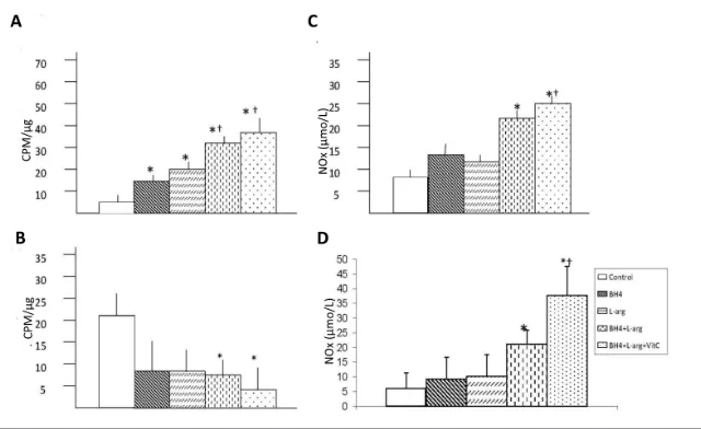
Effects of Oral BH4, L-Arginine and Vitamin C on Gastrocnemius Ca2+-Dependent NOS Activity
Dietary supplements affected Ca2+ dependent NOS activity in the ischemic gastrocnemius muscle (Figure 2A). Rats given a single dietary supplement (BH4 or L-arginine) demonstrated Ca2+ dependent NOS activities that were sim-ilar to each other and greater than that
Figure 1.Effects of BH4, L-arginine (L-arg) and vitamin C (VitC) on eNOS and P-eNOS ex-pression in the ischemic gastrocnemius muscle. Muscle was harvested 14 d after the in-duction of hindlimb ischemia and supernatants from muscle homogenates were used in these assays. (A) Expression of eNOS in gastrocnemius was increased by all dietary addi-tives, although the greatest increase was noted in rats that received BH4 + L-arginine + vi-tamin C. (B) Expression of eNOS in gracilis was not changed by any diet supplementation. (C) Expression of P-eNOS was increased in rats fed BH4 + L-arginine or BH4 + L-arginine + vitamin C. (D) Expression of P-eNOS was significantly increased in rats fed BH4 + L-arginine +
vitamin C. Control rats were fed a standard diet, whereas the four treatment groups con-sumed diets supplemented with additives noted in the bar graph. Concentrations of these additives are noted in the text. Data are means ± standard deviation (sd); n = 5–6. *p < 0.05 versus control; †p < 0.05 versus BH4 or
of rats fed normal chow. Rats given two (BH4 + L-arginine) or three (BH4 + L-arginine + vitamin C) supplements displayed significantly greater Ca2+ dependent NOS activity than rats given a single dietary supplement. The combi-nation of BH4 and L-arginine generated an additive effect. The addition of vita-min C further increased Ca2+-dependent NOS activity, although it did not reach statistical significance.
Effects of Oral BH4, L-Arginine and Vitamin C on Gastrocnemius Ca2+-Independent NOS Activity
Ca2+-independent NOS activity was greater in the ischemic gastrocnemius from rats fed normal chow than in the is-chemic gastrocnemius of dietary fed rats (Figure 2B). Rats fed the two dietary sup-plements (BH4 + L-arginine) or three di-etary supplements (BH4 + L-arginine +
vitamin C) demonstrated Ca2+ -indepen-dent NOS activity levels in the ischemic gastrocnemius muscle that were lower than in rats fed normal chow.
Effects of Oral BH4, L-Arginine and Vitamin C on Calf and Thigh Muscle NOx Levels
The final products of NO in vivois ni-trite (NO2
–
) and nitrate (NO3 –
). NOx is the sum of NO2
–
and NO3 –
and it is the best index of total NO production. The NOx concentration in the ischemic calf region (gastrocnemius) and collateral ar-tery region (gracilis) was significantly higher than in rats fed with BH4 and L-arginine or BH4, L-arginine and vita-min C (Figures 2C, D). No changes oc-curred in rats fed with either agent alone. The addition of vitamin C further increased NOx levels, although it did not reach statistical significance.
Effects of Oral BH4, L-Arginine and Vitamin C on Calf and Thigh Muscle Oxidant Stress
Dietary supplementation affected ni-trotyrosine accumulation and the ratio of GSH versus GSSG in the ischemic calf re-gion (gastrocnemius) and collateral ar-tery region (gracilis) (Figure 3). Rats given a single dietary supplement (BH4 or L-arginine) or the combination of these dietary agents displayed similar nitroty-rosine levels in the ischemic calf region (gastrocnemius) (Figures 3A, C). These levels were significantly less than the level noted in rats fed normal chow. Rats provided with all three dietary supple-ments (BH4 + L-arginine + vitamin C) ex-hibited a nitrotyrosine level significantly less than rats given a single supplement (BH4 or L-arginine) or the combination of these two agents. The ratio of reduced versus oxidized glutathione (GSH-to-GSSG) was also measured as another index of oxidant stress in the ischemic calf region (gastrocnemius). GSH-to-GSSG ratio was increased in the ischemic calf region of rats fed two dietary combi-nations (BH4 + L-arginine) or three di-etary combinations (BH4 + L-arginine + vitamin C). Addition of vitamin C had a beneficial effect in increasing GSH-to-GSSG ratio. However, in the collateral ar-tery region (gracilis) (Figures 3B, D), none of the dietary supplementation reg-imens affected nitrotyrosine accumula-tion, but in rats fed a triple dietary com-bination (BH4 + L-arginine + vitamin C), the GSH-to-GSSG ratio was increased significantly. Taken together, three di-etary supplements given in combination (BH4 + L-arginine + vitamin C) are more effective in decreasing ischemic muscle oxidant stress after induction of hindlimb ischemia in rats.
Effects of BH4, L-Arginine and Vitamin C on Hindlimb Blood Flow
Dietary supplementation significantly increased the recovery of hindlimb blood flow after induction of severe ischemia, and this effect was regimen- and time-dependent (Figure 4). Rats given a single supplement (BH4 or L-arginine) showed
Figure 2.Effects of BH4, L-arginine (L-arg) and vitamin C (VitC) on bioavailable NO in the ischemic gastrocnemius and gracilis muscles. Muscle was harvested 14 d after the in-duction of hindlimb ischemia, and supernatants from muscle homogenates were used in these assays. (A) Ca2+-dependent NOS activity was increased by all dietary additives,
although the greatest increase was noted in mice receiving BH4 + L-arginine or BH4 +
L-arginine + vitamin C. Data are means ± sd; n = 5–6. *p < 0.05 versus control; †p < 0.05
versus BH4 or L-arginine groups. (B) Ca2+-independent NOS activity was less in mice
re-ceiving BH4 + L-arginine or BH4 + L-arginine + vitamin C than in control mice. Data are
means ± sd; n = 5–6. *p < 0.05 versus control. (C, D) NOx was increased in gastrocemius (C) and gracilis (D) in mice receiving BH4 + L-arginine or BH4 + L-arginine + vitamin C.
Data are means ± sd; n = 5. *p < 0.05 versus control; †p < 0.05 versus BH4 or
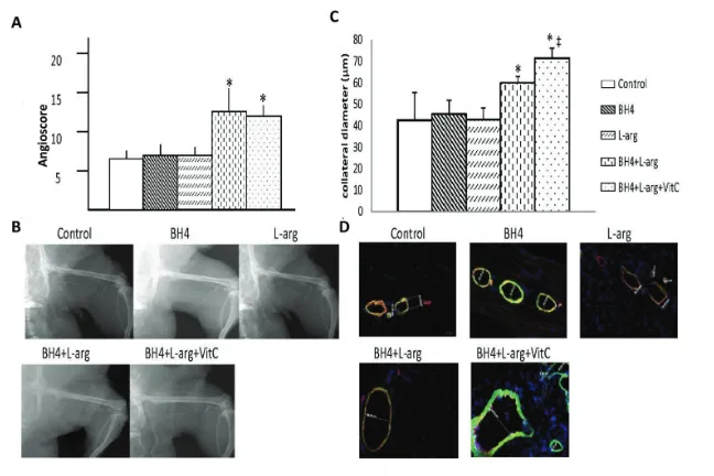
similar degrees of perfusion recovery in the foot, and this level was also similar to that noted in rats fed normal chow. Rats provided with two (BH4 + L-arginine) or three (BH4 + L-arginine + vitamin C) supplements showed significantly greater recovery of foot perfusion than rats fed normal chow or rats given a single dietary supplement. This differ-ence was evident at the later phase of re-covery, on d 21, 28 or 42 after induction of ischemia and also at the time of maxi-mal collateral artery wall remodeling (21). A similar pattern was noted for col-lateral artery angioscores and colcol-lateral diameters determined on d 42 after in-duction of ischemia (Figures 5A–D). Hence, the angioscore and collateral di-ameters were significantly greater in rats given two (BH4 + L-arginine) or three (BH4 + L-arginine + vitamin C) supple-ments than in rats fed normal chow or in rats given a single supplement. In ad-dition, rats fed BH4 + L-arginine + vita-min C showed significantly greater an-gioscores and collateral diameters than rats fed BH4 + L-arginine. Despite this in-crease in collateral artery diameters when all three supplements were given, we did not detect a statistically signifi-cant increase in resting foot blood flow. Resting blood flow is low in skeletal muscle and increases up to 10-fold dur-ing exercise. On the basis of our previous work, we would anticipate detecting sig-nificant increases in blood flow under ex-ercise conditions (22).
Effects of Oral BH4, L-Arginine and Vitamin C on Gastrocnemius Muscle Necrosis
The extent of gastrocnemius necrosis was affected by the provision of dietary supplements. Rats given a single supple-ment (BH4 or L-arginine) or the combina-tion of these agents manifest a similar degree of gastrocnemius muscle necrosis. Moreover, the extent of necrosis noted in these dietary intervention groups was similar to that noted in rats fed normal chow, that is, these dietary regimens did not improve postischemic muscle in-tegrity. However, rats provided with all
Figure 3.Effects of BH4, L-arginine (L-arg) and vitamin C (VitC) on oxidative stress in the is-chemic gastrocnemius and gracilis muscles. Muscle was harvested 14 d after the induc-tion of hindlimb ischemia and supernatants from muscle homogenates were used in these assays. (A, B) Nitrotyrosine expression was decreased by all dietary additives, al-though this decrease was greatest in rats receiving BH4 + L-arginine + vitamin C in ische-mic gastrocnemius muscles (A), but no change was observed in ischeische-mic gracilis muscles (B). (C, D) The GSH-to-GSSG ratio was increased in ischemic gastrocnemius (C) and gra-cilis (D) in rats receiving BH4 + L-arginine; the addition of vitamin C to this regimen caused
an additional increase in this ratio. Data are means ± sd; n = 5–6. *p < 0.05 versus control;
†p < 0.05 versus BH4 or
L-arginine groups; ‡p < 0.05 versus BH4 + L-arginine groups.
three dietary supplements had signifi-cantly less gastrocnemius necrosis than rats fed normal chow, rats provided with a single dietary supplement or rats given the combination of BH4 + L-arginine. This difference was evident on macro-scopic and micromacro-scopic levels. The per-centage of the cut surface of the ischemic gastrocnemius muscle that was necrotic, determined by NBT staining, was signifi-cantly less in the BH4 + L-arginine + vita-min C group than in all other groups (Figures 6A, B). Groups fed normal chow, or supplemented with BH4, or L-arginine, or both agents, demonstrated similar histological evidence of severe necrosis: muscle nuclei were nearly ab-sent, intra-myofiber vacuolization was substantial and the distance between my-ofibers was large (Figure 6C). A pro-nounced inflammatory infiltrate was also present in these groups. In contrast, the BH4 + L-arginine + vitamin C group demonstrated good preservation of
mus-cle histology and only a limited inflam-matory cell infiltrate.
DISCUSSION
The study hypothesis that dietary co-supplementation with BH4, L-arginine and vitamin C act synergistically to de-crease oxidant stress, inde-crease NO and thereby improve limb perfusion and tis-sue recovery in response to acute hindlimb ischemia was supported by our findings. Interestingly, two patterns of ef-fect emerged. Cosupplementation with BH4 + L-arginine increased the depen-dent variables NO bioavailability, foot perfusion, the collateral artery angioscore and collateral artery diameters more than the addition of either component sepa-rately, whereas the addition of vitamin C provided a further beneficial effect on these variables. In addition, coadminis-tration of all three dietary supplements had a significantly greater effect than BH4 or L-arginine, given individually or
in combination, when the dependent var-iables of oxidative stress (nitrotyrosine accumulation and GSH-to-GSSG ratio) or muscle necrosis were measured.
eNOS and P-eNOS expression, Ca2+ -dependent NOS activity, tissue NOx lev-els, foot perfusion, the collateral artery angioscore and collateral diameters are linked by established cause-and-effect re-lationships. eNOS-derived NO is a po-tent vasodilator (2); hence, the increased eNOS expression and activity present in the BH4 + L-arginine group should result in an NO-dependent increase in foot per-fusion, and this expectation was realized by our findings. Moreover, eNOS- derived NO is a critical determinant in the re-sponse to hindlimb ischemia (23–25). This effect is direct, because we and others have shown that eNOS- derived NO is es-sential to collateral artery remodeling (21,26). These effects include mobiliza-tion of mononuclear cells from the bone marrow and their subsequent homing to the ischemic hindlimb (27). Once there, those cells participate in postischemic ar-teriogenesis, the process wherein existing collateral arteries undergo remodeling designed to restore vascular conductance (28). This process was evidenced by the increased angioscores and collateral di-ameters, quantitative markers of collat-eral artery enlargement, in rats provided with BH4 + L-arginine + vitamin C di-etary supplements.
Vitamin C likely exerted its beneficial effects in this study through a variety of molecular mechanisms. In its capacity as an antioxidant, it enhances NO bioavailability by quenching O2
– , thus limiting the inactivation of NO that oc-curs when O2
–
and NO combine to pro-duce OONO–(3). Vitamin C also stabi-lizes existing BH4 (8) and increases endothelial BH4 synthesis (29), thus minimizing eNOS “uncoupling,” which, in turn, lessens generation of O2
– by eNOS and reduces vascular oxidant stress (7). However, BH4 is itself a po-tent antioxidant (7), and administration of exogenous BH4 has been established to increase endothelial BH4 levels (30). Moreover, L-arginine directly stimulates
Figure 5.Effects of BH4, L-arginine (L-arg) and vitamin C (VitC) on collateral enlargement after induction of hindlimb ischemia. (A) The angioscore, determined 42 d after the in-duction of hindlimb ischemia and calculated as described in the text, was greater in rats receiving BH4 + L-arginine or BH4 + L-arginine + vitamin C than in all other groups. Data
are means ± sd; n = 6. *p < 0.05 for BH4 + L-arginine or BH4 + L-arginine + vitamin C groups versus all other groups. (B) Representative angiogram from each study group. (C) Greater collateral artery diameters were observed in rats receiving BH4 + L-arginine; addition of vi-tamin C further increased collateral diameter. Data are means ± sd; n = 6. *p < 0.05 for BH4 + L-arginine or BH4 + L-arginine + vitamin C groups versus all other groups; ‡p < 0.05
eNOS expression (31); enhances eNOS activity by a receptor-dependent, G protein–linked process (32); and lim-its the inhibitory effect of asymmetric dimethyl arginine on eNOS-derived NO production (33). A series of studies proved that metabolic intervention with antioxidants (vitamin C) and L-arginine can promote the beneficial effects in is-chemia-induced vasculogenesis beyond that provided by bone marrow mononu-clear cells alone due to increased NO/eNOS bioactivity, decreased oxida-tive stress and antiinflammatory action in ischemic tissue (34–38). We propose that under the experimental conditions imposed by hindlimb ischemia, addition of vitamin C to BH4 + L-arginine
signifi-cantly decreased oxidative stress, in-creased NO bioavailability, inin-creased collateral artery diameters and accord-ingly decreased tissue necrosis. It may also restore BH4 or NO levels, since we observed an increase in P-eNOS expres-sion and NO bioavailability.
Ca2+-independent NOS activity and tissue nitrotyrosine accumulation were significantly lower in rats receiving all three dietary supplements than in rats receiving BH4 or L-arginine, or a combi-nation of the two. When measured by methods used herein, Ca2+-independent NOS activity is an authentic reflection of iNOS activity, inasmuch as the assay was conducted in vitro, in the absence of shear stress that can activate eNOS in
the absence of Ca2+via phosphorylation (39). The marked elevation of iNOS ac-tivity in rats fed normal chow indicates the presence of postischemia inflamma-tion, which is also evidenced by the cel-lular inflammatory infiltrate in this group. Nitrotyrosine accumulation is in-dicative of OONO–-induced necrosis (40), and it is interesting that the group that exhibited the least amount of tissue necrosis (that is, rats provided with all three supplements) also had the least ni-trotyrosine accumulation. Moreover, rats in the triple therapy group demon-strated a virtual absence of nitrotyro-sine. We interpret the present findings to indicate that vitamin C provided an antioxidant effect that limited tissue in-jury generated by inflammatory cells for which action depends, in part, on oxi-dant production (for example, neu-trophils and macro phages). This effect could be direct because of the antioxi-dant activity of vitamin C, or indirect, because of the beneficial effect of vita-min C on BH4 levels (8,29), insofar as BH4 also exhibits potent antioxidant ac-tivity (7).
Figure 7.Schematic illustration that a regi-men of three dietary suppleregi-ments com-bined can increase NO bioavailability and decrease oxidative stress, accordingly in-crease blood flow recovery and reduce tissue necrosis. A•–, ascorbate; AH–,
dehy-droascorbate. BH3•, BH3–.
Figure 6.The effects of BH4, L-arginine (L-arg) and vitamin C (VitC) on necrosis in the
ische-mic gastrocnemius muscle. (A) Gastrocnemius muscle was removed 7 d after induction of hindlimb ischemia. Transverse sections of this muscle were stained with nitroblue tetrazolium to determine the ratio of necrotic versus viable surface area. This ratio was less in rats re-ceiving BH4 + L-arginine + vitamin C than in all other groups. Data are means ± sd; n = 6.
Although it is well established that en-dothelial dysfunction related to vascular oxidant stress is a critical factor in PAD pathogenesis, dietary supplementation with L-arginine or antioxidants, such as vitamin C, has had equivocal effects on long-term outcome (12,13,16). Dietary supplementation with L-arginine alone has a beneficial effect when given acutely, that is, via intravenous infusion (14) or for short duration (2 months) (15), and these clinical results are consis-tent with the positive effects observed in rats provided with dietary L-arginine. However, long-term administration of L-arginine (6 months) not only failed to demonstrate a beneficial effect, but re-sulted in a degree of eNOS-dependent vascular reactivity significantly less than that of the placebo group (16). The pres-ent findings demonstrated increased iNOS activity after induction of ische-mia. If a similar circumstance is present in PAD, then the singular dietary sup-plementation with L-arginine, the sub-strate for all NOS isoforms, might serve to worsen vascular inflammation, a criti-cal participant in the pathogenesis of PAD (1). In addition, if L-arginine is ad-ministrated in a state of oxidant stress, as is present in many patients with PAD, eNOS is uncoupled and produces ONOO–rather than NO. Vitamin C re-duces vascular inflammation (41) and improves redox balance (11) and eNOS-dependent vascular reactivity (10), but these effects have only been evaluated on a short-term basis, whereas retrospec-tive cross-sectional studies have failed to confirm that dietary supplementation with antioxidants improves PAD out-come (12,13). We interpret the recent findings to indicate that provision of BH4 + L-arginine + vitamin C acting synergistically might prove to be a use-ful therapeutic alternative in PAD treat-ment. To this end, the use of sapropterin dihydrochloride, a synthetic form of (6R)-L -erythro-5,6,7,8-tetrahydro-biopterin recently approved for the treatment of phenylalanine hydroxylase deficiency (42), might provide a practical means for the provision of BH4.
CONCLUSION
Cosupplementation with BH4 + L-arginine + vitamin C resulted in increased eNOS activity and NO con-centration as well as greater foot blood flow recovery than rats receiving nor-mal chow or either agent separately. The addition of vitamin C to the BH4 + L-arginine regimen further reduced oxi-dant stress and increased collateral di-ameters and reduced tissue injury in is-chemic muscles (Figure 7). The clearly superior outcome of rats provided with BH4 + L-arginine + vitamin C warrants investigation of a cosupplementation strategy as a therapeutic alternative in PAD. However, additional preclinical studies need to be undertaken to estab-lish proof of principle in experimental models of hindlimb ischemia in which ischemia is induced gradually and under conditions of systemic oxidant stress such as type 2 diabetes or hyper-cholesterolemia (43).
ACKNOWLEDGMENTS
This work was supported by National Institutes of Health, National Heart, Lung, and Blood Institute grant RO-1 HL-75353 (to LM Messina), as well as grants from the the Wayne and Gladys Valley Foundation (to LM Messina).
DISCLOSURE
The authors declare that they have no competing interests as defined by Molec-ular Medicine, or other interests that might be perceived to influence the re-sults and discussion reported in this paper.
REFERENCES
1. Coutinho T, Rooke TW, Kullo IJ. (2011) Arterial dysfunction and functional performance in pa-tients with peripheral artery disease: a review.
Vasc. Med. 16:203–11.
2. Marletta MA. (1993) Nitric oxide synthase struc-ture and mechanism. J. Biol. Chem.268:12231–4. 3. Pacher P, Beckman JS, Liaudet L. (2007) Nitric
oxide and peroxynitrite in health and disease.
Physiol. Rev.87:315–424.
4. Peterson TE, et al.(1999) Opposing effects of re-active oxygen species and cholesterol on en-dothelial nitric oxide synthase and enen-dothelial cell caveolae. Circ. Res.85:29–37.
5. Kuzkaya N, Weissmann N, Harrison DG, Dikalov S. (2003) Interactions of peroxynitrite, tetrahydrobiopterin, ascorbic acid, and thiols: im-plications for uncoupling endothelial nitric-oxide synthase. J Biol. Chem.;278:22546–54.
6. Bevers LM, et al.(2006) Tetrahydrobiopterin, but not L-arginine, decreases NO syn-thase uncou-pling in cells expressing high levels of endothe-lial NO synthase. Hypertension.47:87–94. 7. Schmidt TS, Alp NJ. (2007) Mechanisms for the
role of tetrahydrobiopterin in endothelial func-tion and disease. Clin. Sci.113:47–63.
8. Heller R, et al.(2001) L-ascorbic acid potentiates endothelial nitric oxide synthesis via a chemical stabilization of tetrahydrobiopterin. J. Biol. Chem.
276:40–7.
9. Langlois M, Duprez D, Delanghe J, De Buyzere M, Clement DL. (2001) Serum vitamin C concen-tration is low in peripheral arterial disease and is associated with inflammation and severity of atherosclerosis. Circulation.103:1863–8. 10. Silvestro A, et al.(2002) Vitamin C prevents
en-dothelial dysfunction induced by acute exercise in patients with intermittent claudication. Athero-sclerosis.165:277–83.
11. Wijnen MH, et al.(2001) Antioxidants reduce ox-idative stress in claudicants. J. Surg. Res.
96:183–7.
12. Donnan PT, Thomson M, Fowkes FG, Prescott RJ, Housley E. (1993) Diet as a risk factor for periph-eral arterial disease in the genperiph-eral population: the Edinburgh Artery Study. Am. J. Clin. Nutr.
57:917–21.
13. Klipstein-Grobusch K, et al.(2001) Dietary an-tioxidants and peripheral arterial disease: the Rotterdam Study. Am. J. Epidemiol. 154:145–9. 14. Böger RH, et al.(1998) Restoring vascular nitric
oxide formation by L-arginine improves the symptoms of intermittent claudication in pa-tients with peripheral arterial occlusive disease.
J. Am. Coll. Card.32:1336–44.
15. Maxwell AJ, Anderson BE, Cooke JP. (2000) Nu-tritional therapy for peripheral arterial disease: a double-blind, placebo-controlled, randomized trial of HeartBar. Vasc. Med.5:11–9.
16. Wilson AM, Harada R, Nair N, Balasubramanian N, Cooke JP. (2007) L-arginine supplementation in peripheral arterial disease: no benefit and pos-sible harm. Circulation.116:188–95.
17. Ueda S, et al.(2000) Tetrahydrobiopterin restores endothelial function in long-term smokers. J. Am. Coll. Card.35:71–5.
18. Heitzer T, Krohn K, Albers S, Meinertz T. (2000) Tetrahydrobiopterin improves endothelium-de-pendent vasodilation by increasing nitric oxide activity in patients with type II diabetes mellitus.
Diabetologia.43:1435–8.
19. Yan J, et al.(2010) Oral tetrahydrobiopterin im-proves the beneficial effect of adenoviral-medi-ated eNOS gene transfer after induction of hindlimb ischemia. Mol. Ther.18:1482–9. 20. Hong HJ, Hsiao G, Cheng TH, Yen MH. (2001)
spontaneously hypertensive rats. Hypertension.
38:1044–8.
21. Park B, et al.(2010) Endothelial nitric oxide syn-thase affects both early and late collateral arterial adaptation and blood flow recovery after induc-tion of hindlimb ischemia in mice. J. Vasc. Surg.
51:165–73.
22. Brevetti LS, et al.(2001) Exercise induced hyper-emia unmasks regional blood flow deficit in ex-perimental hindlimb ischemia. J. Surg. Res.
98:21–6.
23. Yan J, Tang GL, Wang R, Messina LM. (2005) Op-timization of adenovirus-mediated endothelial nitric oxide synthase delivery in rat hindlimb is-chemia. Gene Ther. 12:1640–50.
24. Yu J, et al.(2005) Endothelial nitric oxide syn-thase is critical for ischemic remodeling, mural cell recruitment, and blood flow reserve. Proc. Natl. Acad. Sci. U. S. A.102:10999–1004. 25. Lloyd PG, Yang HT, Terjung RL. (2001)
Arterio-genesis and angioArterio-genesis in rat ischemic hindlimb: role of nitric oxide. Am. J. Physiol. Heart Circ. Physiol.281:H2528–38.
26. Mees B, et al.(2007) Endothelial nitric oxide syn-thase activity is essential for vasodilation during blood flow recovery but not for arteriogenesis.
Arterioscler. Thromb. Vasc. Biol.27:1926–33. 27. Aicher A, et al.(2003) Essential role of endothelial
nitric oxide synthase for mobilization of stem and progenitor cells. Nat. Med.9:1370–6. 28. Helisch A, Schaper W. (2003) Arteriogenesis: the
development and growth of collateral arteries.
Microcirculation. 10:83–97.
29. Huang A, Vita JA, Venema RC, Keaney JF Jr. (2000) Ascorbic acid enhances endothelial nitric-oxide synthase activity by increasing intracellular tetrahydrobiopterin. J. Biol. Chem.275:17399–406. 30. Sawabe K, Wakasugi KO, Hasegawa H. (2004)
Tetrahydrobiopterin uptake in supplemental ad-ministration: elevation of tissue tetrahydro-biopterin in mice following uptake of the exoge-nously oxidized product 7,8-dihydrobiopterin and subsequent reduction by an anti-folate-sensi-tive process. J. Pharmacol. Sci.96:124–33. 31. Javanmard SH, Nematbakhsh M, Nahomoodi F,
Mohajeri MR. (2009) L-Arginine supplementation enhances eNOS expression in experimental model of hypercholesterolemic rabbits aorta.
Pathophysiology.16:9–13.
32. Joshi MS, et al.(2007) Receptor-mediated activa-tion of nitric oxide synthesis by arginine in en-dothelial cells. Proc. Natl. Acad. Sci. U. S. A.
104:9982–7.
33. Böger RS, Ron ES. (2005) L-arginine improves vascular function by overcoming the deleterious effects of ADMA, a novel cardiovascular risk fac-tor. Altern. Med. Rev. 10:14–23.
34. Napoli C, et al.(2005) Beneficial effects of concur-rent autologous bone marrow cell therapy and metabolic intervention in ischemia-induced an-giogenesis in the mouse hindlimb. PNAS.
47:17202–6.
35. De Nigris F, et al.(2007) Therapeutic effects of concurrent autologous bone marrow cell infusion
and metabolic intervention in ischemia-induced angiogenesis in the hypercholesterolemic mouse hindlimb. Int. J. Cardiol.117:238–43.
36. De Nigris F, et al.(2007) Therapeutic effects of autologous bone marrow cells and metabolic intervention in the ischemic hindlimb of spon-taneously hypertensive rats involved reduced cell senescence and CXCR4/Akt/eNOS path-ways. J. Cardiovasc. Pharmacol.50:424–33. 37. Napoli C, et al.(2008) Beneficial effects of
autolo-gous bone marrow cell infusion and antioxidants/ L-arginine in patients with chronic critical limb is-chemia. Eur. J. Cardiovasc. Prev. Rehabil.15:709–18. 38. Balestrieri M, et al.(2010) Therapeutic
angiogene-sis in diabetic apolipoprotein E-deficient mice using bone marrow cells, functional heman-gioblasts and metabolic intervention . Atheroscle-rosis.209:403–14.
39. Ayajiki K, Kindermann M, Hecker M, Fleming I, Busse R. (1996) Intracellular pH and tyrosine phosphorylation but not calcium induce shear stress-induced nitric oxide production in native endothelial cells. Circ. Res. 78:750–8.
40. Beckman JS, Koppenol WH. (1996) Nitric oxide, superoxide, and peroxynitrite: the good, the bad, and ugly. Am. J. Physiol.271:C1424–37. 41. Aguirre R, May JM. (2008) Inflammation in the
vascular bed: importance of vitamin C. Pharma-col. Ther.119:96–103.
42. Yamamizu K, Shinozaki K, Ayajiki K, Gemba M, Okamura T. (2007) Oral administration of both tetrahydrobiopterin and L-arginine prevents en-dothelial dysfunction in rats with chronic renal failure. J. Cardiovasc. Pharmacol.49:131–9. 43. Yagai Y, et al.(2008) Cellular and molecular