ANALYSIS OF MICRORNA FUNCTION IN THE GASTROINTESTINAL TRACT
Bailey Cristina Eileen Peck
A dissertation submitted to the faculty at the University of North Carolina at Chapel Hill in partial fulfillment of the requirements for the degree of Doctor of Philosophy in the Curriculum
in Genetics & Molecular Biology in the School of Medicine.
Chapel Hill 2016
Approved by
Brian J Bennett
Pauline K Lund
Scott T Magness
John F Rawls
ii © 2016
iii
ABSTRACT
Bailey Cristina Eileen Peck: Analysis of microRNA function in the gastrointestinal tract
(Under the direction of Praveen Sethupathy)
microRNAs (miRNAs) are a group of small non-coding RNAs that regulate gene
expression through post-transcriptional targeting of messenger RNAs (mRNAs). Discovered in
mammals in 2001, miRNAs have since become appreciated as both biomarkers and drivers of
disease, including metabolic diseases such as type 2 diabetes. Metabolic diseases are
characterized by systemic energy imbalance, which involve diverse tissues such as liver,
pancreas, adipose, brain, muscle, and intestine. Understanding the role of miRNAs in the
regulation of these organ systems during normal physiology and disease pathogenesis is a
necessary step to help develop effective miRNA-based therapeutics. Toward this goal, in my
dissertation research I identify miRNAs that act as biomarkers of metabolic and gastrointestinal
(GI) diseases and evaluate their role in gene regulatory networks in the liver and small intestine.
miRNAs are severely understudied in the intestine compared to most other metabolic tissues, and
specifically in the intestinal epithelium, so I extended my research objective to help bridge this
gap by identifying diet- and microbiota-sensitive miRNAs in distinct cell populations of the
intestinal epithelium. I found that intestinal epithelial stem cells (IESCs) respond most robustly
to these environmental stimuli. Furthermore, I demonstrated that specific microbiota-sensitive
miRNAs regulate IESC proliferation, which is a key process underlying intestinal homeostasis.
The findings of my research represent key advances in the GI field, and serve as a strong
iv
v
ACKNOWLEDGMENTS
A doctoral dissertation is never completed in solitude, and there are many, many people
whom I have to thank for providing me support, encouragement, mentorship, friendship and the
resources necessary to complete this stage of my education.
I will start by thanking my generous funding sources that have taken a chance on me and
made my graduate studies possible through various fellowship awards. Specifically, the Initiative
for Maximizing Student Diversity (IMSD), the Curriculum in Genetics & Molecular Biology, the
Sarah Graham Kenan-Edwards Hobgood fellowship, the National Institute of Diabetes and
Digestive and Kidney Disorders (NIDDK), the University of North Carolina at Chapel Hill
(UNC), and the UNC Biological and Biomedical Sciences Program.
My dissertation would not have been completed without the endless support and
mentorship from my advisor, Praveen Sethupathy. Praveen is a one-of-a-kind mentor whom I am
so thankful to have met even before starting my graduate career. Several years ago, he selflessly
guided me not only to UNC, but also to his lab, where I have had one of the most positive and
inspiring graduate careers. As a PI, Praveen has kept me confident in the midst of the
discouraging, helped identify hidden gems among dead ends, and taught me the art of telling
stories of great scientific discoveries. As a mentor and advisor, Praveen has been my champion,
encouraging my professional and personal development and cheering my successes, and my
vi
Kay Lund, my mentor and chair, who has been one of my favorite sources of advice
throughout my graduate career, also deserves special thanks. Her guidance and support over the
past four years have helped shape my career aspirations. She is a constant source of inspiration,
and I am so thankful that she listened to my risky project idea and helped me turn it into an
exciting reality. As her career has progressed, I am also thankful for the generous amount of time
she has lent to read my manuscripts and grant applications, and talk me through various career
choices and pitfalls. I am so grateful for her ongoing grounded, constructive, thorough, and much
needed guidance.
I would also like to thank the other members of my thesis committee, Brian Bennett,
Scott Magness, and John Rawls, who despite being in different institutions, departments, and
campuses were always available to meet, provide advice, critique my research, and offer diverse
and interesting perspectives and suggestions. I would like to thank Brian for providing his
expertise on diet and nutrition, giving me genuine career advice, and for advocating for me
throughout my graduate career. I would like to thank Scott for challenging me, questioning me,
and, above all, supporting me. His advice and innovative perspectives have constantly inspired
me to do better science and take my questions one step further. Finally, I would like to thank
John for the many, many hours of discussion and debate regarding microbiota, the
host-epithelium, conventionalization, and overall strategy during the initial stages of my thesis
project. He opened up the world of the microbiome to me and has since been an avid supporter
and priceless mentor. A committee is a family, and I am glad I picked these five!
Several past and present mentors who guided me and encouraged me to continue this
journey deserve recognition. I’d like to start by acknowledging Peggy Daugherty of Colorado
vii
Jackson Laboratory, for introducing me to experimental biology and mouse genetics; Francis
Collins of the National Institutes of Health, for the ongoing, thoughtful guidance and inspiration;
and Samir Kelada of UNC, for preparing me for the challenges of research.
I have recognized my mentors, but I also need to thank all of my lab mates over the past
decade that have listened to my problems and provided advice on troubleshooting, next steps,
and interpretation. Lab mates have laughed with me, sympathized with me, and have generally
been my favorite people I have had the pleasure to see every day. I want to give a special shout
out to Jeanette Baran-Gale and Sara Selitsky, who as senior graduate students in Praveen’s lab
have been there for me every step of the way. I would like to thank them for the advice,
assistance, mentorship, critical ear, and helping hands. They are not only my lab mates, but also
two of my closest friends. Jeanette has provided countless hours of technical support and many
refreshing lunch dates and coffee breaks discussing science, the future, career paths, and life in
general. I would like to thank her for being by my side both in and out of the lab and classroom.
Despite the oceans that may separate us, I know that she will be a constant friend, colleague,
collaborator, and mentor. Since joining Praveen’s lab, I would also like to thank Lisa Kurtz, who
has always been ‘home base’ in the lab, keeping us grounded, but not too serious. Thanks Tim
Dinh, Alisha Coffey, Nev Kazgan, Eva Vitucci, Rowan Beck, Wendy Pitman, Ben Keith, Mi
Zhou, Emily Fannin, John Sincavage, Sydney Feinstein, and the rest of the miRcats for keeping
lab real. I would also like to thank the members of Kay’s lab, including Amanda Mah, Emily
Moorefield, Shengli Ding, Eric Blue, Jim Simmons, and Sara Andres, for teaching me about the
gut, supporting my projects, and lending hours of troubleshooting advice and assistance.
The experiments and published manuscript described in Chapter 2 would not have been
viii
Landstreet, Shengli Ding, Vandana Turaga, Kay Lund, Scott Turner, Sudha B. Biddinger, and
Kasey C. Vickers. Additional acknowledgment must also be given to Michael Erdos, Samir
Kelada, Jeanette Baran-Gale, and Jonathan Haldeman for their helpful suggestions regarding the
study and manuscript, Stanley Lemon for generously sharing Huh7 cells, and Brad Hoffman of
the University of British Columbia for sharing the chromatin occupancy sites for FOXA2 in
mouse liver and islet based on a previously published study.
I would like to thank Shehzad Sheikh for sharing his exciting Crohn’s disease data, his
knowledge and excitement for IBD research, and his mentorship over the last couple years; and
Terry Furey for his ability to simplify complex ideas and his close eye for details. Chapter 3
would not have been possible without their assistance and support. I also want to thank my
co-authors for the research presented in Chapter 3, including Matt Weiser, Eric Lee, Greg Gipson,
Vishal Iyer, Balfour Sartor, Hans Herfarth, Millie Long, Jonathan Hansen, Kim Isaacs, Dimitri
Trembath, Reza Rahbar, and Timothy Sadiq.
Many helping hands assisted the experiments and published manuscript described in
Chapter 4 over the past couple of years. This ‘side-project’ became quite a beast with plot twists,
dead-ends, and a few surprises, which I have my many co-authors to thank for helping untie.
They include John Sincavage, Sydney Feinstein, Amanda Mah, Jim Simmons, and Kay Lund. I
also must acknowledge the Beaulieu laboratory for provision of the HIECs, the UNC Flow
Cytometry Core Facility, the UNC High Throughput Sequencing Facility, the UNC
Bioinformatics Core Facility, and specifically Barry Udis, Joel Parker, Eric Blue, and Scott
Magness for technical assistance and useful discussions.
The data included in Chapter 5 could not have been generated without Amanda Mah,
ix
intestine, troubleshoot FACS, and manage the challenges of graduate school. She has been a
patient and supportive mentor, despite my flaws, and I appreciate all of her help on the various
projects I have undertaken. Additionally, many thanks are due to Eric Blue for his help in
developing the GF Sox9-EGFP resource; Elaine Glenny for critical assistance with the anaerobic
chamber and the protocol for conventionalization; Lisa Kurtz, Emily Moorefield, Shengli Ding,
and Jeanette Baran-Gale for technical assistance and training; John Rawls and Scott Magness for
helpful discussions; as well as Felicia Heyward and UNC Flow Cytometry Core Facility, the
UNC Gnotobiotic Core Facility, Dr. Zhao Lai and the UTHSCSA Genome Sequencing Facility,
and Dr. Bob Bagnell and the UNC Microscopy Services Laboratory for critical services rendered
for the experiments presented in Chapter 5.
Toward the completion of this dissertation as a whole, I also must thank the many friends
and colleagues who volunteered their time to read through and copyedit the very rough versions
of this document. Specifically, thank you to Deirdre Sackett and Jonathan Susser, both of whom
served with me on the executive board of the UNC Science Writing and Communication club;
Sydney Feinstein, a fellow miRcat who worked with me on the results presented in Chapter 4;
Kelsey Gray and Michelle Engle, who are both GMB peers and friends; and Kate Kutchko, a
close friend and colleague.
Outside of lab, I have my peers in GMB, FYG, and IMSD to thank for their friendship,
support, and shoulders to lean on (during both the tough times and the best of times). In
particular, I would like to mention Kate Kutchko, a fellow CC Tiger, who became one of my
closest friends during my time at UNC. I want to thank her for the many passionate and
thought-provoking debates on science, policy, and life, which I am ever thankful we can continue our
x
dates, and happy hours, all of which have helped me get through graduate school in one piece.
Finally, I must acknowledge my family, who has truly made the greatest sacrifices to get
me to and through grad school. They are my constant support network. My father, who passed
away in 2015, was a lifelong inspiration. His intelligence, love of science, hard work ethic, and
many still top-secret projects are the traits and memories that continue to push me to do better.
My mom, who has been my greatest supporter for as long as I can remember, deserves special
recognition. Her love of life, thoughtfulness, patience, drive, and confidence are traits that I will
forever attempt to emulate. My Gramma Ruby, who immigrated to the US to pursue higher
education, has always been a present advocate and supporter. Her sacrifices paved the way for
my successes, and I am truly thankful for everything she has done for me. My stepfather, Tom, I
would like to thank for his quick wit, unique perspectives, friendship, and support. My siblings
Kilian, Teghan, Ivan, and Sara, who have all pursued and achieved successful careers in their
own right, deserve thanks for celebrating the good times and supporting me during the difficult
times. I would also like to thank my best friend, Ashley, for the last 18 years of friendship.
Despite the distance, my love of vegetables, and her two days of age maturity over me, she has
been one of my greatest cheerleaders, confidantes, and companions. I have Ashley to thank for
the many rides home from the airport, hours-long phone conversations, personal advice, and
loving support over the years. May we forever be related through our dogs.
Finally, I would like to thank my partner, David, for his love and support over the last
decade. His quick humor, love, thoughtfulness, patience, presence, and companionship
persevered despite the long work hours, missed dates, and staggered schedules. These past four
years had their challenges and blessings, and I am so thankful for his generosity, kindness,
xi
our dog-children, Getty and Maccabee. In 2010, David chose to move across the country with
me, and I am forever appreciative that he chooses to stand by my side, to share and celebrate our
xii
PREFACE
In each chapter, there are portions of work that were completed by other talented
scientists, and most chapters contain published work that I have been granted access to use as
part of this dissertation.
The full citation for the published work found in Chapter 2 is as follows:
Kurtz, C. L.*, Peck, B. C. E.*, Fannin, E. E., Beysen, C., Miao, J., Landstreet, S. R., Ding, S., Turaga, V., Lund, P. K., Turner, S., Biddinger, S. B., Vickers, K. C., and Sethupathy, P. (2014) MicroRNA-29 fine-tunes the expression of key FOXA2-activated lipid metabolism genes and is
dysregulated in animal models of insulin resistance and diabetes. Diabetes. 63, 3141–3148.
This project evolved from results generated during my first graduate research rotation in
the Sethupathy lab. Lisa Kurtz, who serves as a co-first author with me on the manuscript (as
designated by the asterisk), continued and confirmed many of the initial transfection studies
using LNAs, FOXA2 ORF, and luciferase vectors after the completion of my research rotation.
After I joined the Sethupathy lab for my thesis work, she and I were responsible for completing
the remaining experiments, editing the manuscript, and generating figures. Permission to include
the published article in its entirety is given by the journal Diabetes for educational purposes as
explained on their website (http://www.diabetesjournals.org/content/reprints-permissions).
The full citation for the published work found in Chapter 3 is as follows:
Peck, B. C. E., Weiser, M., Lee, S. E., Gipson, G. R., Iyer, V. B., Sartor, R. B., Herfarth, H. H., Long, M. D., Hansen, J. J., Isaacs, K. L., Trembath, D. G., Rahbar, R., Sadiq, T. S., Furey, T. S., Sethupathy, P., and Sheikh, S. Z. (2015) MicroRNAs Classify Different Disease Behavior
Phenotypes of Crohn's Disease and May Have Prognostic Utility. Inflammatory Bowel Diseases.
21, 2178–2187.
xiii
Bowel Diseases, Wolters Kluwer Health, and Lippincott Williams & Wilkins through RightsLink, which can be found in the online version of the article
(http://journals.lww.com/ibdjournal/Fulltext/2015/09000/MicroRNAs_Classify_Different_Disea
se_Behavior.20.aspx).
The full citation for the published work found in Chapter 4 is as follows:
Peck, B. C. E., Sincavage, J., Feinstein, S., Mah, A. T., Simmons, J. G., Lund, P. K., and Sethupathy, P. (2016) miR-30 family controls proliferation and differentiation of intestinal epithelial cell models by directing a broad gene expression program that includes SOX9 and the
ubiquitin ligase pathway. J. Biol. Chem, 291(31), 15975-15984.
The Journal of Biological Chemistry automatically grants authors the right to reuse their published material in a thesis and/or dissertation. Details of this permission can be found on their
website (http://www.jbc.org/site/misc/Copyright_Permission.xhtml).
The material included in Chapter 5 has been submitted for publication. Rights and usage
will be discussed with the publisher upon acceptance. A full citation for the current version of
this work, which can be found on the preprint server bioRxiv, is as follows:
Peck, B. C. E., Mah, A. T., Pitman, W. A., Ding, S., Lund, P. K., and Sethupathy, P. (2016) Functional transcriptomics in diverse intestinal epithelial cell types reveals robust gut microbial
sensitivity of microRNAs in intestinal stem cells. bioRxiv. 10.1101/087882
xiv
TABLE OF CONTENTS
LIST OF TABLES ... xviii
LIST OF FIGURES ... xix
LIST OF ABBREVIATIONS ... xxi
CHAPTER 1 – INTRODUCTION ... 1
microRNA biogenesis and function ... 3
microRNAs in metabolic disease ... 6
microRNA-33 ... 6
microRNA-375 ... 7
microRNA-378 ... 7
microRNA-24 ... 8
microRNA-30 ... 8
microRNAs as therapeutics and therapeutic targets in the treatment of metabolic disease ... 9
The intestinal epithelium as a metabolic tissue ... 10
Gut microbiota and diet in metabolism and metabolic disease ... 12
CHAPTER 2 – microRNA-29 fine-tunes the expression of key FOXA2-activated lipid metabolism genes and is dysregulated in animal models of insulin resistance and diabetes ... 19
Research Design and Methods ... 21
Animal studies ... 21
xv
Transfection studies ... 22
Small RNA-sequencing analysis ... 22
Gene expression (RNA) analysis ... 22
Western blotting ... 23
Reporter gene (Luciferase) assays ... 24
Bioinformatics ... 25
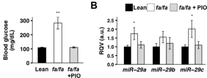
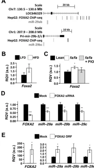
Results ... 25
Hepatic miR-29 is up-regulated in animal models of insulin resistance and is corrected by treatment with the insulin-sensitizing drug Pioglitazone ... 25
Hepatic miR-29 expression is controlled in part by the insulin-regulated transcription factor FOXA2 ... 28
miR-29 fine-tunes FOXA2 mediated regulation of key hepatic lipid metabolism genes ... 29
Discussion ... 32
CHAPTER 3 – microRNAs classify different disease behavior phenotypes of Crohn’s disease and may have prognostic utility ... 37
Research Design and Methods ... 39
Patient Population ... 39
Phenotyping ... 40
Clinical Phenotype ... 40
Identification of formalin-fixed, paraffin-embedded (FFPE) sections in patients with CD ... 40
RNA isolation ... 41
Small RNA and mRNA-sequencing and expression analysis ... 42
Quantitative reverse transcriptase PCR ... 43
miRhub analysis ... 43
xvi
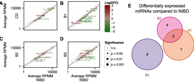
Small RNA-sequencing reveals distinct miRNA signatures in the non-inflamed
colon of CD patients with different disease behaviors ... 43
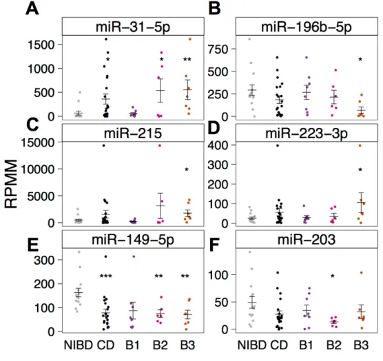
Inflammation is not a primary driver of miR-31-5p, miR-215, and other CD- associated miRNAs ... 46
miR-215 is a potential predictor of a penetrating disease phenotype in CD ... 49
Whole transcriptome analysis reveals that miR-31-5p and miR-203 are candidate drivers of the colonic gene expression profile in CD ... 53
Discussion ... 54
CHAPTER 4 – miR-30 family controls proliferation and differentiation of intestinal epithelial cell models by directing a broad gene expression program that includes SOX9 and the ubiquitin ligase pathway ... 59
Research Design and Methods ... 60
Animals ... 60
IEC dissociation for flow cytometry and FACS ... 61
Quantitative Reverse Transcription PCR (qRT-PCR) ... 61
Cell culture and transfections ... 62
Caco-2 differentiation ... 62
Western blot ... 63
RNA-sequencing ... 63
Bioinformatics ... 64
Results ... 64
miR-30 is predicted to target SOX9 and is robustly expressed in the IE ... 64
Knockdown of miR-30 in vitro results in increased SOX9 mRNA expression but decreased levels of SOX9 protein ... 66
Next-generation high-throughput RNA-sequencing reveals that miR-30 regulates genes enriched in the ubiquitin ligase pathway ... 67
miR-30 promotes IEC proliferation and inhibits IEC differentiation ... 73
xvii
CHAPTER 5 – microRNA profiling in intestinal epithelial subpopulations and functional studies in enteroids identify gut microbiota-responsive miR-375 as a
candidate regulator of stem cell proliferation ... 80
Research Design and Methods ... 81
Animals ... 81
Conventionalization (CV) ... 82
IEC isolation and fluorescence-activated cell sorting (FACS) ... 83
mRNA library preparation and sequencing ... 84
Small RNA library preparation and sequencing ... 84
Bioinformatics ... 85
Enteroid culture ... 86
Validation of miRNA expression levels ... 87
Linear Model ... 87
Results ... 87
Germ-free (GF) animals have fewer stem and more EECs ... 87
Conventionalized stem cells show enrichment for genes involved in proliferation. ... 88
Small RNA-sequencing of each IEC population reveals cell type-specific expression of miRNAs ... 92
miR-375 is robustly and DE in IESCs of GF and conventionalized mice ... 94
Knockdown of miR-375 in ex vivo enteroids results in increased proliferation ... 95
HFD induces robust changes in IESC gene expression but minimal changes in miRNA expression across Sox9-EGFP populations ... 101
Discussion ... 106
CHAPTER 6 – DISCUSSION, CONCLUSIONS, & FUTURE DIRECTIONS ... 111
xviii
LIST OF TABLES
Table 5.1. microRNAs enriched at least 2-fold in one intestinal epithelial cell (IEC)
xix
LIST OF FIGURES
Figure 1.1. The gastrointestinal tract and type 2 diabetes. ... 2
Figure 1.2. Cartoon depicting microRNA and mRNA biogenesis. ... 5
Figure 2.1. Hepatic miR-29 levels are upregulated in diet-induced insulin resistance
in mice. ... 26
Figure 2.2. Hepatic miR-29 levels are elevated in a rat model of diabetes and corrected
by treatment with the insulin-sensitizing drug pioglitazone (PIO). ... 27
Figure 2.3. FOXA2 regulates miR-29 expression. ... 30
Figure 2.4. miR-29 fine-tunes FOXA2-mediated regulation of key lipid metabolism
genes. ... 32
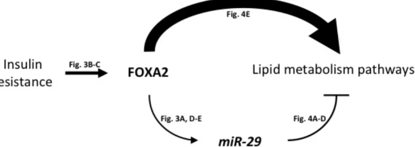
Figure 2.5. Schematic of the FOXA2:miR-29 regulatory circuit in the liver. ... 34
Figure 2.6. The intestine is an understudied metabolic organ. ... 36
Figure 3.1. Colonic miRNAs are differentially expressed between Crohn’s disease
patients and controls. ... 45
Figure 3.2. miRNAs are variably expressed across Crohn’s disease subtypes. ... 47
Figure 3.3. qRT-PCR confirms differential expression of four colonic miRNAs in
Crohn’s disease patients and controls. ... 48
Figure 3.4. Differential expression of miRNAs is not dependent on inflammation status. ... 50
Figure 3.5. miR-215 may have prognostic utility. ... 51
Figure 3.6. RNA-sequencing of matched RNA reveals commonly dysregulated genes
across disease subtypes, and potential master regulatory miRNAs. ... 52
Figure 4.1. miR-30 is predicted to target the 3’UTR of SOX9 and is differentially
expressed across functionally distinct cell types of the intestinal epithelium. ... 68
Figure 4.2. Knockdown of miR-30 increases SOX9 mRNA and decreases SOX9 protein
expression. ... 69
Figure 4.3. Next-generation high-throughput RNA-sequencing of LNA30bcd treated
HIECs. ... 71
Figure 4.4. LNA30bcd treated HIECs undergo robust changes in gene expression over
xx
Figure 4.5. miR-30 target genes in intestinal epithelial cells are over-represented in the
ubiquitin ligase pathway. ... 74
Figure 4.6. miR-30 promotes proliferation and inhibits enterocyte differentiation. ... 75
Figure 5.1. The Sox9-EGFP mouse model can be used to define cell type-specific
responses to microbiota. ... 89
Figure 5.2. Biological data from conventionalized (CV) and GF (GF) animals. ... 90
Figure 5.3. RNA-seq of the Sox9Low population from GF and conventionalized animals. ... 91
Figure 5.4. miRNAs in the intestinal epithelium show cell type-specific expression
and responses to microbiota. ... 96
Figure 5.5. Similar clustering of CVGF samples is seen using RPMMM normalization. ... 97
Figure 5.6. Cell-type-specific response of miRNAs in response to conventionalization
revealed through linear modeling analysis. ... 98
Figure 5.7. miR-375-3p is highly expressed in Sox9Low intestinal epithelial stem cells
and is significantly downregulated upon conventionalization. ... 100
Figure 5.8. Schematic of miRNA knockdown in enteroids using gymnosis. ... 102
Figure 5.9. Ex vivo knockdown of miR-375 in enteroids results in increased proliferation. ... 103
Figure 5.10. Current working model of miR-375-3p mediation of the effects of
microbiota on intestinal epithelial stem cell (IESC) proliferation. ... 104
Figure 5.11. Mice on a 20-week high-fat diet show increase weight gain, blood glucose,
and liver weight. ... 105
Figure 5.12. Global miRNA expression profile is sufficient to cluster HFD, CV, and GF
xxi
LIST OF ABBREVIATIONS
3’UTR 3-prime untranslated region
B1 Crohn’s disease subtype based on Montreal scoring system that describes B1
patients as having a non-stricturing and non-penetrating disease phenotype
B2 Crohn’s disease subtype based on Montreal scoring system that describes B2
patients as having a stricturing phenotype
B3 Crohn’s disease subtype based on Montreal scoring system that describes B3
patients as having a penetrating phenotype
CD Crohn’s disease, see UC and IBD
cDNA complementary DNA, produced upon reverse transcription of RNA
CPM Counts per million, a normalization method for messenger RNA-sequencing
DE Differential expression, or differentially expressed
ECM Extracellular matrix genes, which include COL6A1, COL6A3, COL21A1, and
ELN
EEC Enteroendocrine cells, a population of intestinal cells with endocrine function,
including the release of hormone and signaling molecules in response to environmental stimuli
FC Fold change, see RQV
FDR False discovery date, a multiple testing correction
FFPE Formalin-fixed and paraffin-embedded
GEO Gene Expression Omnibus repository
GI Gastrointestinal
GLM Generalized linear model
GO Gene ontology
GRN Gene regulatory network
GWAS Genome-wide associate study
xxii
HIECs Human intestinal epithelial cells, acquired from the Beaulieu laboratory
IBD Inflammatory bowel diseases, which include Crohn’s disease and ulcerative
colitis
IE Intestinal epithelium
IEC Intestinal epithelial cell
IESC Intestinal epithelial stem cells, includes reserve IESCs (abbreviated rIESCs) and
actively-cycling IESCs (abbreviated aIESCs)
IF Inflamed tissue, as defined by the presence of inflammatory immune cells
IQR Interquartile range
LFD Low-fat diet
LNA Locked nucleic acid. If followed by a number, refers to complementary LNA to
the miRNA. If followed by ‘Scr,’ refers to a control scramble LNA
miRNA microRNA. Individual miRNAs are abbreviated as miR followed by a numeric
identifier. For example, miR-29.
MG132 Z-Leu-Leu-Leu-al, a potent proteasome inhibitor
mRNA Messenger RNA
NI Non-inflamed tissue, see IF
NIBD Non-inflammatory bowel disease control sample or patient
NGS Next-generation sequencing. Typically refers to high-throughput sequencing
technologies
PIO Pioglitazone, an antidiabetic prescription medication
qRT-PCR Quantitative reverse-transcription polymerase chain reaction
RIN RNA integrity index, ranges from 0 to 10
RPMM Reads per million mapped, a normalization means for small RNA-sequencing
RPMMM Reads per million mapped to microRNAs, a normalization means for microRNAs
RQV Relative quantitative value. Similar to fold change, in this case first normalizing
xxiii
SI Small intestine, which consists of duodenum, jejunum, and ileum. If italicized,
refers to gene SI, which codes for sucrase isomaltase
SNP Single nucleotide polymorphism
TER Transepithelial electrical resistance, a tool commonly used to evaluate the
differentiation of Caco-2 and other intestinal cells, which by forming a single- layer with tight junctions, show increased TER
TF Transcription factor
TLR5 Toll-like receptor 5
TMAO Trimethylamine N-oxide
TSS Transcription start site
UC Ulcerative colitis, see CD and IBD
1
CHAPTER 1 – INTRODUCTION
Metabolic diseases, including obesity and type 2 diabetes, represent a global health crisis.
Particularly prevalent in the Western world, these diseases affect approximately one in three
adults in the United States(Aguilar et al. 2015). During disease progression, systemic
dysregulation occurs in tissues including the liver(Rottiers and Naar 2012), adipose(Grundy
2015), skeletal muscle(Marette et al. 2014), brain(Codocedo et al. 2016), and intestine(Changting
Xiao et al. 2015). There are clear environmental risk factors for disease onset, including diet and
gut microbiota(Tremaroli and Bäckhed 2012), as well as diverse and numerous genetic
components that modify and personalize metabolic disease progression.
In my thesis research, I sought to understand the role of a subclass of regulatory RNAs,
microRNAs, in regulating and driving metabolic disease pathogenesis. I began my studies in the
liver, one of the most well studied organ systems with regard to type 2 diabetes and microRNAs
(Figure 1.1). My research eventually led me to the intestine, where nearly 25% of the body’s
glucose is produced(Mithieux et al. 2009), a feat which is vital to maintaining fasting glycaemia
levels(Penhoat et al. 2014). Importantly, the intestine houses the gut microbiota, a metabolic
organ in its own right. microRNAs are known to respond to a wide variety of environmental
stimuli and are released into the blood stream where they may communicate with distant tissues.
Yet little is known about the role of microRNAs in regulating normal physiological, let alone
2
biomarkers of type 2 diabetes (liver) and Crohn’s disease (CD; colon), identify two microRNAs
of critical importance in maintaining physiological homeostasis in the liver and intestine, and
establish microRNAs as key microbiota-sensitive regulators of physiological processes in the
intestine (Figure 1.1C).
Figure 1.1. The gastrointestinal tract and type 2 diabetes. (A) Medical illustration showing the interconnected organ systems that compose the GI tract. The liver receives blood,
metabolites, and other molecules from the pancreas, visceral adipose (not shown), stomach, and intestine through a vascular network that feeds into the portal vein (blue). Image labeled free for
reuse and acquired via Wikimedia. (B) The number of results are shown following a PubMed
search for “type 2 diabetes” AND [tissue], which was conducted on 7/11/16. (C) Outline
depicting the flow of my dissertation investigating miRNAs as biomarkers and drivers of disease, as well as responders to environmental risk factors for the development of metabolic diseases.
6501
5663
4900
819
0 1000 2000 3000 4000 5000 6000 7000
Liver Pancreas Adipose Intestine
# of P
u
b
M
ed
r
es
u
lts
for
"
typ
e
2 d
iab
ete
s"
A
N
D
[ti
ss
u
e]
A
B
miRNAs
Liver Intestine
Biomarkers
Diabetes Crohn’s
Disease
Chapter 2 Chapter 3
Drivers
Diabetes Physiology Epithelial
Liver Intestine
Chapter 2 Chapter 4
Responders
Intestine
Microbiota Diet
Chapter 5
3
microRNA biogenesis and function
microRNAs (miRNAs) are a population of small (18-24 nucleotide), noncoding RNAs
that regulate gene expression through post-transcriptional targeting of messenger RNAs(Bartel
2009). This relatively recently discovered class of RNA can be used as biomarkers of
physiological states, but have also emerged as drivers of disease and responders to environmental
stimuli. miRNAs have their own promoters and are endogenously transcribed (Figure 1.2),
typically in a RNA polymerase II-dependent manner, before being capped, polyadenylated,
cleaved by the RNase III Drosha, and actively transported to the cytoplasm(X. Liu et al. 2008).
Once in the cytoplasm, miRNAs are processed into their mature form by Dicer, which loads
them onto the multiprotein RNA-induced silencing complex (RISC)(X. Liu et al. 2008). An
active metazoan miRNA on RISC will scan the transcriptome searching for accessible target
sites typically located within 3’UTRs that are complementary to the miRNA’s seed region, bases
2-8 at the 5’-end of a miRNA. A bound miRNA typically acts to repress a target gene by
sequestering and preventing translation or by destabilizing the target RNA(X. Liu et al. 2008).
Due to the short sequence-specific targeting requirement, a single miRNA has the
potential to target hundreds of genes, and a single gene may be targeted by several different
miRNAs(Mukherji et al. 2011; Bhajun et al. 2016). Complex miRNA and gene regulatory
networks (GRNs) exist and may help a cell or organism respond to a diverse array of stimuli.
miRNA degeneracy, which describes how multiple different miRNAs can perform the same or
highly overlapping functions, contributes to the ability of a cell or system to adapt to diverse
environmental perturbations(Bhajun et al. 2016). Most miRNAs singularly have a relatively
modest repressive effect. They generally work to fine-tune and buffer gene expression(Tsang et
4
output of large GRNs(X. Liu et al. 2008; H. Guo et al. 2010; Mukherji et al. 2011; Bhajun et al.
2016). Researchers believe that single nucleotide polymorphisms (SNPs) located within miRNA
target sites and miRNA promoter/enhancer regions may underlie some of the identified
genome-wide association study (GWAS) hits across various complex diseases(Bulik-Sullivan et al. 2013;
Delay et al. 2016; Mullany et al. 2016), which strengthens evidence for a strong functional role
of miRNAs in disease pathogenesis. Indeed, one prominent example is a SNP in the target site of
miR-196 within the gene IRGM that significantly increases risk for CD(Brest et al. 2011).
miRNAs may also act as intercellular communicators. Cells package miRNAs into
exosomes and other microparticles, which may be released and taken up by distant cells to
regulate gene expression(Mittelbrunn et al. 2011; Boon and Vickers 2013; L. Xu et al. 2013).
miRNAs in exosomes appear to be targeted for loading, as specific miRNAs may be enriched
and/or depleted relative to intracellular levels(Squadrito et al. 2014). miRNAs have been found
circulating in various bodily fluids, including blood, breast milk, and feces, potentiating wide
regulation of distant cells within and without the body. Moreover, patients with certain metabolic
diseases have altered expression of miRNAs in these fluids, which provides a possible
mechanism by which miRNAs may be involved in the systemic dysregulation(Karolina et al.
2012). Environmental stimuli, such as hormones, cytokines, and nutrients/metabolites, are also
established modulators of miRNA expression(Dalmasso et al. 2011; Dumortier et al. 2013;
García-Segura et al. 2013; Nguyen et al. 2014). Because metabolic diseases include both genetic
5
Figure 1.2. Cartoon depicting microRNA and mRNA biogenesis. This simplified cartoon contrasts canonical miRNA (left) and mRNA biogenesis (right).
miRNA
Promoter Pri-miRNA Promoter Gene 5’UTR 3’UTR Gene X
Genome
Nucleus
Cytoplasm
Primary miRNA (pri-miRNA) transcript
5’cap poly-A tail
Mature messenger RNA (mRNA)
5’cap poly-A tail
Processed pre-miRNA
XPO
5
Exported
pre-miRNA Dicer
N PC
Active mature miRNA
RISC
Exported mRNA
miRNA targeting a
mRNA RISC
6
microRNAs in metabolic disease
A number of miRNAs have already been identified as biomarkers and regulators of
metabolic disease, and multiple reviews have been published on the topic(Rottiers and Naar
2012; Dumortier et al. 2013; Rotllan et al. 2016). However, in this section I will briefly detail a
few of the most recognized miRNAs associated with metabolic disease, as well as others more
recently identified. I hope to emphasize the diversity of roles for miRNAs across tissues, cell
types, and developmental time points, all of which can lead to disease when dysregulated.
microRNA-33
miR-33a and miR-33b are isomiRs, miRNAs with identical targeting seed regions, and
best known for their sophisticated regulation of lipid metabolism in the liver. These miRNAs are
located within and processed from their respective host genes, SREBF1 and SREBF2, which
encode transcription factors (TFs) that serve as master regulators of lipid homeostasis(Horie et
al. 2010; Najafi-Shoushtari et al. 2010) in part by driving the expression of genes that encode
enzymes in the fatty acid and cholesterol synthesis pathways in the liver, including FASN, SCD,
SQLE,and HMGCR(Rottiers and Naar 2012; Dumortier et al. 2013; Rotllan et al. 2016). miR-33 directly targets a number of hepatic mRNAs coding for fatty acid oxidation proteins, as well as
negative regulators of fat production, cholesterol efflux, and glucose metabolism(Ramírez et al.
2013). miR-33 acts to maintain lipid homeostasis, but when dysregulated can also lead to
diseases such as hypercholesterolemia and atherosclerosis(Rayner, Sheedy, et al. 2011).
Moreover, research shows that miR-33 mediated regulation of lipid metabolism pathways is
conserved in both rodents(Rayner, Sheedy, et al. 2011) and non-human primates(Rayner, Esau,
et al. 2011), strengthening the importance of research into its role and use as a potential
7
cholesterol pathways has also been linked to the control of the cell cycle and
proliferation(Cirera-Salinas et al. 2012; Inukai and Slack 2012).
microRNA-375
The first miRNA to be attributed to taking part in insulin secretion, miR-375 was
originally described as pancreatic islet cell specific. It has critical roles in regulating both the
development and function of the pancreatic β-cells(Poy et al. 2004; Poy et al. 2009) and
therefore is an important modulator of glucose homeostasis. More recently, miR-375 has been
identified outside of the islet. Similar to its role in development, miR-375 has been shown to be
downregulated in various cancer subtypes, which enhances proliferation(Yan et al. 2013). In
healthy intestinal tissue, it drives enteroendocrine cell (EEC)(Knudsen et al. 2015) and possibly
Goblet cell differentiation(Biton et al. 2011). Both the pancreas and intestine function as
important metabolic and endocrine organ systems, making miR-375 a particularly relevant
miRNA in the study of type 2 diabetes and other metabolic diseases.
microRNA-378
miR-378 is a notable miRNA in that both the -5p and -3p ends of the precursor miRNA
are loaded onto RISC and have regulatory function. Interestingly, PPARGC1B, which codes for
the energy metabolism transcription factor PGC-1β, is the host gene for miR-378. In adipocytes,
miR-378-5p and miR-378-3p regulate differentiation and function of white adipose
tissue(Romao et al. 2011). Knockout mice for miR-378a are resistant to HFD-induced weight
gain and have increased oxidative capacity, and mitochondrial function across multiple
metabolic tissues(Carrer et al. 2012). miR-378 is also established as a regulator of angiogenesis,
8
microRNA-24
miR-24 was more recently identified as a metabolic disease-relevant miRNA, and has
been shown to be dysregulated in the blood plasma and tissues of type 2 diabetics(Xiang et al.
2015) and in animal models of metabolic disease(Zhu et al. 2013). miR-24 is a direct regulator of
diabetes-linked TF Neurod1, which is involved in islet and endocrine development(Zhu et al.
2013). miR-24 targets von Willdebrand factor, of which elevated levels are associated with
thrombotic cardiovascular diseases, the leading cause of death for patients with type 2 diabetes
(Xiang et al. 2015). Expression levels of miR-24 are responsive to glucose(Zhu et al. 2013;
Xiang et al. 2015) and fatty acids(Ng et al. 2014), which is interesting given that miR-24 also
acts to regulate lipid metabolism, in part through the targeting of Insig1 in the liver(Ng et al.
2014). Knockdown of miR-24 in mice on a HFD, improves circulating plasma and hepatic
triglyceride and cholesterol levels(Ng et al. 2014). miR-24, much like miR-33, miR-378, and
miR-375, has also been attributed a role in regulating proliferation and differentiation, including
in adipocytes and T-cells(Kang et al. 2013; Cho et al. 2016; Jin et al. 2016).
microRNA-30
miR-30 has recently been identified as a regulator of cholesterol synthesis and secretion
in the liver. In 2013, Soh et al. showed that miR-30 directly targets MTP to reduce lipid
synthesis in the liver(Soh et al. 2013). They showed in mice fed a HFD that hepatic
overexpression of miR-30 reduced lipid synthesis. Moreover, in mice at risk for atherosclerosis
(ApoE-/-), transduction of mice with a miR-30 overexpression vector reduced plasma cholesterol
and resulted in fewer atherosclerotic plaques(Soh et al. 2013). Recently, researchers evaluated
treatment with miR-30 mimic in models of metabolic disease. This approach has the benefit of
9
vehicles. In this paper, on which I serve as co-author, we showed similar beneficial effects of
reducing hypercholesterolemia, hepatic lipid synthesis, and atherosclerosis progression(Irani et
al. 2016), suggesting miR-30 may be a highly promising therapeutic for patients with
cardiovascular diseases. Outside of the regulation of lipid synthesis and secretion, miR-30 has
also been linked to the regulation of proliferation and differentiation in a number of tissues and
in the progression of cancer(F. Yu et al. 2010; T. Wu et al. 2012; Guess et al. 2015; B.-W. Zhang
et al. 2016).
microRNAs as therapeutics and therapeutic targets in the treatment of metabolic disease
miRNAs are particularly attractive therapeutic targets. They can be inhibited using
antisense oligonucleotides, or antimiRs, which sequester or inhibit miRNA action, and target
miRNAs for degradation. Chemical modifications to these oligonucleotides, such as locked
nucleic acids, increase the stability of the antimiRs in vitro and in vivo(van Rooij et al. 2012).
Pharmaceutical companies have already designed antimiRs that have entered clinical trials for
the treatment of various diseases. For example, miR-122, one of the first miRNAs to be
developed as a therapeutic, is in phase II trials for the treatment of hepatitis C(Lindow and
Kauppinen 2012; Sethupathy 2016). miR-122 is hijacked by the hepatitis C viral genome to
assist in replication and miR-122 inhibitors have been shown to be effective at reducing viral
activity(Lindow and Kauppinen 2012). Since this landmark development in miRNA therapeutics,
several additional antimiR drugs have entered the preclinical arena, including several for the
treatment of metabolic diseases including obesity and type 2 diabetes, such as miR-103/107 for
10
mimics may also have some therapeutic potential. Anti-cancer miR-34 mimics, such as the drug
MRX34, are in Phase 1 clinical trials and are showing promising results(Beg et al. 2015).
A major identifiable theme across these metabolically relevant miRNAs is their
context-specific actions. A miRNA can have very different (though not necessarily incoherent) functions
across diverse tissues. This is due to the very different gene expression profiles, 3’UTRs, and the
environmental stimuli encountered by each cell population. Due to these considerations, several
challenges still exist toward effective delivery and disease amelioration using miRNA
therapeutics. As miRNAs have diverse, context-dependent functions, off-target effects may
prove problematic. Additionally, certain organs like liver and kidney take up oligos more
effectively than other tissues like brain and pancreas(Sethupathy 2016), identifying delivery
vehicles or oligo modifications that improve this uptake with be key to solidifying the
therapeutic potential of miRNAs in the future.
Importantly, many of these metabolically-relevant miRNAs appear to have roles in both
energy homeostasis and in regulating cell proliferation and fate decisions. While these diverse
functional roles may complicate treatment strategies due to off-target effects, they also have
important implications in terms of disease etiology. There is no tissue more pertinent to
metabolism and proliferative capacity than the intestinal epithelium (IE), which is an essential
regulator of energy homeostasis and the most rapidly renewing tissue in adult mammals.
The intestinal epithelium as a metabolic tissue
The IE is vital for a wide range of physiological functions, including pathogen defense,
nutrient absorption, and metabolic homeostasis. It is also the most rapidly renewing tissue in the
11
stability of intestinal epithelial stem cells (IESCs), which give rise to transit amplifying
progenitor cells that go on to differentiate into various types of mature intestinal epithelial cells
(IECs), such as enterocytes, EECs, Paneth cells, and goblet cells. Precisely regulating gene
expression in these cell types is critical for properly balancing proliferation and differentiation in
the IE.
While fasting, select tissues must carry out gluconeogenesis to maintain normoglycaemia
levels. In addition to absorbing nutrients from diet during the fed state, the IE is responsible for
20-25% of the body’s glucose production during fasting, which once released into the portal vein
by the IE is able to signal to the brain through the periportal neural system to induce
hunger-stimulating hormone secretion(Mithieux and Gautier-Stein 2014). Interestingly, Roux-en-Y
gastric bypass surgery ameliorates type 2 diabetes by improving glycemic control, and the effect
can be seen in the hours following surgery before any weight loss is observed(le Roux et al.
2007; Schauer et al. 2012; Schauer et al. 2014). Recent reports have linked IE gluconeogenesis
and hormone production to this effect(le Roux et al. 2007; Reinehr et al. 2007; Troy et al. 2008;
Schauer et al. 2014).
The functions of miRNAs in the IE are understudied, particularly in relation to other
metabolic tissues such as the liver and muscle. The limited number of published studies suggest
that miRNAs are likely important in shaping IE architecture, barrier function, and
proliferation(McKenna et al. 2010; Dalmasso et al. 2011; Ye et al. 2011). More recently, it has
been proposed that miRNAs likely produced from IECs may regulate resident microbial
communities(S. Liu et al. 2016). Because miRNAs are attractive therapeutic targets in an
increasing array of disorders(van Rooij et al. 2012), it is important to identify specific miRNAs
12
undertaking would provide a strong foundation for the development of novel, effective
miRNA-based therapeutic targets for gastrointestinal (GI) and metabolic disease.
Gut microbiota and diet in metabolism and metabolic disease
The intestine houses the gut microbiota, a collection of greater than 1012 commensal and
symbiotic of bacteria, fungi, viruses, and more(Tremaroli and Bäckhed 2012; Devaraj et al.
2013; Aron-Wisnewsky and Clément 2016), the largest density of which reside in the large
intestine. The gut microbiome influences the host by modulating nutrient absorption(Semova et
al. 2012), hormone secretion(Tolhurst et al. 2012; Chimerel et al. 2014),
inflammation(Bonamichi-Santos et al. 2015; Zaiss et al. 2015), angiogenesis(Schirbel et al.
2013), and intestinal physiology(Larsson et al. 2012; Aidy et al. 2013; Becker et al. 2013).
High-throughput sequencing technology has expanded our ability to analyze the complexity of the
microbiome in real time without independent culturing, which has allowed researchers to
evaluate how microbial diversity changes over time and in response to various stimuli. As such,
a number of factors are known to influence the microbiome, including diet(David et al. 2014),
ethnicity(Prideaux et al. 2013), drug and antibiotic use(Carvalho et al. 2012; Forslund et al.
2015; Mikkelsen et al. 2015), age(Odamaki et al. 2016), and disease status(Tlaskalová-Hogenová
et al. 2011; Qin et al. 2012; Forslund et al. 2015). To characterize the diversity of microbiomes,
researchers in 2011 took a modeling approach to identify enterotypes, which are classified by the
presence and abundance of three genera of bacteria(Arumugam et al. 2011). However,
intra-individual microbial composition varies widely based on localization within the intestinal tract
13
not only the relationships within the microbial community, but also how various compositions
might affect human health and disease.
Metabolites generated by certain microbial species have also been associated with higher
risk for metabolic diseases. One metabolite associated with disease risk is trimethylamine
N-oxide (TMAO). Hepatic flavin monooxygenase converts a metabolite of microbial digestion of
phosphotidylcholine/choline/betaine into TMAO, which is a strong risk factor for adverse
cardiovascular events(Z. Wang et al. 2011; Loscalzo 2013). Other byproducts are thought to be
protective. For example, short chain fatty acids like butyrate act against colorectal cancer(Hu et
al. 2011) and conjugated linoleic acid modulates hepatic and adipose fatty acid
composition(O'Shea et al. 2012). Given the diversity of microbial metabolites, understanding the
mechanism of their effect on other microbes and on host-physiology is a daunting but extremely
important area of scientific research.
Certain gut microbial profiles and byproducts are well appreciated as key environmental
risk factors in metabolic disease pathogenesis(Larsen et al. 2010; Qin et al. 2012; Karlsson et al.
2013; X. Zhang et al. 2013) and may also be important in developing personalized therapeutics.
As mentioned in the previous section, Roux-en-Y surgery results in the rapid amelioration of
type 2 diabetes. Robust changes in microbial composition have also been observed following
surgery(Furet et al. 2010; J.V. Li et al. 2011), suggesting an additional role for the microbiota in
mediating the beneficial effect of the surgery. Mice deficient for Toll-like receptor 5 (TLR5),
which is expressed on IECs and recognizes bacterial flagellar ligands, will develop severe colitis,
obesity, hypercholesterolemia, insulin resistance, and elevated blood pressure(Vijay-Kumar et al.
2010). These metabolic phenotypes are corrected by treatment with antibiotics, implicating gut
14
Interestingly, a dominant nonsense mutation in human TLR5 is also associated with type 2
diabetes, but was found to be protective against obesity. The slight phenotypic dissimilarities
between mice and humans lacking TLR5 may stem from differences in microbial composition or
from slight functional variances(Al-Daghri et al. 2013).
Notably, even microbially-derived drug byproducts are relevant in the study of metabolic
disease pathogenesis. Recently, a meta-analysis of published metagenomic data found that
Metformin treatment was a significant confounding variable(Forslund et al. 2015) in the
comparison of microbiomes from type 2 diabetics and controls. Understanding the effect of
Metformin on the GI tract is therefore relevant to the treatment of diabetes. Treatment with
Metformin in mice reduces overall microbial diversity, but increases the amount of Akkermansia
muciniphila, a bacterial species already linked to the improvement of metabolic disease(Everard et al. 2013; H. Lee and Ko 2014; Shin et al. 2014), which provides a possible mechanism of
action and explanation for the variability of Metformin efficacy. These preliminary studies
suggest a substantial role for microbiota in mediating the beneficial and off-target effects of
Metformin. Understanding how the microbiome helps regulate metabolic disease, drug response,
and IE physiology are important next steps toward developing novel, effective therapeutics for
treating GI and metabolic disease.
To study the role of gut microbiota, researchers have developed germ-free (GF) animals,
which are housed and bred under gnotobiotic conditions. GF mice are leaner than conventional
mice and are resistant to HFD-induced obesity, endotoxaemia and inflammation, steatosis, and
insulin resistance(Rabot et al. 2010; Everard et al. 2013; Aron-Wisnewsky and Clément 2016).
Antibiotic treatment of mice on a HFD has similar beneficial effects(Carvalho et al. 2012),
15
Conventionalization of GF animals using one or more microbial species provides great insight
into the effect of these species on host-physiology. For example, conventionalization of GF
animals with a full cohort of microbiota from an obese human or mouse induces more weight
gain and insulin resistance than when conventionalized with microbiota from lean
individuals(Turnbaugh et al. 2008). Similarly, monocolonization studies have shed light on the
role of the innate and adaptive immune system(Duan et al. 2010; D. Kim et al. 2016), both of
which demonstrate the role of microbiota in host physiology.
GF rodents have substantially altered intestinal physiology. Morphologically, rodents
lacking microbiota have reduced IE renewal, increased passage rate, reduced IEC migration,
shorter villi, longer microvilli, and reduced surface area, though some regional differences have
been reported(Khoury et al. 1969; Gordon and Pesti 1971; Clarke 1975; Abrams 1977). Because
microbiota are important in the conversion of primary bile acids, it is no surprise that GF animals
have reduced secondary bile acids within the ileum, despite having greater total levels of bile
acids(Wostmann 1973; Wostmann 1996). In other GF mammals, increased numbers of Goblet
cells have been reported relative to conventionalized or conventionally-raised counterparts. GF
mice have also been shown to have increased mucin production(Wostmann 1996). Elevated
levels of hormones, including peptide YY and enteroglucagon, have been observed in GF
animals, as well as increased numbers of EECs(Uribe et al. 1994; Arantes and Nogueira 1997),
though this may be diet dependent(Sharma and Schumacher 1996). Nutrient absorption is also
altered: GF animals better absorb calcium and magnesium(Reddy 1972), exhibit reduced
intestinal tyrosine(Lifshitz et al. 1978) and cholesterol absorption, and increased sterol
excretion(Zhong et al. 2015). Moreover, GF mice have impaired mucosal barrier
16
proliferation, differentiation, nutrient absorption, and hormone secretion, most of which is
rescued upon conventionalization.
The role of miRNAs in responding to microbiota in the intestine is very poorly
characterized. Interestingly, mice deficient for miRNAs show some similar phenotypes as GF
animals, suggesting that perhaps miRNAs contribute to some of the microbiota-driven effects on
intestinal physiology. For example, mice with inducible whole body knockout of Dicer1, a key
enzyme in miRNA biogenesis pathway (Figure 1.2), undergo severe GI distress days after
homozygous deletion, leading to death. Histological analyses of their intestines revealed
distorted and disorganized IE morphology and lipid accumulation in small intestine enterocytes,
which corresponds to the dysregulation of small intestinal proteins involved in fatty acid and
triglyceride metabolism in these mice(T.-C. Huang et al. 2012). In IEC-specific Dicer1 knockout
mice, crypt-villus architecture is disorganized, and mice have impaired barrier function and
infiltration of immune cells into the lamina propria and reduced numbers of Goblet cells and
mucin production(McKenna et al. 2010; Biton et al. 2011; Yoshikawa et al. 2013). These mice
show reduced growth, impaired intestinal barrier function(McKenna et al. 2010; Biton et al.
2011; S. Liu et al. 2016), and altered gut microbiota composition(S. Liu et al. 2016).
Interestingly, mice heterozygous for IEC-specific Dicer1 deletion, but not homozygous deletion,
show elevated risk for tumor development in a colitis-associated tumor mouse model, though no
change in inflammation is observed(Yoshikawa et al. 2013). IEC-specific Dicer1 knockout mice
have increased apoptosis in the crypt zone(McKenna et al. 2010; Nakato et al. 2016), unusual
proliferation near the top of the crypt-villus junction(McKenna et al. 2010), and an overall
elevated rate of cellular migration(McKenna et al. 2010), all of which suggest that miRNAs are
17
Moreover, given the similarities that can be drawn between GF and miRNA-deficient mice, it is
possible that miRNAs mediate communication between gut microbes and the host epithelium.
In this dissertation, I explore miRNAs as biomarkers and drivers of hepatic and intestinal
physiology and disease (Figure 1.1C). As miRNAs are important signaling and gene regulatory
molecules that have therapeutic potential, understanding their expression and action in key
metabolic tissues of relevance is of critical importance. Leveraging the power of cell culture
lines, mouse and human patient samples, with in silico, in vitro, ex vivo, and in vivo approaches,
I make important contributions to the understanding of hepatic and intestinal miRNAs in health
and disease. Both the liver and intestine play key roles in maintaining systemic energy
homeostasis. The liver responds to circulating hormones and molecular ligands by modulating
gluconeogenic, lipogenic, and glucogenolytic pathways. In Chapter 2, I evaluate miR-29, a
biomarker of type 2 diabetes, as a regulator of hepatic insulin-responsive and lipogenic
pathways. Recently, the contribution of intestinal microbiota in regulating metabolic disease has
gained wider appreciation. The intestine, and specifically the IE, provides a barrier between
enteric bacteria, food and other ingested material, and the rest of the body. Importantly, it also is
responsible for nutrient absorption, gluconeogenesis, release of hormones and other signaling
molecules in response to luminal stimuli (including diet), and communicating with the gut
microbiota. Yet, little is understood about the role of miRNAs in regulating these fundamental
processes in the intestine. To work toward addressing this limitation, I profile miRNAs in
colonic mucosa to identify potential tissue biomarkers and prognostic indicators of CD in
Chapter 3, evaluate the role of miR-30 in regulating key intestinal physiological processes in
Chapter 4, and in Chapter 5, profile the intestinal epithelial miRNA landscape, and determine
18
interdisciplinary dissertation research lays the groundwork for understanding the role of miRNAs
in the IE as both drivers of physiological processes, and also as signaling molecules mediating
19
CHAPTER 2 – MICRORNA-29 FINE-TUNES THE EXPRESSION OF KEY FOXA2-ACTIVATED LIPID METABOLISM GENES AND IS DYSREGULATED IN ANIMAL
MODELS OF INSULIN RESISTANCE AND DIABETES*
Type 2 diabetes is characterized in part by resistance to insulin action in the liver and
other metabolic tissues(Kadowaki 2000). MicroRNAs (miRNAs) are widely recognized as
important regulators of a diverse array of biological processes(Bartel 2009), including
metabolism(W. Kim and Kyung Lee 2012). Recently, miRNAs have also emerged as stable
plasma biomarkers of physiologic and metabolic status(P.S. Mitchell et al. 2008; Karolina et al.
2012), etiological factors in complex disease(Couzin 2008 Mar 28), and promising therapeutic
targets(Jackson and A.A. Levin 2012; van Rooij et al. 2012). miRNA-mediated gene regulation
occurs principally at the post-transcriptional level and has been the subject of intense research
over the past decade. Several miRNAs have been implicated in the pathobiology of a variety of
metabolic disorders, including type 2 diabetes(Fernandez-Valverde et al. 2011), cardiovascular
disease(Quiat and Olson 2013), and obesity(Williams and G.M. Mitchell 2012). We reported that
miR-27b is a post-transcriptional regulatory hub in liver lipid metabolism and is altered in
dyslipidemia(Vickers et al. 2013). Another group of studies demonstrated that miR-33 modulates
lipoprotein metabolism in mice(Horie et al. 2010; Marquart et al. 2010; Najafi-Shoushtari et al.
* Portions of this chapter previously appeared as a journal article in Diabetes. The original
citation is as follows: *Kurtz, C. L., *Peck, B. C. E., Fannin, E. E., Beysen, C., Miao, J.,
Landstreet, S. R., Ding, S., Turaga, V., Lund, P. K., Turner, S., Biddinger, S. B., Vickers, K. C., and Sethupathy, P. (2014) MicroRNA-29 fine-tunes the expression of key FOXA2-activated lipid metabolism genes and is dysregulated in animal models of insulin resistance and diabetes.
Diabetes. 63, 3141–3148. http://doi.org/10.2337/db13-1015
20
2010; Rayner et al. 2010; Rayner, Sheedy, et al. 2011) and non-human primates(Rayner, Esau, et
al. 2011), as well as insulin signaling in cultured human hepatocytes(Dávalos et al. 2011). Most
recently, both miR-103/107 and miR-802(Kornfeld et al. 2013) were shown to regulate insulin
sensitivity and glucose tolerance in mice(Trajkovski et al. 2011; Kornfeld et al. 2013). These
findings strongly support the notion that miRNAs are critical players in pathways that underlie
metabolic disease etiology, thus raising the possibility that miRNA-based therapy could be
relevant for type 2 diabetes and related metabolic syndromes.
miR-29 has been demonstrated to be an important regulator of numerous biological
processes, including neuronal maturation(Kole et al. 2011), fibrosis(van Rooij et al. 2008)3,
hematopoiesis(Han et al. 2010), replicative senescence(Martinez et al. 2011), and immune
response(Ma et al. 2011). Our recent in silico work identified miR-29 as the strongest candidate
miRNA regulatory hub in the type 2 diabetes gene network(Baran-Gale et al. 2013). Other
groups have shown that miR-29 is highly responsive to glucose and may regulate β-cell
proliferation and insulin secretion(Pullen et al. 2011; Bagge et al. 2012). We sought to
investigate miR-29 in the liver, which is a metabolic tissue of critical relevance to type 2 diabetes
etiology.
In this study, we demonstrate that: 1) hepatic miR-29 and Foxa2 mRNA are significantly
up-regulated in two different animal models of insulin resistance; 2) the insulin-sensitizing drug
Pioglitazone corrects hepatic miR-29 and Foxa2 levels in the Zucker Diabetic Fatty (ZDF) rat
model of diabetes; 3) miR-29 levels in hepatocytes are controlled in part by the insulin-regulated
transcription factor FOXA2; and 4) miR-29 fine-tunes FOXA2-mediated regulation of key lipid
metabolism genes. Taken together, our findings implicate miR-29 as an important regulatory
21
tissue biomarker of type 2 diabetes drug efficacy, as well as a potential therapeutic target in
metabolic syndromes.
Research Design and Methods
Animal studies
Female C57BL/6J mice were from a UNC Chapel Hill colony and started at 4 weeks of
age on high-fat diet (D01060502, 45% kcal from fat) or matched low-fat diet (D01060501, 10%
kcal from fat) (Research Diets, New Brunswick, NJ). Livers were isolated after 16 weeks of diet
and RNA was isolated using the Norgen Total RNA Purification kit (Thorold, ON, Canada).
Male Zucker Diabetic Fatty (ZDF) rats (Charles River, Wilmington MA) were acclimated for
two weeks and had access to a standard chow diet (Lab Diet). Four weeks of Pioglitazone
treatment (30 mg/kg/day) started at 8 weeks of age. Blood was collected once per week during
treatment in order to measure glucose levels. Livers were isolated at 12 weeks of age and RNA
was isolated using TRiZol.
Cell culture
Huh7 cells (human hepatoma) were obtained from Dr. Stanley Lemon’s laboratory at
UNC Chapel Hill. Huh7 cells were maintained in 5 mM glucose DMEM (Sigma-Aldrich, St.
Louis, MO) supplemented with 10% FBS, 2 mM L-glutamine, 1 mM Na-pyruvate and 1X
NEAA (Invitrogen, Grand Island, NY), in 100 mm collagen 1 coated cell culture dishes
(Becton-Dickinson, Bedford, MA). For transfections, cells were split into 6-well collagen-1 coated plates
(Becton-Dickinson) to approximately 70-80% confluency and allowed 24 hours to adhere. All
22
Transfection studies
Huh7 cells were plated on collagen-1-coated 6-well plates (Becton-Dickinson, Bedford,
MA) 1 day before transfection. At ~70-80% confluency, the cells were transfected with either 10
ηM miRIDIAN hsa-miR-29a mimic (ThermoScientific; Waltham, MA), 10 ηM
mmu-miR-29a-3p LNA inhibitor (Exiqon; Woburn, MA), or 100 ηM ON-TARGET plus human siRNA against
FOXA2 (ThermoScientific; Waltham, MA) using either Dharmafect 4 (ThermoScientific;
Waltham, MA) or Lipofectamine 2000 (Life Technologies, Grand Island, NY) transfection
reagent. A human FOXA2 expression vector containing FOXA2 transcript variant 1 in the
pCMV6-XL5 plasmid (Origene, Rockville, MD) was transfected (1 µg) using Dharmafect Duo
transfection reagent (ThermoScientific; Waltham, MA). Forty-eight hours after transfection, total
RNA was isolated from the cells using the Total RNA Purification Kit from Norgen (Thorold,
ON, Canada).
Small RNA-sequencing analysis
Total RNA was extracted from mouse liver tissue using the Norgen total RNA
purification kit (Norgen; Thorold, ON, Canada). RNA quality was assessed by Agilent 2100
Bioanalyzer, and only very high quality samples with RNA Integrity Number (RIN) above 8.0
were considered further. Small RNA libraries (n = 2 for each of HFD-fed and LFD-fed mice)
were generated using the Illumina TruSeq small RNA library preparation kit. These libraries
were then sequenced on the Illumina HiSeq 2000 platform (50 bp reads). miRNA and isomiR
identification and quantitation was performed as described previously(Baran-Gale et al. 2013).
Gene expression (RNA) analysis


![Figure 2.6. The intestine is an understudied metabolic organ. The number of PubMed search results are shown for the terms, “type 2 diabetes” AND [tissue]](https://thumb-us.123doks.com/thumbv2/123dok_us/8246830.2185330/59.918.298.672.655.898/figure-intestine-understudied-metabolic-number-pubmed-results-diabetes.webp)