Human Telomerase Holoenzyme Assemblage as an Anticancer Drug Target
by
Brian Raymond Keppler
A dissertation submitted to the faculty of the University of North Carolina at Chapel Hill in partial fulfillment of the requirements for the degree of Doctor of Philosophy in the
School of Pharmacy (Pharmaceutical Sciences).
Chapel Hill 2005
Approved By:
Advisor: Professor Michael B. Jarstfer
Reader: Professor Kenneth F. Bastow
Reader: Professor Jian Liu
Reader: Professor Scott Singleton
2005
ABSTRACT
Brian Raymond Keppler: Human Telomerase Holoenzyme Assemblage as an Anticancer Drug Target
(Under the direction of Michael B. Jarstfer, Ph.D.)
Telomerase is an RNA-dependent DNA polymerase that extends the 3’ ends of linear chromosomes. Almost 90% of all cancers require telomerase activity in order to maintain their immortal phenotype. This thesis describes a novel platform for human telomerase inhibition as an anticancer approach. Preliminary studies included testing the feasibility of perturbing proper human telomerase assemblage as a means of inhibiting enzymatic activity. This methodology was validated using oligonucleotides targeted at specific domains of the telomerase RNA subunit (hTR), which were found to inhibit telomerase activity in a direct assay by preventing the association of hTR with the telomerase protein subunit (hTERT) when added prior to assembly. Following these proof-of-principle studies, this approach was further authenticated with the use of known nucleic acid-binding ligands. Various compounds, including DNA minor groove binders and aminoglycoside antibiotics, were found to decrease telomerase activity to a greater extent when added prior to hTR/hTERT assembly as compared to their addition after hTR an hTERT were allowed to associate.
II-A. This compound completely inhibited activity prior to assembly, however, could only partially inhibit activity after assembly indicating that its mode of action could be preventing telomerase assemblage.
Studies here also further characterize and elucidate the roles of the telomerase-associated protein Hsp90 and reveal that the presence of Hsp90 is unremittingly required in order to maintain telomerase in an active conformation. Results show that the N-terminus of Hsp90 may be involved in preparing telomerase for telomeric primer loading while the role of the C-terminus may be to stabilize the telomerase holoenzyme complex.
This new technique of identifying novel telomerase inhibitors was converted into a high-throughput format. Scintillation proximity assay technology was utilized in order to design and develop a screen capable of identifying compounds that perturb a specific interaction between hTR and hTERT. The assay was optimized and validated using an oligonucleotide previously shown to preclude this interaction.
This dissertation is dedicated to
my wife, Caterina, whose unconditional love and unyielding support is a true source of inspiration
and
ACKNOWLEDGMENTS
The author wishes to express his sincere gratitude to:
Dr. Michael B. Jarstfer for his tutelage, guidance, patience, enthusiasm and encouragement throughout my graduate career
Drs. Kenneth Bastow, Jian Liu, Scott Singleton and Kevin Weeks for their participation and invaluable contribution to this endeavor
Dr. Eric J. Jarvi for his mentorship and friendship
The American Foundation for Pharmaceutical Education (AFPE) for their fellowship during my graduate career
TABLE OF CONTENTS
Chapter I. Introduction... 1
A. Telomerase... 1
1. Significance and background... 1
2. The catalytic subunit of human telomerase, hTERT (human Telomerase Reverse Transcriptase ... 3
3. The RNA subunit of human telomerase, hTR (human Telomerase RNA) ... 5
4. Telomere structure and function ... 6
5. Telomerase-associated proteins ... 7
a. Hsp90 ... 8
i. Structure and function... 9
ii. Hsp90-associated proteins ... 10
iii. Hsp90 and cancer... 12
iv. Hsp90 inhibition ... 13
6. The secondary role of telomerase in tumorigenesis... 15
7. Regulation of telomerase ... 16
a. Transcriptional regulation ... 16
i. Positive regulators of hTERT transcription ... 16
ii. Negative regulators of hTERT transcription ... 19
c. Telomeric accessibility... 23
B. Previously documented telomerase inhibitors ... 25
1. Reverse transcriptase inhibitors ... 26
2. Antisense oligonucleotides directed against hTR ... 27
3. G-quadruplex stabilizing compounds ... 29
4. Natural products... 30
5. Small molecules ... 31
6. Pitfalls of previous approaches to inhibit telomerase ... 33
C. Targeting human telomerase assemblage ... 33
1. Significance... 33
D. Specific aims of this research ... 34
1. (Chapter II) Determine the feasibility of inhibiting human telomerase activity by affecting assemblage with hTR-targeting oligonucleotides in a direct assay ... 34
2. (Chapter III) Test the hypothesis that known RNA-binding ligands will affect human telomerase assemblage and activity... 34
3. (Chapter IV) Evaluate the human telomerase assemblage inhibiting effects of the natural product tanshinone II-A and a group of novel derivatives... 34
4. (Chapter V) Characterize the roles of Hsp90 in human telomerase assemblage and activity ... 34
5. (Chapter VI) Design and develop a high-throughput screen capable of identifying novel inhibitors of telomerase assemblage using scintillation proximity assay (SPA) technology ... 34
Chapter II. Inhibiting telomerase assemblage with specific hTR-targeted oligonucleotides... 35
B. Results... 36
1. Identification of susceptible regions of hTR... 36
2. Pre- and post-assemblage assays ... 39
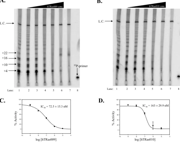
a. Concentration dependence of hTRas009 and hTRas010 inhibition... 39
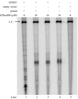
b. Direct telomerase assay in the presence of hTRas009 and hTRas010 ... 41
3. hTRas009 and hTRas010 affect the association of hTR with hTERT ... 42
4. hTRas009 affects hTR homo-dimerization... 45
C. Discussion ... 46
D. Materials and methods ... 53
1. Oligonucleotides ... 53
2. pET-28c-hTERT and phTR+HH expression plasmids ... 53
3. In vitro transcription and purification of hTR... 54
4. Synthesis of hTERT... 55
5. Reconstitution of telomerase... 55
6. Direct telomerase assay... 55
7. Assemblage assay ... 57
8. Synthesis of 32P-labeled CR4-CR5 and pseudoknot RNA fragments and full-length hTR... 57
9. Immunoprecipitation/RNA pull-down... 58
10. Inhibition studies... 59
Chapter III. RNA-binding ligands affect telomerase assemblage ... 61
A. Introduction... 61
B. Results... 63
1. Pre- and post-assemblage assays ... 63
2. Binding studies of Hoechst 33258 to hTR... 65
C. Discussion ... 66
D. Materials and methods ... 69
1. Chemical reagents... 70
2. T. thermophila telomerase assays ... 70
3. E. aediculatus telomerase assays ... 70
4. Fluorescence spectroscopy... 70
5. Calculation of dissociation constant ... 71
Chapter IV. The natural product tanshinone II-A and novel derivatives inhibit telomerase assemblage ... 72
A. Introduction... 72
B. Results... 74
1. Tanshinone II-A is a potent and specific inhibitor of telomerase assemblage... 74
2. Tanshinone II-A inhibition of various nucleic acid polymerases... 76
3. The tanshinone quinone moiety is essential for inhibition ... 76
4. Tanshinone II-A inhibits telomerase in cultured cells and is selectively cytotoxic towards telomerase positive cells... 77
C. Discussion ... 78
1. Chemical reagents... 82
2. Cells and cell culture... 82
3. Telomerase activity of cell extracts ... 83
4. Cytotoxicity assay... 84
5. Testing the effects of tanshinone II-A on T7 RNA polymerase, M-MLV reverse transcriptase and the Klenow fragment of DNA polymerase I ... 84
6. Natural product isolation and derivative synthesis ... 85
Chapter V. Hsp90 is required to maintain telomerase in an active conformation... 86
A. Introduction... 86
B. Results... 88
1. Hsp90 inhibitors affect in vitro reconstituted human telomerase activity both before and after assembly... 88
2. Telomerase maintains hTR-hTERT interactions in the presence of geldanamycin and novobiocin after hTERT translation... 90
3. Primer binding overcomes GA, but not NB, inhibition of human telomerase ... 93
4. Hsp90 is associated with hTERT after telomeric primer binding ... 95
5. Hsp90 inhibition alters telomerase holoenzyme structure ... 96
6. NB destabilizes hTERT ... 98
7. Hsp90 is associated with hTERT following geldanamycin or novobiocin treatment ... 99
C. Discussion ... 100
1. Antibodies and chemical reagents ... 106
2. In vitro transcription and purification of hTR... 106
3. Synthesis of [32P]-labeled pseudoknot and CR4-CR5 RNA fragments and full-length hTR... 106
4. Synthesis of biotinylated pseudoknot or CR4-CR5 RNA fragment ... 107
5. Inhibition studies... 107
6. Association of hTR and hTERT in the presence of GA and NB ... 108
7. Association of hTR and hTERT when GA or NB was present during translation of hTERT... 108
8. Immunoprecipitation of telomerase using an Hsp90 antibody ... 110
9. Lys-C proteolysis of hTERT... 111
10. Pulse-chase... 111
Chapter VI. Development of a high-throughput screen capable of identifying novel inhibitors of a specific hTERT/hTR interaction ... 113
A. Introduction... 113
B. Results... 116
1. Biotin-labeled CR4-CR5 domain binds hTERT ... 116
2. A Scintillation Proximity Assay for hTERT-hTR interactions... 118
3. Validation of SPA ... 119
4. Optimization of SPA... 120
C. Discussion ... 122
D. Materials and methods ... 127
2. Biochemical assay for the interaction between hTERT and
the CR4-CR5 domain of hTR ... 128
3. Scintillation Proximity Assay ... 128
4. Inhibition studies... 130
5. Z-factor determination ... 130
Chapter VII. Conclusions and future directions of this research ... 131
Representative works ... 136
LIST OF TABLES
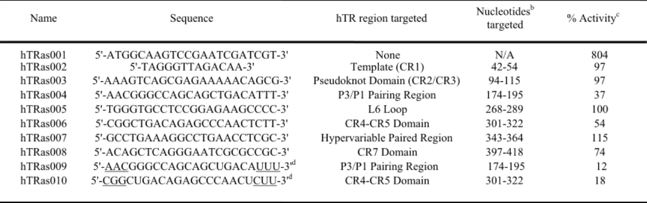
Table 2.1 Summary of inhibition data with hTR-targeted
oligonucleotides ... 37 Table 3.1 Percent inhibition of telomerase by selected nucleic acid-binding
LIST OF FIGURES
Figure 1.1 Mechanism of telomerase-mediated telomere extension ... 2
Figure 1.2 Telomere length versus time for different cell types... 3
Figure 1.3 Human telomerase RNA... 5
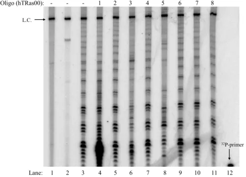
Figure 2.1 Assemblage assay using hTR-targeted oligonucleotides ... 38
Figure 2.2 Concentration dependence of telomerase inhibition by hTRas009 and hTRas010 when added before assemblage ... 39
Figure 2.3 High concentrations of hTRas009 in an assemblage assay produce telomerase-independent artifacts ... 40
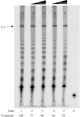
Figure 2.4 Concentration dependence of telomerase inhibition by hTRas009 and hTRas010 when added after assemblage ... 41
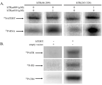
Figure 2.5 Inhibition of hTR/hTERT interactions by hTRas009 and hTRas010 ... 43
Figure 2.6 Oligonucleotide and RNA specificity ... 44
Figure 2.7 hTRas009 inhibits hTR homo-dimerization ... 45
Figure 2.8 hTRas009 and hTR010 prevent proper telomerase assemblage by blocking essential interactions between hTR and hTERT... 52
Figure 3.1 Compounds tested as telomerase inhibitors ... 62
Figure 3.2 RNA binding ligand-induced inhibition of telomerase pre- and post-assemblage ... 64
Figure 3.3 Hoechst 33258 binds to hTR but not to other nucleic acid components present in the telomerase reaction ... 66
Figure 4.1 Effectors of telomerase assemblage ... 72
Figure 4.3 Inhibition of telomerase by tanshinone II-A ... 75
Figure 4.4 Effects of tanshinone II-A on different nucleic acid
processing enzymes ... 76
Figure 4.5 Consequences of treating immortalized cells with
tanshinone II-A ... 78
Figure 5.1 GA and NB inhibit telomerase when added before
or after assembly... 89 Figure 5.2 p23 has a slight rescue effect on GA-induced telomerase
inhibition... 90 Figure 5.3 hTR/hTERT interactions are unaffected by the presence
of GA and NB after translation of hTERT, but hTR/hTERT interactions are effected by the presence of GA and NB
during translation ... 92
Figure 5.4 Pre-Incubation with primer rescues GA-induced but not
NB-induced telomerase inhibition ... 94
Figure 5.5 Hsp90 remains associated with hTERT after primer loading ... 95
Figure 5.6 NB, but not GA, increases the proteolysis rate of hTERT ... 97
Figure 5.7 hTERT stability, but not RNA/protein complex half-life,
is affected by NB ... 98
Figure 5.8 Hsp90/hTERT interactions are unaffected by the presence
of GA and NB after hTERT translation... 99 Figure 5.9 Conceptual model for Hsp90-mediated human telomerase
maturation and the effects of GA and NB ... 103 Figure 6.1 Structure and hTERT-binding inhibition of the CR4-CR5
Figure 6.2 Assay schematics for scintillation proximity assay... 118
Figure 6.3 Concentration dependence of hTRas010 in an affinity
purification and a scintillation proximity assay ... 119
Figure 6.4 Effects of hTRas010 and hTRas010MM on the interaction
between hTERT and the CR4-CR5 fragment of hTR... 121
Figure 6.5 Precision of scintillation proximity assay for assembly of
LIST OF ABBREVIATIONS
17-AAG 17-allylamino-17-demethoxygeldanamycin 2’-MOE 2’-O-(2-methoxyethyl)
5-FAM, SE 5-carboxyfluorescein, succinimidyl ester
5-TAMRA, SE 5-carboxytetramethylrhodamine, succinimidyl ester ALT Alternative Lengthening of Telomeres
AZT 3′-azido-3′-deoxythymidine
BIBR1532 2-[(E)-3-naphtalen-2-yl-but-2-enoylamino]-benzoic acid BRACO-19 9-[4-(N,N
-dimethylamino)phenylamino]-3,6-bis(3-pyrrolodinopropionamido) acridine
DAPI 4’,6-diamidino-2-phenylindole DHBV Duck Hepatitis B Virus
GA Geldanamycin
HDAC Histone deacetylase
Hsp70 Heat shock protein-70
Hsp90 Heat shock protein-90
hTERT human Telomerase Reverse Transcriptase
hTR human Telomerase RNA
HTS High-throughput screen
NB Novobiocin NPS N3'→ P5' thio-phosphoramidate
PKC Protein Kinase C
RNP Ribonucleoprotein RRL Rabbit Reticulocyte Lysate
RT Reverse Transcriptase
SPA Scintillation Proximity Assay Tan II-A Tanshinone II-A
TRAP Telomeric Repeat Amplification Protocol
TUNEL Terminal deoxynucleotidyl transferase (TdT)-mediated dUTP Nick End Labeling
Chapter I. Introduction
A. Telomerase
1. Significance and background
Cancer is a growing health problem for people all over the world. Most cancer
research focuses on curing or subduing a specific type of cancer (e.g. breast, prostate, lung,
colon, etc.). Recently, though, a large proportion of cancer cell types were found to share a
common trait as approximately 90% of all human tumors express telomerase activity (Cong
et al., 2002). Telomerase, or telomere terminal transferase, is a ribonucleoprotein complex
responsible for the maintenance of telomeric DNA. Telomeres are DNA-protein complexes,
which posses a 3' single-stranded overhang of short, repeated, guanosine-rich sequences
(5'-TTAGGG for humans). Human telomerase is composed minimally of a protein (hTERT)
and an RNA (hTR) subunit (Feng et al., 1995; Meyerson et al., 1997; Nakamura et al., 1997).
The RNA subunit is used as a template for the G-rich sequence addition while the protein
subunit employs its reverse transcriptase activity to extend telomeric DNA (Figure 1.1).
Telomerase activity is extinguished in most human tissues during embryonic
development (Wright et al., 1996b), however, certain cells in the human body express
telomerase activity through all stages of life such as germline cells and some stem cells
(Cong et al., 2002). Most normal healthy human somatic cells, on the other hand, are
deficient in telomerase activity. The telomeres of these somatic cells therefore undergo
(Olovnikov, 1971; Watson, 1972). When a cell's telomeres get too short, the cell will either
senesce or enter crisis and die. The majority of cancerous cells, however, can escape crisis
5’- GGTTAGGGTTAGGGTTAG
3’- CCAAT UCCCAAUC
3’ 5’
5’- GGTTAGGGTTAGGGTTAGGGTTAG
3’- CCAAT UCCCAAUC
3’ 5’
5’- GGTTAGGGTTAGGGTTAGGGTTAG
3’- CCAAT UCCCAAUC
3’ 5’
5’- GGTTAGGGTTAGGGTTAGGGTTAGGGTTAG
3’- CCAAT UCCCAAUC
3’ 5’
Telomere with single-stranded 3’ overhang
Telomerase RNA template Telomerase Reverse
Transcriptase
Telomere Extension
Translocation
Telomere Extension
Figure 1.1 Mechanism of telomerase-mediated telomere extension. The telomerase reverse transcriptase (hTERT) and the telomerase RNA (hTR) work together to extend the 3’ end of the telomere. Nucleotides added to the telomere by telomerase are bold. This figure was adapted from White et al., 2001.
by up-regulating telomerase expression (White et al., 2001) (Figure 1.2). As a result,
telomerase promotes tumorigenicity in part by helping maintain telomeric DNA length.
Importantly, inhibiting telomerase activity in cancer cells causes telomere shortening and
cessation of cell growth (Hahn et al., 1999). The successful exploitation of this common trait
could therefore lead to the development of a universal anticancer agent based on inhibiting
Telomerase Inhibition
Time/cell divisions
T
el
om
er
e
le
n
gt
h
Germline Cells Stem Cells
Normal Somatic Cells
Senescence
Crisis
Telomere Stabilization
Telomerase Activation
Crisis
Figure 1.2 Telomere length versus time for different cell types. Germline cells constitutively express telomerase activity and thus their telomeres do not decrease in length over time. Most normal somatic cells do not express telomerase and thus their telomeres decrease in length over time. The telomeres of certain stem cells also decrease in length over time, but at a slower rate than that of most normal somatic cells because they express low levels of telomerase activity. Though the administration of a telomerase inhibitor to the human body would affect all telomerase-positive cells, the cancerous cells would die off first before the healthy cell's telomeres get critically short. This figure was adapted from White et al., 2001.
2. The catalytic subunit of human telomerase, hTERT (human Telomerase Reverse Transcriptase
The hTERT gene, discovered in 1997 (Harrington et al., 1997; Kilian et al., 1997;
Meyerson et al., 1997; Nakamura et al., 1997), is found on the short arm of chromosome 5
(5p15.33). The hTERT protein is 1132 amino acids in length and contains a number of
distinguishable features including reverse transcriptase motifs within the C-terminal half of
the gene, a conserved telomerase-specific region (T-motif) located just 5' of the reverse
transcriptase motifs, and a large N-terminal region (Cong et al., 2002; Harrington, 2003).
The telomerase catalytic subunits from different organisms represent a distinct subgroup
are phylogenetically conserved with other reverse transcriptases, but are more closely related
within their own subgroup than with other members of the reverse transcriptase family
(Eickbush, 1997; Nakamura et al., 1997; Nakamura and Cech, 1998). The T-motif has been
found to be required for hTR binding in vitro (Bryan et al., 2000) and is also thought to be
involved in the recruitment of hTERT to the nucleolus, which has been proposed to be an
important step in telomerase biogenesis (Etheridge et al., 2002; Bosoy et al., 2003). The
large N-terminal region contains conserved residues that are functionally important for hTR
binding, telomerase complex assemblage, and catalysis, as well as a number of other
functions (Cong et al., 2002; Harrington, 2003).
Importantly, hTERT expression usually correlates closely with telomerase activity,
cancer initiation, progression, and metastasis (Cong et al., 2002). In fact, while hTR and
other required subunits of the telomerase holoenzyme are ubiquitously expressed in most
cells, many independent studies have shown that hTERT is the limiting determinant of
telomerase activity (Meyerson et al., 1997; Weinrich et al., 1997; Ito et al., 1998; Kanaya et
al., 1998; Nakayama, J. et al., 1998; Takakura et al., 1998). Furthermore, elevated
telomerase levels oftentimes directly correlate with poor tumor prognosis and vice versa,
though this is not the case for all cancer cell types (Granger et al., 2002).
The hTERT gene contains 16 exons and 15 introns and can be differentially spliced
(Kilian et al., 1997) to generate both the full-length protein as well as multiple splice
variants, however, only the full-length transcript has been found to correlate with telomerase
activity (Ulaner et al., 1998; Ulaner et al., 2000). Although the expression of the variants is
deliberate and may serve a dominant negative function, the role of the alternately spliced
3. The RNA subunit of human telomerase, hTR (human Telomerase RNA)
hTR was the first core component of human telomerase to be cloned (Blasco et al.,
1995; Feng et al., 1995). Unlike hTERT, hTR is typically expressed ubiquitously in most
tissues, even those lacking telomerase activity (Blasco et al., 1995; Avilion et al., 1996;
Harrington, 2003). The mature human transcript is 451 nucleotides long and is transcribed
by RNA Polymerase II (Feng et al., 1995).
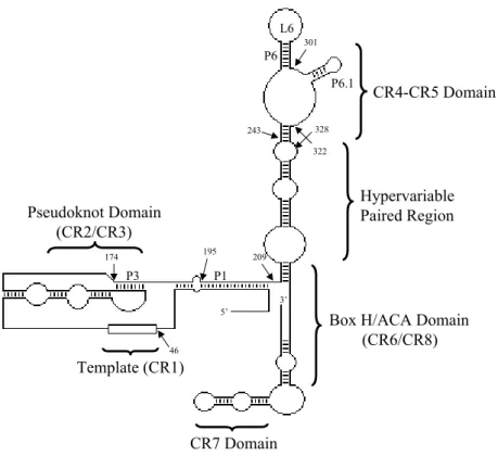
Extensive comparative phylogenic analysis of the vertebrate telomerase RNA allowed
a proposed secondary structure of hTR (Chen, J. L. et al., 2000). Vertebrate telomerase
RNAs contain several conserved regions including the template-containing pseudoknot
domain and the CR4-CR5 domain (Figure 1.3). Previous studies have shown that the
P6
P6.1
CR4-CR5 Domain
Hypervariable Paired Region
L6
Pseudoknot Domain (CR2/CR3)
P3
Template (CR1)
Box H/ACA Domain (CR6/CR8)
CR7 Domain
P1
5’
3’
46
209
243 328
301
322
174 195
pseudoknot and CR4-CR5 domains are essential for telomerase activity and interact
separately with hTERT (Tesmer et al., 1999; Beattie et al., 2000; Mitchell and Collins, 2000;
Bachand and Autexier, 2001; Martin-Rivera and Blasco, 2001; Chen, J. L. et al., 2002b;
Chen, J. L. and Greider, 2003). In addition, Chen et al. revealed the presence of an additional
secondary structure within the CR4-CR5 domain of hTR, the P6.1 stem-loop (nucleotides
302-314), which appears to be essential for telomerase activity as well as for binding to
hTERT (Chen, J. L. et al., 2002b). The roles of hTR were further extended by the work of
Ly et al. (Ly et al., 2003) and Ren et al. (Ren et al., 2003). Ly et al. showed that hTR can
homo-dimerize via the P3 pairing region to form a trans-pseudoknot and that mutations
preventing the P3-trans interaction led to loss of enzymatic activity but did not appear to
prevent binding of hTR to hTERT (Ly et al., 2003). Similarly, Ren et al. propose that hTR
can homo-dimerize via the internal loop J7b/8a within the CR7 domain to form a "kissing
complex," which may be functionally important for hTR accumulation and telomerase
holoenzyme assemblage in vivo (Ren et al., 2003). Theimer et al., however, reported
evidence contradictory to the previously proposed hTR dimerization models (Theimer et al.,
2005). Using NMR spectroscopy to solve the solution structure of a fragment of the human
telomerase RNA pseudoknot they showed that telomerase activity actually correlates with the
presence of a conserved pseudoknot tertiary structure and not with pseudoknot-mediated hTR
dimerization (Theimer et al., 2005).
4. Telomere structure and function
The human telomere ranges in length from 5-15 kilobases (White et al., 2001) of the
overhang (McElligott and Wellinger, 1997; Wright et al., 1997). Telomeric DNA, which is
not replicated efficiently by the normal DNA replication machinery, is lost at a rate of 50-100
base pairs per cell cycle in cells with limited telomerase activity. With the help of various
specialized telomere-binding proteins, such as TRF1, TRF2 and Pot1, the single-stranded
portion of the telomere can be inserted into the double-stranded portion to form what is
called a t-loop (Griffith, J. D. et al., 1999). The telomeric repeat binding factors, TRF1 and
TRF2 (van Steensel and de Lange, 1997; van Steensel et al., 1998; Griffith, J. D. et al., 1999;
Smogorzewska et al., 2000; Stansel et al., 2001), bind to the double-stranded region of the
telomere while Pot1 (protection of telomeres) (Baumann and Cech, 2001; Colgin et al., 2003;
Loayza and De Lange, 2003) binds to the single-stranded region and plays a critical role in
both t-loop integrity and regulating telomere length. The t-loop therefore appears to function
as a protective "cap" to guard the telomere from degradation, chromosome end-to-end
fusions and mistaken DNA repair (Griffith, J. D. et al., 1999; Cong et al., 2002). Though
telomere shortening has been found to induce cellular senescence, it is actually the disruption
in the telomeric capping structure that alters the cell's replicative potential (Karlseder et al.,
2002). In other words, when a cell's telomeres get too short the t-loop cannot form and a
signal triggers the cell to enter senescence. Therefore, telomere capping is extremely
important for cell viability as loss of capping can lead to genetic instability and cell death
(Rhodes et al., 2002).
5. Telomerase-associated proteins
Though human telomerase is minimally composed of hTR and hTERT, there are
other proteins that associate with the holoenzyme and are required for in vivo telomerase
multi-subunit holoenzyme containing hTERT, hTR, Hsp90, p23, Hsp70, p60 and Hsp40/ydj,
Ku, TEP1 and dyskerin, as well as other telomerase-associated proteins (Forsythe et al.,
2001; Cong et al., 2002; Harrington, 2003). The addition of these and other factors to in
vitro experiments, however, is not necessary when expressing hTERT in reticulocyte lysate
as the reticulocyte lysate already contains these factors (Holt et al., 1999; Cong et al., 2002).
a. Hsp90
The molecular chaperone Hsp90, which directly associates with hTERT, has a
demonstrable role in establishing telomerase activity both in vitro and in vivo, and previous
reports indicate that Hsp90 is required for the reconstitution of telomerase activity from
recombinant hTERT and hTR (Holt et al., 1999). Hsp90, perhaps the most abundant
housekeeping protein, is believed to account for almost 2% of all cytosolic proteins. The
human chaperone exists as two isoforms, Hsp90α and Hsp90β, which share 85% identity,
however, no functional differences have been identified (Hickey et al., 1986). The main
function of Hsp90 is to stabilize its clients in denaturing environments and times of stress,
however, the unwavering involvement of this heat shock protein in membrane translocation,
turnover, folding and activation of numerous targets reveals a much larger responsibility.
Hsp90 is highly conserved in both prokaryotes and eukaryotes, and its client list includes
proteins involved in signal transduction (Richter and Buchner, 2001; Pratt and Toft, 2003),
steroid receptors (Cheung and Smith, 2000; Kimmins and MacRae, 2000; Smith, 2000;
Bledsoe et al., 2002), kinases (Rose et al., 1987; Matts and Hurst, 1989; Stancato et al., 1993;
Palmquist et al., 1994; Wartmann and Davis, 1994; Jaiswal et al., 1996; Sato et al., 2000;
Pratt and Toft, 2003) and reverse transcriptases (Hu and Seeger, 1996; Holt et al., 1999), as
i. Structure and function
The Hsp90 protein consists of three main domains; the N-terminus, the C-terminus
and a central charged region. The N-terminal of Hsp90 is the most conserved and therefore
the most studied domain in the chaperone (Prodromou et al., 1997). Though the C-terminal
portion of Hsp90 is less well understood, it has been shown to contain a dimerization domain
that is required to achieve an active functional homodimer (Iannotti et al., 1988; Minami et
al., 1991). Both the N-terminal (Grenert et al., 1997; Prodromou et al., 1997; Stebbins et al.,
1997) and C-terminal (Marcu et al., 2000a; Garnier et al., 2002; Soti et al., 2002) contain
ATP-binding pockets. The central charged region acts as a flexible linker and is believed to
mediate cross-talk between the two termini (Scheibel et al., 1999; Marcu et al., 2000a).
There is also some evidence that this middle segment regulates access of nucleotides to the
N- and C-termini by interacting with the γ-phosphate of ATP (Marcu et al., 2000a; Soti et al.,
2002; Meyer et al., 2003).
The N-terminal pocket shares sequence homology with MutL proteins, histidine
kinase and bacterial gyrase B, an ATP-dependent DNA topoisomerase (Dutta and Inouye,
2000). Hydrolyzable ATP must be able to bind to this domain in order to convert Hsp90 into
its active conformation capable of binding client proteins and co-chaperones (Grenert et al.,
1997; Prodromou et al., 1997; Stebbins et al., 1997). In fact, binding of a non-hydrolyzable
ATP analog to Hsp90 or chaperone mutations resulting in the loss of ATP binding
capabilities eradicates Hsp90 function both in vitro and in vivo (Obermann et al., 1998;
Panaretou et al., 1998; Grenert et al., 1999). When the N-terminal ATP binding site is
unoccupied, the chaperone is “open” and able to bind clients. As ATP binds, the chaperone
2000; Richter et al., 2002). In fact, it is believed that the two N-termini of the homodimer
interact transiently upon ATP binding, and that this interaction is required for ATP
hydrolysis (Maruya et al., 1999; Prodromou et al., 2000). Furthermore, additional evidence
suggests that the capturing of clients is also dependent on the participation of
Hsp90-associated co-chaperones (Wegele et al., 2004; Young et al., 2004).
The C-terminal of Hsp90 is responsible for holding the homodimer in an antiparallel
arrangement (Maruya et al., 1999). The dimer formation of this chaperone is required for
functionality as C-terminal truncations have been shown to prevent ATP hydrolysis
(Prodromou et al., 2000). This terminus, like the N-terminal, is capable of binding ATP.
Some data suggests that the C-terminal binding site only becomes available for nucleotide
binding once the N-terminal ATP binding site is occupied (Chiosis et al., 2004). Because
Hsp90 exists as a dimer and both termini are capable of binding substrate, it is possible that
the functional homodimer may be capable of binding four substrates simultaneously (Pratt
and Toft, 2003).
ii. Hsp90-associated proteins
Although Hsp90 is typically the major player in chaperoning cellular activities it does
not act alone, but rather functions in larger multi-protein complexes with accessory proteins
and other chaperones. One of the most important partners of Hsp90 is the acidic
phosphoprotein p23 (Johnson and Toft, 1994). This 23-kDa protein is known to bind to the
N-terminal domain of ATP-bound Hsp90 (Sullivan et al., 1997; Fang et al., 1998; Chadli et
al., 2000). Studies have also provided evidence that additional p23 binding sites on Hsp90
exist outside of the N-terminus though the precise locations have yet to be identified (Grenert
active telomerase from recombinant sources (Holt et al., 1999). The principal function of
p23 is to suppress Hsp90’s ATPase activity and thus aid Hsp90 in binding and holding onto
its clients by maintaining the chaperone in a closed state (McLaughlin et al., 2002; Panaretou
et al., 2002). The presence of p23, however, is not essential for functional assembly of
Hsp90 complexes (Johnson and Toft, 1994; Bohen, 1998).
Hsp70, another cellular chaperone involved in protein folding and preventing protein
aggregation, is another Hsp90-associated protein. This heat shock protein binds to the
C-terminus of Hsp90 (Young et al., 1998; Carrello et al., 1999; Marcu et al., 2000a; Murphy, P.
J. et al., 2001) and is necessary to run the ATP hydrolysis cycles of Hsp90 (Wegele et al.,
2004; Young et al., 2004). In fact, both Hsp90 and Hsp70 are known to work in conjunction
in order to prepare certain clients, such as steroid receptors, for ligand binding. It is believed
that, in the case of steroid receptors, Hsp70 first binds to the client in an ATP-dependent step
to “prime” the receptor, followed by transfer of the client to Hsp90 in a second
ATP-dependent step, which fully activates the receptor (Morishima et al., 2001; Hernandez et al.,
2002; Kanelakis et al., 2002). Furthermore, Hsp70 is released from the Hsp90 multi-protein
chaperone complex during the assembly of steroid receptors (Smith, 1993). Similarly, in the
case of human telomerase, Hsp70 is only transiently associated while Hsp90 and p23 are
stably associated with the holoenzyme (Forsythe et al., 2001). As for p23, the precise
binding site(s) for Hsp70 on Hsp90 have not been mapped out, however, recent studies are
beginning to shed some light on this question (Young et al., 1997; Scheibel et al., 1998).
Evidence reveals that both Hsp70 and Hsp90 are essential components of the active
chaperone complex while p23 is non-essential, though the presence of p23 has the potential
proteins that are involved in client maturation, however, they will not be discussed in detail
here.
iii. Hsp90 and cancer
Over-expression of Hsp90 and other heat shock proteins have been found to lead to
poor prognosis in a variety of cancers (Jameel et al., 1992; Ciocca et al., 1993; Yano et al.,
1996; Conroy et al., 1998). Targeting Hsp90 as an anticancer approach has therefore
received much attention (Beliakoff and Whitesell, 2004; Chiosis et al., 2004; Workman,
2004). Because Hsp90 is ubiquitously expressed in all cell types, normal and cancerous, it is
also the functions of Hsp90 and not only its presence that defines its involvement in cancer
(Bagatell and Whitesell, 2004). Hsp90 has been shown to stabilize mutated proteins found in
cancer cells which contribute to apoptotic pathways. For example, this heat shock protein
chaperones Akt (Sato et al., 2000; Basso et al., 2002), NF-κB (Chen, G. et al., 2002a),
survivin (Fortugno et al., 2003), Raf-MAPK (Schulte et al., 1995), Apaf-1 (Pandey et al.,
2000), IKKα/β (Lewis et al., 2000), TNF-α (Zhao and Wang, 2004) and many other clients
to maintain transformed cells in an immortalized state. By buffering these mutations, Hsp90
acts as an anti-apoptotic and growth promoter contributing to tumor cell survival (Chiosis et
al., 2004).
Because of Hsp90’s role in so many different cancer progression pathways, it is
hypothesized by some that inhibitors of this chaperone have the potential to induce a
wide-range of anticancer effects (Vilenchik et al., 2004). The use of Hsp90 inhibitors as a means
of cancer therapy, however, has revealed indiscriminate toxicity for normal and malignant
cells as the chaperone is present in all cell types and is involved in so many cellular pathways
with the fact that transformed cells express higher levels of Hsp90 than normal cells, has led
to the identification of new drugs that are selective for cancer cells and display little
target-associated toxicity (Schulte and Neckers, 1998). It is also believed that cancer cells
exercising Hsp90-mediated mutant protein stabilization may be more sensitive to toxicity
than normal cells (Chiosis et al., 2004). Furthermore, it was determined that some Hsp90
inhibitors exhibit dose limiting toxicity (Goetz et al., 2003). Inhibiting Hsp90 has been
found to not only prevent stabilization, but also initiate ubiquitination of its clients,
eventually leading to growth arrest (Xu, W. et al., 2002; McDonough and Patterson, 2003).
Additionally, the signal transduction inhibitor hypericin causes ubiquitination of Hsp90
leading to the release and proteasome-independent degradation of clients such as mutant p53
(Blank et al., 2003).
iv. Hsp90 inhibition
Functional activation of Hsp90 as well as substrate and client binding are not
permanent but require multiple ATP hydrolysis cycles to maintain. Therefore, targeting the
ATP binding sites of Hsp90 is a widely accepted anticancer approach.
One class of compounds known to bind the N-terminus of Hsp90 is benzoquinone
ansamycin antibiotics, including geldanamycin (GA) and herbimycin (Whitesell et al., 1994;
Stebbins et al., 1997; Neckers et al., 1999; Roe et al., 1999). The most studied of these
compounds is GA. Described as an ATP/ADP mimetic (Prodromou et al., 1997) or a
competitive inhibitor of client-protein binding, GA interacts with the N-terminal ATP
binding site, thus displacing the nucleotide (Grenert et al., 1997). This release of ATP
disrupts the Hsp90 chaperone complexes on steroid hormones and results in the
1995). GA binding to Hsp90 also causes the release of p23 from the chaperone (Prodromou
et al., 1997). This is believed to occur because GA seizes Hsp90 in its ADP-dependent state,
which is incapable of binding p23 (Johnson and Toft, 1995; Grenert et al., 1997; Sullivan et
al., 1997). In human telomerase, the same effect is seen as the Hsp90 interaction, but not the
p23 interaction, is maintained with hTERT in the presence of GA (Holt et al., 1999). GA has
also been shown to promote degradation of the heat shock protein’s clientele prior to their
complete activation by preventing Hsp90-client dissociation (Schneider et al., 1996; Schulte
et al., 1997). Specifically, GA treatment of H1299 cells resulted in the perturbation of Hsp90
and the ubiquitination and degradation of hTERT in a proteasome-dependent fashion (Kim, J.
H. et al., 2005). Besides being used to elucidate the roles of Hsp90, GA has also been tested
as a potential therapeutic drug. In certain cell lines this ansamycin causes growth arrest,
differentiation and apoptosis (Hostein et al., 2001; Munster et al., 2001). However, despite
being a potent inhibitor of cellular Hsp90, clinical testing of GA and related compound has
revealed a significant toxicity profile (Supko et al., 1995). Therefore, in terms of cancer
therapy, new compounds had to be developed. One such compound is the GA derivative
17-allylamino-17-demethoxygeldanamycin (17-AAG) (Schulte and Neckers, 1998). 17-AAG,
now in phase II of FDA clinical trials, exhibits potent anti-Hsp90 activity at sub-toxic doses
in various animal models (Kelland et al., 1999; Solit et al., 2002).
Aside from GA, another commonly used Hsp90 inhibitor employed to elucidate the
cellular functions of Hsp90 is novobiocin (NB). NB is a coumarin-type antibiotic and a
known inhibitor of DNA gyrase B. As opposed to GA, NB binds to the C-terminus of Hsp90
(Marcu et al., 2000a; Marcu et al., 2000b; Soti et al., 2002). Upon NB treatment, Hsp90
and p23 (Young et al., 1998; Carrello et al., 1999; Marcu et al., 2000a). This fact had led to
the hypothesis that NB may therefore provoke additional deleterious effects on Hsp90
function as compared to GA (Marcu et al., 2000a), which only induces the release of p23
from Hsp90 (Prodromou et al., 1997), though this premise has yet to be substantiated.
Studies have also shown that NB binding to the carboxy terminus of Hsp90 prevents both
nucleotide binding (Pratt and Toft, 2003) and GA binding (Marcu et al., 2000b) to the amino
terminus, providing evidence for collaboration between the two termini. In cells, NB
undermines a number of Hsp90-associated client proteins including Her2, Raf-1, v-src and
mutant p53 and downregulates multiple downstream targets, though it binds to Hsp90 with
poor affinity (Marcu et al., 2000a; Marcu et al., 2000b).
6. The secondary role of telomerase in tumorigenesis
As previously discussed, telomerase aids in tumorigenesis by extending telomeric
DNA and thus maintaining telomeric integrity (Masutomi and Hahn, 2003). Interestingly,
however, recent evidence suggests that telomerase is involved in a secondary, anti-apoptotic
role in tumorigenesis (Cao et al., 2002; Stewart et al., 2002). Cao et al. demonstrated that
breast cancer cells undergoing apoptosis could be rescued by the expression of a mutant
hTERT that lacked enzymatic activity and thus did not have the ability to elongate telomeres
(Cao et al., 2002). The work by Stewart et al. also suggests that telomerase plays at least two
roles in tumorigenesis (Stewart et al., 2002). Their research focused on an alternative
mechanism of telomere maintenance (ALT – Alternative Lengthening of Telomeres) and its
ability to substitute for telomerase expression. The ALT pathway, which is found in 7-10%
of human cancers, elongates telomeres by inter-chromosomal recombination (Stewart et al.,
cell transformation assay (Stewart et al., 2002). Together, these independent reports suggest
that telomerase has a secondary, anti-apoptotic role in tumorigenesis separate from its
telomere-elongating activity (Saretzki, 2003). One proposed explanation for these results is
that telomerase is involved in telomere capping (Blackburn, E., 1999; Blackburn, E. H.,
2000; Blackburn, E. H., 2001; Masutomi et al., 2003). Therefore, the mere presence of
telomerase, whether it is active not, may elicit an anti-apoptotic effect as it interacts with and
helps to maintain functional telomeres (Saretzki, 2003). We predict that inducing telomerase
misassemblage will alter these other roles of telomerase in cancer biology.
7. Regulation of telomerase
Telomerase regulation is a multi-faceted process taking place at the levels of
transcription, hTERT maturation, subcellular localization and telomeric accessibility.
Because hTERT expression is the rate-limiting factor in determining enzymatic activity, its
regulation is of particular importance. In most normal cells hTERT is transcriptionally
repressed. During immortalization, the gene is activated and upregulated allowing for
unlimited cell proliferation.
a. Transcriptional regulation
i. Positive regulators of hTERT transcription
Transcriptional regulation of hTERT at its promoter is thought to be a major
determinant in protein expression (Meyerson et al., 1997; Nakamura et al., 1997). In fact,
hTERT transient transfection with a promoter-luciferase reporter reveals that the hTERT
promoter is active in immortalized cells, but inactive in normal cells (Cong et al., 1999;
have both positive and negative regulatory connotations (Cong et al., 1999). One such
binding site is that for Myc/Mad, which contains an E-box and is a transcriptional target of
the oncogene c-myc (Wang, J. et al., 1998; Greenberg et al., 1999; Wu et al., 1999). c-Myc
has been linked to a number of different human cancers as it promotes proliferation, growth
and apoptosis (DePinho et al., 1991; Grandori et al., 2000). In normal human mammary
epithelial cells and primary fibroblasts, as well as other cell types, c-myc induction has been
shown to correlate with hTERT expression (Wang, J. et al., 1998). Conversely, c-myc and
hTERT are typically downregulated in non-proliferative cells. Furthermore, over-expression
of the c-Myc antagonist Mad1 results in the down-regulation of hTERT (Gunes et al., 2000;
Oh, S. et al., 2000). Although c-myc expression is an important aspect of hTERT regulation,
additional transcription factors are usually required to account for the full transforming
activity of c-Myc.
The general transcription factor Sp1 is also involved in hTERT up-regulation. It does
so by binding GC-boxes within the core of the hTERT promoter (Kyo et al., 2000). In fact,
these Sp1 binding sites are necessary components of the hTERT promoter as evidenced by
the fact that mutations in the GC-boxes eradicate promoter activity (Cong and Bacchetti,
2000; Kyo et al., 2000). The reason for this requirement is because the hTERT promoter
contains no TATA box. Sp1 therefore helps to initiate transcription of the hTERT promoter,
as well as other TATA-less promoters, by binding general transcription machinery such as
the TATA-box binding protein (TBP) and associated factors (Pugh and Tjian, 1991; Hoey et
al., 1993; Emili et al., 1994). Sp1 has also been found to work in concert with c-myc in order
One factor known to activate hTERT transcription independent of c-myc induction is
the human papillomavirus 16 E6 protein (Gewin and Galloway, 2001; Oh, S. T. et al., 2001;
Veldman et al., 2001). The protein’s oncogenic variants have been found to induce
telomerase activity in primary human keratinocytes and mammary epithelial cells
(Klingelhutz et al., 1996).
Several sex hormones have also been linked to telomerase activity regulation. For
example, estrogen activates telomerase through direct and indirect transcriptional activation
of hTERT expression in hormone-sensitive tissues such as mammary epithelial cells that
express the estrogen receptor (Kyo et al., 1999; Misiti et al., 2000). Human ovary epithelial
cells also show hTERT mRNA induction in the presence of the hormone, however, this
transcriptional activation is dependent on the -950 estrogen response element of estrogen
receptor α and not estrogen receptor β (Misiti et al., 2000). The hTERT promoter can also
be activated in MCF-7 breast cancer cells. Estrogen in these cells has been found to have an
indirect effect on hTERT activation via c-myc induction (Kyo et al., 1999). Furthermore, the
estrogen-specific initiation of hTERT transcription has also been confirmed by demonstrating
that the anti-estrogen drug tamoxifen reduces telomerase activity in certain cell lines (Aldous
et al., 1999; Nakayama, Y. et al., 2000). Progesterone, another sex hormone, also has an
antagonistic effect on estrogen-induced hTERT expression (Wang, Z. et al., 2000). Though
the mechanism of progesterone-mediated hTERT regulation is complex and still widely
unknown, evidence suggests that progesterone targets the hTERT promoter and that the
reversal of estrogen-mediated hTERT activation by progesterone may be indirect (Wang, Z.
et al., 2000; Cong et al., 2002). Androgens also possess regulatory capabilities over hTERT,
fully elucidated. Androgen depletion in dependent cell lines, but not
androgen-independent cell lines, has been found to reduce telomerase activity. This activity in
androgen-sensitive cells, however, can be restored by the addition of testosterone as
androgen signaling upregulates hTERT expression (Guo et al., 2003).
ii. Negative regulators of hTERT transcription
Transcriptional repression of hTERT is thought to be the main reason that most
normal somatic cells do not express telomerase activity. Transformation of cancerous cells,
therefore, is usually due to the loss of such repression. This hypothesis is supported by
results from cell fusion experiments in which normal somatic cells repress telomerase
activity in telomerase-positive immortalized cells (Wright et al., 1996a; Ishii et al., 1999).
These results suggest that normal somatic telomerase-negative cells may express
transcriptional repressors of hTERT (Shay, 1999; Cong et al., 2002).
Some normal human chromosomes contain transcriptional repressors of hTERT.
Transfer of chromosomes 3 (Oshimura and Barrett, 1997) 6 (Steenbergen et al., 2001) and 10
(Nishimoto et al., 2001) into various human telomerase-positive cancer cell lines has been
shown to downregulate telomerase activity and cause telomere shortening, suggesting the
presence of alleged telomerase repressors on these chromosomes. Aside from these as yet
undiscovered transcriptional repressors of hTERT, many transcription factors have already
been identified as negative regulators of hTERT transcription.
One negative regulator already mentioned above is Mad 1. Mad, c-Myc and Max are
transcription factors that dimerize in different combinations and bind to the E-box on the
hTERT promoter to either upregulate or downregulate hTERT expression. While Max is
Mad is upregulated in telomerase-negative normal somatic cells. The expression and
dimerization of Myc and Mad with Max therefore results in increased or decreased hTERT
transcription, respectively (Xu, D. et al., 2001).
Another negative regulator of hTERT transcription is the tumor suppressor p53. This
53-kDa protein is of central importance in terms of cellular transformation and immortality
as over 50% of all human cancers contain dysfunctional p53 (Hollstein et al., 1994; Asker et
al., 1999). p53 functions in part by inducing cell cycle arrest or apoptosis in order to keep
uncontrolled growth in check (Levine, 1997). As a transcription factor, p53 perturbs
telomerase activity by transcriptionally repressing hTERT, possibly independent of cell cycle
arrest or apoptosis (Kusumoto et al., 1999; Kanaya et al., 2000; Xu, D. et al., 2000b).
Counter intuitively, the transcription factor Sp1, an activator of hTERT transcription, is
required for p53-dependent hTERT repression (Kanaya et al., 2000; Xu, D. et al., 2000b).
Though it has been established that Sp1 binding to the hTERT promoter is impeded by p53
binding to Sp1, the precise mechanism of p53-mediated transcriptional repression of hTERT
remains a conundrum (Xu, D. et al., 2000b). Several hypotheses have been proposed
including the notion that p53 and Sp1 may impede the access of transcriptional activators
(Avantaggiati et al., 1997) or recruit repressor complexes to the hTERT promoter (Murphy,
M. et al., 1999).
p53 works closely with two other transcription factors during cell cycle regulation.
pRB (Nguyen and Crowe, 1999; Crowe and Nguyen, 2001) and E2F (Henderson et al.,
2000), when over expressed, have the potential to repress hTERT transcription in partnership
with one another and possibly even independently. Despite the fact that there is no
describes the involvement of histone deacetylase (HDAC) complexes (Harbour and Dean,
2000). Shown to be negative regulators of hTERT transcription (Cong and Bacchetti, 2000;
Takakura et al., 2001; Xu, D. et al., 2001; Hou et al., 2002), histone deacetylases may be
interacting with pRB and DNA-bound E2F at the promoter in order to stifle hTERT
transcription.
Another tumor suppressor involved in hTERT repression is Wilms’ tumor 1 (WT1).
WT1 has been found to decrease levels of hTERT mRNA in 293 kidney cells as a result of
direct interaction with the hTERT promoter (Oh, S. et al., 1999). Menin (Lin, S. Y. and
Elledge, 2003), Interferon-α (Xu, D. et al., 2000a) and TGF-β (Yang et al., 2001) are also
negative regulators of telomerase activity, and the list continues. Transcriptional control of
hTERT is undoubtedly a complex and cell cycle-dependent process involving transcription
factors, hormones, oncogenic proteins, viral proteins and tumor suppressors, as well as many
other known and unknown cell cycle regulators.
b. Posttranslational regulation
The suggestion of posttranslational hTERT modifications as a means of telomerase
regulation arose when various cells lacking telomerase activity were found to express both
hTR and hTERT mRNA (Liu et al., 1999; Tahara et al., 1999; Rohde et al., 2000; Ulaner et
al., 2000; Minamino et al., 2001). This inconsistency supposes that telomerase activity does
not always correlate with the presence of hTERT, but rather the presence of correctly
modified and active hTERT. The most prevalent modification responsible for
posttranslational regulation of a great deal of proteins including hTERT is reversible
Activators and inhibitors of various kinases have been shown to be determinant
factors in telomerase activity regulation. Protein kinase C (PKC) is a
phospholipid-dependent kinase that functions to regulate growth, differentiation and carcinogenesis. In
certain cells types, the PKC inhibitor phorbol myristate acetate upregulates telomerase
activity and the PKC inhibitor bisindolylmaeimide I downregulates telomerase activity
(Bodnar et al., 1996). Similarly, activators and inhibitors of phosphatases have the opposite
effect on hTERT modification. Phosphatase 2A has been shown to downregulate telomerase
activity in a breast cancer cell line. Conversely, okadaic acid upregulates telomerase activity
in vivo by means of phosphatase 2A inhibition (Li et al., 1997). These reports, as well as
others, demonstrate that phosphorylation of hTERT is a common and effective means of
telomerase activity regulation, though the effectiveness is PKC isoform-dependent (Ku et al.,
1997; Li et al., 1998; Yu et al., 2001).
A second kinase with significant involvement in hTERT regulation is Akt protein
kinase, or protein kinase B. It has been suggested the hTERT protein contains two distinct
phosphorylation sites, which, when phosphorylated by Akt kinase, increase telomerase’s in
vitro enzymatic activity (Kang et al., 1999). Accordingly, wortmannin downregulates
cell-type specific telomerase activity via Akt kinase inhibition (Kang et al., 1999).
The tyrosine kinase c-Abl also possesses hTERT phosphorylation potential, however,
its effects on telomerase activity oppose those of protein kinase C and Akt kinase. Via its
SH3 domain, c-Abl directly interacts with and phosphorylates hTERT resulting in a decrease
in telomerase activity (Kharbanda et al., 2000). Therefore, cells over-expressing c-Abl will
typically have low levels of telomerase activity whereas cells lacking c-Abl will show signs
Importantly, experiments studying T-lymphocyte activation revealed that the
up-regulation of telomerase activity after hTERT phosphorylation may be the result of
subcellular localization of hTERT from the cytoplasm into the nucleus (Liu et al., 2001). It
is believed that phosphorylating hTERT tags it for nuclear import to bring it into close
contact with the chromosomes and extend the telomeres.
c. Telomeric accessibility
Telomere accessibility is the salient determinant in telomerase activity regulation. If
the 3’ overhang of the chromosome is not exposed and available for telomerase-mediated
extension, then telomerase activity is not detectable. The formation of a t-loop at the end of
the telomere dictates accessibility and controls telomere homeostasis. The accessibility of
the 3’ overhang of the telomere to telomerase is therefore the last line of regulation and is
accomplished by the involvement and collaboration of a number of telomeric binding
proteins (Evans and Lundblad, 2000; de Lange, 2002).
The telomeric repeat binding factors TRF1 and TRF2 are the chief components of
telomere homeostasis (van Steensel and de Lange, 1997; Smogorzewska et al., 2000). As
mentioned above, these two proteins bind duplex telomeric DNA. The over-expression of
either factor causes telomere shortening in telomerase-positive cells as a result of preventing
telomere-telomerase interaction (Smogorzewska et al., 2000). The role of TRF1 is to
evaluate and regulate telomere length at each chromosome (van Steensel and de Lange, 1997;
Smogorzewska et al., 2000; Ancelin et al., 2002). TRF1 is also implicated in parallel pairing
of telomeric tracks as it has been shown by electron microscopy to seize and arrest telomeric
repeat arrays and preclude the unwinding of DNA (Griffith, J. et al., 1998). Furthermore, not
polymerase-mediated C-strand synthesis, thus inhibiting telomere elongation by multiple
mechanisms (Smucker and Turchi, 2001). Over-expression of a dominant negative TRF1
mutant in telomerase-positive cells therefore initiates telomere elongation as the endogenous
TRF1 is displaced from the telomere (van Steensel and de Lange, 1997; Karlseder et al.,
2002). TRF2, like TRF1, when over-expressed, results in telomere shortening
(Smogorzewska et al., 2000; Karlseder et al., 2002). Interestingly, some data suggests that
TRF2 also has the potential to initiate a telomeric DNA degradation pathway (Ancelin et al.,
2002).
TRF1 and TRF2, however, are not the only proteins involved in telomerase regulation
as Pot1 also plays a role in dictating telomeric accessibility. Pot1 regulates telomere length
by binding to single-stranded TTAGGG repeats at the chromosome ends (Baumann and
Cech, 2001), binding to TRF1 (Loayza and De Lange, 2003) and perhaps recruiting
telomerase to the telomere (Evans and Lundblad, 1999; Evans and Lundblad, 2000). Pot1
itself is actually recruited to the telomere by TRF1 (Loayza and De Lange, 2003).
Furthermore, the amount of Pot1 bound to the telomere, which depends on the length of
single-stranded DNA, is regulated by TRF1 (Loayza and De Lange, 2003). TRF1, which
binds to the double stranded region of the telomere, evaluates overall telomere length and,
through its interaction with Pot1, relays this information to the telomere terminus (Loayza
and De Lange, 2003). This collaboration therefore allows Pot1 to control
telomerase-mediated telomere elongation (Loayza and De Lange, 2003). By over-expressing Pot1 splice
variants in telomerase-positive human cell lines Colgin et al. first revealed that Pot1 acts as a
positive regulator of telomere length (Colgin et al., 2003). Lei et al. further defined the role
telomere extension in vitro (Lei et al., 2005). Depending on the number of nucleotides that
are free from Pot1 binding on the 3’ overhang, telomere accessibility, and therefore
telomerase activity, will either be promoted or hindered (Lei et al., 2005). Additionally,
Loayza et al. have shown that Pot1(∆OB), a Pot1 mutant that is deficient in single-stranded
DNA binding but still able to be recruited to the telomere by TRF1, perturbs TRF1-mediated
telomere length regulation and induces telomere elongation in human HTC75 cells (Loayza
and De Lange, 2003). These results are expected as Pot1(∆OB) is unable to cap the telomere
or aid in t-loop formation, thus allowing telomerase unlimited access to the telomere. There
exist many other telomeric binding proteins which are no doubt associated with telomere
integrity, however, the roles described here for TRF1, TRF2 and Pot1 are the most
well-defined and the most compelling in terms of telomerase accessibility and regulation.
B. Previously documented telomerase inhibitors
Since the majority of cancer cell types require telomerase-mediated telomere
extension for survival (Masutomi and Hahn, 2003), the inhibition of telomerase should result
in cancer cell mortality. In order for a drug to be considered an optimal telomerase inhibitor
it must act through a telomere-dependent mechanism and accomplish a number of goals
leading to the death of cancer cells (White et al., 2001). The inhibitor must first decrease
telomerase activity and then, following a lag phase, progressively lead to telomere shortening
after each cell division. Second, the addition of telomerase inhibitors should ultimately lead
to the growth arrest or death of cancer cells. Finally, if a telomerase inhibitor is in fact acting
through a telomere-dependent mechanism, then the amount of time it takes a cell to decrease
initial telomere length (White et al., 2001). This last requirement, however, is changing and
being redefined as the new secondary roles of telomerase in tumorigenesis described above
are being discovered and elucidated.
Several methods to affect telomerase inhibition have been documented (White et al.,
2001; Saretzki, 2003). These include, but are not limited to, reverse transcriptase inhibitors,
antisense oligonucleotides directed at the RNA subunit, particularly the template,
G-quadruplex-stabilizing compounds, natural products and small molecules. These inhibitors
generally target either the telomerase-catalyzed primer extension reaction or the telomerase
primer (for some G-quadruplex-interacting molecules).
1. Reverse transcriptase inhibitors
Reverse transcriptase inhibitors, used widely in the treatment of Human
Immunodeficiency Virus (HIV), have been studied as anti-telomerase agents, though the
results of these studies are inconsistent (White et al., 2001). The most extensively studied
drug of this class of inhibitor in terms of telomerase inhibition is 3′-azido-3′-deoxythymidine
(AZT) (Figure 1.4). Some studies using AZT revealed inhibition of telomerase activity and
decreased proliferation of cells in culture, but no change in telomere length and no growth
arrest (Strahl and Blackburn, 1996; Gomez et al., 1998; Melana et al., 1998; Murakami et al.,
1999). Other studies using the reverse transcriptase inhibitor dideoxyguanosine did reveal
telomere shortening in specific cell lines but no noticeable changes in cell viability were
observed (Strahl and Blackburn, 1996). One possible explanation for these mixed results
may be that these reverse transcriptase inhibitors are not acting through a selective inhibition
of telomerase but are eliciting toxic effects on the cells by inhibiting mitochondrial DNA
2. Antisense oligonucleotides directed against hTR
The use of antisense oligonucleotides targeting hTR became an attractive approach to
inhibiting telomerase once Feng et al. identified the sequence of the RNA subunit in 1995
(Feng et al., 1995). The use of oligonucleotides as "drugs" has been difficult, however,
because of their high cost and their low intracellular bioavailability caused by problems with
cellular uptake and degradation. Recent advances in oligonucleotide modifications, though,
have begun to address these problems. Several classes of modified oligonucleotides have
been tested for their roles in telomerase inhibition including simple phosphodiesters
(Glukhov et al., 1998), 2-5A linked phosphodiesters (Kondo et al., 1998),
phosphorothioate-substituted molecules (Pitts and Corey, 1998; Elayadi et al., 2001), 2'-O-methyl molecules
(Pitts and Corey, 1998), 2'-O-(2-methoxyethyl) (2'-MOE) molecules (Elayadi et al., 2001),
N3'→P5' thio-phosphoramidate (NPS) molecules (Figure 1.4) (Asai et al., 2003), PNA
molecules (Norton et al., 1996) and hammerhead ribozymes (Wan et al., 1998). The most
promising results thus far have come from antisense oligonucleotides that target the
template portion of hTR (Elayadi et al., 2001; Corey, 2002; Asai et al., 2003). By
forming a stable duplex between an antisense molecule and the template, the telomeric
primer is unable to interact with the telomerase complex and thus cannot be extended.
Elayadi et al. used template-targeting 2'-MOE RNA molecules to inhibit telomerase and
found that some had IC50 values ranging from 5-10 nM in cell extracts (Elayadi et al., 2001).
Asai et al. used a NPS oligonucleotide, GRN163, to target the template which yielded an IC50
of ~1 nM in various tumor cell lines in the presence of carriers (e.g. Lipofectamine) (Figure
1.4) (Asai et al., 2003). GRN163, developed by Geron Corporation, not only inhibited
N N H O N N H N O N N H N S S N
O C2H5
CH3 S
N C2H5
CH3 Cl -+ N O O N H OH O O N O N O N O N O N O N CH3 O N CH3 N S CH3 N H O COOH R 5’-TAGGGTTAGACAA N H N O O N N N OH O AZT GRN163 NPS linkage BRACO-19 O HN O P O S _ HN O O Base Base Telomestatin FJ5002 BIBR CGK1026 +
-Figure 1.4 Previously documented telomerase inhibitors. GRN163 is a N3'→P5' thio-phosphoramidate (NPS) oligonucleotide which contains a 3-terminal amino group. BIBR1502, R=H.
senescence or apoptosis following a lag phase that correlated with initial telomere length
(Asai et al., 2003). As evidence of the clinical usefulness of telomerase inhibition, Geron has
filed an investigational new drug application and has begun clinical trials on GRN163.
Furthermore, Djojosubroto et al. have revealed that GRN163L, a lipid-modified derivative of
the parent compound also presently in human FDA clinical trials, is a more potent and
efficacious inhibitor of telomerase activity and tumor growth in vitro and in vivo
3. G-quadruplex stabilizing compounds
G-quadruplex-stabilizing compounds are promising telomerase inhibitors. G-rich,
single-stranded DNA, such as that found at the end of the telomere, has been shown to form
coplanar tetraplex structures. These structures, termed G-quadruplexes, are stabilized by
pairs of Hoogsteen hydrogen bonds between adjacent guanine bases and are able to form
both intra- and inter-molecular structures (Neidle and Read, 2000). Because telomeric DNA
can fold into a quadruplex structure, quadruplex-stabilizing compounds can prevent
telomerase from accessing the telomere and therefore inhibit telomere extension. A variety
of G-quadruplex-interacting molecules have been tested and found to inhibit telomerase
activity including anthraquinones (Sun et al., 1997; Read, M. A. et al., 1999; Read, M. et al.,
2001), acridine derivatives (Harrison et al., 1999), porphyrin derivatives (Han et al., 2001;
Shi et al., 2001), perylenes (Fedoroff et al., 1998; Han et al., 1999; Rangan et al., 2001),
ethidium derivatives (Koeppel et al., 2001), fluoroquinophenoxazines (Duan et al., 2001) and
fluorenone-based compounds (Perry et al., 1999). Though this class of telomerase inhibitor
has been shown to inhibit telomerase activity and induce growth arrest, telomere shortening
is often not an observed effect (Neidle and Read, 2000; White et al., 2001; Saretzki, 2003).
In addition, some of these compounds have been found to exhibit nonspecific cytotoxicity
and low relative affinities for quadruplex versus duplex DNA (Gowan et al., 2001). Despite
these pitfalls, one potentially therapeutically relevant G-quadruplex-stabilizing compound is
the natural product telomestatin (Figure 1.4), which selectively interacts with the human
telomeric intramolecular G-quadruplex (Shin-ya et al., 2001; Kim, M. Y. et al., 2002). This
fungal derivative interacts with the quadruplex in a 2:1 stacking complex, similar to the