LONG TERM OUTCOMES OF ENDODONTIC TREATMENT PERFORMED WITH RESILON/EPIPHANY SYSTEM
PART 1: RADIOGRAPHIC OUTCOME ASSESSMENT OF ENDODONTIC TREATMENT WITH RESILON/EPIPHANY SYSTEM
PART 2: POST OPERATIVE FACTORS EFFECT ON OUTCOMES OF ENDODONTIC TREATMENT WITH RESILON/EPIPHANY SYSTEM
Krista Andersen Strange
A thesis submitted to the faculty of the University of North Carolina at Chapel Hill in partial fulfillment of the requirements for the degree of Master of Science in the Endodontics
Department in the School of Dentistry
Chapel Hill 2018
Approved by:
Peter Z. Tawil
Ceib Phillips
ã2018
ABSTRACT
Krista Andersen Strange: Long Term Outcomes of Endodontic Treatment Performed with Resilon/Epiphany System
Part 1: Radiographic Outcome Assessment of Endodontic Treatment Performed with Resilon/Epiphany System
Part 2: Post-Operative Factors Effect on Outcomes of Endodontic Treatment performed with Resilon/Epiphany System
(Under the direction of Peter Z. Tawil)
The purpose of this retrospective cohort study was to evaluate the radiographic and
clinical outcome of the Resilon/Epiphany TM obturation system and to determine if Resilon
differed in healing. In part 1, 125 teeth were radiographically evaluated using Orstavik’s PAI; 80
treated with Resilon and 45 with gutta percha. Age, gender, tooth position and number of months
to follow up were documented and a multivariate analysis was performed. Resilon treated teeth
were more likely to have a lesion at follow up when compared to gutta percha (p=0.009). Teeth
presenting with pre-operative lesions, regardless of material used, were also more likely to
present with a lesion at follow up (p=0.04). In part 2, 38 subjects were clinically evaluated.
Bivariate analysis showed no difference in any clinical signs between Resilon and gutta percha
and placement of a final restoration within 3 months of root canal completion, regardless of
AKNOWLEDGEMENTS Many thanks to the following people:
Tarheel Endodontic Association for their commitment to our research
Dr. Peter Z. Tawil for his mentorship, friendship and overwhelming optimism throughout my entire study
Dr. Ceib Phillips for her tenacity and endless support
Dr. Ashraf Fouad for his passion for research and instilling in me its importance
Dr. Lesleigh Payne for her camaraderie and commitment to this project for the past 3 years
Dr. Dan Crossen for his optimism and encouragement
Dr. Elisa Arnarsdottir, Dr. Nicholas Pettit, Dr. Michael Mittelsteadt for their help in my research study
UNC SOD endodontic staff for their support and clinical assistance
My parents, Dr. David and Nancy Andersen for their unwavering support and love
TABLE OF CONTENTS
LIST OF FIGURES………...vii
LIST OF TABLES………...viii
LIST OF ABBREVIATIONS……… ……ix
Thesis Introduction………..1
References………...7
Manuscript 1: Radiographic Outcome Assessment of Endodontic Treatment Performed with Resilon/Epiphany System……… ……12
Introduction………. ……12
Materials and Methods……… ……12
Statistical Analysis………..15
Results………. ……15
Discussion………... ……19
Conclusion………...22
References………... ……22
Manuscript 2: Post-Operative Factors Effect on Outcomes of Endodontic Treatment performed with Resilon/Epiphany System………... ……25
Introduction………. ……25
Materials and Methods……… ……25
Results………. ……27
Discussion………...29
Conclusion………..30
References………... ……32
Thesis Summary………. ……33
References………... ……35
Appendix A: Patient Consent Form………...36
Appendix B: HIPPA Consent Form………...41
Appendix C: IRB Approval Letter………. ……44
LIST OF FIGURES
Figure 1: Post treatment (A,C,E) and follow up (B,D,F) radiographs of (A) Resilon treated #30 with PAI score of 1 (B) 3.5 year follow up of #30 with PAI score of 5 (C) Resilon treated #18 with PAI score of 3 (D) 11.9 year follow up of #18 with PAI score of 1 (E) Gutta percha treated #19 with PAI
LIST OF TABLES
Table 1: Characteristics for All Subjects and a Comparison of the Two
Obturation Materials………...15
Table 2: Odds Ratio from the Multivariate Analysis of the Presence of a
Follow Up Lesion………..16
Table 3: Characteristics for All Clinically Evaluated Subjects and a
LIST OF ABBREVIATIONS
Ant Anterior
CBCT Cone beam computed tomography
GP Gutta percha
PA Periapical
PAI Periapical Index
PCL Polycaprolactone
Post Posterior
R Resilon
RCT Root canal treatment
THESIS INTRODUCITON
Endodontics is the field of dentistry specifically designed to prevent and treat injuries and
diseases to the dental pulp and periapical region (1). A proper chemomechanical preparation of
the root canal with antibacterial irrigation and mechanical instrumentation is critical to properly
disinfect the canal space. Placement of a root canal filling and coronal restoration are equally as
important steps in treatment to ensure a proper barrier from a secondary bacterial infection (2).
The root canal filling should be biocompatible, dimensionally stable, able to seal both apically
and coronally, insoluble to tissue fluids, bacteriostatic, radiopaque, and removable from the canal
if a re-treatment is needed (1).
Gutta percha is one of the most widely used obturation material in practice today (3). It is
composed of 20% gutta percha, 66% zinc oxide, 11% radiopacifier, and 3% plasticizer (4). It has
multiple beneficial properties, such as biocompatibility, thermoplasticity, and ease of removal
(5). Several methods of obturation with gutta percha have been used. Effective root canal
obturation can be achieved with the injection-molded, thermoplasticized gutta percha method (6)
and also cold lateral method (7).In an in vitro study using injection-molded, thermoplasticized
gutta percha, radiographs showed uniformed density, close adaptation to the walls, a detailed
replication of the canal system, and a good apical seal (6). Although its benefits are great, there is
Different obturation materials have been introduced into the market that claim to have
superior if not equivocal results to gutta percha. Resilon (Resilon Research LLC, Madison, CT)
was introduced in 2004 as a thermoplastic synthetic polymer alternative to gutta percha. It is
composed primarily of a parent polymer polycaprolactone (25-40%), which is a biodegradable
aliphatic polyester. The remaining fillers are bioactive glass, bismuth oxychloride and barium
sulfate (10). Both Sodium and Calcium ions are released upon setting. The sealer, EpiphanyTM
Sealer (Pentron Clinical Technologies, Wallingford, CT), is a dual curable dentin resin
composite sealer (11). Its bond occurs under chemical reaction, a halogen curing light, and with
the Primer, which prepares the canal wall to get in contact with Resilon and the sealer (12).
When Epiphany TM sealer is used with Resilon, a bond is said to be created to both the canal wall
and the core canal filling material. This type of obturation system is considered a single entity
which forms a “Monoblock”(13). This “monoblock” is due to Resilon containing 3-10%
dimethacrylates, which enables it to bond to methacrylate-based resin sealers to create a
continuous chemical union (14). This method claimed to have less leakage than the traditional
gutta-percha with sealer (11).
Although this obturation system had received much support after its introduction, there
are several undesirable properties that have been discovered over time including its degradation,
lack of a true monoblock, shrinkage of the sealer, and lack of antibacterial properties. Resilon
consists of 25-40% polycaprolactone (PCL), which is itself biodegradable in nature and
susceptible to enzymatic hydrolysis by endodontic bacteria and fungi (15)(16). Gutta percha was
found to be inert against the activities of these enzymes and did not degrade when exposed to
between AH Plus sealer and gutta-percha as well as gaps present between Resilon and the
Epiphany TM sealer. This indicated that a hermetic apical seal, which Resilon based its superiority
on, was not occurring. The finding was likely due to rapid polymerization contraction of the
Epiphany TM sealer (18). When bonding to the narrow root canal the configuration factor
(C-factor) imposes many challenges including great polymerization shrinkage. C-factor is the ratio
of bonded surface area to the unbonded surface area in a cavity. In a long narrow root canal, the
unbonded surface area becomes smaller and has insufficient stress relief creating a high
probability that multiple bonded areas will debond. The C-factor in a canal has been shown to be
extremely high (over 1000) when compared to indirect intracoronal restorations (19). It is
doubtful that the Resilon sealer bonds can resist this shrinkage stress (20). The presence of
sodium hypochlorite, the primary irrigant used in almost every endodontic procedure, has also
been shown to significantly reduce the bond strength of resin to dentin. The technique of riding
the canal of sodium hypochlorite for better bonding of Resilon presents as another challenge. It is
not only difficult to fully visualize the canal space but hard to fully rid the canal of moisture,
since the narrow canals can hold water by surface tension (21). In addition to studies on the
bonding effectiveness of Resilon, its antibacterial properties have also been reviewed. An in vitro
study showed that Resilon did not display any antibacterial properties, whereas gutta percha
inhibited F. nucleatum and A. naeslundii (22). It seems the properties that attracted many clinicians to use Resilon are not being supported by recent findings and their claims of its
superiority to gutta percha are weak.
Even with a favorable prognosis and proper techniques re-infection of the canal space can
the canal system through exposed dentin at the gaps of restorations, which can result in
periapical inflammation (23). The root canal system can also be re-infected during or after
treatment in several ways. Delaying the placement of the coronal restoration, leakage of the
temporary filling, tooth fracture and recurrent decay can all subject the tooth to re-infection.
Radiographs can be used to assist in determining the treatment outcome. When root
resorption is observed, when a new periapical lesion is found or when an existing periapical
lesion has grown, the prognosis of the tooth changes and can be considered unfavorable (1). In
studies evaluating primary endodontic treatment success, teeth with pre-operative radiographic
lesions consistently have lower success rates than those without any periapical pathosis (24).
When taking into account studies without bias and with high levels of evidence the proportion of
completely healed root canal treated tooth after initial treatment ranges from 75% to 86% (25).
Rates of failure of traditional gutta-percha obturation techniques is dependent on whether an
apical radiolucency is present pre-operatively. In cases with no lesion, success ranges from
89.5-95.4% In teeth with apical radiolucencies the success ranges from 75.5-82.7% (26)(27). The
healing rate of Resilon vs gutta percha in an in vivo 12-month minimum follow up was found to
have no significant difference (28). Another retrospective study with 12-25 month follow-ups
also found indistinguishable differences in clinical outcome between the two obturation methods
(29). Resilon’s outcome is clearly not superior to gutta percha, as many have tried to show. It is
apparent that this material has no short-term benefit to the traditionally used gutta percha.
A recent study out of Texas A&M found that Resilon obturated teeth had 5.7 times
greater chance of failure when compared to gutta percha (30). This material was used at Texas
Resilon used for a 9-year span at UNC but it was first introduced in an academic setting at
UNC-SOD. Its precise technique was taught to faculty and students by the pioneers behind this
material. By examining the outcome of Resilon treated cases at its primarily used University, and
through a longer 9-year period, this study will establish a stronger level of evidence to assess the
healing ability of this material when compared to gutta percha. The objective of study is to
determine if the long-term outcome of Resilon differs from classic gutta percha.
Healing following non-surgical root canal treatment is dependent on multiple factors.
These factors can occur before any treatment is rendered, during root canal shaping and filling,
and after completion of root canal therapy. Although we know many prognostic factors of
healing and failure, it would be beneficial to the clinician to know whether a particular obturation
system’s mode of failure was the same or different from another. This would aid that practitioner
in better choosing treatment options if the primary non-surgical root canal treatment was
unsuccessful and further treatment needed to be rendered.
In 1979 Crump created the mnemonic POOR PAST to assist clinicians in their
differential diagnosis in endodontic failure. P-perforation; O-obturation; O-overfill; R-root canal
missed; P-periodontal disease; A-another tooth; S-split tooth; T-trauma (31). Although these are
not the only causes of root canal failure it is a wonderful basis to begin ones thought process on
the etiology of failure.
Radiographs are commonly used to evaluate the health of the periapical structure. When a
periapical lesion is present pre-operatively there is a negative impact on treatment outcome
(32)(33)(34). In a prospective study the treatment outcome was most affected by pre-operative
demographics have also been shown to affect the outcome of root canal treatment. In a 2 year
follow up of over 100,000 teeth treated with primary endodontic treatment the incidence of
subsequent extraction increased with patient age (35). An epidemiological study also suggested
that increasing age can contribute to decreased retention of endodontically treated teeth (36). The
tooth position has also been evaluated in regards to effect on treatment outcome. In a prospective
study, tooth type was shown to have a strong effect on healing, maxillary and mandibular molars
being the location with higher success (37). After root canal treatment is completed the presence
and quality of coronal restoration has been found to be even more important than the quality of
the root canal fill (38). In a prospective study of factors affecting outcomes of non-surgical root
canal treatment good-quality coronal restoration significantly increased the odds success by
11-fold (37). Leakage into the coronal restoration can be due to delay in a final restoration, fracture
of the existing restoration or if less than 5mm of gutta percha remains apical to a post space (39).
If the coronal seal is compromised the literature states a range of time in which exposure to the
oral environment can contaminate the canal(s). In vitro studies show in as early as 3 days (40),
30 days (41) or up to 3 months (42) of exposure, the root canal system can be irreversibly
contaminated. In addition to the coronal seal, the apical seal is also of concern if leakage occurs,
leading to treatment failure (43)(44). If there are voids present, especially more apically, failure
is more common than if no voids existed (45). Azim et. al in a prospective outcome study
evaluated over 400 teeth for 2 years and determined the density of root fillings significantly
affected the treatment outcome (46). It is clear that a quality coronal restoration completed
obturation technique that mimic’s the original canal and exhibits proper length and density of
fill.
By comparing the modes of failure of Resilon and gutta percha cases, this study will
determine if one material has a less favorable outcome. Having this knowledge can assist in
deciding the best treatment option for non-healing Resilon filled root canals and how to secure
better outcomes. By having a greater understanding of the etiology of the failure, the decision
REFERENCES
1 European Society of Endodontology. Quality guidelines for endodontic treatment: consensus report of the European Society of Endodontology. Int Endod J
2006;39(12):921–30.
2 Young GR, Parashos P, Messer HH. The principles of techniques for cleaning root canals. Aust Dent J 2007;52(1 Suppl):S52-63.
3 Nguyen TN. Pathways Of The Pulp. 6th ed. 1994.
4 Friedman Charles E, Sandrik James L, Heuer Michael A, Rapp Gustav W. Composition and physical properties of gutta-percha endodontic filling materials. J Endod
1977;3(8):304–8. Doi: 10.1016/S0099-2399(77)80035-6.
5 Miner Marcus R, Berzins David W, Bahcall James K. A Comparison of Thermal Properties Between Gutta-Percha and a Synthetic Polymer Based Root Canal Filling Material (Resilon). J Endod 2006;32(7):683–6. Doi: 10.1016/j.joen.2006.01.008. 6 Yee FS, Marlin J, Krakow AA, Gron P. Three-dimensional obturation of the root canal
using injection-molded, thermoplasticized dental gutta-percha. J Endod 1977;3(5):168–74. Doi: 10.1016/S0099-2399(77)80091-5.
7 Peng Li, Ye Ling, Tan Hong, Zhou Xuedong. Outcome of root canal obturation by warm gutta-percha versus cold lateral condensation: a meta-analysis. J Endod 2007;33(2):106–9. Doi: 10.1016/j.joen.2006.09.010.
8 Ingle JI, Beveridge EE, Glick DH, JA Weichman-, 1994 undefined. Endodontics. 4th ed. 1994.
9 Jose’ de Souza Filho Francisco, Gallina Giuseppe, Gallottini Livio, Russo Riccardo, Maria Cumbo Enzo. Innovations in Endodontic Filling Materials: Guttapercha vs Resilon. Curr Pharm Des 2012;18(34):5553–8. Doi: 10.2174/138161212803307635.
10 Lotfi Mehrdad, Ghasemi Negin, Rahimi Saeed, Vosoughhosseini Sepideh, Saghiri Mohammad Ali, Shahidi Atabak. Resilon: a comprehensive literature review. J Dent Res Dent Clin Dent Prospects 2013;7(3):119–30. Doi: 10.5681/joddd.2013.020.
11 Shipper Guy, Ørstavik Dag, Teixeira Fabricio Batista, Trope Martin. An evaluation of microbial leakage in roots filled with a thermoplastic synthetic polymer-based root canal filling material (Resilon). J Endod 2004;30(5):342–7. Doi:
10.1097/00004770-200405000-00009.
12 Teixeira Fabricio B, Teixeira Erica CN, Thompson Jeffrey Y, Trope Martin. Fracture resistance of roots endodontically treated with a new resin filling material. J Am Dent Assoc 2004;135(5):646–52.
13 Shipper Guy, Teixeira Fabricio B, Arnold Roland R, Trope Martin. Periapical
resilon. J Endod 2005;31(2):91–6. Doi: 10.1097/01.DON.0000140569.33867.BF. 14 Tay Franklin R, Pashley David H, Loushine Robert J, et al. Susceptibility of a
polycaprolactone-based root canal filling material to degradation. Evidence of biodegradation from a simulated field test. Am J Dent 2007;20(6):365–9.
15 Mochizuki M, Hirami M. Structural Effects on the Biodegradation of Aliphatic Polyesters. Polym Adv Technol 1997;8(4):203–9. Doi:
10.1002/(SICI)1099-1581(199704)8:4<203::AID-PAT627>3.0.CO;2-3.
16 Tsuji Hideto, Ishizaka Takeharu. Porous biodegradable polyesters. II. Physical properties, morphology, and enzymatic and alkaline hydrolysis of porous poly(?-caprolactone) films. J Appl Polym Sci 2001;80(12):2281–91. Doi: 10.1002/app.1333.
17 Tay Franklin R, Pashley David H, Yiu Cynthia KY, et al. Susceptibility of a polycaprolactone-based root canal filling material to degradation. II. Gravimetric evaluation of enzymatic hydrolysis. J Endod 2005;31(10):737–41.
18 Tay Franklin R, Loushine Robert J, Weller R Norman, et al. Ultrastructural evaluation of the apical seal in roots filled with a polycaprolactone-based root canal filling material. J Endod 2005;31(7):514–9.
19 Tay Franklin R, Loushine Robert J, Lambrechts Paul, Weller R Norman, Pashley David H. Geometric factors affecting dentin bonding in root canals: a theoretical modeling approach. J Endod 2005;31(8):584–9. Doi: 10.1097/01.DON.0000168891.23486.DE.
20 Tay Franklin R, Hiraishi Noriko, Pashley David H, et al. Bondability of Resilon to a methacrylate-based root canal sealer. J Endod 2006;32(2):133–7. Doi:
10.1016/j.joen.2005.10.026.
21 Bouillaguet Serge, Troesch Sabra, Wataha John C, Krejci Ivo, Meyer Jean Marc, Pashley David H. Microtensile bond strength between adhesive cements and root canal dentin. Dent Mater 2003;19(3):199–205.
22 MELKER K, VERTUCCI F, ROJAS M, PROGULSKEFOX A, BELANGER M. Antimicrobial Efficacy of Medicated Root Canal Filling Materials. J Endod 2006;32(2):148–51. Doi: 10.1016/j.joen.2005.10.032.
23 Bergenholtz G, Cox CF, Loesche WJ, Syed SA. Bacterial leakage around dental restaurations: its effect on the dental pulp. J Oral Pathol 1982;11:439–50.
24 Bystrom A, Happonen RP, Sjogren U, Sundqvist G. Healing of periapical lesions of pulpless teeth after endodontic treatment with controlled asepsis. Endod Dent Traumatol 1987;3(2):58–63.
25 Friedman Shimon. Chapter 15 Prognosis of Healing in Treated Teeth with Endodontic
Infections. Second. John Wiley & Sons, Inc.; 2017.
years. Oral Surgery, Oral Med Oral Pathol Oral Radiol Endodontology 2011;112(6):825– 42. Doi: 10.1016/J.TRIPLEO.2011.08.003.
27 Peters OA, Barbakow F, Peters CI. An analysis of endodontic treatment with three nickel-titanium rotary root canal preparation techniques. Int Endod J 2004;37(12):849–59. Doi: 10.1111/j.1365-2591.2004.00882.x.
28 Tehrany Arya M, Rivera Eric M, Teixeira Fabricio B, Caplan Daniel J. OUTCOME STUDY OF GUTTA-PERCHA AND RESILON FILLED ROOT CANALS: A
RADIOGRAPHIC AND CLINICAL ANALYSIS. University of North Carolina, 2009.
29 Cotton Taylor P, Schindler William G, Schwartz Scott A, Watson William R, Hargreaves Kenneth M. A retrospective study comparing clinical outcomes after obturation with Resilon/Epiphany or Gutta-Percha/Kerr sealer. J Endod 2008;34(7):789–97. Doi: 10.1016/j.joen.2008.03.018.
30 Barborka Benjamin J, Woodmansey Karl F, Glickman Gerald N, Schneiderman Emet, He Jianing. Long-term Clinical Outcome of Teeth Obturated with Resilon. J Endod
2017;43(4):556–60. Doi: 10.1016/j.joen.2016.12.005.
31 Crump MC. Differential diagnosis in endodontic failure. Dent Clin North Am 1979;23(4):617–35.
32 Sundqvist G, Figdor D, Persson S, Sjögren U. Microbiologic analysis of teeth with failed endodontic treatment and the outcome of conservative re-treatment. Oral Surg Oral Med Oral Pathol Oral Radiol Endod 1998;85(1):86–93.
33 Chugal Nadia M, Clive Jonathan M, Spångberg Larz SW. Endodontic infection: some biologic and treatment factors associated with outcome. Oral Surg Oral Med Oral Pathol Oral Radiol Endod 2003;96(1):81–90. Doi: 10.1067/moe.2003.S1079210402917038. 34 FRIEDMAN S, ABITBOL S, LAWRENCE H. Treatment Outcome in Endodontics: The
Toronto Study. Phase 1: Initial Treatment. J Endod 2003;29(12):787–93. Doi: 10.1097/00004770-200312000-00001.
35 LAZARSKI M, WALKERIII W, FLORES C, SCHINDLER W, HARGREAVES K. Epidemiological Evaluation of the Outcomes of Nonsurgical Root Canal Treatment in a Large Cohort of Insured Dental Patients. J Endod 2001;27(12):791–6. Doi:
10.1097/00004770-200112000-00021.
36 Mindiola Michael J, Mickel André K, Sami Chogle, Jones Jefferson J, Lalumandier James A, Nelson Suchitra S. Endodontic Treatment in an American Indian Population: A 10-Year Retrospective Study. J Endod 2006;32(9):828–32. Doi: 10.1016/j.joen.2006.03.007. 37 Ng YL, Mann V, Gulabivala K. A prospective study of the factors affecting outcomes of
nonsurgical root canal treatment: Part 1: Periapical health. Int Endod J 2011;44(7):583– 609. Doi: 10.1111/j.1365-2591.2011.01872.x.
technical quality of the root filling and the coronal restoration. Int Endod J 1995;28(1):12– 8.
39 Saunders W, Saunders EM, Saunders WP. Endodontics & Dental Traumatology Coronal leakage as a cause of failure in root- canal therapy: a review. Endod Dent Traumatol 1994;10:105–8.
40 Swanson Kimberly, Madison Sandra. An evaluation of coronal microleakage in endodontically treated teeth. Part I. Time periods. J Endod 1987;13(2):56–9. Doi: 10.1016/S0099-2399(87)80155-3.
41 Khayat Akbar, Lee Seung-Jong, Torabinejad Mahmound. Human saliva penetration of coronally unsealed obturated root canals. J Endod 1993;19(9):458–61. Doi:
10.1016/S0099-2399(06)80533-9.
42 Magura Mark E, Kafrawy Abdel H, Brown Cecil E, Newton Carl W. Human saliva coronal microleakage in obturated root canals: An in vitro study. J Endod
1991;17(7):324–31. Doi: 10.1016/S0099-2399(06)81700-0.
43 Strindberg L. The Dependence of the Results of Pulp Therapy on Certain Factors. Acta Odontol Scand 1956;14(21):1–175.
44 Ingle, JI, Beveridge, EE, Click, DH, Weichman, JA, Abou-Rass M. Modern endodontic therapy. Int Endod 1985:27–49.
45 Adenubi JO, Rule DC. Success rate for root fillings in young patients. A retrospective analysis of treated cases. Br Dent J 1976;141(8):237–41.
MANUSCRIPT 1: RADIOGRAPHIC OUTCOME ASSESSMENT OF ENDODONTIC TREATMENT PERFORMED WITH RESILON/EPIPHANY SYSTEM
Introduction
Resilon with EpiphanyTM Sealer was brought to market in 2004 as a new method of root
canal obturation and was introduced at the University of North Carolina – Chapel Hill (UNC) in
that same year. Both predoctoral and graduate endodontic students were taught the bonding
technique required for its use. This material, as well as the traditionally used gutta percha
(Diadent Group International Burnaby, BC Canada) with AH Plusã sealer (Dentsply, De-Trey GmbH, Konstanz, Germany), was in use over a 9-year span in the UNC endodontic clinics.
While Resilon was initially thought to create a “monoblock” seal between the material and the
canal(1), in vitro studies later suggested this concept to be flawed (2). Potential drawbacks of
Resilon have been reported in several publications. Degradation to oral enzymes, shrinkage of its
sealer, and lack of antibacterial properties compared to gutta percha were all reported (3)(4)(5).
The long-term outcome of Resilon in comparison to gutta percha using a proven radiographic
index has not been assessed. The purpose of this study was to radiographically evaluate the
outcome of Resilon treated root canals to traditional gutta percha.
Materials and Methods
Institutional Review Board approval for this retrospective clinical study was obtained
Patients who were 18 years or older, had completed root canal treatment (dental codes D3310,
D3320, D3330) during the time period of 08/2004 – 08/2013, and had been treated in the
predoctoral or graduate Endodontic clinic were identified through a search of the electronic
patient records. Patients whose dental records did not include a radiograph immediately after the
original root canal treatment or specify which material was used for obturation (Resilon or gutta
percha) were excluded.
Patient record review indicated that 7,376 patients were seen for primary root canal
treatment during the specified time period in either the predoctoral or graduate Endodontic clinic.
Five hundred eighty patients that met the inclusion and exclusion criteria were randomly selected
and telephone calls were made to the primary number on file. If there was no answer, a scripted
voice message was left and the secondary number on file was then called. One hundred
twenty-five (21.6%) patients agreed to come into the Endodontic clinic for a follow up visit and signed a
consent form.
Sample Size Estimation
After a preliminary sample of 50 teeth, using NQuery ®, a sample size estimation
indicated that a two-group chi square test with a 0.05 two-sided significance level would have
greater than 80% power to detect a 25% difference in healing between Resilon and gutta percha
using an unequal sample size ratio of 1.5. An unequal sample size was used as Resilon treated
cases were more numerous during the time frame studied.
Treatment
The follow up examination was performed under supervision of board certified
GP) were recorded and the presence or absence of a pre-operative periapical lesion was also
documented. Two digital periapical images of each tooth were collected for evaluation using
Photostimulable Phosphor Plates (Gendex: DenOptix QST PSP #2 Plates) with Rinn XCP
precision instrument (Rinn Corp., Elgin, IL). The plates were scanned into the company
recommended scanning device (Gendex: DenOptix QST Class 1 Laser Scanner). The first image
was taken immediately after root canal treatment, and the second at the most recent follow up.
The time from initial root canal treatment to the most recent follow up was recorded to the
nearest month. At the recall visit, diagnostic tests were performed on the treated tooth and patient
symptoms were recorded.
Recording of Data
The information gathered at the follow up examination was recorded during the
appointment on an assessment form specific for each patient. This information was then
transferred to a Microsoft Excel database (ã 2017 Microsoft). Before the periapical images were evaluated, two board certified endodontists (P.T.) (H.W.) were calibrated to interpret the images
using Orstavik’s PAI calibration kit of 100 periapical radiographs. Intra- and interexaminer
reliability was done using the calibration kit with study radiographs and assessed using Cohen’s
Kappa statistic. The inter-examiner reliability was found to be in nearly perfect agreement
(k=0.87). The intra-examiner reliability was also excellent (k=0.90).
The post-treatment and follow up images were viewed and assessed under similar
lighting and monitor screens. The index was scored from 1 to 5 with the following descriptions
apical periodontitis; 3: bone structural changes with some mineral loss characteristics of apical periodontitis; 4: well-defined radiolucency; 5: radiolucency with radiating expansions of bone structural changes
The examiners (P.T.) (H.W.) were masked to the material used for obturation. They
evaluated and ranked the radiograph immediately following root canal treatment and the follow
up radiograph of each tooth according to the PAI criteria. Multi-rooted teeth were given one
score: the highest score of any of the roots. If there was disagreement greater than 1 rank, the two
examiners met at a later time to discuss the images until consensus agreement was reached.
Outcome Assessment
The radiographic data was dichotomized into no lesion present (PAI scores of 1 and 2)
and lesion present (PAI scores of 3,4 and 5).
Statistical Analysis
Bivariate analysis to compare the obturation materials was performed using chi-square for
nominal variables and Wilcoxon rank-sum for continuous variables. Proc-Genmod (SAS ® Vers
9.3), as a conditional logistic regression analysis, was used to assess the effect of material,
months to follow up, presence of a pre-operative lesion, age, gender and tooth position on the
presence of a follow up lesion. The level of significance was established as p < 0.5.
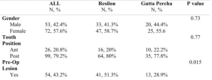
Results
The majority of the 125 subjects were female and had a root canal of a posterior tooth
(Table 1). Forty-three percent of the subjects had presented with a pre-op lesion and 36% had a
percha. There were no statistically significant differences between the two obturation materials
with respect to gender, tooth position, or age (Table 1). The two materials were statistically
significantly different with respect to presence of a pre-operative lesion, presence of a follow up
lesion and months to follow up (Table 1). Subjects with Resilon had a higher percentage of
pre-operative lesions, a higher percentage of follow up lesions, and a longer time to follow up.
Follow up periapical radiographs of Resilon and gutta percha treated teeth showed varying levels
of healing (Figure 1).
In the multivariate analysis, age, gender, tooth position, and months to follow up were not
statistically significantly associated with the presence of a follow up lesion when the presence of
an initial lesion and material were controlled for (Table 2). The presence of a pre-operative
lesion and the type of material used for obturation were statistically significant when controlling
for the age, gender, tooth position, and months to follow up (Table 2). Both the lack of an initial
lesion and having gutta percha were protective i.e. individuals with an initial lesion and those
receiving Resilon were more likely to have a follow up lesion.
Table 1: Characteristics for All Subjects and a Comparison of the Two Obturation Materials
ALL
N, % Resilon N, % Gutta Percha N, % P value
Gender 0.73
Male 53, 42.4% 33, 41.3% 20, 44.4%
Female 72, 57.6% 47, 58.7% 25, 55.6
Tooth Position
0.77
Ant 26, 20.8% 16, 20% 10, 22.2%
Post 99, 79.2% 64, 80% 35, 77.8%
Pre-Op Lesion
0.015
No 71, 56.8% 39, 48.7% 32, 71.1% Follow Up
Lesion
0.0004
Yes No
45, 36% 80, 64%
38, 47.5% 42, 52.5%
7, 15.6% 38, 84.4%
ALL Resilon Gutta Percha P value
N Median IQR N Median IQR N Median IQR
Age 125 56 19 80 57.5 19.5 45 54 20 0.70
Months to
Follow Up 125 49 63 80 62.5 62 45 26 19 <0.0001
Table 2: Odds Ratio from the Multivariate Analysis of the Presence of a Follow Up Lesion. A negative estimate indicates a protection factor.
Variable Estimate Error 95% CI P value
Pre-Op Lesion -0.87 0.42 -1.7 – (-0.05) 0.04
Material -1.67 0.64 -2.9 – (-0.42) 0.009
Months to FU -0.16 0.24 -0.6 – 0.3 0.49
Age -0.02 0.02 -0.05 – 0.02 0.34
Gender -0.56 0.49 -1.5 – 0.39 0.25
Figure 1: Post treatment (A,C,E) and follow up (B,D,F) radiographs of (A) Resilon treated #30 with PAI score of 1 (B) 3.5 year follow up of #30 with PAI score of 5 (C) Resilon treated #18 with PAI score of 3 (D) 11.9 year follow up of #18 with PAI score of 1 (E) Gutta percha treated #19 with PAI score of 4 (F) 1.3 year follow up of #19 with PAI score of 1
A
B
DISCUSSION
This study represents the longest outcome data available for a comparison of Resilon and
gutta percha materials. The use of Resilon was introduced at UNC by those who pioneered this
product and its use was continued for almost a decade. Fortunately, this meant that the sensitive
technique of the bonded material was taught to both the faculty and students at UNC at the
introduction of the material into clinical practice. This puts this data set in a unique advantage in
providing a good level of evidence in the long-term outcome of Resilon.
All subjects treated at UNC were treated under standard protocol, one for Resilon, and one
for gutta percha, that was taught to predoctoral and graduate students by instructors that were
familiar with both obturation techniques. From the 580 subjects that fit our inclusion and
exclusion criteria, 125 subjects had a follow up visit for a follow up rate of 21.6%, which is
comparable to other long-term outcome studies (6). The minimum follow up for both materials
was 12 months, as it has been shown that initiated healing can be observed in 89% of cases in as
early as 1 year (7). The maximum follow up of Resilon and gutta percha was 12.4 years and 12.1
years respectively. While both Resilon and gutta percha were in use from 2004-2013, gutta
perhca was more common clinically in the later years. Resilon patients were recalled primarily
from the beginning of Resilon’s implementation as well as the last year of its use which explains
why the follow up for gutta percha was on average less than Resilon.
The primary outcome for this study was radiographic healing. This was established by
using Orstavik’s proven PAI to evaluate the periapical structure of the treated teeth. We chose to
dichotomize the data into either a lesion not being present (PAI 1,2) or a lesion being present
(PAI 3,4,5). By dichotomizing our data these ranks could be sorted into two distinctive and
radiographically separate groups. The multivariate analysis, shown in Table 2, controlled for all
other explanatory variables. We found that Resilon treated teeth were more likely to have a
periapical lesion at follow up than the control material, which was found to be statistically
significant (p=0.009). We also determined that a tooth with a pre-operative lesion, regardless of
material used, was more likely to have a follow up lesion (p=0.04). This agrees with many
classic and current studies that pre-operative pathosis has a negative effect on treatment success
(8)(9).
Regardless of the positive findings reported in in-vitro studies of Resilon (10)(11) clinical
studies should influence the materials used in the clinic setting. Resilon and gutta percha showed
an indistinguishable difference in healing outcome in a 12-25 month retrospective follow-up
(12). This study evaluated 103 teeth, 68 of which were evaluated between 18 and 25 months. The
other 35 were evaluated less than 18 months. The only other long-term outcome study on Resilon
was a 5.6 month follow up recently published in 2017 by Barborka et al (13). There were
al. used Orstavik’s PAI to score the periapical images of teeth at time of treatment and at follow
up. Gutta percha was used as a control for comparison with Resilon in the Cotton et al, Barborka
et al, and our paper. A large difference between Barborka et al. and our paper was how they
radiographically assessed healing. Instead of using a proven periapical index they chose to
evaluate the images side by side with a study derived definition of success. They also did not
actively recruit patients with phone calls or use a power analysis to determine the amount of
cases needed in each group.
There are potentially several reasons for the decreased outcome of Resilon. The first being
the composition of the material. Polycaprolactone, the biodegradable polyester compromising a
majority of Resilon, was suggested to result in severe surface pitting and erosion (3). The
adhesive property of Resilon was also shown to not be as predictable in the long narrow canal
even with aid from a surgical microscope. The effectiveness of the bond was also a concern as it
is difficult to avoid over-thinning of the adhesive (14). In an in vitro study, the presence of gaps
along the core/sealer/dentin interface was shown to potentially create an environment for leakage
and re-infection (2). A retrospective study used data from PA radiographs and CBCT scans to
examine various factors affecting the outcome of root canal treatment. The density of the root
filling was identified in both PA and CBCT as a predictor that significantly influenced the
treatment outcome (15). Figure 1 shows several periapical radiographs of Resilon and gutta
percha treated teeth. While recalling patients we noticed that despite the highly dense and
radiopaque appearance of Resilon treated teeth (A), large periapical lesions were seen upon
follow up (B). Resilon was advertised as more radiopaque than gutta percha (16), so it is possible
Resilon treated case developed a periapical lesion. Resolution of periapical lesions were noted as
well (D).
Although Resilon is no longer on the market there are countless patients that still retain Resilon
treated teeth. As this study has shown that Resilon treated teeth present with more lesions at
follow up we must ask the question if a “recall” of all Resilon treated cases is indicated. It may
be necessary to advise these patients that their treatment may be compromised
CONCLUSION
Within the limitations of the study, the results demonstrate that teeth presenting with
pre-operative lesions are more likely to have a lesion at follow up regardless of obturation material.
Teeth that are obturated with Resilon present with more lesions at follow-up compared to gutta
percha obturated teeth suggesting that there is no long-term benefit to this material as the healing
capability of Resilon is inferior to gutta percha. It may be indicated to share these findings with
REFERENCES
1 Shipper Guy, Ørstavik Dag, Teixeira Fabricio Batista, Trope Martin. An evaluation of microbial leakage in roots filled with a thermoplastic synthetic polymer-based root canal filling material (Resilon). J Endod 2004;30(5):342–7. Doi:
10.1097/00004770-200405000-00009.
2 Tay Franklin R, Loushine Robert J, Weller R Norman, et al. Ultrastructural evaluation of the apical seal in roots filled with a polycaprolactone-based root canal filling material. J Endod 2005;31(7):514–9.
3 Hiraishi Noriko, Yau Joyce YY, Loushine Robert J, et al. Susceptibility of a
Polycaprolactone-based Root Canal–filling Material to Degradation. III. Turbidimetric Evaluation of Enzymatic Hydrolysis. J Endod 2007;33(8):952–6. Doi:
10.1016/j.joen.2007.05.004.
4 Tay Franklin R, Loushine Robert J, Lambrechts Paul, Weller R Norman, Pashley David H. Geometric factors affecting dentin bonding in root canals: a theoretical modeling approach. J Endod 2005;31(8):584–9.
5 MELKER K, VERTUCCI F, ROJAS M, PROGULSKEFOX A, BELANGER M. Antimicrobial Efficacy of Medicated Root Canal Filling Materials. J Endod 2006;32(2):148–51. Doi: 10.1016/j.joen.2005.10.032.
6 FRIEDMAN S, ABITBOL S, LAWRENCE H. Treatment Outcome in Endodontics: The Toronto Study. Phase 1: Initial Treatment. J Endod 2003;29(12):787–93. Doi:
10.1097/00004770-200312000-00001.
7 Orstavik D. Time-course and risk analyses of the development and healing of chronic apical periodontitis in man. Int Endod J 1996;29(3):150–5.
8 Bergenholtz G, Lekholm U, Milthon R, Heden G, Odesjö B, Engström B. Retreatment of endodontic fillings. Scand J Dent Res 1979;87(3):217–24.
9 Ng YL, Mann V, Gulabivala K. A prospective study of the factors affecting outcomes of nonsurgical root canal treatment: Part 1: Periapical health. Int Endod J 2011;44(7):583– 609. Doi: 10.1111/j.1365-2591.2011.01872.x.
10 Shipper Guy, Teixeira Fabricio B, Arnold Roland R, Trope Martin. Periapical
inflammation after coronal microbial inoculation of dog roots filled with gutta-percha or resilon. J Endod 2005;31(2):91–6. Doi: 10.1097/01.DON.0000140569.33867.BF. 11 Tay Franklin R, Pashley David H, Loushine Robert J, et al. Susceptibility of a
polycaprolactone-based root canal filling material to degradation. Evidence of biodegradation from a simulated field test. Am J Dent 2007;20(6):365–9.
10.1016/j.joen.2008.03.018.
13 Barborka Benjamin J, Woodmansey Karl F, Glickman Gerald N, Schneiderman Emet, He Jianing. Long-term Clinical Outcome of Teeth Obturated with Resilon. J Endod
2017;43(4):556–60. Doi: 10.1016/j.joen.2016.12.005.
14 Tay Franklin R, Pashley David H. Monoblocks in root canals: a hypothetical or a tangible goal. J Endod 2007;33(4):391–8. Doi: 10.1016/j.joen.2006.10.009.
15 Liang Yu-Hong, Li Gang, Wesselink Paul R, Wu Min-Kai. Endodontic Outcome
Predictors Identified with Periapical Radiographs and Cone-beam Computed Tomography Scans 2011. Doi: 10.1016/j.joen.2010.11.032.
16 Pentron Clinical Technologies LLC. Epiphany ® Soft Resin Endodontic Obturation System TM Dentin Tubules Epiphany ® Sealer/Hybrid Zone Resilon ® Material
Introducing the new standard in obturation…. Available at:
MANUSCRIPT 2: POST OPERATIVE FACTORS EFFECT ON OUTCOMES OF ENDODONTIC TREATMENT WITH RESILON/EPIPHANY SYSTEM
Introduction
The outcome of root canal treatment is influenced by many factors once the procedure is
completed. Presence and quality of a final restoration has shown to be of equal importance to the
quality of obturation (1). A full coverage crown after root canal treatment has been shown to
increase fracture resistance (2) and the tooth survival rate against fracture when compared to
teeth restored with resin composite (3). The timing of restoration placement is also a factor that
has shown to have an effect of healing. Long-term survival rates of initial root canal treatment
were adversely affected when the placement of the final restoration was delayed according to a
national insurance database (4).
A thorough follow up clinical exam is an ideal way to determine the presence and type of
final restoration and evaluate other sign and symptoms of disease. The objective of this study
was to assess whether the difference in healing of Resilon and gutta percha treated root canals
was related to the clinical signs of disease (swelling, sinus tract, mobility, percussion, and
probing) or the presence, the type, and the timing of final restoration.
Materials and Methods
Institutional Review Board approval for this retrospective cohort clinical study was
(16-1069). Patients who were 18 years or older, had completed root canal treatment (dental codes
D3310, D3320, D3330) during the time period of 08/2004 – 08/2013, had been treated in the
predoctoral or graduate Endodontic clinic, and had a full comprehensive clinical evaluation with
an endodontic resident were identified through a search of the electronic patient records. Patients
whose dental records did not include a radiograph immediately after the original root canal
treatment or specify which material was used for obturation (Resilon or gutta percha) were
excluded.
Patient record review indicated that 7,376 patients had primary endodontic treatment in
either the predoctoral or graduate Endodontic clinic during the specified time period. Five
hundred eighty patients were randomly selected and telephone calls were made to the primary
number on file. If there was no answer, a scripted voice message was left and the secondary
number on file was then called. One hundred twenty-five (21.6%) patients agreed to come into
the Endodontic clinic for a follow up visit and signed a consent form. Of these, thirty-eight
(30.4%) patient received a comprehensive intra- and extra-oral exam.
Treatment
Two endodontic residents performed all of the clinical exams under supervision of a
board-certified endodontist. In addition to the data gained from the radiographic interpretation,
other post-operative factors were collected to evaluate for potential prognostic factors. Presence
of mobility, sensitivity to percussion, and probing depths greater than 3mm obtained with
standard periodontal probe were noted. Presence of a final restoration and whether or not that
restoration was a crown was recorded. The time from completion of the root canal to placement
considered to be ideal if placed within 3 months of root canal treatment completion. If the tooth
being evaluated showed signs of re-infection the proper referrals were provided to the patient.
Recording of Data
The post-operative factors and patient information gathered at the follow up examination
was recorded during the appointment on an assessment form specific for each patient. This
information was then transferred to a Microsoft Excel database (ãMicrosoft 2017).
Statistical Analysis
Bivariate analysis was performed using Fisher’s exact test for nominal variables and
Wilcoxon rank-sum for continuous variables. The level of significance was established as p <
0.5. The power was established at 80.
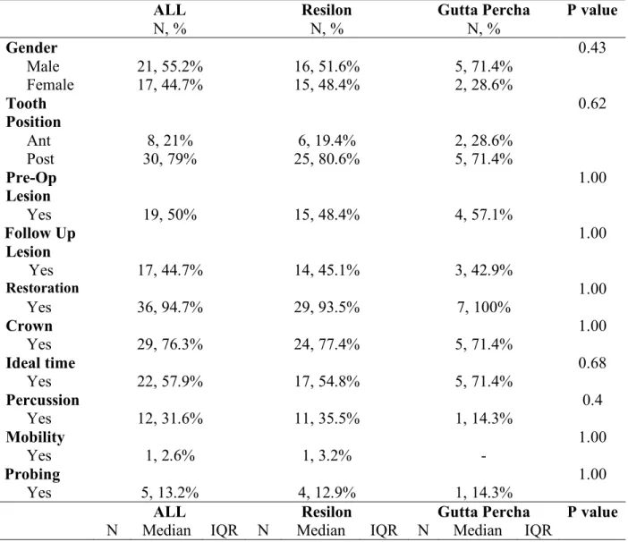
Results
The majority of the 38 patients were male and had a root canal of a posterior tooth (Table
3). Fifty percent of the subjects had presented with a pre-op lesion and forty-five percent had
follow up lesion. Thirty-one subjects had Resilon used as the obturation material and seven gutta
percha. There were no statistically significant differences between the two obturation materials
with respect to gender, tooth position, or age (Table 3). The majority of the 38 patients had a
final restoration, and one that was a full coverage crown placed in the ideal amount of time.
There were not statistically significant differences between the two obturation materials with
respect to presence of percussion sensitivity, mobility, or deep probing depths (Table 3).Time to
placement of restoration was dichotomized into within 0 to 3 months of root canal completion
obturation materials with respect to placement of the final restoration in an ideal time. When
evaluating the effect of time to restoration on follow up lesion, teeth presenting without a lesion
had a median time to restoration of 2 months and teeth presenting with a lesion had a median
time to restoration of 4 months. A shorter time to placement of final restoration (within 3
months) was found to be beneficial (p=0.047), regardless of the material used to obturate.
Table 3: Characteristics for All Clinically Evaluated Subjects and a Comparison of the Two Obturation Materials ALL N, % Resilon N, % Gutta Percha N, % P value
Gender 0.43
Male 21, 55.2% 16, 51.6% 5, 71.4%
Female 17, 44.7% 15, 48.4% 2, 28.6%
Tooth
Position 0.62
Ant 8, 21% 6, 19.4% 2, 28.6%
Post 30, 79% 25, 80.6% 5, 71.4%
Pre-Op Lesion
1.00
Yes 19, 50% 15, 48.4% 4, 57.1%
Follow Up Lesion
1.00
Yes 17, 44.7% 14, 45.1% 3, 42.9%
Restoration 1.00
Yes 36, 94.7% 29, 93.5% 7, 100%
Crown 1.00
Yes 29, 76.3% 24, 77.4% 5, 71.4%
Ideal time 0.68
Yes 22, 57.9% 17, 54.8% 5, 71.4%
Percussion 0.4
Yes 12, 31.6% 11, 35.5% 1, 14.3%
Mobility 1.00
Yes 1, 2.6% 1, 3.2% -
Probing 1.00
Yes 5, 13.2% 4, 12.9% 1, 14.3%
ALL Resilon Gutta Percha P value
Age 38 57.5 19 31 57 20 7 60 17 0.88 Months to
Follow Up 38 95 72 31 102 54 7 49 92 0.07
Discussion
In order to truly determine healing following non-surgical root canal treatment, an
extra-oral and intra-extra-oral clinical exam is indicated. Clinical findings along with updated radiographs
give the practitioner everything needed to properly diagnose the tooth and assess the treatment’s
outcome. From our initial 125 subjects, 38 received a full comprehensive clinical exam. The
majority of subjects evaluated clinically were Resilon (31/38). Since 80/125 of the initial
subjects were Resilon, it would be expected that more Resilon treated subjects were able to have
a full clinical exam. The presence of a restoration and the type of restoration, filing or crown,
was documented. This was important, as endodontically treated teeth that have not been crowned
after obturation have been shown to be lost at a 6.0 times greater rate than those crowned (5).
Only 2 teeth total did not present with a final restoration, both Resilon treated. The first of the
two teeth still contained the temporary. The second tooth’s restoration was completely missing.
If the root canal treatment was performed through a crown, and a core filling was placed
afterwards, the tooth was considered to have a crown as its final restoration. The majority of
Resilon and gutta percha treated teeth that had final restorations were full coverage crowns
(77.4%, 71.4%). No statistical significance was found between the two materials and presence or
type of restoration. The ideal time to restoration was an important factor to consider, as a delay
in restoration can adversely affect a tooth’s survival rate (4). A recent study showed teeth that
received a crown 4 months after RCT were almost 3 times more likely to get extracted(6). We
recommended at UNC dental school to place the final restoration on a root canal treated tooth
within 3 months.
In the clinical exam, none of the 38 subjects had any signs of extra-oral/intra-oral swelling
or presented with a sinus tract. Only 1 subject treated with Resilon presented with tooth mobility
which was determined to be anything greater than Class 1 mobility. It was important to test
percussion sensitivity as this can indicate inflammation of the periodontal ligament. A greater
number of Resilon treated teeth exhibited sensitivity on percussion, however, this was not
statistically significant (p=0.4). Using the biological width measurements of Gargiulo, any
isolated probing depth greater than 3mm was considered to be positive for a periodontal probing.
There was no statistically significant difference between the two materials and the number of
teeth with probing defects (p=1.00). When our primary outcome was evaluated, regardless of the
material used, we found teeth that had a final restoration placed within 3 months of root canal
completion were less likely to have a PA lesion at follow up (p=0.047). When there was no
lesion, the median time for placement of the restoration was 2 months. When a lesion was
present the median time for placement of the restoration was 4 months.
Conclusion
Within the limited scope of this study there were no statistically significant differences in
the clinical presentation of teeth obturated with Resilon or gutta percha. There were no
statistically significant differences between the two obturation materials with respect to presence
of a restoration, type of restoration, or placement of the final restoration in an ideal time. When
all 38 teeth were evaluated, placement of the final restoration within 3 months of root canal
likely to have a periapical lesion at follow up. This finding stresses the importance and benefit of
a faster placement of the final restoration after root canal treatment has been completed. None of
the other explanatory variables showed a statistical significance between the two materials. It
appears that future studies with a higher subject number would allow for greater power to
potentially find other significant factors and/or differences in prognostic factors between Resilon
REFERENCES
1 Ray HA, Trope M. Periapical status of endodontically treated teeth in relation to the technical quality of the root filling and the coronal restoration. Int Endod J 1995;28(1):12– 8.
2 Hoag EP, Dwyer TG. A comparative evaluation of three post and core techniques. J Prosthet Dent 1982;47(2):177–81.
3 Suksaphar Warattama, Banomyong Danuchit, Jirathanyanatt Titalee, Ngoenwiwatkul Yaowaluk. Survival Rates from Fracture of Endodontically Treated Premolars Restored with Full-coverage Crowns or Direct Resin Composite Restorations: A Retrospective Study. J Endod 2017. Doi: 10.1016/j.joen.2017.09.013.
4 Yee Kandace, Bhagavatula Pradeep, Stover Sheila, et al. Survival Rates of Teeth with Primary Endodontic Treatment after Core/Post and Crown Placement. J Endod 2017. Doi: 10.1016/j.joen.2017.08.034.
5 Aquilino Steven A, Caplan Daniel J. Relationship between crown placement and the survival of endodontically treated teeth. J Prosthet Dent 2002;87(3):256–63.
6 Pratt Isaac, Aminoshariae Anita, Montagnese Thomas A, Williams Kristin A,
THESIS SUMMARY
Resilon was brought to market in the dental community with its support mainly being
from in vitro leakage and structural strength studies (1)(2). In vitro studies are very helpful from
a proof of concept perspective and to aid the manufacturers in finding weaknesses and strengths
of a material. It is however, critical to base the use of newly introduced clinical materials from
clinical in vivo research so that the effectiveness and safety can be properly determined.
Our retrospective study evaluated the radiographic outcome of 125 subjects, and the
clinical outcome of 38. When controlling for all explanatory variables, teeth treated with the
Resilon/Epiphany TM system were more likely to have periapical lesions at follow up when
compared to gutta percha (p=0.009). This finding implies that Resilon treated teeth are not
healing at a similar rate to the control material and its use is not warranted as there is no
long-term treatment benefit. Although this material is no longer on the market, its use in the country
spanned over a decade. There are countless patients that still have Resilon treated teeth. It may
be necessary to advise patients of the new clinical findings and urge them to have a follow up
examination. These patients may not be having any signs of infection or re-infection. In a
recently published 20-year cohort study, of the teeth that presented with re-infection of the root
canal system, 62% were asymptomatic (3). Our clinical evaluation did not show any significant
difference in presence of percussion sensitivity, deep probing pockets, or tooth mobility between
Teeth that presented with apical lesions at the time of initial root canal treatment were also
more likely to have a periapical lesion at their follow up, regardless of material used for
obturation (p=0.047). The presence of a pre-operative lesion has been shown by many studies to
be a major prognostic factor in treatment success(4)(5)(6), which this study also confirmed.
At UNC, the patient is advised to receive their final restoration within 3 months of the root
canal treatment completion. At times the restoration can be placed immediately following RCT,
other times a separate appointment is needed with the restorative provider. The data collected
was dichotomized into teeth receiving final restoration within 0 to 3 months and those receiving
the final restoration after 3 months. The clinical evaluation was able to show that the presence of
a final restoration placed within 3 months after completion of root canal treatment was beneficial
to the tooth’s outcome, regardless of obturation material (p=0.047). Teeth without a lesion at
follow up had a median time to restoration of 2 months, whereas teeth with a lesion at follow up
had a median time to restoration of 4 months. A proper coronal seal placed in an appropriate
amount of time is critical to root canal treatment healing.
These results should add to the current knowledge of Resilon and help aid practitioners
who are following up with Resilon treated root canals. It appears that Resilon treated teeth
present with decreased long-term healing when compared to gutta percha. Future studies to
REFERENCES
1 Shipper Guy, Ørstavik Dag, Teixeira Fabricio Batista, Trope Martin. An evaluation of microbial leakage in roots filled with a thermoplastic synthetic polymer-based root canal filling material (Resilon). J Endod 2004;30(5):342–7. Doi:
10.1097/00004770-200405000-00009.
2 Teixeira Fabricio B, Teixeira Erica CN, Thompson Jeffrey Y, Trope Martin. Fracture resistance of roots endodontically treated with a new resin filling material. J Am Dent Assoc 2004;135(5):646–52.
3 Prati C, Pirani C, Zamparini F, Gatto MR, Gandolfi MG. A 20-year historical prospective cohort study of root canal treatments. A Multilevel analysis. Int Endod J 2018. Doi: 10.1111/iej.12908.
4 Bystrom A, Happonen RP, Sjogren U, Sundqvist G. Healing of periapical lesions of pulpless teeth after endodontic treatment with controlled asepsis. Endod Dent Traumatol 1987;3(2):58–63.
5 Ricucci Domenico, Russo John, Rutberg Michael, Burleson Josef A, Spångberg Larz SW. A prospective cohort study of endodontic treatments of 1,369 root canals: results after 5 years. Oral Surgery, Oral Med Oral Pathol Oral Radiol Endodontology 2011;112(6):825– 42. Doi: 10.1016/J.TRIPLEO.2011.08.003.
APPENDIX A: PATIENT CONSENT FORM University of North Carolina at Chapel Hill
Consent to Participate in a Research Study Adult Participants
Consent Form Version Date: 07/22/2016 IRB Study # 16-1069
Title of Study: Long-term outcome assessment and treatment of Resilon obturation system compared to gutta percha.
Principal Investigator: Peter Tawil
Principal Investigator Department: Endodontics Principal Investigator Phone number: 919-537-3403 Principal Investigator Email Address: [email protected]
Co-Investigators: Steven Card, Lesleigh Payne, Krista Andersen
_________________________________________________________________
What are some general things you should know about research studies?
You are being asked to take part in a research study. To join the study is voluntary.
You may refuse to join, or you may withdraw your consent to be in the study, for any reason, without penalty.
Research studies are designed to obtain new knowledge. This new information may help people in the future. You may not receive any direct benefit from being in the research study. There also may be risks to being in research studies.
Details about this study are discussed below. It is important that you understand this information so that you can make an informed choice about being in this research study.
You will be given a copy of this consent form. You should ask the researchers named above, or staff members who may assist them, any questions you have about this study at any time.
What is the purpose of this study?
The purpose of this study is to evaluate the long-term outcome of root canal therapy at the UNC graduate and undergraduate endodontic clinics. The materials used for treatment will be
evaluated as well as other factors that could affect the success of treatment. Teeth that need retreatment will be examined for canal filling break down, flare-ups or other post-operative complications.
You are being asked to be in the study because you have had root canal therapy completed at UNC school of dentistry at a time where different materials were in use.
You should not be in this study if you are under the age of 18.
How many people will take part in this study?
There will be approximately 6,855 people in this research study.
How long will your part in this study last?
Your visits may range from one to four appointments over a maximum of three months with the investigators throughout the research study. The first interaction will be the standard of care follow up evaluation and introduction of the research study. It will last no more than 30 minutes. If re-treatment of the tooth/teeth in question is needed and deemed appropriate a questionnaire will be given for you to answer in between appointments. The completion of the questionnaire will take no more than 5 minutes and will be given to the investigator at the final appointment.
What will happen if you take part in the study?
As part of this research study, the researchers will also examine your dental records involving your treatment.
After your initial follow-up appointment, blinded and calibrated dental examiners who are apart of the research team will review your dental radiographs.
Following any treatment rendered, a questionnaire will be given to you between
appointments, which should be returned to your investigator at the final appointment. This questionnaire will ask you to record any post-operative pain or swelling that occurred after the initial visit. You may choose not to answer a question for any reason.
The clinician who the patient is assigned to will be responsible for the patients welfare during the study.
What are the possible benefits from being in this study?
Research is designed to benefit society by gaining new knowledge. You will not benefit personally from being in this research study.
What are the possible risks or discomforts involved from being in this study? There are no foreseen immediate or long-term physical, psychological, or social risks/discomforts.
What if we learn about new findings or information during the study?
You will be given any new information gained during the course of the study that might affect your willingness to continue your participation.
How will information about you be protected?
Every effort will be made to protect your privacy. All information collected in this study will remain confidential and only those directly involved in the study will have access to this information. All participants will be assigned numbers and all electronic data collected will be password protected on a secure UNC server. Paper documents will be stored in a locked filing cabinet in a locked office where only research participants will have access.
Participants will not be identified in any report or publication about this study. Although every effort will be made to keep research records private, there may be times when federal or state law requires the disclosure of such records, including personal information. This is very unlikely, but if disclosure is ever required, UNC-Chapel Hill will take steps allowable by law to protect the privacy of personal information. In some cases, your information in this research study could be reviewed by representatives of the University, research sponsors, or government agencies (for example, the FDA) for purposes such as quality control or safety.
What will happen if you are injured by this research?
All research involves a chance that something bad might happen to you. This may include the risk of personal injury. In spite of all safety measures, you might develop a reaction or injury from being in this study. If such problems occur, the researchers will help you get medical care, but any costs for the medical care will be billed to you and/or your insurance company. The University of North Carolina at Chapel Hill has not set aside funds to pay you for any such reactions or injuries, or for the related medical care. You do not give up any of your legal rights by signing this form.
What if you want to stop before your part in the study is complete?
You can withdraw from this study at any time, without penalty. The investigators also have the right to stop your participation at any time. This could be because you have had an unexpected reaction, or have failed to follow instructions, or because the entire study has been stopped.
Will you receive anything for being in this study?
Will it cost you anything to be in this study? It will not cost you anything to be in this study.
What if you have questions about this study?
You have the right to ask, and have answered, any questions you may have about this research. If you have questions about the study (including payments), complaints, concerns, or if a research-related injury occurs, you should contact the researchers listed on the first page of this form.
What if you have questions about your rights as a research participant?
All research on human volunteers is reviewed by a committee that works to protect your rights and welfare. If you have questions or concerns about your rights as a research subject, or if you would like to obtain information or offer input, you may contact the Institutional Review Board at 919-966-3113 or by email to [email protected].
Participant’s Agreement:
I have read the information provided above. I have asked all the questions I have at this time. I
voluntarily agree to participate in this research study.
______________________________________________________
Signature of Research Participant
____________________
Date
______________________________________________________
Printed Name of Research Participant
______________________________________________________
Signature of Research Team Member Obtaining Consent
____________________
Date
______________________________________________________
Printed Name of Research Team Member Obtaining Consent