SURVIVAL MOTOR NEURON PROTEIN INTERACTION PARTNERS
IN
DROSOPHILA MELANOGASTER
Kelsey Marie Gray
A dissertation submitted to the faculty of the University of North Carolina at Chapel Hill in partial fulfilment of the requirements for the degree of Doctor of Philosophy in the
Curriculum in Genetics and Molecular Biology.
Chapel Hill 2018
Approved By: Frank Conlon Blossom Damania A. Gregory Matera Mark Peifer
ABSTRACT
Kelsey Marie Gray: Survival Motor Neuron protein interaction partners
in Drosophila melanogaster
(Under the direction of A. Gregory Matera)
Spinal Muscular Atrophy (SMA) is a neuromuscular disorder that results from biallelic loss-of-function mutations in the human survival motor neuron 1 (SMN1) gene. Tissue-specific and housekeeping functions have been ascribed to SMN; however, their relevance to SMA pathology is not well understood. We generated transgenic Drosophila
melanogaster that express only flag-tagged wild-type SMN. Our objective is to
characterize novel protein-protein interactions of SMN. We collected embryos and analyzed Flag-purified lysates by mass spectrometry. We identified Flag-SMN along with other known interactors such as the Sm proteins and the Gemins.
We also identified Slmb, SkpA, and Cullin 1 as being highly enriched in Flag-SMN samples as compared to the control sample. Together, these proteins comprise the SCFSlmb E3 ubiquitin ligase. These interactions were verified in Drosophila S2 cells and human cells. In vitro experiments revealed Slmb and SMN can directly interact.
degron is indeed mediating degradation of SMN. Finally, expression of truncated SMN stabilized by the mutation modifies viability of a mild SMA mouse model.
We identified additional protein interactions of SMN with CG2941, nucleosome assembly protein 1 (Nap1), and Bendless (Ben). Each of these interactions was verified in cell culture or using antibodies generated specifically for the protein of interest.
Preliminary investigation of CG2941 has revealed it is an essential gene that produces protein that localizes to both the nucleus and the cytoplasm.
We have examined SMN protein interactions in the context of developing
Drosophila melanogaster embryos, with follow-up studies in mouse, and human systems.
To my parents, my brother, Dorian,
ACKNOWLEDGEMENTS
First and foremost, I would like to thank my parents. I was raised in an incredibly fortunate home where I knew I would be supported through earning a graduate degree from the time I was born. They have always been there to motivate me and to support my love of science. They taught me to persevere, and I am incredibly thankful for that today. I appreciate my brother for always being there to ask me questions to which I don’t know the answer, as a reminder that I always have more to learn.
I want to acknowledge my mentor, Greg Matera, for conversations about many topics in biology. He fostered an environment that enabled me to grow as an independent scientist and thinker, which is crucial for my future success as an educator. I want to thank Greg also for the opportunities he gave me to mentor students in the lab. These opportunities were instrumental to my development as a teacher of science.
Surviving graduate school would not have been possible without my lab mates and friends. I am especially grateful for the “Matera Lab Ladies” and all of their love, support, and scientific insights. I am also thankful for our previous lab manager, Ying Wen, whose help was invaluable in completing this dissertation.
TABLE OF CONTENTS
LIST OF FIGURES ... x
LIST OF ABBREVIATIONS ... xii
CHAPTER I ... 1
Spinal Muscular Atrophy ... 1
SMA Genetic Etiology ... 2
SMN Protein and the SMN Complex ... 5
SMN Function ... 10
snRNP Biogenesis ... 10
snRNP independent functions of SMN ... 15
Modeling SMA ... 18
Drosophila SMA models ... 18
Other animal models of SMA ... 20
Oligomeric properties of SMN complexes ... 22
SMN Protein Stability ... 23
Ubiquitin Proteasome System (UPS) and SMN ... 24
Mindbomb1 (Mib1) ... 26
Itch ... 27
SCFSlmb/SCFB-TrCP ... 27
CHAPTER II ... 30
Experimental procedures ... 33
Fly stocks and transgenes ... 33
Drosophila embryo protein lysate and mass spectrometry ... 34
Tissue culture and transfections ... 35
In vitro binding assay ... 36
In vivo ubiquitylation assay ... 37
Cycloheximide treatment ... 37
Immunoprecipitation ... 37
Antibodies and Western blotting ... 38
Larval locomotion ... 39
SMA Mouse Models ... 39
Human iPSC Cell culture ... 39
Immunocytochemistry ... 40
Immunocytochemical Analysis ... 41
Results ... 41
Flag-SMN interacts with UPS (ubiquitin proteasome system) proteins ... 41
SCFSlmb is a bona fide SMN interaction partner that ubiquitylates SMN ... 45
Depletion of Slmb/B-TrCP results in a modest increase in SMN levels ... 48
Identification and characterization of a Slmb/B-TrCP degradation signal in SMN ... 50
SMN self-oligomerization regulates access to the Slmb degron ... 52
Mutation of the Slmb degron rescues viability and locomotion defects in SMA model flies ... 54
GFP-SMN∆7 overexpression stabilizes endogenous SMN and SMN∆7 in cultured human cells ... 56
SMN∆7A is a protective modifier of intermediate SMA mouse phenotypes ... 59
Discussion ... 63
The SCFSlmb degron is exposed by SMN2 exon skipping ... 64
SMN targeting by multiple E2 and E3 systems ... 64
Does SMN function as a signaling hub? ... 65
Phosphorylation of the Slmb degron within SMN ... 66
Supplementary Data ... 68
CHAPTER III ... 72
Introduction ... 72
Experimental Procedures ... 75
Fly stocks ... 75
Antibodies and Western blotting ... 75
Immunoprecipitation ... 76
Drosophila embryo protein lysate and mass spectrometry ... 76
Tissue culture and transfections ... 77
Immunofluorescence ... 77
Results ... 78
Flag-SMN interacts with proteins involved in a variety of cellular processes ... 78
CG2941-Flag localizes to both the cytoplasm and nucleus ... 80
CG2941 RNAi reduces fly viability ... 81
Discussion ... 83
CG2941 as a member of the core SMN complex? ... 84
Chromatin remodelling and transcriptional regulation in SMA ... 85
SMN and cellular signalling pathways ... 87
Molecular characterization of SMN function using SMA patient mutations ... 89
The SMN complex and protein stability ... 91
Ubiquitylation of SMN and endocytosis ... 97
Additional functions of SMN ... 100
Role of SMN as a signaling hub ... 102
Summary ... 103
LIST OF FIGURES
Figure 1.1. SMN1 and SMN2 ... 4
Figure 1.2. Major domains in the SMN protein ... 6
Figure 1.3. SMN complex members in human (mammal) and Drosophila ... 8
Figure 1.4. Overview of the mammalian snRNP biogenesis pathway ... 11
Figure 1.5. The role of SMN in transcriptional regulation ... 16
Figure 1.6. SMA patient mutations in the DrosophilaSmn gene ... 19
Figure 1.7. The ubiquitin proteasome system (UPS) ... 25
Figure 2.1. Flag-SMN immunopurified lysates ... 44
Figure 2.2. Interaction between SMN and the SCFSlmb/B-TrCP E3 ubiquitin ligase ... 47
Figure 2.3. Depletion of Slmb/B-TrCP results in an increase of SMN levels ... 49
Figure 2.4. Putative Slmb/B-TrCP phospho-degron ... 53
Figure 2.5. Mutation of the Slmb degron rescues defects in SMA model flies ... 55
Figure 2.6. Stabilization of endogenous SMN and SMNΔ7 in cultured human cells ... 58
Figure 2.7. SMN∆7A is a protective modifier of intermediate SMA phenotypes ... 61
Figure 2.8. Model of SMN as a substrate of SCFSlmb E3 ubiquitin ligase ... 63
Figure S2.1. Flag-vSmn and Flag-vSmnS201A fly viability ... 68
Figure S2.2. Transfection controls ... 69
Figure S2.3. Flag-vSmn and Flag-vSmnS201A protein levels ... 70
Figure S2.4 SMN∆7A expression in the severe Delta7 mouse model ... 71
Figure 3.1. Flag-SMN immunopurified lysates contain novel interaction partners ... 79
Figure 3.2. CG2941 is in both the nucleus and the cytoplasm ... 81
LIST OF ABBREVIATIONS
AAV9 Adeno-associated virus serotype 9
ATP Adenosine triphosphate
Ben Bendless
CHX Cycloheximide
Cul1 Cullin 1
dsRNA Double-stranded RNA
DNA Deoxyribonucleis acid
dSMN Drosophila SMN
EDTA Ethylenediaminetetraacetic acid
EMS Ethyl methane sulphonate
FLAG Polypeptide epitope
GF Giant fiber neuron
GFP Green fluorescent protein
GST Glutathione S-transferase
HEK Human embryonic kidney cells
Impβ Importinβ
IP Immunoprecipitation
iPSC Induced pluripotent stem cells
m7G 7-methylguanosine
m3G Trimethylguanosine
Nap1 Nucleosome assembly protein
NP-40 Nonident P-40
mRNA messenger RNA
mRNP messenger RNP
NMJ Neuromuscular junction
OR Oregon-R
PBS Phosphate buffered saline
PHAX Phosphorylated adaptor for snRNA export
PLS3 Plastin 3
PRMT5 Protein arginine methyltransferase
RIG Rigor Mortis
RNA Ribonucleic acid
RNAi RNA interference
RNP Ribonucleoprotein
S2 Schneider 2
scaRNP small cajal body specific RNP
SETX Senataxin
siRNA Short interfering RNA
SkpA Skp1-related A
Slmb Supernumerary limbs
SMA Spinal muscular atrophy
SMN Survival motor neuron
snRNA small nuclear RNA
snRNP small nuclear RNP
SPN1 Snurportin1
Tgs1 Trimethylguanosine synthase
TMG 2,2,7-trimethylguanosine
UAS Upstream activating sequence
Unrip unr-interacting protein
UPS Ubiquitin proteasome system
U-snRNA Uridine rich snRNA
UTR Untranslated region
CHAPTER I: Introduction
Spinal Muscular Atrophy
Spinal muscular atrophy (SMA) is a common neuromuscular disorder that is the most prevalent genetic cause of infant mortality (Pearn 1980). SMA has a carrier
frequency of 1 in 50 and an incidence rate of 1 in 6,000-10,000 (Ogino et al. 2002). The disease presents clinically as the degeneration of motor neurons in the anterior horn of the spinal cord (Crawford and Pardo 1996). Without stimulation from the neuron, the
proximal muscles atrophy, leading to subsequent loss of motor function, and ultimately symmetrical paralysis. Since the timing of the onset of symptoms and their severity can vary, SMA has historically been classified into subtypes (Ogino S 2004). More recently, clinicians have recognized that SMA is better characterized as a continuous spectrum disorder, ranging from severe (prenatal onset) to nearly asymptomatic (Tiziano et al. 2013)
While there are several different ways to classify SMA, one of the most
Symptoms begin to appear as early as in utero and are clearly detectable during the first 6 months of life (Wee et al. 2010; Prior 2010). Affected infants experience progressive muscle weakness, which correlates with a reduced ability to swallow. These infants most frequently die from complications such as progressive and restrictive respiratory failure by 2 years of age (Kolb and Kissell 2015). Type II SMA is less severe with symptom onset occurring between 6 and 18 months of age. These children experience
developmental motor delays and are unable to stand or walk, although they can sit unsupported. The lifespan of type II patients can vary from 2 to 30 years, with death usually occurring as a result of respiratory infections. SMA type III, also known as Kugelberg-Welander disease, is the mildest form with an age of onset after 2 years. Most type III patients are able to stand and walk, but often need to use a wheelchair in
adulthood due to muscle weakness. Many type III patients have a normal life expectancy since the disease progress is slow.
SMA Genetic Etiology
In 1990, linkage analysis was used to map the SMA-causing gene to chromosome 5q11.2-13.3 in several affected families (Brzustowicz et al. 1990; Melki et al. 1990). Lefebvre et al. (1995) discovered a gene in that chromosomal region, survival of motor
neuron 1 (SMN1), that was identified to be the causative gene in SMA (Bussaglia et al.
1995; Rodrigues et al. 1995; van der Steege et al. 1995; Chang et al. 1997). At this time, it was determined that over 95% of SMA patients have deletions of SMN1 (Lefebvre et al. 1995; Campbell et al. 1997).
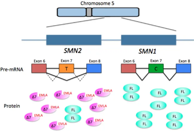
located at 5q11-13 that resulted in a telomere-proximal copy (SMN1) and a centromere-proximal copy (SMN2) of the SMN gene (Lefebvre et al. 1995). Five base pair differences distinguish the two SMN copies at the nucleotide level, but each of these nucleotide changes leave the amino acid sequence of the protein unchanged. The nucleotide
difference that affects the functionality of the proteins produced by each of the genes is a change from C to T in exon 7 of the SMN2 gene (Lorson et al. 2010). This change affects splicing and results in the exclusion of exon 7 (SMNΔ7) in ~90% of the SMN2 transcripts (Fig. 1.1). This SMNΔ7 transcript encodes a truncated and unstable protein. The last 16 amino acids of SMN are replaced in SMNΔ7 by four amino acids, EMLA, encoded by exon 8.
Current estimates suggest the remaining ~10-15% of transcripts are full length and encode protein that is fully functional and indistinguishable from that produced by
SMN1 (Lorson et al. 1999; Monani et al. 1999; Lorson et al. 2000). Thus, both SMN1 and
Figure 1.1. SMN1 and SMN2 are found on human chromosome five. SMN2 cannot fully compensate for loss of SMN1, but is the most significant modifier of the disease phenotype. A base change from C to T in the SMN2 gene results in the exclusion of exon 7 and production of a truncated and unstable protein with the addition of four amino acids (EMLA) (Δ7) in ~90% of the transcripts and full-length (FL) protein in ~10% of the transcripts.
SMA is a gene dosage disorder with SMN2 being the primary genetic modifier of the phenotype. There is an inverse correlation between the number of SMN2 copies in the genome and disease severity (Vitali et al. 1999). Mildly affected patients generally have more copies of SMN2 than those with more severe phenotypes. Consistent with this observation, levels of SMN protein in cells from SMA type I patients are reduced to 5-20% of levels in controls (Lefebvre et al. 1997; Vitali et al. 1999). In contrast, type III SMA patient cells have SMN levels that are comparable to controls. One potential explanation for this finding is that type I SMA is caused by deletions and/or mutations in the SMN1 gene, whereas type III SMA results from gene conversion events that convert
SMN1 to SMN2 (Campbell et al. 1997). In the latter case, there would be more copies of
SMN2, since SMN1 was converted, and thus more functional SMN protein. This would result in a milder SMA phenotype. While SMA typically results from homozygous deletion of SMN1 gene (Lefebvre et al. 1995), a small fraction of SMA patients in all three categories of severity have lost one copy of SMN1 and the remaining copy contains a missense mutation (Burghes and Beattie 2009). While the genetic etiology of the disease is well-established, the molecular role of SMN in the disease is largely unknown. Several animal models of SMA, including Drosophila melanogaster, are used to address this open question.
SMN Protein and the SMN Complex
proteins, and the C-terminal region called the YG box (Fig. 1.2). The Tudor domain is thought to be involved in binding of SMN to Sm proteins (Buhler et al. 1999). The YG box is the most well conserved region in the protein and is involved in SMN self-oligomerization (Lorson et al. 1998). Interestingly, primates are the only species that have more than one copy of the SMN gene, and only humans have the C to T base change that defines the SMN2 gene. Researchers have failed to detect any SMNΔ7 mRNA in our closest relative, the chimpanzee (DiDonato et al. 1997; Rochette et al. 2001).
Consistent with its function in the essential process of snRNP biogenesis, SMN is ubiquitously expressed (Coovert et al. 1997; Burlet et al. 1998) and localizes to the cytoplasm as well as the nucleus. In the cytoplasm, SMN is diffuse, whereas SMN is found in nuclear foci called Cajal bodies in the nuclei of most tissues (Carvalho et al. 1999). Cajal bodies contain high levels of snRNPs, small nucleolar ribonucleoproteins (snoRNPs), small Cajal body specific ribonucleoproteins (scaRNPs) as well as other proteins involved in RNP metabolism (reviewed in Matera et al. 2006).
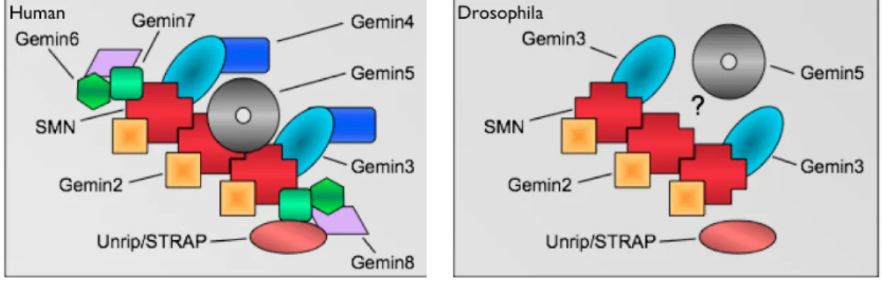
SMN is found as part of a large multimeric complex consisting of SMN, the Gemins, and Unrip in the cytoplasm (Charroux et al. 1999; Charroux et al. 2000; Baccon et al. 2002; Gubitz et al. 2002; Pellizzoni et al. 2002; Grimmler et al. 2005; Carissimi et al. 2006) (Fig. 1.3). The entire SMN complex is required for proper snRNP assembly in vivo. SMN and Gemin2 alone are not sufficient to restore RNP assembly activity in
Xenopus egg extracts immunodepleted for these proteins (Meister et al. 2001). A number
Figure 1.3. Comparison of core SMN complex members in human (mammal) and
Drosophila. Only verified members are shown in each representation and the
stoichiometry is not intended to reflect true relative amounts of protein. The human SMN complex includes Gemin2/3/4/5/6/7/8 and Unrip/STRAP. Drosophila has a lesser verified SMN complex, consisting only of Gemin2/3 and Unrip. The Drosophila homolog of Gemin5, rig, is likely a member of the complex. However, rig has not been definitively shown to play a role in snRNP biogenesis. Figure from Greg Matera, unpublished.
While orthologs of Gemin2 can be identified in all species, the other Gemins are not as well conserved (Fig. 1.3). A bioinformatic investigation to identify Gemin
homologs suggested that Gemins 3 and 5 are the most ancestral Gemins in the complex (Kroiss et al. 2008). Putative homologs of Gemins 4, 6, 7, and 8 were only found in animals at the time, suggesting they are newer additions to the complex. Both Dipterans that were analysed, D.melanogaster and A.gambiae, seemed to have only Gemins 2, 3 and 5 (Fig. 1.3).
snRNA complex throughout the cytoplasmic phase of assembly (Massenet et al. 2002) and is also important for re-import of the immature snRNP into the nucleus (Narayanan et al. 2002; Narayanan et al. 2004). This presents the possibility that Gemins have functions in steps following the assembly of Sm proteins onto snRNAs.
The Gemins are very structurally different, and the precise function of most of them in the SMN complex is not clear. However, there are suggestions for the functions of several of them. For example, Gemin2 has been reported to stabilize SMN by
enhancing SMN self-association through the N-terminal Gemin2 binding domain (Ogawa et al. 2007). Additionally, Gemin2 binds a pentamer of the Sm proteins D1, D2, E, F and G directly, as visualized in a crystal structure of Gemin2 with the Gemin2-binding domain of SMN (Zhang et al. 2011). Gemin3 contains a DEAD box domain with
potential helicase activity; therefore, Gemin3 may perform the ATP dependent step of the assembly reaction (Charroux et al. 1999). Gemin5 has been shown to bind snRNAs directly, which could contribute to distinguishing them from other RNAs (Battle et al. 2006). This mechanism provides specificity to the assembly reaction (Battle et al. 2006). Gemins 6 and 7 form a heterodimer that is similar to the structure created by
SMN Function
A role of SMN in RNA metabolism was the first suggested cellular function (Liu et al. 1996). This was determined due to the observation that SMN protein can associate with the RNA binding domain of hnRNP U, an RNA binding protein. The role of SMN in RNA metabolism was bolstered when, in 1997, Liu et al. showed that SMN, along with Gemin2, co-purified with a set of proteins that bind to uridine-rich small nuclear ribonucleoproteins (U-snRNPs). These proteins are called Sm proteins. Fischer et al. (1997) determined the functional significance of the interaction by using Xenopus
oocytes to show that SMN and Gemin2 were involved in an early step in spliceosomal U-snRNP biogenesis. These data were corroborated in 2001 by Meister et al. who showed that the SMN complex was required for in vivo assembly of Sm proteins onto U-snRNAs.
snRNP Biogenesis
SMN is expressed in all tissues of animals (Matera and Wang 2014; Tripsianes et al. 2011; Li et al. 2014). The best-characterized function for the ubiquitous SMN protein is in the assembly of Sm-class snRNPs. Sm-class snRNPs are made up of uridine-rich snRNAs, non-coding RNAs that perform diverse roles in RNA metabolism (Mattaj et al. 1993; Tern and Steitz, 1997). Sm-class snRNPs also contain several specific proteins that are unique to each snRNA and a set of 7 common Sm proteins.
require higher levels of the components of the major-class than there are of the minor-class. Indeed, the major-class snRNAs are ~100 fold more abundant than the snRNAs that make up the minor-class spliceosome (Zieve and Sauterer 1990). U1, U2, U4 and U6 snRNAs make up the major-class and U11, U12, U4atac and U6atac snRNAs comprise the minor-class (Levine and Durbin 2001). The U5 snRNA is shared by both
spliceosomes (Patel and Steitz 2003).
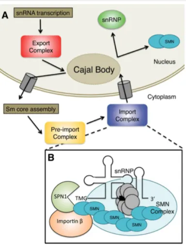
Importin β, where it localizes to the Cajal body. There, the snRNA binds other proteins
and acquires further modifications. Figure from Raimer, Gray, and Matera 2016.
The life cycle of the Sm-class U-snRNAs takes place in both the cytoplasm and the nucleus (Fig. 1.4). Sm-class snRNAs are transcribed by RNA polymerase II and contain additional nucleotides at the 3’ end and a monomethylated m7GpppG (m7G) cap structure at the 5’ end (Cougot et al. 2004) (Fig 1.4A). After 3’ end processing to remove the extraneous nucleotides, pre-snRNA transcripts are exported from the nucleus by a set of factors that includes the cap-binding complex (CBP80 and CBP20), the snRNA-specific export adaptor phosphorylated adaptor RNA export (PHAX), and arsenite resistance 2 (ARS2) (Hallais et al. 2013; Frey et al. 1995; Frey et al. 1999; Ohno et al. 2000; Frey et al. 2001; Suzuki et al. 2010). These proteins link the 5’ cap of the snRNA to the nuclear export receptor chromosome region maintenance 1 (CRM1/Exportin1). This entire complex interacts with nuclear pore proteins to promote export to the cytoplasm (Fornerod et al. 1997). The snRNA nuclear export complex dissociates upon phosphorylation of PHAX in the cytoplasm (Kitao et al. 2008; Ohno et al. 2000). The SMN protein complex regulates the entire cytoplasmic phase of the snRNP cycle (Fig. 1.4B). Specific phases of snRNP biogenesis in the cytoplasm regulated by SMN include Sm core assembly, trimethylguanosine (TMG) cap formation, and Snurportin1 binding to the TMG cap structure.
pre-snRNA called the ‘Sm-site’ to form a ring (Kambach et al. 1999; Will et al. 2001; Meister et al. 2002; Pellizzoni et al. 2002; Yong et al. 2004; Golembe et al. 2005; Paushkin et al. 2002). Assembly of the Sm core not only stabilizes the snRNA by protecting it from nucleases, but also is required for downstream RNA-processing steps.
The Sm proteins are delivered to the SMN complex due to the activity of the protein arginine methyltransferase 5 (PRMT5) complex, consisting of PRMT5, pICln, and WD45 (Mep50) (Brahms et al. 2000; Brahams et al. 2001; Friesen et al. 2001; Meister et al. 2001; Friesen et al. 2002). The PRMT5 complex symmetrically
dimethylates C-terminal arginine residues within SmB, SmD1, and SmD3 (Meister et al. 2001; Friesen et al. 2001). These methylation marks enhance the interaction between the Sm proteins and SMN. In Drosophila, Sm protein methylation is not necessary for snRNP assembly (Gonsalvez et al. 2008). Thus, while many of the biochemical properties of snRNP biogenesis are conserved between flies and mammals, this is an important caveat to consider.
Following Sm-core assembly, an RNA methyltransferase called
trimethylguanosine synthase (TGS1) is recruited to the m7G cap (Mouaikel et al. 2002; Verheggen et al. 2002). The SMN complex does not immediately dissociate from the RNA after Sm-core assembly, suggesting that SMN may play a role in the recruitment of TGS1 to the complex (Mouaikel et al. 2003). Additionally, it has been shown that TGS1 directly interacts with SMN both in vivo and in vitro supporting the role of SMN in recruitment. TGS1 hypermethylates the cap to form a 2,2,7-trimethylguanosine (TMG) cap structure. A properly assembled Sm core is required for this process as well as for 3’- end maturation (Mouaikel et al. 2002; Mattaj 1986; Neuman de Vegvar and Dahlberg 1990).
Once the cytoplasmic phase of snRNP biogenesis is complete, Importinβ (Impβ)
binds the import adaptor Snurportin 1 (SPN1) that attaches to the TMG cap and imports the partially assembled pre-snRNP, along with the SMN complex, back into the nucleus (Fig. 1.4B) (Palacios et al. 1997; Huber et al. 1998; Narayanan et al. 2004). Interaction between SMN and Impβ, and observations that snRNP import is defective in the presence
of some SMN mutations, indicate that SMN may also function in facilitating snRNA nuclear import (Narayanan et al. 2002; Narayanan et al. 2004). This means it is possible that SMN plays a role in every stage of snRNP development, in both the cytoplasm and the nucleus.
The process of snRNP assembly and import is rapid, taking place in
stored in nuclear domains called speckles, where snRNPs are thought to be kept while not participating in splicing (Sleeman and Lamond 1999; Fakan 1994). snRNPs may also localize to active transcription sites in perichromatin fibrils where they actively participate in splicing.
Two spliceosomal snRNAs, U6 and U6atac, have not been discussed thus far and do not follow the same assembly pathway as the others. U6 and U6atac are transcribed by RNA polymerase III and acquire a γ-monomethyl cap after transcription. They bound
by seven Sm-like (Lsm) proteins (Lsm2-Lsm8) (Achsel et al. 1999), and are referred to as “Lsm-class” snRNAs. These Lsm proteins are used in place of the Sm proteins that assemble around the other snRNAs. The assembly of U6 and U6atac is thought to occur entirely within the nucleus and appears to be independent of SMN.
snRNP independent functions of SMN
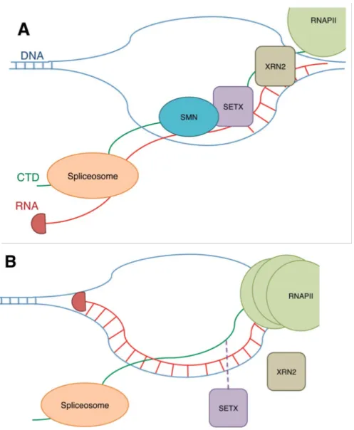
Although snRNP biogenesis remains by far the most well established function of SMN, the protein has also been implicated in other global and tissue-specific roles. For example, SMN has been reported to influence the activity of a viral transcription activator (Strasswimmer et al. 1999). SMN has also been suggested to interact with RNA helicase A and RNA polymerase II (Pellizzoni et al. 2001), invoking the possibility that it may function in transcriptional regulation. Further evidence supporting this function of SMN was provided when Zhao et al. (2015) demonstrated that SMN binds the RNA
XRN2 exonuclease to be recruited, thereby contributing to transcription termination (Skourti-Stathaki et al. 2011).
Figure 1.5. The potential role of SMN in transcriptional regulation. A. SMN interaction with R1810me2s on the C-terminal domain (CTD) of RNA polymerase II (RNAPII) stabilizes SETX, an RNA/DNA helicase, interaction with the CTD. SETX allows the spliceosome to access and splice the RNA by preventing R-loop formation. B. Following reduction of SMN or loss of R1810 methylation, SETX is not recruited as efficiently resulting in an increase in R-loop formation. RNAPII accumulates at the transcription termination site. Figure from Raimer, Gray, and Matera 2016.
connections to Profilin and Plastin3, which are actin bundling proteins (Giesemann et al. 1999; Sharma et al. 2005; Oprea et al. 2008; Ackermann et al. 2013). This function of SMN may be the primary cause for defects observed in SMA, such as problems with endocytosis at the synapse (Custer and Androphy 2014; Dimitriadi et al. 2016;
Hosseinibarkooie et al. 2016). The localization of SMN to growth cones in differentiating neurons (Fan and Simard 2002; Sharma et al. 2005) along with defects in axonal growth of motor neurons in mouse and zebrafish models of SMA (McWhorter et al. 2003;
Rossoll et al. 2003) indicate a possible function for SMN in neurite outgrowth and axonal pathfinding.
One of the most well-supported snRNP independent roles for SMN is in mRNP assembly and transport along motor axons (Rossoll et al. 2003). Evidence for this role includes defects in localization of β-actin mRNA in developing motor neurons of SMA
mice. SMN may regulate levels of other proteins through a connection with phosphatase and tensin homolog-mediated (PTEN-mediated) protein synthesis pathways (Ning et al. 2010) and other modes of translational regulation (Sanchez et al. 2013). SMN has also been implicated in neuromuscular junction (NMJ) formation and function (Fan et al. 2002; Chan et al. 2003; Kariya et al. 2008; Kong et al. 2009; Voigt et al. 2010).
It is challenging to distinguish between primary and secondary effects in neurons and muscles in SMA models since cell autonomy is difficult to establish when the functions of motor neurons and muscles are highly interconnected. Nonetheless, SMN has been suggested to play a role in some muscle-specific functions. Mouse cells
The severity of these defects was proportional to the level of SMN expression. Rajendra et al. (2007) observed localization of SMN to sarcomeres, the contractile units of muscle fibers, in Drosophila and in mice. The final piece of evidence for a muscle specific function of SMN is that flight muscles in an adult Drosophila model of SMA were severely disorganized, suggesting a role for SMN in maintenance of muscle architecture (Walker et al. 2008; Bowerman et al. 2009).
The central question facing researchers in the SMA field is how the loss of a protein with an essential and ubiquitous function can cause a primarily neuromuscular disease. Current hypotheses suggest that certain tissues, such as neurons or muscles, may have a greater requirement for snRNPs thus making them more sensitive to low levels of SMN and more susceptible to defects in snRNP biogenesis. It is also possible that disruptions to the tissue-specific functions of SMN lead to SMA. These hypotheses need not be mutually exclusive.
Modeling SMA
Drosophila SMA models
Integral aspects of cell and developmental biology in humans are conserved in
Drosophila. For example, approximately 75% of disease-causing loci in humans have
resembles that of vertebrates in many ways, thus making Drosophila well-suited for study of disorders such as SMA (Fernandes et al. 1999).
The DrosophilaSmn ortholog is an intron-less gene that codes for a 226 amino
acid protein. Due to the absence of introns, the mechanism of splicing of SMN2 that occurs in humans is not optimally studied in this model system. The human and fly homologs share 23.5% identity and 36.7% similarity; therefore, several other elements of SMN biology can be effectively modeled. The regions of the protein showing greatest conservation correspond to the Gemin2 binding site near the N-terminus, the Tudor domain, and the YG box self-oligomerization domain at the C-terminus (Fig. 1.6) (Miguel-Aliaga et al. 2000). The Drosophila SMN complex participates in the assembly of Sm proteins onto snRNAs, indicating that the function of human SMN in snRNP biogenesis is conserved in the fly (Rajendra et al. 2007).
Figure 1.6. SMA patient mutations in the Drosophila Smn gene. Many point mutations in SMN1 that cause SMA are at amino acid residues that are conserved in Drosophila. These point mutations span all three known functional domains of SMN, the Gemin2 binding domain, the tudor domain, and the YG Box self-oligomerization domain. This point mutations lead to mild, intermediate, and severe defects in the fly, thus representing the varying severity of the disease observed in humans. Figure modified from the thesis of Kavita Praveen (Praveen, 2012).
Chan et al. (2003) isolated two point mutations in the YG box
mobility and coordination. Several additional Smn mutants have also been generated via transposon-mediated mutagenesis (Rajendra et al. 2007; Shpargel et al. 2009). These animals vary in the severity of their phenotype based on the location of the insertion. Using one of these lines that contained a transposon insertion upstream of Smn, an Smn null allele was used in an imprecise excision screen to generate a deletion that removes the promoter, open reading frame, and part of the 3` UTR of SMN (Chang et al. 2008). This fly model of SMA, called SmnX7, is useful for the study of larval phenotypes but not adult phenotypes since animals die as larvae without pupating. In addition, these models do not recapitulate human SMA, which is caused by a reduction, but not complete loss, of functional protein.
The number of Drosophila melanogaster SMA models expanded when a series of flies were developed where the endogenous Smn gene is replaced with a Flag-Smn
transgene expressing either wild-type SMN or SMN containing patient-derived point mutations at conserved amino acids (Fig. 1.6) (Praveen et al. 2012; Praveen et al. 2014). Although it is highly similar to human SMN1 and SMN2, the entire open reading frame of fruitfly Smn is contained within a single exon, and so only full-length SMN protein is expressed in Drosophila (Rajendra et al. 2007). Without complications introduced by splicing, the fruitfly is an excellent model for investigating the functions of the protein in isolation. When modeled in the fly, SMA-causing point mutations recapitulate the range of phenotypic severity seen in humans (Praveen et al. 2014; Garcia et al. 2016).
Other animal models of SMA
2000; Owen et al. 2000; McWhorter et al. 2003). Severe reduction or complete knockout of Smn in all of these organisms is lethal. Moderate reductions in SMN levels in C.
elegans resulted in an uncoordinated phenotype and lack of muscle tone. This ultimately
resulted in paralysis in this model system. Moderate loss of SMN in zebrafish led to defects in motor axon pathfinding. This occurred without defects in muscles or overt movement problems.
To achieve a milder reduction in SMN levels and obtain a mouse model that more closely resembled the human disorder, two groups generated mice that expressed the human SMN2 gene in the background of a homozygous mouse Smn mutation (Smn-/-;
SMN2) (Hsieh-Li et al. 2000; Monani et al. 2000). The inclusion of the human SMN2
gene in the mouse genome meant that the splicing pattern of the gene in humans was also a factor in this model. This rescued the embryonic lethality of Smn-/- mice and they presented with many of the pathological features observed in SMA patients. These include a shorter lifespan, motor neuron degeneration, and developing muscle weakness over time (Monani et al. 2000). As observed in the human disease, varying the number of copies of SMN2 varied the severity of the phenotype from that resembling type I (1 or 2 copies) patients to complete rescue (8-10 copies). In this way, the full spectrum of the human disease can be represented in this model.
SMNΔ7 transcript, meaning that its splicing cannot be modulated, in addition to having a copy of human SMN2. The 2B/– mouse (Smn2B/–) is a model of intermediate SMA (Bowerman et al. 2012; Rindt et al. 2015) and these animals survive much longer before dying, typically between P25 and P45. This provides a longer window for conducting experiments, which can provide insights into disease pathology that cannot be gained when animals die very early.
Oligomeric properties of SMN complexes
The C-terminal YG box self-oligomerization domain of SMN has been examined using X-ray crystallographic studies (Martin et al. 2012; Gupta et al. 2015). Hydrophobic interactions, similar to those found in glycine zipper domains of certain transmembrane channel proteins, are responsible for dimerization of the protein. (Fig. 1.2). The core of this YG box helical domain contains a highly conserved YxxxYxxxY motif.
More than half of the known SMA patient missense mutations are found within the YG box (Burghes et al. 2009; Li et al. 2014; Wirth 2000). Speaking to the importance of oligomerization in SMN function, mutations that completely disrupt SMN’s ability to self-interact display severe phenotypes in human SMA patients and in animal models (Lefebvre et al. 1997; Lorson et al. 1998; Praveen et al. 2014; Clermont et al. 2004; Wirth et al. 1999; Pellizzoni et al. 1999; Workman et al. 2009).
tetramers and octamers (Gupta et al. 2015). There are two possibilities for the way in which SMN tetramers are formed: by a dimer of dimers or by forming symmetric bundles. Further analysis of this question has revealed they are composed of a dimer of dimers.
Only dimers and tetramers are detected in fission yeast, in contrast to human SMN-Gem2, which forms dimers to octamers and possibly even larger complexes. Octamers appear to form due to self-association of tetramers, although a hexameric SMN complex cannot be ruled out. Human SMN-Gem2 co-sediments with Gemins3–8 in vivo (Paushkin et al. 2000), however, the relative stoichiometries of these proteins are unknown.
SMN Protein Stability
The stability of full-length SMN and SMNΔ7 protein has been the subject of many studies, given that SMA is caused by low levels of SMN protein. SMNΔ7 protein produced from the SMN2 gene is known to be unstable. The SMNΔ7 protein has a twofold shorter half-life than full-length SMN in cells (Burnett et al. 2009). SMNΔ7 is also not thought to be fully functional compared with full-length SMN protein (Lorson et al. 1998; Pellizzoni et al. 1999). Biochemical studies have shown that SMNΔ7 protein does not oligomerize well and interactions with established binding partners, such as the snRNP Sm proteins, are decreased. Mutations in full-length SMN that inhibit
oligomerization and prevent complex formation also reduce half-life, suggesting that SMN protein stability is modulated by complex formation (Burnett et al. 2009).
the C-terminus of SMNΔ7 (268YMSGYHTGYYMEMLA282). It was thought to be created in SMN∆7 by SMN2 alternative splicing since it includes the EMLA amino acids that are specific to SMNΔ7. Identified through scanning alanine mutagenesis of the C-terminus, mutation of serine 270 to alanine was shown to stabilize SMN∆7 constructs in human cells. Overexpression of SMN∆7S270A in SMN-deficient chicken DT40 cells rescued their viability, indicating some functionality of stabilized SMN∆7 protein. Factors responsible for specifically mediating SMN∆7 degradation have not been identified.
Intracellular proteins can be degraded or cleaved by a variety of different proteolytic systems including the calcium-activated neutral protease (calpain) system, lysosomal proteases, autophagy, and the ubiquitin proteasome system (UPS). SMN has been shown to be a proteolytic target of Calpain (Walker et al. 2008; Fuentes et al. 2010). Inhibition of the lysosome, autophagy, and the proteasome revealed that only proteasome inhibition significantly increased SMN protein levels. This and other experiments show ubiquitylation pathways regulate the stability and degradation of SMN (Chang et al. 2004; Burnett et al. 2009; Hsu et al. 2010).
Ubiquitin Proteasome System (UPS) and SMN
Figure 1.7. The ubiquitin proteasome system (UPS) involves three types of proteins: E1 ubiquitin activating enzymes, E2 ubiquitin conjugating enzymes, and E3 ubiquitin ligase enzymes. E3 ubiquitin ligases recognize target substrates and ubiquitylate them, leading to their degradation by the proteasome. In other cases, ubiquitylation occurs without leading directly to proteasome targeting and degradation. Figure adapted from Elledge lab, Harvard.
Ubiquitin homeostasis is thought to be especially important for neuromuscular pathology in SMA (Groen and Gillingwater 2015). X-linked infantile SMA is caused by mutations in ubiquitin-like modifier activating enzyme 1 (UBA1) (Ramser et al. 2008; Schmutzler et al. 2008). Furthermore, mouse models of SMA have disrupted levels of the E1 protein, UBA1 (Wishart et al. 2014). Finally, ubiquitylation pathways have been shown to specifically affect axonal and synaptic stability (Korhonen and Lindholm 2004).
part of the UPS for their ubiquitylation targets, investigators have studied E3 ligases of SMN using candidate approaches (Han et al. 2016; Hsu et al. 2010; Kwon et al. 2013). As outlined below, researchers have discovered E3 ligases that target SMN for
degradation in cultured human cells through these studies. It is therefore likely that SMN is targeted by multiple E3 ubiquitin ligases, as this mechanism of regulation has been demonstrated for a number of proteins (e.g. p53; Jain and Barton 2010). Ubiquitylation does not always result in immediate destruction of the target (Mukhopadhyay and Riezman 2007; Ikeda and Dikic 2008; Liu and Walters 2010). Different functions arise based on the type of lysine linkage between ubiquitin molecules and the length of the ubiquitin chain that is created.
Mindbomb1 (Mib1)
Kwon et al. (2013) identified the E3 ubiquitin ligase, mind bomb 1 (Mib1), as interacting with and ubiquitylating SMN, leading to subsequent degradation. It was originally chosen as a candidate E3 ubiquitin ligase due to its role in inhibition of the outgrowth of neurites in cultured neurons (Choe et al. 2007). Additionally, loss of Mib1
in Drosophila melanogaster had been shown to increase the number of synaptic boutons
at neuromuscular junctions, producing synaptic overgrowth. Evidence for Mib1 targeting SMN for degradation included the experiment that revealed Mib1 knockdown in cultured cells increases SMN protein levels. Additionally, in SMN deficient C. elegans,
SMN, demonstrating the interaction is mediated by the N-terminal domain of Mib1 and the part of SMN protein encoded for by exon 6.
Itch
In some cases, protein ubiquitylation does not result in destruction of the target. Different types of ubiquitin lysine linkages or different chain lengths can affect other aspects of protein function within the cell, including cellular localization. SMN is found in both the cytoplasm and nucleus. In the nucleus, SMN is concentrated in nuclear bodies – Cajal bodies, gems, and the nucleolus of neurons (Stanek and Neugebauer, 2006; Liu and Dreyfuss 1996). Posttranslational modifications are thought to affect this cellular localization. Han et al. (2016) reported that the E3 ubiquitin ligase called Itch directly interacts with and monoubiquitylates SMN. This had a modest effect on protein degradation and a more pronounced effect on cellular localization of SMN. This
mislocalization of SMN was shown to impair Cajal body integrity and findings suggested impairment of snRNP maturation.
SCFSlmb/SCFB-TrCP
essential for interaction of Cul1 with the F-box protein (Patton et al. 1998a; Patton et al. 1998b).
The SCFSlmb/SCFB-TrCP complex is one of the best-characterized SCF E3 ligases in animals (Willems et al. 2004). SCFSlmb is the Drosophila melanogaster homolog of SCF
B-TrCP in mammals (Bocca et al. 2001). Initial analysis of this SCF pathway revealed that
SCFSlmb/SCFB-TrCP catalyzes the phosphorylation-dependent ubiquitylation of the NFKB inhibitor IKB and the transcription factor B-catenin (Yaron et al. 1998; Winston et al. 1999). Most known substrates contain the amino acid sequence DGSXXS degron motif or a variant thereof. Both serine, or in some cases threonine, residues need to be
phosphorylated for efficient recognition of the degron in the substrate by the E3 ligase. Since the discovery and characterization of these two substrates of SCFSlmb/SCFB-TrCP numerous additional substrates have been identified using substrate trapping proteomics analysis and other approaches (Kim et al. 2015; Skwarek et al. 2014). The work
described here describes the identification of a novel SCFSlmb/SCFB-TrCP substrate, SMN protein.
Research Objectives
Spinal muscular atrophy is a neuromuscular disorder caused by loss of, or
mutation in, the SMN1 gene. The best-characterized function of SMN is in the biogenesis of snRNPs, core components of the spliceosome. The mechanism of SMA disease
CHAPTER II: Self-oligomeriation regulates stability of Survival Motor
Neuron (SMN) protein isoforms by sequestering an SCF
Slmbdegron
1Introduction
Spinal muscular atrophy (SMA) is a common neuromuscular disorder, recognized as the most prevalent genetic cause of early childhood mortality (Pearn 1980). Patients with the most severe form of the disease, which is also the most common, become
symptomatic in the first six months of life and rarely live past two years (Wee et al. 2010; Prior 2010). Because the onset of symptoms and their severity can vary, SMA has
historically been classified into three subtypes (Ogino and Wilson 2004). More recently, clinicians have recognized that SMA is better characterized as a continuous spectrum disorder, ranging from acute (prenatal onset) to nearly asymptomatic (Tiziano et al. 2013). Clinically, SMA patients experience degeneration of motor neurons in the anterior horn of the lower spinal cord (Crawford and Pardo 1996). This leads to progressive atrophy of proximal muscle groups, ultimately resulting in loss of motor function and symmetrical paralysis. The cause of death is often restrictive respiratory failure (Kolb and Kissell 2015).
SMA typically results from homozygous deletion of the survival motor neuron 1 (SMN1) gene (Lefebvre et al. 1995). A small fraction of SMA patients have lost one copy of SMN1 and the remaining copy contains a point mutation (Burghes and Beattie 2009).
1 This chapter previously appeared as an article in Molecular Biology of the Cell. The original citation is as
Humans have two SMN paralogs, named SMN1 and SMN2, both of which contribute to total cellular levels of SMN protein. SMN2 exon 7 contains a silent base change that alters splicing to primarily produce a truncated, unstable protein product called SMN∆7 (Lorson et al. 1999; Monani et al. 1999; Lorson and Androphy 2000). The last 16 amino acids of SMN are replaced in SMNΔ7 by four amino acids, EMLA, encoded by exon 8. Current estimates suggest that SMN2 produces 10-15% of the level of full-length protein produced by SMN1 (Lorson et al. 2010). Complete loss of SMN is lethal in all organisms investigated to date (O’Hearn et al. 2016). Although the amount of full-length protein produced by SMN2 is not enough to compensate for loss of SMN1, SMN2 is sufficient to rescue embryonic lethality (Monani et al. 2000). SMA is therefore a disease that arises due to a hypomorphic reduction in SMN levels (Lefebvre et al. 1995). Furthermore, relative levels of the SMN protein correlate with the phenotypic severity of SMA (Coovert et al. 1997; Lefebvre et al. 1997).
Whereas a causative link between SMN1 and SMA was established over 20 years ago, the molecular role of SMN in disease etiology remains unclear. SMN is the central component of a multimeric protein assemblage known as the SMN complex (Matera and Wang 2014; Li et al. 2014). The best-characterized function of this complex, which is found in all tissues of metazoan organisms, is in the cytoplasmic assembly of small nuclear ribonucleoproteins (snRNPs), core components of the spliceosome (Fischer et al. 1997; Meister et al. 2001; Pellizzoni et al. 2002).
Simard 2002; McWhorter et al. 2003; Sharma et al. 2005), axonal transport of β-actin
mRNP (Rossoll et al. 2003), phosphatase and tensin homolog-mediated (PTEN-mediated) protein synthesis pathways (Ning et al. 2010), translational regulation
(Sanchez et al. 2013), neuromuscular junction formation and function (Chan et al. 2003; Kariya et al. 2008; Kong et al. 2009; Voigt et al. 2010), myoblast fusion (Shafey et al. 2005) and maintenance of muscle architecture (Rajendra et al. 2007; Walker et al. 2008; Bowerman et al. 2009).
Ubiquitylation pathways have been shown to regulate the stability and
degradation of SMN (Chang et al. 2004; Burnett et al. 2009; Hsu et al. 2010) as well as axonal and synaptic stability (Korhonen and Lindholm 2004). In the ubiquitin proteasome system (UPS), proteins destined for degradation are tagged by linkage to ubiquitin
through the action of three factors (Petroski 2008). E1 proteins activate ubiquitin and transfer it to the E2 enzyme. E2 proteins conjugate ubiquitin to their substrates. E3 proteins recognize the substrate and assist in the transfer of ubiquitin from the E2. Because E3 ligases confer substrate specificity, they are typically considered as candidates for targeted inhibition of protein degradation. Ubiquitin homeostasis is thought to be particularly important for neuromuscular pathology in SMA (Groen and Gillingwater 2015). Indeed, mouse models of SMA display widespread perturbations in UBA1 (ubiquitin-like modifier activating enzyme 1) levels (Wishart et al. 2014).
Furthermore, mutations in UBA1 are known to cause X-linked infantile SMA (Ramser et al. 2008; Schmutzler et al. 2008).
partners. Previously, we developed Drosophila melanogaster as a model system wherein the endogenous Smn gene is replaced with a Flag-Smn transgene (Praveen et al. 2012). Although it is highly similar to human SMN1 and SMN2, the entire open reading frame of fruitfly Smn is contained within a single exon, and so only full-length SMN protein is expressed in Drosophila (Rajendra et al. 2007). When modeled in the fly, SMA-causing point mutations recapitulate the full range of phenotypic severity seen in humans
(Praveen et al. 2014; Garcia et al. 2016). Using this system, we carried out proteomic profiling of Flag-purified embryonic lysates and identified the SCFSlmb E3 ubiquitin ligase complex as a novel SMN interactor. Importantly, this interaction is conserved from flies to humans. We show that SCFSlmb binding requires a phospho-degron motif located within the SMN self-oligomerization domain, mutation of which stabilizes SMN∆7 and, to a lesser extent, full-length SMN. Additional studies in flies, mice and human cells elucidate a disease-relevant mechanism whereby SMN protein stability is regulated by self-oligomerization. Other E3 ligases have been reported to target SMN for degradation in cultured human cells (Han et al. 2016; Hsu et al. 2010; Kwon et al. 2013). Given our findings in fruit fly embryos, SMN is likely targeted by multiple E3 ubiquitin ligases.
Experimental procedures
Fly stocks and transgenes
constructs were injected into embryos by BestGene Inc. (Chino Hills, CA) as described in Praveen et al. 2014. In short, a ~3kb fragment containing the entire Smn coding region was cloned from the Drosophila genome into the pAttB vector (Bischof et al. 2007). A 3X FLAG tag was inserted immediately downstream of the start codon of dSMN. Point mutations were introduced into this construct using Q5 (NEB) site-directed mutagenesis according to manufacturer’s instructions. The basal Smn construct used, vSmn, contained three single amino acid changes and the addition of the MGLR motif to make fruitfly Smn more similar to the evolutionarily conserved vertebrate Smn. Subsequently generated constructs used vSmn as a template and consist of the amino acid changes detailed in Figure 4. Y203C, G206S, and G210V were previously published in Praveen et al. 2014.
Drosophila embryo protein lysate and mass spectrometry
total of six times using buffer with KCl concentrations ranging from 100mM to 250mM with rotation for 1 min at 4C in between each wash. Finally, Flag proteins were eluted 3 consecutive times with one bed volume of elution buffer (Tris 20mM pH 8, 100 mM KCl, 10% glycerol, 0.5 mM DTT, PMSF 0.2 mM) containing 250ug/mL 3XFLAG peptide (sigma). The entire eluate was used for mass spectrometry analysis on an Orbitrap Velos instrument, fitted with a Thermo Easy-spray 50cm column.
Tissue culture and transfections
S2 cell lines were obtained from the Drosophila Genome Resource Center (Bloomington, IL). S2 cells were maintained in SF900 SFM (Gibco) supplemented with 1% penicillin/streptomycin and filter sterilized. Cells were removed from the flask using a cell scraper and passaged to maintain a density of approximately 106-107 cells/mL. S2 cells were transferred to filter sterilized SF900 SFM (Gibco) without antibiotics prior to transfection with Cellfectin II (Invitrogen). Transfections were performed according to Cellfectin II protocol in a final volume of 4 mL in a T-25 flask containing 107 cells that were plated one hour before transfection. The total amount of DNA used in transfections was 2.5ug. Human embryonic kidney HEK-293T and HeLa cells were maintained at 37C with 5% CO2 in DMEM (Gibco) supplemented with 10% FBS and 1%
penicillin/streptomycin (Gibco). 1x10⁶ - 2x10⁶ cells were plated in T-25 flasks and transiently transfected with 1-2ug of plasmid DNA per flask using Lipofectamine
(Invitrogen) or FuGENE HD transfection reagent (Roche Applied Science, Indianapolis, IN) according to the manufacturer’s protocol. Cells were harvested 24-72 h
For siRNA transections, HeLa cells were plated subconfluently in T-25 flasks and transfected with 10nm of siRNA (Gift from Mike Emanuele lab) and 17uL Lipofectamine RNAi MAX (Invitrogen) in 5mL total media according to manufacturers instructions. After 48h of transfection cells were harvested. For RNAi in S2 cells using dsRNA, 107 cells were plated in each well of a 6-well plate in 1 mL of media. Cells were treated ~ every 24h with 10ug/mL dsRNA targeted against Slmb, Oskar, or Gaussia Luciferaese (as controls) as described in Rogers and Rogers 2008.
In vitro binding assay
GST and GST-SMN were purified from E. coli. In brief, cells transformed with BL21*GST-SMN were grown at 37˚C overnight and then induced using 1 mM IPTG. Recombinant protein was extracted and purified using Glutathione sepharose 4B beads. GST-B-TrCP1 was purchased from Novus Biologicals (cat# H00008945). SMNGem2 complexes were co-expressed in E. coli using SMN∆5 and Gemin2(12-280) constructs, as described in Gupta et al. (2015). Glutathione sepharose 4B beads were washed 3x with PBS. GST alone, GST-SMN, or GST-B-TrCP1 were attached to beads during
In vivo ubiquitylation assay
The in vivo ubiquitylation assay was performed as described previously
(Choudhury et al. 2016). Briefly, HEK-293T cells were transfected as indicated in 10 cm dishes using Lipofectamine2000 (Thermo Fisher Scientific). The day after, cells were treated with 20 µM of MG132 for 4 hours and then harvested in PBS. 80% of the cell suspension was lysed in 6M Guanidine-HCl containing buffer and used to pull down His-Ubiquitinated proteins on Ni2+-NTA beads, while the remaining 20% was used to prepare inputs. Ni2+ pull down eluates and inputs were separated through SDS-PAGE and analyzed by western blot.
Cycloheximide treatment
Following RNAi treatment, S2 cells were pooled, centrifuged and resuspended in fresh media. 1/3 of these cells were frozen and taken as the 0h timepoint. The remainders of the cells were replated in 6 well plates. 100ug/mL cycloheximide (CHX) was added to each sample, and cells were harvested at 2 and 6 hours following treatment.
Immunoprecipitation
Antibodies and Western blotting
Larval and adult lysates were prepared by crushing the animals in lysis buffer (50mM Tris-HCl, pH 7.5, 150 mM NaCl, 1mM EDTA, 1% NP-40) with 1X (adults) or 10x (larvae) protease inhibitor cocktail (Invitrogen) and clearing the lysate by
centrifugation at 13,000 RPM for 10 min at 4ºC. S2 cell lysates were prepared by suspending cells in lysis buffer (50mM Tris-HCl, pH 7.5, 150 mM NaCl, 1mM EDTA, 1% NP-40) with 10% glycerol and 1x protease inhibitor cocktail (Invitrogen) and disrupting cell membranes by pulling the suspension through a 25 gauge needle (Becton Dickinson). The lysate was then cleared by centrifugation at 13,000 RPM for 10 min at 4ºC. Human cells (293Ts and HeLas) were first gently washed in ice-cold 1X PBS, then collected in ice-cold 1X PBS by scraping. Cells were pelleted by spinning at 1000 rpm for 5 min. The supernatant was removed and cells were resuspended in ice cold lysis buffer (50mM Tris-HCl, pH 7.5, 150 mM NaCl, 1mM EDTA, 1% NP-40) and allowed to lyse on ice for 30 min. After lysing, the lysate was cleared by centrifuging the cells for 10 min at 13000 at 4C. Western blotting on lysates was performed using standard protocols. Rabbit anti-dSMN serum was generated by injecting rabbits with purified full-length dSMN protein (Pacific Immunology Corp, CA), and was subsequently affinity purified. For Western blotting, dilutions of 1 in 2,500 for the affinity purified anti-dSMN, 1 in 20,000 (fly) or 1 in 5,000 (human) for anti-α tubulin (Sigma), 1 in 10,000 for monoclonal
Larval locomotion
Smn control and mutant larvae (73-77 hours post egg-laying) were placed on a 1.5% agarose molasses tray five at a time. The tray was then placed in a box with a camera and the larvae were recorded moving freely for 60 seconds. Each set of larvae was recorded three times, and one video was chosen for analysis based on video quality. The videos were then converted to AVI files and analyzed using the wrMTrck plug-in of the Fiji software. The "Body Lengths per Second" was calculated in wrMTrck by dividing the track length by half the perimeter and time (seconds). P-values were generated using a multiple comparison ANOVA.
SMA Mouse Models
Two previously developed SMA mouse models were utilized. The ‘Delta7’ mouse (Smn−/−;SMN2;SMNΔ7), is a model of severe SMA (Le et al. 2005). The ‘2B/–‘ mouse (Smn2B/–) is a model of intermediate SMA (Bowerman et al. 2012; Rindt et al. 2015). Adeno-associated virus serotype 9 (AAV9) delivered SMN cDNA isoforms to these SMA mice, as previously described (Foust et al. 2010; Passini et al. 2010; Valori et al. 2010; Dominguez et al. 2011; Glascock et al. 2012). Gross motor function was
measured using a modified tube-test which tests the ability of mice to splay their legs and maintain a hanging position.
Human iPSC Cell culture
medium Stemline (Sigma) supplemented with 100ng/ml human basic fibroblast growth factor (FGF-2, Miltenyi), 100ng/ml epidermal growth factor (EGF, Miltenyi), and 5µg/ml heparin (Sigma-Aldrich) in ultra-low attachment flasks. Aggregates were passaged using a manual chopping technique as previously described (Svendsen et al. 1998; Ebert et al. 2013). To induce motor neuron differentiation, neural progenitor cells were cultured in neural induction medium (1:1 DMEM/F12 (Gibco), 1x N2 Supplement (Gibco), 5µg/mL
Heparin (Sigma), 1x Non-Essential Amino Acids (Gibco), and 1x Antibiotic-Antimycotic (Gibco)) plus 0.1µM all-trans retinoic acid (RA) for two weeks; 1µM Purmorphamine
(PMN, Stemgent) was added during the second week. Spheres were then dissociated with TrypLE Express (Gibco) and plated onto Matrigel-coated 12mm coverslips in NIM plus 1µM RA, 1µM PMN, 1x B27 Supplement (Gibco), 200ng/mL Ascorbic Acid (Sigma),
1µM cAMP (Sigma), 10ng/mL BDNF (Peprotech), 10ng/mL GDNF (Peprotech)). One
week post-plating, cells were infected with lentiviral vectors (MOI = 5) expressing mCherry alone or SMN S270A-IRES-mCherry. Transgenes in both viruses were under the control of the EF1α promoter. Uninfected cells served as controls. Cells were analyzed at 1 and 3 weeks post-infections, which was 4 and 6 weeks of total differentiation (Ebert et al. 2009; Sareen et al. 2013).
Immunocytochemistry
room temperature. Finally, nuclei were labeled with Hoechst nuclear stain (Sigma) to label DNA and mounted onto glass slides using FluoroMount medium
(SouthernBiotech). Primary antibodies used were mouse anti-32 (Covance SMI-32R, 1:1000) and rabbit anti-mCherry (ThermoFisher, 1:1000). Secondary antibodies used were donkey anti-rabbit Cy3 (Jackson Immunoresearch 711-165-152) and donkey anti-mouse AF488 (Invitrogen A21202).
Immunocytochemical Analysis
Images were acquired from five random fields per coverslip using an inverted fluorescent microscope (Nikon) and NIS Elements software. Images were blinded and manually analyzed for antigen specificity with NIS Elements software.
Results
Flag-SMN interacts with UPS (ubiquitin proteasome system) proteins
We collected (0-12h) embryos from Flag-SmnWT/WT,SmnX7/D (SMN) and Oregon-R (Ctrl) animals and analyzed Flag-purified lysates by ‘label-free’ mass spectrometry. In addition to Flag-SMN, we identified SMN complex components Gemin2 and Gemin3, along with all seven of the canonical Sm-core snRNP proteins (Fig. 1A). We also identified the U7-specific Sm-like heterodimer Lsm10/11 (Pillai et al. 2003) and the Gemin5 orthologue, Rigor mortis (Gates et al. 2004). Previous studies of Schneider2 (S2) cells transfected with epitope-tagged Smn had identified most of the proteins listed above as SMN binding partners in Drosophila (Kroiss et al. 2008). However, despite
bioinformatic and cell biological data indicating that Rigor mortis is part of the fruit fly SMN complex, this protein failed to co-purify with SMN in S2 cells (Kroiss et al. 2008; Cauchi et al. 2010; Guruharsha et al. 2011). On the basis of our purification data, we conclude that the conditions are effective and that Rigor mortis/Gemin5 is an integral member of the SMN complex in flies.
F-box protein and is the substrate recognition component (Jiang and Struhl 1998). SkpA is a bridging protein essential for interaction of Cul1 with the F-box protein (Patton et al. 1998a; Patton et al. 1998b). Because of its role in substrate recognition, Slmb is likely to be the direct interacting partner of SMN within the SCFSlmb complex. For this reason, we focused on Slmb for the initial validation. As shown, Slmb was easily detectable in Flag-purified eluates from embryos expressing Flag-SMN and nearly undetectable in those from control embryos (Fig. 1D). SmB and SmD3 were also easily detectable by western blot in Flag-purified embryonic lysates and were used as positive controls for known protein interaction partners of SMN. Tubulin and α-Actinin were not detected as
SMN interaction partners. Flag-SMN was successfully purified from SMN embryos, but was undetectable in the control. As positive controls for known protein interaction partners of SMN, SmB and SmD3 were also easily detectable by western blotting using anti-Sm antibodies. The presence of Slmb was verified using anti-Slmb. α-Actinin and Tubulin were not enriched in our purification and are used as negative controls to demonstrate specificity.
SCFSlmb is a bona fide SMN interaction partner that ubiquitylates SMN
As an E3 ubiquitin ligase, the SCFSlmb complex is a substrate recognition
component of the ubiquitin proteasome system. As outlined in Fig. 2A, E3 ligases work with E1 and E2 proteins to ubiquitylate their targets. The interaction of SCFSlmb with SMN was verified in a reciprocal co-immunoprecipitation, demonstrating that Flag-tagged SCF components form complexes with endogenous SMN (Fig. 2B) in S2 cells. SCF complexes are highly conserved from flies to humans: SkpA is 77% identical to human Skp1, Cul1 is 63% identical, and Slmb is 80% identical to its human homologs, B-TrCP1 and B-TrCP2. Slmb/B-TrCP is the SCF component that directly contacts substrates of the E3 ligase. We therefore tested the interaction of recombinant human SMN in complex with Gemin2 (SMNGem2) (Gupta et al. 2015) with GST-tagged B-TrCP1 and -SMN proteins in an in vitro binding assay. As shown in Fig. 2C,
demonstrate a conserved interaction between SMN and the SCFSlmb/B-TrCP E3 ubiquitin ligase complex.
followed by immunodetection of SMN. F. Protein lysate from HEK 293T cells transfected with 6xHis-Flag-ubiquitin (6HF-Ub) and GFP-SMN was purified using a Ni2+ pull down for the tagged ubiquitin. Baseline levels of ubiquitylated GFP-SMN were detected using anti-GFP antibody. Following transfection of TrCP1 or Flag-B-TrCP2, the levels of ubiquitylated SMN markedly increased. Ubiquitylation levels were further increased following addition of both proteins together. In the input, GFP-SMN was detected using anti-GFP antibody, Flag-B-TrCP1 and Flag-B-TrCP2 were detected using anti-Flag antibody, and GAPDH was detected by anti-GAPDH antibody. In the Ni2+ pull down, ubiquitylated GFP-SMN was detected using anti-GFP antibody and 6HF-Ub was detected using anti-Flag antibody to verify successful pull down of tagged ubiquitin.
Depletion of Slmb/B-TrCP results in a modest increase in SMN levels
Given that one of the primary functions of protein ubiquitylation is to target proteins to the proteasome, we examined whether depletion of Slmb by RNA interference (RNAi) using dsRNA in S2 cells would increase SMN levels (Fig. 3A). Following Slmb RNAi, endogenous SMN levels were modestly increased as compared to cells treated with control dsRNA. We obtained similar results using an siRNA that targets both B-TrCP1 and B-TrCP2 in HeLa cells. As shown in Fig. 3B, we detected a modest increase in levels of full-length SMN following B-TrCP RNAi, but not control RNAi. Next, we treated S2 cells with cycloheximide (CHX), in the presence or absence of dsRNA targeting Slmb, to determine whether differences in SMN levels would be exacerbated when production of new proteins was prevented (Fig. 3C). SMN protein levels were also specifically targeted using dsRNA against Smn as a positive control for the RNAi
Identification and characterization of a Slmb/B-TrCP degradation signal in SMN
Studies of numerous UPS substrates in a variety of species have revealed the presence of degradation signals (called degrons) that are required for proper E3 target recognition and binding. Slmb/B-TrCP canonically recognizes a consensus
DpSGXXpS/T degron, where p indicates a phosphoryl group (Jin et al. 2005; Frescas and Pagano 2008; Fuchs et al. 2004). There are also several known variants of this motif, for example: DDGFVD, SSGYFS, TSGCSS (Kim et al. 2015). As shown in Fig. 4A, we identified a putative Slmb/B-TrCP degron (269MSGYHT274) in the highly conserved self-oligomerization domain (YG Box) of human SMN. Interestingly, this sequence was previously identified as part of a larger degron motif (268YMSGYHTGYYMEMLA282) that was thought to be created in SMN∆7 by SMN2 alternative splicing (Cho and Dreyfuss 2010). In particular, mutation of S270 (S201 in flies) to alanine was shown to dramatically stabilize SMN∆7 constructs in human cells, and overexpression of
SMN∆7S270A in SMN-deficient chicken DT40 cells rescued their viability (Cho and Dreyfuss 2010). However, factors responsible for specifically mediating SMN∆7 degradation have not been identified.
protein (Fig. 4A). To test the effects of overall protein length and distance of the putative degron from the C-terminus, we also generated vSmn constructs that are the same length as SMNΔ7, replacing the MEMLA* motif (the amino acids introduced by human SMN2 splicing) with MGLRQ*, see Fig. 4A. The S201A mutation was created in this construct as well (MGLRQ*S201A). To mimic a constitutively phosphorylated state, we also
introduced serine to aspartate mutations, vSmnS201D and vSmn∆7D. We transfected each of these constructs, Flag-tagged and driven by the native Smn promoter, into S2 cells and measured protein levels by western blotting (Fig. 4B). We note that these constructs are expressed at levels far below those of endogenous SMN protein in S2 cells; moreover, they do not affect levels of endogenous SMN (Fig. S2). As shown, the vSmnS201A and vSMNΔ7A constructs exhibited increased protein levels compared to their serine containing counterparts, whereas levels of the S201D mutants were reduced, suggesting that the phospho-degron motif identified in human SMN∆7 (Cho and Dreyfuss 2010) is also conserved in the fly protein. In addition to examining protein levels of each of these constructs in cell culture, transgenic flies expressing vSmn, vSmnS201A, vSmnΔ7S, and vSmnΔ7A were created. Here again, we observed that the S201A mutation increased protein levels of both full-length SMN and SMN∆7 (Fig. S3).