ONCOGENIC KRAS, GSK-3, NF-κB AND TBK1: THE INTERPLAY AND CONSEQUENCES IN PROMOTING PANCREATIC AND LUNG CANCER
Deepali Bang
A dissertation submitted to the faculty of the University of North Carolina at Chapel Hill in partial fulfillment of the requirements for the degree of Doctor of Philosophy in the Department of Cell and Developmental Biology.
Chapel Hill 2014
Approved by:
Albert Baldwin
Channing Der Jenny Ting
ii © 2014 Deepali Bang
ABSTRACT
Deepali Bang: Oncogenic KRAS, GSK-3, NF-κB and TBK1: the interplay and consequences in promoting pancreatic and lung cancer
(Under the direction of Dr. Albert Baldwin)
The development of KRAS targeted therapy has evolved into targeting the complex signaling pathways activated downstream of oncogenic KRAS and associated with disease progression. Recent focus has been made on targeting the constitutive activation of the transcription factor, NF-κB. However, advancement of this therapeutic strategy is dependent on fully understanding the elusive mechanisms underlying
constitutive NF-κB activity in KRAS driven cancers. Glycogen synthase kinase-3 has previously been implicated in regulating pro-survival NF-κB signaling in pancreatic cancer cells through IκB kinase (IKK), but a distinction between roles of the individual isoforms, GSK-3α and GSK-β has not been done. Moreover, TGF-β activated kinase 1 (TAK1), an upstream regulator of IKK activity, was recently shown to regulate survival in KRAS positive colorectal cancers. Here, we characterize mutant KRAS dependent GSK-3α regulation of NF-κB in pancreatic cancer cells. Our data suggests that mutant KRAS upregulates GSK-3α, which promotes pro-survival canonical NF-κB via
iv
vi
TABLE OF CONTENTS
LIST OF TABLES ... viii
LIST OF FIGURES ... ix
LIST OF ABBREVIATIONS ... xi
CHAPTER I. INTRODUCTION ... 1
1.1 Summary ... 1
1.2 RAS proteins ... 2
1.3 Role of RAS in Cancer ... 3
1.4 Targeting RAS as a therapeutic approach to cancer ... 8
1.5 Constitutive NF-κB in Cancer ... 10
1.6 TAK1-dependent regulation of NF-κB ... 13
1.7 GSK-3-dependent NF-κB Signal Transduction ... 15
1.8 Role of non-canonical IKK, TBK1 in cancer ... 19
1.9 Conclusions ... 21
Figures ... 23
References ... 33
II. GSK-3α PROMOTES ONCOGENIC KRAS FUNCTION IN PANCREATIC CANCER VIA TAK-TAB STABILIZATION AND REGULATION OF NON-CANONICAL NF-κB ... 44
2.2 Introduction ... 45
2.3 Results ... 48
2.4 Discussion ... 58
2.5 Materials and Methods ... 63
Figures ... 67
Supplementary Data ... 76
References ... 87
III. A REQUIREMENT FOR TBK1 IN KRAS-INDUCED LUNG CANCER ... 97
3.1 Summary ... 97
3.2 Introduction ... 97
3.3 Material and Methods ... 100
3.4 Results ... 103
3.5 Discussion and Future Direction ... 107
Figures ... 111
References ... 116
VII. CONCLUSIONS & FUTURE DIRECTIONS 4.1 Conclusions & Future Directions ... 119
viii
LIST OF TABLES
Table
LIST OF FIGURES
Chapter I
1.1 RAS signaling in an active GTP bound state. ... 23
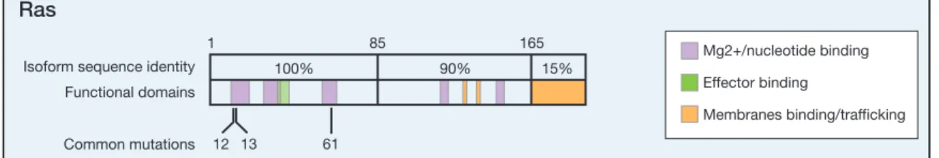
1.2 Oncogenic mutations of RAS. ... 24
1.3 Accumulation of genetic mutations during pancreatic carcinogenesis. ... 25
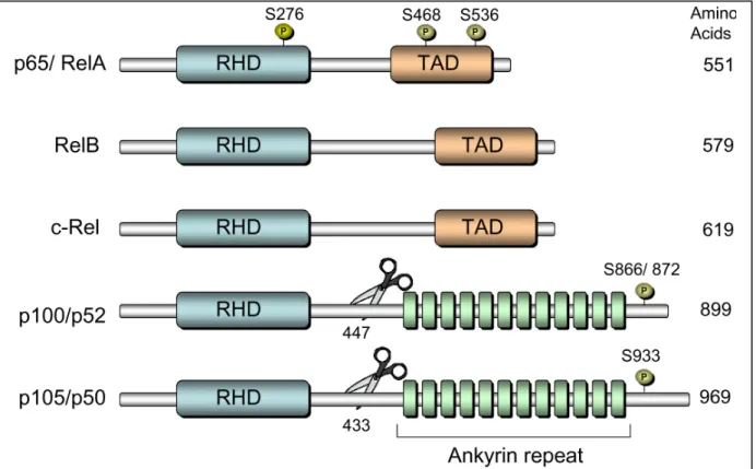
1.4 Domain organization of NF-κB family members. ... 26
1.5 Canonical versus non-canonical NF-κB signal transduction. ... 27
1.6 Domain organization of TAK1 and TAB proteins. ... 28
1.7 Glycogen synthase kinase-3 isoforms. ... 28
1.8 GSK-3 regulatory pathways and substrate targets. ... 29
1.9 TBK1 domain organization. ………30
Chapter II 2.1 GSK-3α and GSK-3β regulate growth and NF-κB activity in pancreatic cancer cells. ... 67
2.2 TAK1 is constitutively active and regulates NF-κB activity in pancreatic cancer cells. ... 69
2.3 GSK-3 inhibition suppresses TAK1 levels. ... 71
2.4 GSK-3α regulates the non-canonical NF-κB pathway in pancreatic cancer cells. ... 72
2.5 GSK-3 inhibition suppresses tumor growth in mice. ... 73
2.6 GSK-3 inhibition leads to changes in NF-κB target gene expression. ... 74
2.7 Model of GSK-3-NF-κB pathway downstream of mutant KRAS in pancreatic cancer ... 75
x
2.2A KRAS regulates TAK1 levels. ... 78
2.2B TAK1 inhibition induces apoptosis. ... 79
2.3 TAK1 inhibition decreases cell proliferation in KRAS+ cells. ... 80
2.4 TAK1 inhibition alters cell cycle regulation. ... 81
2.5 GSK-3 inhibition inhibits TAK1 mediated signaling. ... 82
2.6 GSK-3 inhibition leads to a decrease in TAK1 protein. ... 83
2.7 NF-κB2 depletion leads to increase in PARP cleavage. ... 84
2.8 GSK-3 inhibition leads to a decrease in TAK1 levels and p100-p52 processing in a statistically significant manner. ... 85
2.9 KRAS regulates GSK-3α mRNA levels. ... 86
Chapter III 3.1 Generation of TBK1fl/fl mice. ... 112
3.2 Characterization of TBK1Δ/Δ mice. ... 114
3.3 Deletion of TBK1 in the mouse lung decreases KRAS-induced lung tumorigenesis. ... 115
3.4 Deletion of TBK1 in the mouse lung does not affect the levels of IKKε ... 116
LIST OF ABBREVIATIONS
ATP Adenosine-5-‘triphosphate Cre Cre recombinase enzyme DMSO Dimetheyl sulphur oxide
EGFR Epidermal growth factor receptor FTI Farnesyltransferase inhibitors GAP GTPase activating protein GDP Guanosine-5’diphosphate
GEF Guanine nucleotide exchange factor GEMM Genetically engineered mouse models GSK-3 Glycogen synthase kinase-3
GTP Guanosine -5’triphosphate
ICMT Isoprenylcysteine carboxylmethyltransferase IKK IκB kinase
IκB Inhibitor of κB
MAP3K Mitogen activated protein kinase kinase kinase NAK NFκB activating kinase
NF-κB Nuclear kactor kappa B NSCLC Non-small cell lung cancer
xii
RALGDS Ral guanine nucleotide-disassociation stimulator RCE1 RAS converting enzyme 1
TAB TGFβ activated kinase binding protein TAK1 TGFβ activated kinase 1
TANK TRAF-associated NF-κB activator TBK1 Tank binding kinase 1
TGF-β Transforming growth factor beta TNFR TNF receptor
CHAPTER I
INTRODUCTION
1.1 Summary
KRAS is frequently mutated and drives oncogenesis in a variety of human cancers (1,2). Unsuccessful attempts at direct targeting of KRAS has led investigators to consider targeting the downstream effector pathways of KRAS. The constitutive activation of the transcription factor NF-κB is among the many deregulated signaling pathways
downstream of oncogenic KRAS that has been associated with multiple malignancies, including pancreatic cancer and lung cancer. Consequently, there has been focus on targeting constitutive NF-κB activity in these cancers (3,4) (5). Advancement of this therapeutic strategy is dependent on fully understanding the complex signaling events that drive NF-κB activity. Recently Tank binding kinase-1 (TBK1), an IκB kinase (IKK) related kinase, which regulates NF-κB signaling, was shown to be important for KRAS-driven cancers (6,7). Here I provide new insight into elusive signaling events
downstream of oncogenic KRAS that drive tumorigenesis in pancreatic and lung cancer and discuss glycogen synthase kinase-3α (GSK-3), and TGF-β activated kinase-1 (TAK1), and TBK1 as potential therapeutic targets for these cancers.
2
signaling. Furthermore, I will review the functional significance of constitutive KRAS activity during oncogenesis and discuss the relevance to target this pathway in pancreatic and lung cancer.
1.2 RAS proteins
RAS proteins are proto-oncogenes that are frequently mutated in human cancers. The mammalian genome encodes three RAS genes that give rise to four ubiquitously expressed highly homologous proteins: HRAS (homologous to the oncogene of the Harvey murine sarcoma virus), KRAS- (homologous to oncogene of Kirsten murine sarcoma virus; K-RAS 4A and K-RAS 4B are splice variants of single gene) and NRAS (which does not have a retroviral homolog and was first isolated from a neuroblastoma cell lines) (8).
The ~21kDa RAS proteins cycle between inactive (GDP)-bound and active (GTP)-bound conformations and function as molecular switches that couple cell surface receptor activation to intracellular signaling pathways. (1). The activation of RAS proteins, that is, the exchange of GDP with GTP, is an intrinsically slow process and is catalyzed by guanine nucleotide exchange factors (GEFs). However, this exchange is reversible. The inactivation of RAS involves the hydrolysis of the γ-phosphate of GTP to GDP catalyzed by GTPase-activating proteins (GAPs) and is irreversible. This
resistant to GAP-mediated GTP hydrolysis, which renders them constitutively active. GTP- bound RAS can interact productively with more than 20 effectors, including Raf, phosphatidylinositol 3-kinase (PI3K) and Ral guanine nucleotide-dissociation stimulator (RALGDS), to regulate various cellular responses including proliferation, survival and differentiation (Figure 1.1).
Newly synthesized RAS is a cytosolic protein that undergoes posttranslational modifications, which helps localize RAS to the correct subcellular compartment — principally the inner face of the plasma membrane. These modifications include the covalent attachment of a non-sterol isoprenoid from farnesyl pyrophosphate (FPP) to a cysteine residue that is close to the carboxyl terminus by prenylation. Farnesyltransferase inhibitors (FTIs) block this farnesylation, so RAS remains in the cytosol and is unable to function. After prenylation an endopeptidase, named RAS converting enzyme 1(RCE1), removes the end three amino acids from the carboxyl terminus of the protein. The new carboxyl terminus is then methylated by isoprenylcysteine carboxylmethyltransferase (ICMT), followed by palmitoylation and transfer to the plasma membrane. KRAS4B attaches to the plasma membrane without palmitoylation(1).
The following section will review roles of RAS signaling in cancer with a focus on pancreatic and lung cancer.
1.3 Role of RAS in Cancer
4
(50%), thyroid (50%), lung (30%) and melanoma (25%) having the highest
prevalence(8). Different tumor types have specific associations with individual RAS isoforms. For example KRAS, the most frequently mutated isoform in most cancers, is associated with pancreatic cancer, non-small cell lung cancer (NSCLC), colorectal cancer and seminoma. In contrast HRAS mutations are more strongly associated with tumors of the skin and of the head and neck and NRAS mutations are common in hematopoietic malignancies (9). All these mutations occur most frequently in codons 12, 13, or 61 (Figure 1.2) and stabilize RAS in a constitutively active GTP bound conformation (2). Moreover, several other human cancers harbor alteration in factors that lie upstream of RAS, leading to over expression or mutational activation of growth factor receptor tyrosine kinase, such as epidermal growth factor (EGFR and ERBB2). Alternatively factors downstream of RAS are also commonly mutated, such as mutation of B-RAF in melanoma and colon cancer (1).
Activated mutant RAS in cells can promote several of the characteristics of malignant transformation. These include increased proliferation due to upregulation of several transcription factors that are required for cell cycle entry and progression,
activation of NF-κB (11). Oncogenic RAS-induced effector pathways also impinge on metabolic reprogramming of cancer cells, a known hallmark of cancer (12). The effect on cellular metabolism is exerted through concurrent activation of MAP3K and PI3K
leading to upregulation of HIF1α (13), and through upregulation of autophagy(14,15), both of which stimulate a glycolytic shift. RAS effector pathway also lead to the induction of angiogenesis, mainly by means of ERK mediated transcriptional upregulation of angiogenic factors (like VEGF), matrix metallo proteinases (16).
Oncogenic RAS can also subvert antitumor immunity by down-regulating expression of antigen-presenting major histocompatibility complexes (MHC) on cancer cells(17), and through the recruitment of immunosuppressive regulatory T cells (TRegs) and myeloid-derived suppressor cells (MDSCs) to the tumor site(18,19). Finally RAS -dependent signaling pathways including the RAS–MAPK, RAS–PI3K, RAS–RAL GTPase and RAS–RHO GTPase have been demonstrated to have an essential role in metastatic
progression (9). Targeting RAS and its effector pathway could, therefore, have a potential impact on almost all aspects of malignancy.
The next two sections review the role of KRAS in driving two of the most difficult to treat cancers: Pancreatic and lung cancer.
1.3.1 Role of KRAS in pancreatic ductal adenocarcinoma
6
number of new diagnoses. Approximately 46,420 new cases of pancreatic cancer will be expected in 2014, while 39,590 patients are estimated to die from this disease(20). The nucleoside analog, gemcitabine, is among the most active single-agent chemotherapy currently used to treat advanced stages of pancreatic cancer and is considered to be the first-line therapy for patients (21). Lack of efficient early-detection methods, aggressive metastatic potential, and chemotherapy resistance all account for the dismal mortality rate (a 5-year survival rate of 6% is associated with pancreatic cancer (20). Therefore, it is critical to advance our understanding of the molecular pathogenesis of pancreatic cancer in order to develop effective targeted chemotherapies to treat this disease.
Extensive histological and genetic studies have characterized the pathogenesis of pancreatic cancer into three early stages of pre-invasive lesions known as pancreatic intraepithelial neoplasia (PanIN) (Fig. 1.3) (22). Recent tumor genome sequencing studies have established the prevalence of mutant KRAS in PanINs, (23), and in
pancreatic cancer(24,25) with increased precision. The most common mutation in KRAS
is one amino-acid substitution in position 12 of the KRAS protein, leading to a glycine (G) to aspartic acid (D) substitution, although other variants, such as G to V are also common (Table 1.1) (26-29). Malignant progression from PanINs to invasive and metastatic disease is accompanied by the early acquisition of activating mutations in the KRAS oncogene, which occurs in >90% of cases, and subsequent loss of tumor
1.3.2 Role of KRAS in lung cancer
Lung cancer is the leading cause of cancer-related deaths worldwide (20). The two major forms of lung cancer are non-small cell lung cancer (about 85% of all lung cancers) and small-cell lung cancer (about 15%). Non-small cell lung cancer (NSCLC) has amongst the worst prognoses of all human malignancies. Non-small cell lung cancer can be divided into three major histologic subtypes: squamous-cell carcinoma,
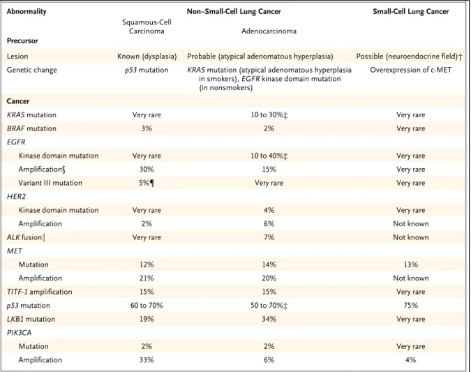
adenocarcinoma, and large-cell lung cancer; adenocarcinoma is the most common type accounting for >40% of all lung cancers (36). Analysis of resected tumors has revealed several frequent molecular changes: KRAS is mutated to an activated form in 30% of NSCLCs, EGFR is up-regulated in 10%–40%, the p53 tumor suppressor gene is mutated or deleted in 50%, and the INK4A/ARF locus is often deleted or hypermethylated (Table 2).
KRAS mutations are also observed in a substantial number of sporadic and chemically induced lung adenocarcinomas in mice (37-39). Further evidence for the role of KRAS in oncogenesis of lung cancer comes from mouse studies where expression of mutant KRAS is sufficient to cause transformation and development of adenocarcinoma (40,41) .
8
Importantly, activating point mutations in KRAS are associated with poor prognosis and therapy resistance (42,43).
Taken together, the role of oncogenic KRAS in initiating and driving pancreatic and lung adenocarcinomas is well established. Thus targeting RAS and its effector pathway has been a focus for cancer biologists seeking to develop rational anti-cancer drugs as explained in the next section.
1.4 Targeting RAS as a therapeutic approach to cancer
For RAS as a therapeutic target, post-translational modifications, known to be required for RAS biological activity has been a very attractive target(44). In this regard, Farnesyl transferase that catalyse attachment of farnesyl isoprenoid group to RAS proteins were an obvious target. Number of highly effective Farnesyl transferase
inhibitors (FTIs) have been identified that inhibit cell growth of a large variety of cancer cell lines in vitro and in vivo as tumor xenografts (45,46). However, KRAS, the most frequently mutated RAS oncogene in human cancers, can undergo alternative prenylation by GGTase I in FTI-treated cells, resulting in a persistent membrane localization of KRAS and concomitant upregulation of downstream signaling. Inhibition of the two post prenylated enzymes RCE1 and ICMT has also met little success (47,48). While blocking RCEI activity in tumor cells or mouse models has shown only moderate effects,
inhibition of ICMT affects multiple pathways besides RAS leading to toxic effects like atherosclerotic vascular injury (8,46).
have, for the most part, not been rewarding. The idea of preventing RAS expression by antisense or RNA interference seems promising, but the successful application of this technology is currently limited by lack of efficient delivery, uptake, and gene silencing. Additionally the observation that tumor cells expressing mesenchymal markers are relatively independent on the presence of RAS oncogene has raised questions on the potential of elimination of RAS expression as an effective cancer therapy (49).
Collectively, developing therapeutic agents to directly block oncogenic RAS activity has been a challenging and unsuccessful endeavor, for reasons discussed above. Therefore, a great deal of effort has been applied to developing therapies that target effector pathways downstream of RAS (Figure 1.1). Understanding which effector pathways are required for RAS-driven oncogenesis is critical for determining which pathways should be targeted for therapeutic purposes. As a result, new insight has emerged in comprehending the network of deregulated signal transduction pathways downstream of oncogenic KRAS. Most RAS effector pathways are comprised of kinase cascades, providing multiple nodes for potential therapeutic intervention. Several RAS effectors have been identified and comprehensively described, including RAF-MEK-ERK and PI3K signaling (1,8,46). In the case of lung and pancreatic cancers, constitutive signaling from PI3K/AKT, EGFR, and RAS/RAF/MAPK pathways have all been
(4,5,51-10
53). However, advancement of this strategy is dependent on fully understanding the signaling events that drive constitutive NF-κB activation.
1.5 Constitutive NF-κB in Cancer
NF-κB was originally discovered over 25 years ago as a nuclear factor that binds the immunoglobulin κ enhancer sequence in B-cells(54). NF-κB has since been
characterized as an inducible family of conserved dimeric transcription factors consisting of: RelA/p65, RelB, c-Rel, p52 (p100 precursor), and p50 (p105 precursor) (Fig. 1.4). The N-terminal region of NF-κB consists of a highly conserved, 300 amino-acid, Rel homology domain (RHD) which promotes dimerization, nuclear localization, and DNA binding. A C-terminal transactivational domain (TAD) is located within RelA/p65, RelB, and c-Rel and facilitates NF-κB transcriptional activity upon inducible posttranslational modifications. The C-terminal region of the precursor NF-κB members (p100 and p105) contains an inhibitor ankryin repeat domain which can undergo proteolytic cleavage, to generate the active p52 and p50 subunits. Although processing of p105 to p50 is known to be constitutive, p100 to p52 processing is tightly regulated (55).
In resting cells, NF-κB is rendered inactive and sequestered in the cytoplasm through interaction with an inhibitor protein called IκB. NF-κB is typically activated through one of two distinct pathways. Canonical NF-κB activity can be triggered by various stimuli including inflammatory cytokines, microbial infection, DNA damage, and mitogens(57). These stimuli promote the activation of the IκB kinase (IKK) complex which consists of a regulatory subunit (IKKγ) and two catalytic, serine/ threonine kinase subunits (IKKα and IKKβ). Once this complex is activated, IKKβ phosphorylates NF-κB-bound IκB proteins which leads to its rapid ubiquitination and subsequent
proteosomal degradation (58,59). Consequently, NF-κB dimers can undergo nuclear translocation and bind to κB consensus sequences within target gene promoters (Fig. 1.5).
In the non-canonical NF-κB signal transduction, diverse stimuli such as B-cell activating factor (BAFF) and CD40 ligand, trigger a signaling cascade which activates the NF-κB inducing kinase (NIK) (55). NIK drives the activation of IKKα homodimers which subsequently phosphorylates p100, leading to its proteolytic processing and the generation of p52 (Fig. 1.5) (60). As a result, activated RelB/p52 heterodimers can translocate to the nucleus to drive NF-κB-dependent gene transcription.
12
breast, colon, lung cancer, melanoma, multiple myeloma, esophageal adenocarcinma, and pancreas (62) (63). NF-κB is often thought to serve as the bridge between inflammation and cancer (63). In colitis-associated cancer, deletion of IKKβ inhibited transcription of proinflammatory cytokines and reduced tumor formation in vivo (64).
Due to the role of NF-κB in tumorigenesis, NF-κB is an attractive target for cancer therapeutics. Several IKKβ inhibitors are available to determine the efficacy of IKK/NF-κB inhibition in various types of cancer. For example, treatment of lung tumor cells ex vivo with an IKKβ inhibitor reduced NF-κB activation (5), and treatment of animals with mutant KRAS-driven lung tumors resulted in tumor regression and
prolonged survival (65). However, IKKβ inhibition also leads to lymphocyte toxicity (66) and granulocytosis (67) in healthy animals. Further studies are required to determine the effects of IKKβ inhibition in other in vivo cancer models, and whether the benefits outweigh the side effects.
1.5.1 RAS and NF-κB
Studies from our group revealed that oncogenic H-RAS induces cellular
showed reduced tumor development both at the time of tumor initiation and after tumor progression. Additionally, our group (5) showed that deletion of NF-κB subunit
p65/RelA reduced the number of KRAS– induced lung tumors both in the presence and absence of p53, and the tumors that emerged in the absence of p65/RelA showed a higher number of apoptotic cells, reduced spread, and showed lower grade. In KRAS-driven pancreatic cancers, both canonical and non-canonical NF-κB have been found to be constitutively active and promote survival and chemoresistance (51,73-78). Recently knockout of IKKβ was shown to suppress tumor growth/progression in a KRAS/INK4a
null animal model of pancreatic cancer (79). Oncogenic RAS also induces inflammatory cytokines that activate NF-κB and STAT3. For example, KRAS-driven non-small cell lung cancers (NSCLC) (7) and pancreatic ductal adenocarcinomas (PDAC) (8) engage cell autonomous IL-1 signaling. Recently IKK related kinases, TBK1 and IKKe have also been implicated in promoting survival of KRAS-driven tumors (6,80).
Collectively, these reports and others underscore the need to target IKK and NF-κB as a chemotherapeutic strategy in KRAS-driven cancers. The following sections will
discuss the regulation of NF-κB by two versatile serine theronine kinases, transforming growth factor β-activated kinase 1 (TAK1) and glycogen synthase kinse-3 (GSK-3).
1.6 TAK1-dependent regulation of NF-κB
14
βligand family, this versatile serine/theronine kinase can be activated by a diverse set of stimuli such as interleukin-1 (IL-1), tumor necrosis factor-α (TNF-α), and
lipopolysaccharide (LPS) (82,83). Upon receptor activation, TAK1 can initiate individual downstream signaling cascades culminating in the activation of IKK and MAPKs (JNK and p38) (83,84). TAK1 plays a central role in NF-κB signaling through direct phosphorylation of IKKβ (85), and promotes expression of various cellular stress and inflammatory-response genes.
TAK1 activity is dependent on various phosphorylation events and interaction with essential signaling adapters called TAK1 binding proteins (TAB1, TAB2, TAB3 and TAB4) (Fig. 1.6) which facilitate autophoshorylation at Thr178, Thr184 and Ser 192 following stimulation (86-88). Notably, TAB proteins have been demonstrated to play a crucial role in facilitating TNF-α and IL-1β-dependent activation of NF-κB and MAPK pathways (87,89).
Deregulated TAK1 signal transduction has been associated with constitutive NF-κB activity in human cancer cell lines (90,91). TAK1 was shown to promote cell
1.7 GSK-3-dependent NF-κB Signal Transduction
Glycogen synthase kinase-3 (GSK-3) was discovered 25 years ago as a
serine/threonine kinase involved in the downregulation of glycogen biosynthesis through the phosphorylation and inactivation of glycogen synthase (94). There are two
mammalian isoforms, GSK-3α and GSK-3β, that are encoded by two independent genes
and share 85% overall sequence identity and 93% similarity within the catalytic domain (95,96) (97). A significant difference between GSK-3 isoforms is observed in the N-terminal region where GSK-3α possesses an extended glycine-rich tail (Fig. 1.7). Although purified GSK-3 isoforms exhibit similar substrate properties (96), evidence from GSK-3α and GSK-3β deficient mouse models suggest non-overlapping substrate specificities10. GSK-3α deficient mice exhibit enhance glucose and insulin
sensitivity(98), while GSK-3β deficiency is embryonic lethal at day E13.5 due to liver degeneration (99).
16
GSK-3 is a multi-tasking kinase that plays an intricate role in diverse signaling pathways. As a consequence, GSK-3 is known to have an expanded range of substrate specificities (Fig. 1.8). The consensus motif for GSK-3 substrates is Ser/Thr-X-X-X-Ser/Thr (X represents any amino acid). GSK-3 has the unique ability of targeting substrates that have previously phosphorylated (primed) at the C-terminal Ser/Thr
residues of the consensus motif (101). Interestingly, there has been an increasing body of literature implicating the role GSK-3 plays in cross regulating the activity of NF-κB.
The first evidence of GSK-3-dependent regulation of NF-κB was provided by James Woodgett and collegues (99). Interestingly, embryonic lethality was observed in GSK-3βhomozygous null mice between E13.5 and E14.5 days due to TNF-α-dependent hepatocytes apoptosis. Notably, this phenotype is consistent with the homozygous deletion of the p65 subunit of NF-κB (102) and IKKβ (103). To demonstrate GSK-3β -dependent NF-κB regulation, mouse embryonic fibroblasts from GSK-3β-deficient mice were also shown to be defective in TNF-α-induced NF-κB activation (99).
The exact mechanism of how GSK-3β regulates inducible NF-κB activity remains controversial. Reports have implicated GSK-3β to either regulate early stages of NF-κB activation (IKK complex activation and IκB degradation) or later stages (NF-κB
transcription activity at target gene promoters). Bharat Aggarwal and colleagues reported that GSK-3β null MEFs are defective in inducing IKK activity and IκBα
role of GSK-3 in regulating IKK-driven NF-κB activity in pancreatic cancer cells (75). Further evidence showed that GSK-3β can directly target the phosphorylation of p65 (amino acids 354-551), thus promoting NF-κB transactivation (106). Overall, GSK-3β plays an essential role in cross regulating cytokine-induced NF-κB activation through either an IKK-dependent or independent mechanism. Due to the known pro-survival functions of NF-κB, studies have also focused on the potential role GSK-3 may play during carcinogenesis.
1.7.1 GSK-3 Inhibitors as a Targeted Therapy for Cancer
GSK-3 has been linked to deregulated signaling pathways leading to the pathogenesis of various human diseases. Consequently, GSK-3 has emerged over the years as a therapeutic target for Alzheimer’s disease, bipolar disorder, dementia, and noninsulin-dependent diabetes mellitus (NIDDM) (107). Moreover, there is increasing evidence linking aberrant GSK-3 signaling with the development of cancer. GSK-3 is generally considered a tumor suppressor due to its role in suppressing the oncogenic effects of the Wnt/ β-catenin pathway in colorectal cancer (108). However, this
paradigm has been challenged by recent reports that suggest GSK-3 plays an oncogenic role by facilitating cell growth, survival, and constitutive NF-κB activity. Advancement of these studies was made possible by the development of pharmacological GSK-3 inhibitors.
18
3(110). SB 216763 (ATP-competitive, IC50 = 34.3 nM), SB 415286 (ATP-competitive,
IC50 = 77.5 nM), AR-A014418 (ATP-competitive, IC50 = 104 nM), and TDZD-8
(non-ATP-competitive, IC50 = 2µM) are among the most recently developed GSK-3 inhibitors
that have been implicated to have therapeutic potential in human cancers (111-113). Notably, these inhibitors do not provide significant selectivity between GSK-3α and GSK-3βisoforms due to sequence identity within the kinase domain.
Studies within the past five years have implicated GSK-3 inhibitors as a potential targeted therapy for prostate cancer, multiple myeloma, leukemia, and pancreatic
cancer(114-117). Although the mechanism of action remains unclear, GSK-3 inhibitors may function partially by suppressing constitutive, pro-survival signaling to NF-κB. Our lab and others have provided evidence of GSK-3-dependent constitutive pro-survival IKK-NF-κB activity in pancreatic cancer cell lines (75,118,119). These reports
demonstrated that GSK-3 is active in pancreatic cancer cells, and that GSK-3 inhibition suppresses constitutive NF-κB reporter activation and target gene expression.
Furthermore, the suppression of constitutive NF-κB activity following GSK-3 inhibition correlated with decreased cell growth/ survival in both in-vitro (75,118) and in-vivo
models (119). All the above studies either focused on GSK-3β or did not distinguish between the two isoforms, GSK-3α and GSK-3β, however, a recent study implicated GSK-3α in promoting acute myelogenous leukemia (120). Taken together, these reports establish GSK-3 inhibition as a potential therapeutic strategy for cancer but needs further investigation to dissect the contribution of the individual isoforms in various
1.8 Role of non-canonical IKK, TBK1 in cancer
In addition to the conventional IKKs, a related pair of non-canonical kinases, IKKε (IKKi, encoded by IKBKE) and TBK1 (NAK), have been identified as important mediators of both inflammatory and oncogenic signaling. TBK1 was identified as an interaction partner with the scaffolding molecule, TRAF-associated NF-κB activator (TANK) (121). It comprises of an N-terminal kinase domain, an ubiquitin- like domain, a C-terminal LZ and a HLH motif (Fig. 1.8) and is a critical inducer of interferon signaling in response to viral infection (80). TBK1 can be activated in a TLR dependent or
independent manner by viral components or ds RNA/DNA respectively, leading to phosphorylation of interferon regulatory factors (IRFs) 3, 5 and 7. This allows for heterodimerization and nuclear translocation of the IRFs and induction of pro-inflammatory and antiviral genes, IFNα/β.
20
cancer by activating autocrine cytokine signaling of CCL5 and IL-6 (7). Moreover, TBK-1 dependent non-canonical NF-κB signaling has been observed to contribute to
autophagy addiction in KRAS driven NSCLC (124).
To sum it all, the above results identify TBK1 as a novel and effective target downstream of oncogenic RAS highlighting the need to further investigate the functions of TBK1.
1.8.1 Mouse Models of TBK1
Studies of TBK1 function have been hindered by the fact that homozygous deletion of TBK1 is lethal in utero at approximately embryonic day 14.5 as a result of massive liver degeneration and apoptosis, a phenotype similar to knockout of p65(125). This embryonic lethality can be rescued by breeding these animals onto a TNF-/- or
TNFR1-/-background and thus most studies in which TBK1 deficient mice were used,
such animals were TBK1-/- TNF-/- or TBK1-/- TNFR1-/-double knockouts. Recently a conditional TBK1fl/flmodel was crossed with B-cell specific Cre recombinase to generate
TBK1-BKO mice heterozygous for the Cre gene, and with B-cell specific ablation of
TBK1(126). Using these mice, TBK1 was shown to negatively regulate non-canonical NF-κB signaling by phosphorylating NIK leading to its degradation. In another study, a conditional TBK1fl/fl was crossed with a line of transgenic mice that carry the Prml-Cre recombinase transgene. This transgene caused conversion of the TBK1fl allele to TBK1Δ
in the sperm of male mice by Cre-mediated deletion of exon 2. TBK1Δ allele expressed low amounts of an inactive and much smaller in size TBK1 protein and mice
but not C57BL6 (127). Thus a true TBK1 deficient mouse model is still not available to establish the role of TBK1 in KRAS driven oncogenesis in vivo.
1.9 Conclusions
Despite continuous research efforts, tumors harboring KRAS mutations, such as pancreatic and non-small cell lung adenocarcinomas, remain the most difficult to treat and lack any effective targeted therapies. The evolution of pancreatic and lung cancer chemotherapy is dependent on the development of agents that specifically target deregulated molecular pathways downstream of mutations associated with disease progression (128). Pancreatic and lung cancers are among many human malignancies that utilize constitutive NF-κB signaling. GSK-3 has been demonstrated to regulate constitutive IKK and the subsequent pro-survival NF-κB activity in pancreatic cancer cells, while the IKK related kinase TBK1 has been implicated in the survival of lung cancer cells through c-Rel. Therefore, chemotherapeutic agents that target GSK-3 and TBK1 activity may be efficacious in the treatment of these malignancies. Advancement of this therapeutic strategy requires full understanding of the complex molecular
mechanisms that regulate constitutive GSK-3-IKK-NF-κB activity in pancreatic cancer cell and TBK1-cRel activity in lung cancer cells.
This chapter reviewed the critical roles GSK-3, TAK1 and TBK1 play in promoting constitutive NF-κB activity in human cancers. Importantly, GSK-3
22
associated with oncogenic KRAS in colorectal cancer (92) and may also be linked to constitutive IKK and NF-κB activity and chemoresistance in pancreatic cancer (93). Despite the evidence of GSK-3-dependent NF-κB activation in pancreatic cancer, the mechanism of regulation and a clear distinction between the role of two isoforms, GSK-3α and GSK-3β remains elusive. Similarly, though TBK1 has been implicated in lung cancer, the effect of TBK1 deletion on KRAS driven lung cancers in situ is
undetermined.
The remaining chapters will characterize the requirements of GSK-3 isoforms (GSK-3α and GSK-3β), and TAK1 for maintaining constitutive NF-κB activity and cell growth/ survival in pancreatic cancer cells. Furthermore, I will investigate the potential link between these signaling components, and propose that constitutive canonical and non-canonical NF-κB activity is driven by a unique GSK-3α- signaling cascade. Collectively, these studies emphasize the significance of NF-κB signaling in pancreatic cancer, and highlight the potential of GSK-3α and TAK1 as novel therapeutic targets for this disease.
Finally, I will the first true TBK1 conditional knockout mouse and use it to investigate and propose the negative effect of TBK1 inhibition on the development of lung
FIGURES
24
Figure 1.2 Oncogenic mutations of RAS: The key oncogenic mutations are in the region that is identical among the 3 isoforms. Of the forty-four separate point mutations characterized for RAS isoforms, 99.2% occur at codons 12, 13 and 61. (2)
With the exception of the salivary gland, screening has focused on the locations and isoforms with the strongest coupling. Of note, however, the mutation rate in the pancreas is 60% rather than the generally quoted 90%. In most cases, 1 isoform dominates the number of mutations scored for a particular cancer. One exception is thyroid cancer, in which large numbers of mutants of all 3 isoforms have been counted. Although these observations confirm known trends, a
com-parison of codon mutations among the Ras isoforms reveals some intriguing deeper patterns.
Codon specificity of Ras isoform mutations
In analogy to the isoform bias we can see in specific
cancers, analyses of codon mutation frequencies reveal that each isoform has a distinctive codon mutation signature (Fig. 2). K-Ras and N-Ras represent 2 extremes of this phenomenon: 80% of K-Ras mutations occur at codon 12, whereas very few mutations are observed at codon 61. In contrast, almost 60% of N-Ras tumors harbor mutations at codon 61, compared with 35% at codon 12. H-Ras displays an intermediate behavior, with an approximately 50%/40% split between mutations at codons 12 and 61, respectively. These data represent averages of the percentages for each cancer in which at least 20 tumors were scored. Of importance, a closer examination of trends within different cancers
con-firms the individuality of each isoform even in
circum-stances in which the isoforms presumably have been exposed to common mutagenic factors (Fig. 2B).
These differences in codon specificity are surprising because
all 3 oncogenic mutations are in amino-acid regions that are
identical among the 3 isoforms and are assumed to generate equivalent effects on protein activity. Of note, even at the DNA level, K-Ras and N-Ras share identical sequences encoding Gly12 and Gln61. Furthermore, individual single-base substi-tutions result in the same amino-acid replacement for all of the isoforms. Nevertheless, an examination of the preferred single-base substitutions collated from all Ras-mutated tumors in the COSMIC database reveals afinal level of difference among the
isoforms (Table 2).
Ras codons 12, 13, and 61 can each be converted to 6 other amino acids via single-base substitutions. However,>60% of the total mutations for each isoform are accounted for by only 3 of the 18 potential mutations across the codons (Table 2). K-Ras mutation patterns are dominated by the 43% of mutations that are G!A transitions at the second base of codons 12 or 13, resulting in G12D or G13D muta-tions. G!T transversions at the second base make up the bulk of the remainder to produce G12V. A special case is observed in lung cancer, where a G!T transversion of the
first base of codon 12 to produce G12C predominates. N-Ras
favors similar types of mutations at codons 12 and 13, albeit at much lower rates compared with K-Ras. In contrast, H-Ras favors G12V in all cancers with codon 12 mutations, and more generally exhibits a 3-fold higher proportion of transversion-to-transition mutations compared with K-Ras and N-Ras. Mutations at codon 61 recapitulate the hetero-geneity that is evident between isoforms at codon 12.
These data reveal that Ras isoforms exhibit differential and preferential coupling to specific cancers, codons, and base
substitutions. The distinct mutation patterns exhibited by Ras
Figure 1. Oncogenic mutations of Ras isoforms. The key oncogenic mutations are in the region that is identical among the 3 isoforms. Forty-four separate point mutations have been characterized in Ras isoforms, with 99.2% of all mutations occurring at codons 12, 13, and 61. Mutations that cluster in and around loops 1, 2, and 4 are responsible for nucleotide binding and result in enhanced GTP binding. Residues that are mutated in cancer are highlighted in red, those that are mutated in developmental disorders are underlined, and those that are variable among isoforms are in gray (26, 65, 66).
Prior et al.
Cancer Res; 72(10) May 15, 2012 Cancer Research
2458
American Association for Cancer Research Copyright © 2012 cancerres.aacrjournals.org on May 17, 2012 Downloaded from
26
28
Figure 1.6. Domain organization of TAK1 and TAB proteins. TAK1 consists of an N-terminal kinase domain which undergoes autophosphorylation at the indicated serine/ threonine residues upon activation. TAK1 exist in complex with TAK1-binding proteins (TAB1, TAB2, TAB3). TAB1 interacts with N-terminal region of TAK1, while TAB2/3 interacts at the C-terminal region. TAB1 consists of an uncharacterized, N-terminal protein phosphatase 2C (PP2C) domain. TAB2/3 contains CUE and zinc-finger domains which facilitates interaction with polyubiquitin chains. (Adapted from Wilson, W, The Regulation of constitutive NF-κB activity by GSK-3 in pancreatic cancer cells, 2008) Adapted with permission.
Figure 1.7. Glycogen synthase kinase-3 isoforms. Glycogen synthase kinase-3 exist a two, closely related mammalian isoforms (GSK-3α and GSK-3β). Gly-Rich indicates the N-terminal glycine-rich region located in GSK-3α. GSK-3 activity is mediated by
Figure 1.8. GSK-3 regulatory pathways and substrate targets. GSK-3 can be negatively regulated by a variety of upstream signaling events through the
30
Figure 1.9. TBK1 domain organization The major domains of each TBK1 are depicted with amino-acid numbers that correspond to the human proteins. Somatic mutations of TBK1 identified in lung adenocarcinomas are marked in red. ULD, Ubiquitin-like domain; LZ, leucine zipper; HLH, helix-loop-helix; NBD, NEMO-binding domain (adapted from Shen, RR and Hahn, WC Oncogene, 2010)
(Figure 1). Despite their similarity in structure, TBK1
and IKK
e
exhibit differential expression patterns.
TBK1, like IKK
a
and IKK
b
, is ubiquitously expressed
(Shimada
et al.
, 1999). In contrast, IKK
e
expression is
restricted to particular tissue compartments, with
high-est levels detected in lymphoid tissues, peripheral blood
lymphocytes and the pancreas (Shimada
et al.
, 1999).
Various epithelial-derived cell lines also exhibit IKK
e
expression (Shimada
et al.
, 1999; Gravel and Servant,
2005; Honda
et al.
, 2005; Bibeau-Poirier
et al.
, 2006).
Mitogenic stimulation with LPS and TNF
a
can also
induce IKK
e
and TBK1 expression in a NF-
k
B
dependent manner (Shimada
et al.
, 1999; Kravchenko
et al.
, 2003; Hemmi
et al.
, 2004). With these partially
overlapping characteristics, IKK
e
and TBK1 are
func-tionally more similar to each other than the canonical
IKKs (Clement
et al.
, 2008).
The non-canonical IKKs coordinate the interferon
response
IKK
e
and TBK1 are critical inducers of interferon
signaling in response to viral infection (Fitzgerald
et al.
,
2003; Sharma
et al.
, 2003). Following activation of
toll-like receptors by viral components, IKK
e
and TBK1
assemble with TRAF3 and TANK to phosphorylate
interferon regulatory factors (IRFs) 3, 5 and 7 at
multiple serine and threonine residues (Pomerantz and
Baltimore, 1999; McWhirter
et al.
, 2004; Mori
et al.
,
2004; Caillaud
et al.
, 2005; Cheng
et al.
, 2006). This
activity allows for heterodimerization and nuclear
translocation of the IRFs and induction of
pro-inflammatory and antiviral genes, including
IFN-
a
/
b
(Lin
et al.
, 1998; Sato
et al.
, 2000). Toll-like
receptor-independent mechanisms also activate IKK
e
and TBK1
to induce the interferon response. In this scenario, viral
double stranded RNA and DNA initiate signaling
through intracellular RNA and DNA sensors such as
RIG-I, MDA-5 and DAI (Andrejeva
et al.
, 2004;
Yoneyama
et al.
, 2004; Kawai
et al.
, 2005; Meylan
et al.
, 2005; Seth
et al.
, 2005; Takaoka
et al.
, 2007).
IFN
b
also activates a toll-like receptor-independent
pathway by stimulating IKK
e
phosphorylation of
STAT1 to facilitate binding with ISGF3, a complex
that serves as the transcriptional machinery important
for activating a subset of interferon response genes
(Tenoever
et al.
, 2007). Moreover, engagement of
both toll-like receptor-dependent and -independent
pathways recruits additional scaffolding molecules
including FADD, TRADD, MAVS, NAP1, HSP90
and
SINTBAD
which
are
necessary
for
IKK
e
and
TBK1-mediated
interferon
activation
(Rothe
et al.
, 1996; Balachandran
et al.
, 2004; Hacker
et al.
,
2006; Oganesyan
et al.
, 2006; Yang
et al.
, 2006; Gatot
et al.
, 2007; Guo and Cheng, 2007; Ryzhakov and
Randow, 2007; Michallet
et al.
, 2008). Thus, IKK
e
and
TBK1 form several protein complexes, the composition
of which is dependent on the type of cellular stimuli.
Ultimately, these signaling complexes share a role in
activating interferon responses for achieving an antiviral
state.
The ability of IKK
e
and TBK1 to activate the
interferon response is also dependent on post
transla-tional modifications by proteasome-independent Lys-63
linked ubiquitin chains (Figure 2). Both IKK
e
and
TBK1 have an ubiquitin like domain, and Lys-63
ubiquitination of both kinases are promoted by TRAF3,
which itself is Lys-63 ubiquitinated. Disruption of this
activity by TAXBP1 and the deubiquitinase A20 ablates
the interferon response (Ikeda
et al.
, 2007; Parvatiyar
et al.
, 2010). Both TBK1 and IKK
e
also mediate
ubiquitination of TANK by an unknown E3 ligase
(Gatot
et al.
, 2007). Although TANK further serves as a
phosphorylation target of TBK1 and IKK
e
, TANK
ubiquitination seems to occur independently of this
kinase activity. The mechanism by which TANK
ubiquitination contributes to the activation of IKK
e
and TBK1 is unknown. Lys-63 ubiquitination also has a
role in the negative regulation of IKK
e
and TBK1
induced responses. For example, during- by RNA
viruses, Lys-63 ubiquitination of MAVS is essential for
the recruitment of IKK
e
and leads to the inhibition of
antiviral and NF-
k
B induced inflammatory genes (Paz
et al.
, 2009). Ultimately, these modifications will likely
dictate a dynamic system of regulating IKK
e
and
TBK1-mediated function in both inflammation and
cancer.
The non-canonical IKKs function as NF-
k
B effectors
Although TBK1 and IKK
e
are not a part of the classical
IKK
a
/
b
/
g
signaling complex, these kinases were
origin-ally characterized as activators of NF-
k
B and target
multiple NF-
k
B members and effectors. Both
IKK-related kinases phosphorylate I
k
B
a
at, one of the
two-serine residues typically targeted on I
k
B
a
. Although
IKK
e
phosphorylates Ser
36, TBK1 phosphorylates Ser
32IKKε 1 Kinase domain ULD LZ HLH 716
TBK1
IKKβ Kinase domain HLH NBD
15 300 458 486 737 742
756
1 ULD LZ
311 388 603 642
IKKα Kinase domain LZ HLH NBD
15 301 599 638
745 1
455 483 738 743
LZ
Kinase domain 730
1 ULD
305 383 591 632
HLH 578 619 350 383 W445S
9 300 G417D 500 527
9 300 499 527 P675L
Figure 1
Structural comparison of the classical and non-canonical
IKKs. The major domains of each IKK kinase are depicted with
amino-acid numbers that correspond to the human proteins. The
kinase domain of IKK
e
exhibits 27% and 24% identity to IKK
a
and IKK
b
, respectively, and TBK1 shares 49% identity and 65%
similarity to IKK
e
. Somatic mutations of IKK
e
and TBK1 recently
identified in lung adenocarcinomas are marked in red. ULD,
Ubiquitin-like domain; LZ, leucine zipper; HLH, helix-loop-helix;
NBD, NEMO-binding domain (May
et al.
, 2004; Hiscott
et al.
,
2006; Perkins, 2007; Kan
et al.
, 2010).
Roles for the non-canonical IKKs in cancer
RR Shen and WC Hahn
632
Table 1.1 Frequency of genetic mutations associated with pancreatic cancer.
32
Table 1.2 Genetic abnormalities in specific lung cancers
(Adapted from Herbst, R et al., Lung Cancer, The New England Journal of Medicine, 2008)
Molecular Origins of Cancer
n engl j med 359;13 www.nejm.org september 25, 2008 1369
Clonal Evolution
Changes in certain genes (e.g., proinflammatory interleukin-8 [IL8] and some DNA-repair genes) occur in nonmalignant lung tissue of smokers and patients with lung cancer, a finding consis-tent with diffuse tissue injury.3,27-29 These chang-es probably precede epithelial clonal evolution, an important element of the molecular origins of lung and other cancers (Fig. 1). Patches of
clon-ally related cells, or clonal patches containing 40,000 to 360,000 cells, have been mapped in the lung.30 The size and number of subclones in a clonal patch may contribute to the cancer risk.31 Early events in the development of non–small-cell lung cancer include loss of heterozygosity at chromosomal region 3p21.3 (site of RASSF1A, a member of the Ras association domain family, and FUS1), 3p14.2 (FHIT, a fragile histidine triad Table1. GeneticAbnormalitiesSpecificintheLungtoNon–Small-CellLungCancerandSmall-CellLungCancer.*
Abnormality Non–Small-CellLungCancer Small-CellLungCancer
Squamous-Cell
Carcinoma Adenocarcinoma
Precursor
Lesion Known (dysplasia) Probable (atypical adenomatous hyperplasia) Possible (neuroendocrine field)† Genetic change p53 mutation KRAS mutation (atypical adenomatous hyperplasia
in smokers), EGFR kinase domain mutation (in nonsmokers)
Overexpression of c-MET
Cancer
KRAS mutation Very rare 10 to 30%‡ Very rare
BRAF mutation 3% 2% Very rare
EGFR
Kinase domain mutation Very rare 10 to 40%‡ Very rare
Amplification§ 30% 15% Very rare
Variant III mutation 5%¶ Very rare Very rare
HER2
Kinase domain mutation Very rare 4% Very rare
Amplification 2% 6% Not known
ALK fusion∥ Very rare 7% Not known
MET
Mutation 12% 14% 13%
Amplification 21% 20% Not known
TITF-1 amplification 15% 15% Very rare
p53 mutation 60 to 70% 50 to 70%‡ 75%
LKB1 mutation 19% 34% Very rare
PIK3CA
Mutation 2% 2% Very rare
Amplification 33% 6% 4%
* Non–small-cell lung cancer includes squamous-cell carcinoma and adenocarcinoma.
† Neuroendocrine fields have been detected only in tissue surrounding tumors and have been characterized by extremely high rates of allelic loss and by c-MET overexpression (Salgia R: personal communication).
‡ Variations are based in part on smoking profiles.
§ The percentages include increased gene copy numbers from amplification or polysomy and represent percentages from resected cancers. The percentages are higher in primary tumors from patients with metastatic disease. Increased copy numbers have been reported in squamous dysplastic lesions but not in adenocarcinoma precursors.
¶ Genomic EGFR variant III mutations have been detected only in lung squamous-cell carcinoma, and these tumors are sensitive preclinically to irreversible EGFR tyrosine kinase inhibitors. The incidence of 5% is substantially lower than that of 30 to 40% for the detection in squamous-cell carcinoma or adenocarcinoma by immunohistochemical analysis or other techniques.
∥ The anaplastic lymphoma kinase (ALK) fusion gene (involving chromosome 2p), consisting of parts of EML4 and ALK, is transforming in fibroblasts and occurs in adenocarcinoma but not in other types of non–small-cell lung cancer or other nonlung cancers.
The New England Journal of Medicine
REFERENCES
1. Downward J. Targeting RAS signalling pathways in cancer therapy. Nat Rev Cancer. 2003;3:11–22.
2. Prior IA, Lewis PD, Mattos C. A Comprehensive Survey of Ras Mutations in Cancer. Cancer Research. 2012;72:2457–67.
3. Zhang Z, Rigas B. kappaB, inflammation and pancreatic carcinogenesis: NF-kappaB as a chemoprevention target (review). Int J Oncol. 2006;29:185–92. 4. Holcomb B, Yip-Schneider M, Schmidt CM. The Role of Nuclear Factor [kappa]
B in Pancreatic Cancer and the Clinical Applications of Targeted Therapy. Pancreas. LWW; 2008;36:225–35.
5. Bassères DS, Ebbs A, Levantini E, Baldwin AS. Requirement of the NF-kappaB subunit p65/RelA for K-Ras-induced lung tumorigenesis. Cancer Research. 2010;70:3537–46.
6. Barbie DA, Tamayo P, Boehm JS, Kim SY, Moody SE, Dunn IF, et al. Systematic RNA interference reveals that oncogenic KRAS-driven cancers require TBK1. Nature. 2009;462:108–12.
7. Zhu Z, Aref AR, Cohoon TJ, Barbie TU, Imamura Y, Yang S, et al. Inhibition of KRAS-driven tumorigenicity by interruption of an autocrine cytokine circuit. Cancer Discovery. 2014.
8. Saxena N, Lahiri SS, Hambarde S, Tripathi RP. RAS: Target for Cancer Therapy. Cancer Invest. 2008;26:948–55.
9. Pylayeva-Gupta Y, Grabocka E, Bar-Sagi D. RAS oncogenes: weaving a tumorigenic web. Nature Publishing Group. 2011;11:761–74.
10. Robles AI, Rodriguez-Puebla ML, Glick AB, Trempus C, Hansen L, Sicinski P, et al. Reduced skin tumor development in cyclin D1-deficient mice highlights the oncogenic ras pathway in vivo. Genes & Development. 1998;12:2469–74. 11. Joneson T, Bar-Sagi D. Suppression of Ras-induced apoptosis by the Rac
GTPase. Molecular and Cellular Biology. 1999;19:5892–901.
12. Hanahan D, Weinberg RA. Hallmarks of Cancer: The Next Generation. Cell. 2011;144:646–74.
34
Expression of Noxa and Beclin-1 Promotes Autophagic Cell Death and Limits Clonogenic Survival. Molecular Cell. 2011;42:23–35.
15. Guo JY, Chen HY, Mathew R, Fan J, Strohecker AM, Karsli-Uzunbas G, et al. Activated Ras requires autophagy to maintain oxidative metabolism and tumorigenesis. Genes & Development. 2011;25:460–70.
16. Rak J, Mitsuhashi Y, Bayko L, Filmus J, Shirasawa S, Sasazuki T, et al. Mutant ras oncogenes upregulate VEGF/VPF expression: implications for induction and inhibition of tumor angiogenesis. Cancer Research. 1995;55:4575–80.
17. Maudsley DJ, Bateman WJ, Morris AG. Reduced stimulation of helper T cells by Ki-ras transformed cells. Immunology. 1991;72:277–81.
18. Clark CE, Hingorani SR, Mick R, Combs C, Tuveson DA, Vonderheide RH. Dynamics of the Immune Reaction to Pancreatic Cancer from Inception to Invasion. Cancer Research. 2007;67:9518–27.
19. Tran Thang NN, Derouazi M, Philippin G, Arcidiaco S, Di Berardino-Besson W, Masson F, et al. Immune Infiltration of Spontaneous Mouse Astrocytomas Is Dominated by Immunosuppressive Cells from Early Stages of Tumor
Development. Cancer Research. 2010;70:4829–39.
20. Siegel R, Ma J, Zou Z, Jemal A. Cancer statistics, 2014. CA Cancer J Clin. 2014;64:9–29.
21. Burris H3, Moore MJ, Andersen J, Green MR, Rothenberg ML, Modiano MR, et al. Improvements in survival and clinical benefit with gemcitabine as first-line therapy for patients with advanced pancreas cancer: a randomized trial. J Clin Oncol. American Society of Clinical Oncology; 1997;15:2403–13.
22. Hruban RH, Goggins M, Parsons J, Kern SE. Progression model for pancreatic cancer. Clin Cancer Res. AACR; 2000;6:2969–72.
23. Kanda M, Matthaei H, Wu J, Hong SM, Yu J, Borges M, et al. Presence of Somatic Mutations in Most Early-Stage Pancreatic Intraepithelial Neoplasia. Gastroenterology. 2012;142:730–9.
24. Jones S, Zhang X, Parsons DW, Lin JCH, Leary RJ, Angenendt P, et al. Core Signaling Pathways in Human Pancreatic Cancers Revealed by Global Genomic Analyses. Science. 2008;321:1801–6.
25. Biankin AV, Waddell N, Kassahn KS, Gingras M-C, Muthuswamy LB, Johns AL, et al. Pancreatic cancer genomes reveal aberrations in axon guidance pathway genes. Nature. 2012;491:399–405.
27. Hruban RH, Van Mansfeld AD, Offerhaus GJ, Van Weering DH, Allison DC, Goodman SN, et al. K-ras oncogene activation in adenocarcinoma of the human pancreas. A study of 82 carcinomas using a combination of mutant-enriched polymerase chain reaction analysis and allele-specific oligonucleotide hybridization. The American journal of pathology. American Society for Investigative Pathology; 1993;143:545.
28. Redston MS, Caldas C, Seymour AB, Hruban RH, da Costa L, Yeo CJ, et al. p53 mutations in pancreatic carcinoma and evidence of common involvement of homocopolymer tracts in DNA microdeletions. Cancer Research. 1994;54:3025– 33.
29. Schutte M, Hruban RH, Geradts J, Maynard R, Hilgers W, Rabindran SK, et al. Abrogation of the Rb/p16 tumor-suppressive pathway in virtually all pancreatic carcinomas. Cancer Research. 1997;57:3126–30.
30. Hezel AF. Genetics and biology of pancreatic ductal adenocarcinoma. Genes & Development. 2006;20:1218–49.
31. Aguirre AJ. Activated Kras and Ink4a/Arf deficiency cooperate to produce metastatic pancreatic ductal adenocarcinoma. Genes & Development. 2003;17:3112–26.
32. Hingorani SR, Petricoin EF, Maitra A, Rajapakse V, King C, Jacobetz MA, et al. Preinvasive and invasive ductal pancreatic cancer and its early detection in the mouse. Cancer Cell. 2003;4:437–50.
33. Guerra C, Mijimolle N, Dhawahir A, Dubus P, Barradas M, Serrano M, et al. Tumor induction by an endogenous K-ras oncogene is highly dependent on cellular context. Cancer Cell. 2003;4:111–20.
34. Collins MA, Bednar F, Zhang Y, Brisset J-C, Galbán S, Galbán CJ, et al.
Oncogenic Kras is required for both the initiation and maintenance of pancreatic cancer in mice. J Clin Invest. 2012;122:639–53.
35. Ying H, Kimmelman AC, Lyssiotis CA, Hua S, Chu GC, Fletcher-Sananikone E, et al. Oncogenic Kras Maintains Pancreatic Tumors through Regulation of Anabolic Glucose Metabolism. Cell. Elsevier Inc; 2012;149:656–70. 36. Fisher GH. Induction and apoptotic regression of lung adenocarcinomas by
regulation of a K-Ras transgene in the presence and absence of tumor suppressor genes. Genes & Development. 2001;15:3249–62.
36
et al. Ki-ras gene mutations and absence of p53 gene mutations in spontaneous and urethane-induced early lung lesions in CBA/J mice. Mol Carcinog.
1998;21:251–60.
39. Tuveson DA, Jacks T. Modeling human lung cancer in mice: similarities and shortcomings. Oncogene. 1999;18:5318–24.
40. Johnson L, Mercer K, Greenbaum D, Bronson RT, Crowley D, Tuveson DA, et al. Somatic activation of the K-ras oncogene causes early onset lung cancer in mice. Nature. 2001;410:1111–6.
41. Schubbert S, Shannon K, Bollag G. Hyperactive Ras in developmental disorders and cancer. Nature Publishing Group. 2007;7:295–308.
42. Herbst RS, Heymach JV, Lippman SM. Lung cancer. N Engl J Med. 2008;359:1367–80.
43. Mills NE, Fishman CL, Rom WN, Dubin N, Jacobson DR. Increased prevalence of K-ras oncogene mutations in lung adenocarcinoma. Cancer Research.
1995;55:1444–7.
44. Seabra MC. Membrane association and targeting of prenylated Ras-like GTPases. Cellular Signalling. 1998;10:167–72.
45. Basso AD. Thematic review series: Lipid Posttranslational Modifications. Farnesyl transferase inhibitors. The Journal of Lipid Research. 2005;47:15–31. 46. Gysin S, Salt M, Young A, McCormick F. Therapeutic Strategies for Targeting
Ras Proteins. Genes & Cancer. 2011;2:359–72.
47. Wahlstrom AM, Cutts BA, Karlsson C, Andersson KME, Liu M, Sjogren AKM, et al. Rce1 deficiency accelerates the development of K-RAS-induced
myeloproliferative disease. Blood. 2007;109:763–8.
48. Winter-Vann AM, Casey PJ. Post-prenylation-processing enzymes as new targets in oncogenesis. Nat Rev Cancer. 2005;5:405–12.
49. Singh A, Greninger P, Rhodes D, Koopman L, Violette S, Bardeesy N, et al. A Gene Expression Signature Associated with ••K-Ras Addiction•• Reveals Regulators of EMT and Tumor Cell Survival. Cancer Cell. Elsevier Ltd; 2009;15:489–500.
50. SARKAR F, BANERJEE S, Li Y. Pancreatic cancer: Pathogenesis, prevention and treatment. Toxicology and Applied Pharmacology. 2007;224:326–36. 51. Bang D, Wilson W, Ryan M, Yeh JJ, Baldwin AS. GSK-3 Promotes Oncogenic
52. Xia Y, Yeddula N, Leblanc M, Ke E, Zhang Y, Oldfield E, et al. Reduced cell proliferation by IKK2 depletion in a mouse lung-cancer model. Nat Cell Biol. Nature Publishing Group; 2012;14:257–65.
53. Meylan E, Dooley AL, Feldser DM, Shen L, Turk E, Ouyang C, et al.
Requirement for NF-κB signalling in a mouse model of lung adenocarcinoma. Nature. Nature Publishing Group; 2009;461:104–7.
54. Sen R, Baltimore D. Multiple nuclear factors interact with the immunoglobulin enhancer sequences. Cell. 1986;46:705–16.
55. XIAO G, RABSON A, YOUNG W, QING G, QU Z. Alternative pathways of NF-κB activation: A double-edged sword in health and disease. Cytokine & Growth Factor Reviews. 2006;17:281–93.
56. Ghosh S, Karin M. Missing pieces in the NF-kappaB puzzle. Cell. 2002;109 Suppl:S81–96.
57. Pahl HL. Activators and target genes of Rel/NF-κB transcription factors. Oncogene. 1999;18.
58. Delhase M. Positive and Negative Regulation of IB Kinase Activity Through IKK Subunit Phosphorylation. Science. 1999;284:309–13.
59. Karin M, Ben-Neriah Y. Phosphorylation meets ubiquitination: the control of NF-κB activity. Annual review of immunology. Annual Reviews 4139 El Camino Way, PO Box 10139, Palo Alto, CA 94303-0139, USA; 2000;18:621– 63.
60. Bonizzi G, Bebien M, Otero DC, Johnson-Vroom KE, Cao Y, Vu D, et al. Activation of IKKα target genes depends on recognition of specific κB binding sites by RelB: p52 dimers. EMBO J. Nature Publishing Group; 2004;23:4202– 10.
61. Karin M, Greten FR. NF-κB: linking inflammation and immunity to cancer development and progression. Nat Rev Immunol. 2005;5:749–59.
62. Bassères DS, Baldwin AS. Nuclear factor-κB and inhibitor of κB kinase
pathways in oncogenic initiation and progression. Oncogene. 2006;25:6817–30. 63. Baldwin AS. Control of oncogenesis and cancer therapy resistance by the
transcription factor NF-kappaB. J Clin Invest. 2001;107:241–6.
64. IKKbeta links inflammation and tumorigenesis in a mouse model of colitis-associated cancer. 2004;118:285–96. Available from:
38
65. Xue W, Meylan E, Oliver TG, Feldser DM, Winslow MM, Bronson R, et al. Response and Resistance to NF- B Inhibitors in Mouse Models of Lung Adenocarcinoma. Cancer Discovery. 2011;1:236–47.
66. Rapid TNFR1-dependent lymphocyte depletion in vivo with a selective chemical inhibitor of IKKbeta. 2006;107:4266–73. Available from:
http://bloodjournal.hematologylibrary.org/cgi/content/full/107/11/4266
67. NF-kappaB is a negative regulator of IL-1beta secretion as revealed by genetic and pharmacological inhibition of IKKbeta. 2007;130:918–31. Available from: http://eutils.ncbi.nlm.nih.gov/entrez/eutils/elink.fcgi?dbfrom=pubmed&id=17803 913&retmode=ref&cmd=prlinks
68. Finco TS, Westwick JK, Norris JL, Beg AA, Der CJ, Baldwin AS. Oncogenic Ha-Ras-induced signaling activates NF-κB transcriptional activity, which is required for cellular transformation. J Biol Chem. ASBMB; 1997;272:24113–6. 69. Mayo MW, Wang CY, Cogswell PC, Rogers-Graham KS, Lowe SW, Der CJ, et al. Requirement of NF-kappaB activation to suppress p53-independent apoptosis induced by oncogenic Ras. Science. 1997;278:1812–5.
70. Kim B-Y, Gaynor RB, Song K, Dritschilo A, Jung M. Constitutive activation of NF-kappaB in Ki-ras-transformed prostate epithelial cells. Oncogene.
2002;21:4490–7.
71. Tang X, Liu D, Shishodia S, Ozburn N, Behrens C, Lee JJ, et al. Nuclear
factor-κB (nf-κB) is frequently expressed in lung cancer and preneoplastic lesions. Cancer. 2006;107:2637–46.
72. Duran A, Linares JF, Galvez AS, Wikenheiser K, Flores JM, Diaz-Meco MT, et al. The Signaling Adaptor p62 Is an Important NF-κB Mediator in
Tumorigenesis. Cancer Cell. 2008;13:343–54.
73. Li L, Aggarwal BB, Shishodia S, Abbruzzese J, Kurzrock R. Nuclear factor-κB and IκB kinase are constitutively active in human pancreatic cells, and their down-regulation by curcumin (diferuloylmethane) is associated with the suppression of proliferation and the induction of apoptosis. Cancer. 2004;101:2351–62.
74. Liptay S, Weber CK, Ludwig L, Wagner M, Adler G, Schmid RM. Mitogenic and antiapoptotic role of constitutive NF-κB/Rel activity in pancreatic cancer. Int J Cancer. 2003;105:735–46.
75. Wilson W, Baldwin AS. Maintenance of Constitutive I B Kinase Activity by Glycogen Synthase Kinase-3 / in Pancreatic Cancer. Cancer Research. 2008;68:8156–63.
factor-κB RelA transcription factor is constitutively activated in human pancreatic adenocarcinoma cells. Clin Cancer Res. AACR; 1999;5:119–27. 77. Nishina T, Yamaguchi N, Gohda J, Semba K, Inoue J-I. NIK is involved in
constitutive activation of the alternative NF-κB pathway and proliferation of pancreatic cancer cells. Biochemical and Biophysical Research Communications. 2009;388:96–101.
78. Wharry C. Constitutive non-canonical NFκB signaling in pancreatic cancer cells. Cancer Biol Therpay. 2009;:1–20.
79. Ling J, Kang Y, Zhao R, Xia Q, Lee D-F, Chang Z, et al. KrasG12D-Induced IKK2/β/NF-κB Activation by IL-1α and p62 Feedforward Loops Is Required for Development of Pancreatic Ductal Adenocarcinoma. Cancer Cell. Elsevier Inc; 2012;21:105–20.
80. Shen RR, Hahn WC. Emerging roles for the non-canonical IKKs in cancer. Oncogene. Nature Publishing Group; 2010;30:631–41.
81. Yamaguchi K, Shirakabe K, Shibuya H, Irie K, Oishi I, Ueno N, et al.
Identification of a member of the MAPKKK family as a potential mediator of TGF-β signal transduction. Science. American Association for the Advancement of Science; 1995;270:2008–11.
82. Irie T, Muta T, Takeshige K. TAK1 mediates an activation signal from toll-like receptor (s) to nuclear factor-κB in lipopolysaccharide-stimulated macrophages. FEBS Lett. Elsevier; 2000;467:160–4.
83. Takaesu G, Surabhi RM, Park K-J, Ninomiya-Tsuji J, Matsumoto K, Gaynor RB. TAK1 is Critical for IκB Kinase-mediated Activation of the NF-κB Pathway. Journal of Molecular Biology. 2003;326:105–15.
84. Karin M, Liu Z-G, Zandi E. AP-1 function and regulation. Current opinion in cell biology. Elsevier; 1997;9:240–6.
85. Wang C, Deng L, Hong M, Akkaraju GR, Inoue J, Chen ZJ. TAK1 is a ubiquitin-dependent kinase of MKK and IKK. Nature. 2001;412:346–51. 86. Omori E, Inagaki M, Mishina Y, Matsumoto K, Ninomiya-Tsuji J. Epithelial
transforming growth factor β-activated kinase 1 (TAK1) is activated through two independent mechanisms and regulates reactive oxygen species. Proc Natl Acad Sci U S A. 2012.