How to cite:
Siahaan P, Prasasty VD, Simanjuntak BMD et al. (2017) Structural Stability of ADTC5 Peptide: Conformational Insights into Dynamics and Its Binding Mode. J. Trop. Life. Science 7 (2): 138 – 145.
*Corresponding author: Parsaoran Siahaan
Department of Chemistry, Faculty of Science and Mathematics, Diponegoro University
Jalan Prof. Soedarto, Tembalang, Semarang, Indonesia 50275 Email: [email protected]
VOL. 7, NO. 2, pp. 138 – 145, April 2017 Submitted October 2016; Revised January 2017; Accepted April 2017
Structural Stability of ADTC5 Peptide:
Conformational Insights into Dynamics and Its Binding Mode
Parsaoran Siahaan 1 *, Vivitri Dewi Prasasty 2, Bungaran M. David Simanjuntak 1, Suci Z. Hildayani 1, Khairul Anam 1 1 Department of Chemistry, Faculty of Science and Mathematics, Diponegoro University, Semarang, Indonesia
2 Faculty of Biotechnology, Atma Jaya Catholic University of Indonesia, Jakarta, Indonesia
ABSTRACT
The cyclic structure of ADTC5 (Ac-CDTPPVC-NH2) peptide is known to have the ability to modulate homodimer E-cadherin interactions to form junction at the intercellular junction. Its function to inhibit E-cadherin interaction has become important to increase paracellular porosity in delivering drug molecules to the target cell. There are two types of ADTC5 state: opened-cyclic state (OCS) and closed-cyclic state (CCS). OCS of ADTC5 is affected by distance constraints, and CCS is formed by disulfide bond from terminal cysteines through force restraint. The purpose of this research is to determine the inhibition activity of ADTC5 peptide upon E-cadherin. Here we used molecular docking, and molecular dynamics approaches. The PyMOL program was generated by the structure of ADTC5 peptide. GROMACS v4.5.5 was utilized to simulate molecular dynamics. The ADTC5 peptide molecule was placed optimum in aqueous or polar condition at physiological pH. Furthermore, ADTC5 was docked with EC1-EC2 coupled domain of E-cadherin using AutoDock 4.2 and refined using molecular dynamic (MD). The result showed that CCS ADTC5 peptide has a stronger affinity and more stable interaction with EC1-EC2 coupled domain than the OCS one.
Keywords: ADTC5 peptide, E-cadherin, molecular dynamics, molecular docking
INTRODUCTION
Delivering drugs into the brain tissue is blocked by the presence of biological barricades, called the blood-brain barrier (BBB) in the paracellular pathway [1, 2]. The paracellular pathway consists of proteins barrier at the zonula occludens (tight junctions), zonula adherens, and desmosomes (gap junctions) [3]. These proteins se-lectively allow the molecules with a diameter only below 11 Å and the molecular weight below 500 Dalton [4] which can pass through the paracellular pathway [5].
The porosity of paracellular pathway can be ad-justed by modulating homodimer E-cadherin interaction in the zonula adherens area at BBB [3]. The ADTC5 peptide is derived from the bulge region on the EC1 do-main. This peptide has the biological activity to enhance the porosity of paracellular pathway [6]. The ADTC5 peptide has the ability to modify the BBB in the delivery of marker molecules such as, 14C-mannitol, gadolinium
-diethylenetriaminepentacetate (Gd-DTPA)] to the brain over the paracellular pathways of the BBB. In vitro studies indicated that the ADTC5 had the best profile to inhibit junction resealing in Madin-Darby canine kidney cell monolayers. Furthermore, the ADTC5 peptide could increase in vivo delivery of Gd-DTPA to the brain of Balb/c mice when administered intravenously. There-fore, the ADTC5 has potential to improve delivery of diagnostic and therapeutic agents to the brain [7].
Table 1. Generated OCS and CCS forms of the ADTC5 peptide by molecular dynamics approach
Code OCS
Distance constraint of S14---S95 (nm)
Force restraint (kJ.mol-1.nm-2) A1 All bond freely rotatable
A2 0.3-0.4 None
A3 0.3-0.4 4,000
A4 0.3-0.4 12,000
A5 0.2-0.3 None
A6 0.2-0.3 4,000
A7 0.2-0.3 12,000
CCS
B1 All bond freely rotatable A
B2 None 4,000
B3 None 12,000
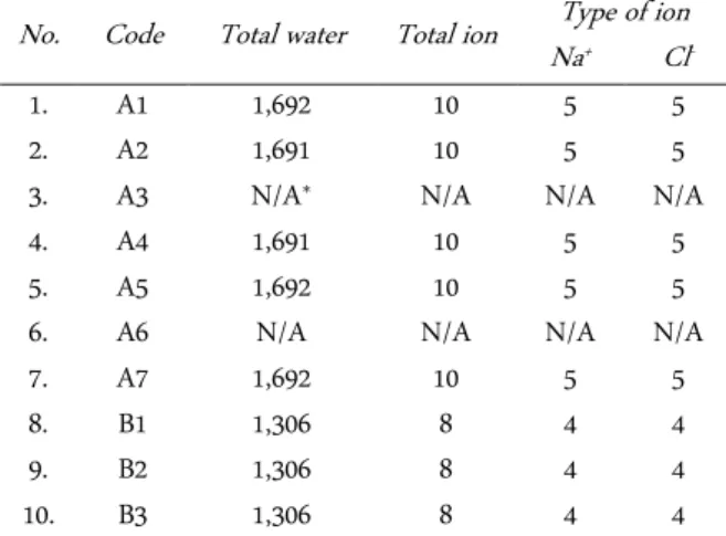
Table 2. The total number of water and ion molecules in each simulation system
No. Code Total water Total ion NaType of ion + Cl
-1. A1 1,692 10 5 5
2. A2 1,691 10 5 5
3. A3 N/A* N/A N/A N/A
4. A4 1,691 10 5 5
5. A5 1,692 10 5 5
6. A6 N/A N/A N/A N/A
7. A7 1,692 10 5 5
8. B1 1,306 8 4 4
9. B2 1,306 8 4 4
10. B3 1,306 8 4 4
*N/A = Not Available
Table 3. The total number and Periodic Boundary Condition (PBC) size
No. Code Total Atom
(unit) PBC size (nm)
1. A1 5,188 3.76329 × 3.76329 × 3.76329 2. A2 5,188 3.92033 × 3.92033 × 3.92033
3. A3 N/A* N/A
4. A4 5,184 3.74830 × 3.74830 × 3.74830 5. A5 5,188 3.75078 × 3.75078 × 3.75078
6. A6 N/A N/A
7. A7 5,188 3.72279 × 3.72279 × 3.72279 8. B1 4,025 3.43038 × 3.43038 × 3.43038 9. B2 4,025 3.43963 × 3.43963 × 3.43963 10. B3 4,025 3.42386 × 3.42386 × 3.42386 *N/A = Not Available
cules [10]. Moreover, study the chemical structure mod-ifications of peptide molecule can be done to increase its biological activity. This peptide structure involve linear and cyclic forms, S--S distance from start-end terminus amino acid on each Cysteine, and force restraint. They will affect to change ADTC5 structure conformation which causes the changes of binding site location upon EC1-EC2 coupled domain. Meanwhile, an understand-ing of non-covalent bonds is required to learn the most favorable molecular interaction [11].
In this work, we mainly focused on the chemical stability features which can differentiate the CCS from OCS form of the ADTC5 peptide. Moreover, we focused on the description of CCS and OCS molecules in the microscopic behavior of physical systems, such as solva-tion system using ion and water molecules, minimiza-tion energy, constrained and unconstrained systems. The aim of this research is to determine the ADTC5 sta-bility and binding affinity of the ADTC5 peptide with E-cadherin’s EC1-EC2 coupled domain.
The hypothesis feature was generated and per-formed as comparison between OCS and CCS models of the ADTC5. In this research, protein target of EC1-EC2 coupled domain was used to find the best binding mode feature of OCS and CCS. To evaluate their stabil-ity and interactions, computational aided-molecular dy-namics simulations were carried out to generate 3D structures of OCS and CCS of the ADTC5 peptides. Molecular docking method was utilized to determine the interaction between OCS and CCS of the ADTC5 with EC1-EC2 coupled domain of E-cadherin protein.
MATERIALS AND METHODS Protein and ligand preparation
The EC1-EC2 coupled domain of E-cadherin struc-ture was retrieved from Protein Data Bank (PDB ID No. 2O72). While the PyMOL program was used to generate the structure of the ADTC5 peptide. Furthermore, this peptide molecule was simulated by molecular dynamics on GROMACS v4.5.5 [12-15]. Therefore, molecular dy-namics was used to obtain the optimized structure in a different fashion, OCS, and CCS of ADTC5 peptide. In preparation of ADTC5 states, sulphuric atoms within Cys1 and Cys6 residues consist of S14 and S95, respec-tively, and these were arranged in several different force restraints and distance constraints as shown in Table 1.
Molecular dynamics were conducted in three steps: (1) System preparation. This aims is to prepare ADTC5 structure and solvation system using energy minimiza-tion and posiminimiza-tion restraint, respectively. This solvaminimiza-tion system uses a simple cubical periodic box (the dimen-sions of the box based upon setting the box edge ap-proximately 1.0 nm from the molecule periphery), TIP3P (Transferable Intermolecular Potential 3-Point) water as a solvent, with ionic concentration is 0.15 M. The total number of water and ions in each simulation system are presented in Table 2.
Trajectory generation was conducted with 120 ns (120,000 ps) running time. Analyzing trajectory by us-ing total energy and RMSD (Root Mean Square
Devia-tion) of a Cα atom in every second ps [11]. The widely
used CHARMM27 force field was implemented in the GROMACS simulation package [16]. The Berendsen weak-coupling algorithm was used in MD simulation which is extremely efficient to minimize the error in the ensemble scales during heating or equilibrium for relax-ing the system to the targeted temperature [17]. The to-tal number of atoms present in each simulation system and the size of the Periodic Boundary Cell (PBC) were specified in Table 2 and 3.
Energy minimization was given after ions addition during MD simulation. It aims to decrease the excessive forces on peptide systems, such as dihedral tension and tight non-binding contact. These appear due to the ad-dition of water molecules and ions in the simulation box, thus cause overlapping within some water molecules and ions, and it might produce high repulsion energy due to unstable interactions.
Molecular docking of the ADTC5 peptide with EC1-EC2 domain of E-cadherin
Molecular docking was run by using Autodock v.4.2 [18, 19] that involves two main parameters: Autogrid and Autodock. Autogrid is a grid box placed at: (1) Ala43, Asp44, Thr45 residues of the EC1 domain with all CCS variations (B1, B2, B3) as depicted at Figure 1 and 2. The entire surface of EC1-EC2 coupled domain, using 11 boxes with one type of CCS (B1). The size of the box is 50 × 50 × 50 (125,000 grid points) with grid spacing set at 0.375 Å (Figure 2). For all working types, the populations towards the genetic algorithm (GA) was
150, a maximum number of evals was 5 × 10⁶, a
maxi-mum number of generations was 27 × 103, and the
num-ber of genetic algorithms (GA) was 150.
Autodock involved two stages: sampling and scor-ing which used Lamarckian genetic algorithm and
Figure 1. A gridbox position of the box A with x = 28.053 y = 4.926 z = 48.111 (Ala43, Asp44, Thr45 residues in EC1 domain)
Figure 2. Eleven grid boxes (box A to K) on the entire surface of E1-EC2 coupled domain
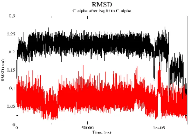
Figure 3. The RMSD value of Cα atom in A4 of OCS form (black) and B2 (red) of CCS form
(a) (b)
scoring, respectively. All default parameters were used by Autodock program. All the hydrogen added to the polar atoms on the peptides as the ligands and EC1-EC2 coupled domain as the receptor. The charge type used was Gasteiger. After running Autodock, the data was grouped into one population based on similarity of RMSD < 2Å, to get the best binding mode [20]. The active site locations of EC1-EC2 macromolecules were determined by lower energy optimization, hydrogen bonds, van der Waals forces, and hydrophobic interac-tion analysis.
RESULTS AND DISCUSSION
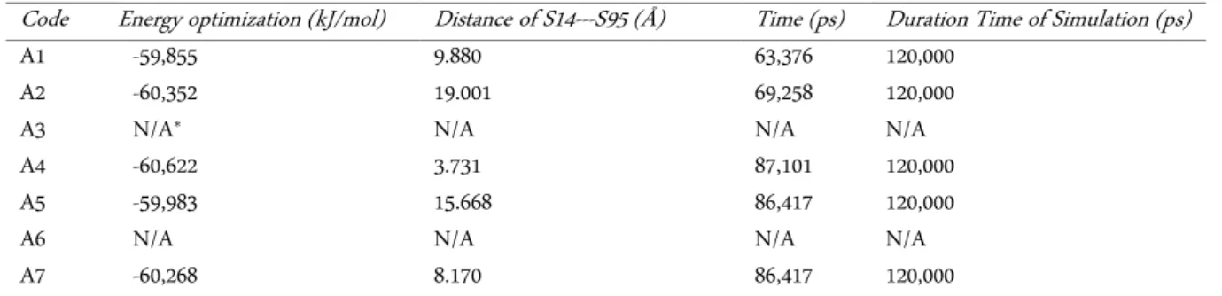
The 3D structures of the ADTC5 peptide were suc-cessfully generated by Pymol program which first built structure as OCS with different energy optimization based on modified OCS arrangements (Table 4). To ob-tain its cyclic conformation, the molecular dynamics simulation was conducted to find the best-optimized structure of CCS which mimics to native form (Table 5).
Molecular dynamics simulation at 120 ns (120,000 ps) was run to obtain the optimized OCS and CCS forms. Based on OCS result, variations of molecular dy-namics simulation were conducted by two different re-arrangement conditions of S14—S95 atom interactions: distance constraint and force restraint. By arranging S14—S95 distances, it showed that the longer distance A5 and shorter distance (A1, A4, A7) of S14---S95 sulted from the different energy optimizations due to re-arranged-force constraints. The region of the ADTC5 peptide was restrained during minimization is atom S14 from the start-terminus amino acid of Cysteine and atom S95 from end-terminus of Cysteine. It was as-sumed that the closer S14---S95 distance would give more stable OCS form based on the energy tions. Thus, A6 A4 showed the lowest energy optimiza-tion compared to others -60,622 kJ/mol). However, the RMSD values from all OCS forms are still over 2 Å. Therefore, these OCS forms are still unstable and it sug-gests that the presence of other non-covalent interac-tions are needed to stabilize these structures, such as the amino acid side-chain interactions between the EC1-EC2 coupled domain with the ADTC5 peptide.
The A4 structure of OCS form was selected for fur further analysis. It did not form the cyclization. There-fore, the sulfide bond addition to S14 and S95 atoms should be arranged manually. Moreover, by retaining force restraint of A4 structure of OCS form at 12,000
kJ.mol-1.nm-2 upon both atoms, it showed that A4
struc-ture has the best OCS form with S14---S95 distance at 3.731 Å through molecular dynamics simulation (Figure 4a).
The CCS form with molecular dynamics simulation showed that in giving two different force restraint (4,000 and 12,000 kJ.mol-1.nm-2) has resulted in similar energy optimization from B1, B2 and B3 forms (-47,044, -47,137 and -47,095 kJ/mol, respectively) as shown in Table 5. The OCS form of the ADTC5 peptide has a rigid cyclic structure (Figure 4b), which does not cause a change in molecular dynamics simulation. To mimic the native peptide structure of the ADTC5 peptide, CCS structure was generated. According to the average energy optimi-zation and S14---S95 distance arrangements, it showed that CCS structure has higher energy optimization than OCS. Thus, CCS conformation is more favorable com-pared to OCS conformation.
Also, to obtain the lowest energy optimization, B2 of CCS form has been chosen due to it has the most stable conformation (-47,137 kJ/mol) compared to A4 of OCS form (Figure 3). The low RMSD value has shown B2 structure is stable in molecular dynamics simulation. Thus, CCS is very stable from the initial to the final molecular dynamics process (RMSD ± 0.05 nm), while OCS is unstable (RMSD ± 0.15 nm). Moreover, CCS structures were further investigated in the way how they bind to EC1-EC2 coupled domain.
Molecular docking peptide ADTC5 with EC1-EC2 cou-pled domain
CCS form of the ADTC5 peptide was predicted has the highest similarity with ADTC5 native structure. Thus, the interaction can represent a cyclic ADTC5 na-tive interaction. Molecular dynamics results provide three conformations (B1, B2, and B3) in different energy optimizations. Furthermore, three CCS forms were docked with EC1-EC2 coupled domain.
In molecular docking, the best binding mode de-pends on: (1) the lowest affinity energy between ligand and protein, which is obtained from B1 binding mode (affinity energy -21,686 kJ/mol); (2) The highest amount of stable structures in a population, which is obtained from B1 binding mode (population of 81 structures); and (3) Validation of re-docking methods, characterized by comparing RMSD < 2 Å [21-23], obtained from B1 binding mode (re-docking up to 76%, better than B2 and B3). The most common parameters used in molec-ular docking is RMSD value with the ADTC5 peptide with the lowest affinity energy.
11 boxes, where 11 boxes are on the entire EC1-EC2 Table 4. The best structure for optimization molecular dynamics of OCS form
Code Energy optimization (kJ/mol) Distance of S14---S95 (Å) Time (ps) Duration Time of Simulation (ps)
A1 -59,855 9.880 63,376 120,000
A2 -60,352 19.001 69,258 120,000
A3 N/A* N/A N/A N/A
A4 -60,622 3.731 87,101 120,000
A5 -59,983 15.668 86,417 120,000
A6 N/A N/A N/A N/A
A7 -60,268 8.170 86,417 120,000
*N/A = Not Available
Table 5. The best structure of CCS form in molecular dynamics optimization
Code Energy optimization (kJ/mol) Distance of S14---S95 (Å) Time (ps) Duration Time of Simulation (ps)
B1 -47,044 2.029 85,453 120,000
B2 -47,137 2.027 17,914 120,000
B3 -47,095 2.029 730 120,000
Table 6. ADTC5 peptide interaction with the entire surface of the EC1-EC2 Best Binding
mode
Affinity energy
(kJ/mol) Hydrogen bond
Box, EC1/EC2 domain
Residues involve in hydrophobic interactions inside binding site of EC1 and EC2 domains
B1 -21.686 1 D of EC1 Arg55, Tyr36, Ile53, Ile52, Gly49,
Ala43, Ile38, Phe35, Val81, Ser37, Glu54
B2 -20.215 2 E of EC1 Trp2, Gln23, Ile24, Met92,
Lys25, Val3, Asn27, Ser26, Ile4
B3 -12.726 1 I of EC2 Asp1, Trp2, Val3, Pro5
coupled domain, where position of box A to G (except C) are within EC1 domain, while boxing H to K are within EC2 domain, and box C placed in the middle region between EC1 and EC2 domains which it contains calcium atoms. From molecular docking results, interac-tions of the ADTC5 peptide occur within three binding sites of EC1-EC2 coupled domain, there are two sites within box D and box E of the EC1 domain, with affin-ity energy -21.686 kJ/mol and -20.215 kJ/mol, respec-tively. The other site of docking interaction was also found within the box I of the EC1 domain, with affinity energy -12.726 kJ/mol (Table 6). Meanwhile, the best docking result of B1 was refined using Molecular Dy-namics (data not shown).
Prediction inhibition of interactions EC1-EC2---EC1-EC2 with ADTC5 peptide
Five repeated extracellular domains (EC1-E2-E3-E4-EC5) of E-cadherin homophilic can interact with other E-cadherin to form cis- and trans-dimer. In the paracel-lular pathway, E-cadherin interaction forms junction is trans-dimer interaction with other E-cadherin. Some studies showed that the interaction could involve the other domains. These interactions are formed in two ways: (1) the interaction of groove region from EC1a domain of cadherin to the other EC1b domain of E-cadherin in the bulge region [6] and (2) interaction from adhesion hands that will bind to the binding pocket [24].
Molecular docking study of the ADTC5 peptide showed that the ADTC5 inhibition upon trans-dimer in-teraction between EC1-EC2 extensively involved several types of chemical interactions, such as: (1) Van der Waals interaction with Ala43, Phe35 and Arg55 Asp44 and Val48 residues in the bulge region of EC1 domain and inhibits this bulge region to interact with grooves region. Also, hydrophobic interaction with Glu54, Ile52, Ile53, Tyr36, Ile38, Ser37, Val 81 and hydrogen bond with Ala43 which is between the bulge-groove regions from the EC1 domain of E-cadherin will help to inhibit interactions trans dimer in the EC1 domain. (2) Van der Waals interactions with Ile4, Lys25, Asn27 and Gln23 residues on adhesion arm region from EC1 domain of E-cadherin, as well as hydrophobic interactions with Trp2, Val3,Met92, Ile24, Ser26 on binding pocket region from EC1 domain of E-cadherin, thus the interaction at binding pocket-adhesion arm region on EC1 domain of E-cadherin will be hindered by ADTC5 peptide and hy-drogen bonds are Gln23 and Trp2; (3) hydrophobic in-teractions of Asp1, Trp2 and Pro3 residues; and (4)
hy-drogen bond interactions with Val3 in the Adhesive Arm of EC1 (Figure 5). These three binding sites would in-hibit the formation of trans-dimer between EC1-EC2 coupled domains on the adjacent cells. Thus the inter-action on junction will be weaker, and the paracellular pathways will be widely opened.
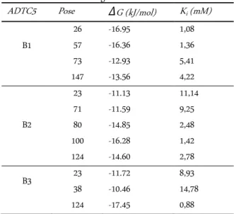
In finding the ΔG value, the ADTC5 peptide in
three CCS forms (B1, B2, B3) have been re-docked over
than 1000 scoring iterations. ΔG value for B1 is within
the range -12.93 to -16.95 kJ/mol; B2 is within the range 11.13 to 16.28 kJ/mol; and B3 is within the range -10.46 to -17.45 kJ/mol. On the other hand, the affinity of the ADTC5 peptide in inhibiting trans-dimer EC1-EC2 also can be determined through inhibition constant values (Ki). The Ki value of B3 is found having the strongest affinity (0.88 mM at pose 124) compared to B1 and B2 (Table 6). Meanwhile, Ki value was found using
formula ΔG = RT ln Ki (where ΔG is Gibbs free energy
(kJ.mol-1), R is ideal gas constant (8.314 J.mol-1.K-1), and Ki is inhibition constant (mM)).
The ADTC5 peptide was re-oriented towards the surface when it interacts with EC1-EC2 coupled do-main. This phenomenon can be seen by a B1 form of the ADTC5 that has the best optimization of energy when the molecular dynamics does not have a good in-teraction energy. It is claimed that structure with the best optimization does not have to produce the best in-teraction energy due to a re-orientation towards the tar-get. The best affinity energy is a B3 form (affinity ener- gy is -17.45 kJ/mol) of the ADTC5 peptide analyzed with molecular docking at the entire surface of the EC1-EC2 coupled domain (Table 5). The result obtained that
Table 7. Molecular docking of ADTC5 peptide (B1-B3) with EC1-EC2 within gridbox A
ADTC5 Pose ΔG (kJ/mol) Ki (mM) B1
26 -16.95 1,08
57 -16.36 1,36
73 -12.93 5,41
147 -13.56 4,22
B2
23 -11.13 11,14
71 -11.59 9,25
80 -14.85 2,48
100 -16.28 1,42
124 -14.60 2,78
B3 23 -11.72 8,93
38 -10.46 14,78
the entire interaction energy is spontaneous by giving negative value on the affinity energy. Moreover, the presence hydrogen bonds, van der Waals forces, and hy-drophobic interactions to stabilize the ADTC5 peptide interaction with the EC1-EC2 domain [25].
The affinity property of the ADTC5 peptide will af-fect the drug delivery that across the paracellular path-ways. In delivering the drug to body and brain cells, the ADTC5 cyclic peptide will increase the porosity of para-cellular pathway and allow the drug molecules to pass through it. However, the modulating of the paracellular pathway will need Ki value less than 100 µM [10]. It will increase the porosity of ADCT5 peptide and allow the drug goes to the target cell and through the paracellular pathway. This study will be very useful in designing more potential cyclic peptides derived from E-cadherin protein.
CONCLUSION
Our study showed that CCS of the ADTC5 pep-tide has more stable interactions with EC1 domain than the OCS one. Pre-experiment proved that ADTC5 which mimics to the native structure is in cyclic confor-mation, with optimization energy at -47,137 kJ/mol and it is very stable from initial to final molecular dynamics simulation process (RMSD ratio ± 0.5Å). Molecular docking study between the ADTC5 with EC1-EC2 cou-pled domain showed the best binding mode to EC1 (Box D), EC1 (Box E), and EC2 (Box I) with the affinity energy at -21.686 kJ/mol, -20.215 kJ/mol, and -12.726 kJ/ mol, respectively.
ACKNOWLEDGMENT
Thanks to Prof. Teruna J. Siahaan, Ph.D. (Phar-maceutical Chemistry Department, University of Kan-sas, US), for the valuable direction and discussion of the cells and drug delivery systems. Thanks to Prof. Krzysztof Kuczera, Ph.D. (Chemistry Department, Uni-versity of Kansas, US) for in depth discussion of a com-putational modeling. Last but not least, thanks to the Indonesian Directorate General of Higher Education, which has funded this research through the Research and Technology Funding scheme 2013.
REFERENCES
1. Brown RC, Davis TP (2002) Calcium modulation of and tight junction function: a potential mechanism for blood-brain barrier disruption after stroke. Stroke 33: 1706-1711. doi: 10.1161/01.STR.0000016405.06729.83
2. Majumdar S, Siahaan TJ (2012) Peptide-mediated targeted drug delivery. Medicinal Research Reviews 32 (3): 637-658. doi: 10.1002/med.20225.
3. Lutz KL, Siahaan TJ (1997) Molecular structure of the api-cal junction complex and its contribution to the paracellu-lar barrier. Journal of Pharmaceutical Sciences 86: 977-984. 4. Menard S, Cerf-Bensussan N, Heyman M (2010) Multiple facets of intestinal permeability and epithelial handling of dietary antigens. Mucosal Immunology 3 (3): 247-259. doi: 10.1038/mi.2010.5.
5. N'Da DD (2014) Review prodrug strategies for enhancing the percutaneous absorption of drugs. Molecules 19 (12): 20780-20807. doi: 10.3390/molecules191220780.
6. Sinaga E, Jois SD, Avery M, Makagiansar IT et al. (2002) Increasing paracellular porosity by E-cadherin peptides: Discovery of bulge and groove regions in the EC1-domain of E-cadherin. Pharmaceutical Research. 19 (8): 1170-1179. 7. Laksitorini MD, Kiptoo PK, On NH et al. (2015)
Modula-tion of intercellular juncModula-tions by cyclic-ADT peptides as a method to reversibly increase blood-brain barrier permea-bility. Journal of Pharmaceutical Sciences 104 (3): 1065-1075. doi: 10.1002/jps.24309.
8. Zheng K, Laurence JS, Kuczera K et al. (2009) Characteri-zation of multiple stable conformers of the EC5 domain of E-cadherin and the interaction of EC5 with E-cadherin pep-tides. Chemical Biology and Drug Design73 (6): 584-598. doi: 10.1111/j.1747-0285.2009.00818.x.
9. Kiptoo P, Sinaga E, Calcagno AM et al. (2011) Enhance-ment of drug absorption through the blood-brain barrier and inhibition of intercellular tight junction resealing by E-cadherin peptides. Molecular Pharmaceutics 8 (1): 239-249. doi: 10.1021/mp100293m
10. van Holde KE, Johnson WC, Ho PS (2006) Principles of physical biochemistry, Second Edition. New Jersey, Pearson Prentice Hall.
11. Siahaan P, Radiman CL, Rahayu SI et al. (2007) Investiga-tion of molecular interacInvestiga-tion between Phenylacetylene and Hexamethylphosphoric triamide by 13C NMR T1relaxa-tion time studies and Ab initio QM calculaT1relaxa-tions. Indone-sian Journal of Chemistry 7 (3): 273-277.
12. Klimovich PV, Mobley DL (2015) A Python tool to set up relative free energy calculations in GROMACS. Journal of Aided Molecular Design. Journal of Computer-Aided Molecular Design 29 (11): 1007-1014. doi: 10.1007/s10822-015-9873-0.
14. van der Spoel D, van Maaren PJ, Caleman C (2012) GROMACS molecule and liquid database. Bioinformatics 28 (5): 752-753. doi: 10.1093/bioinformatics/bts020. 15. van der Spoel D, Lindahl E, Hess B et al. (2015)
GROMACS: Fast, flexible, and free. Journal of Computa-tional Chemistry 26 (16): 1701-1718. doi: 10.1002/jcc.20291.
16. Lindorff-Larsen K, Maragakis P, Piana S et al. (2012) Sys-tematic validation of protein force fields against experi-mental data. PloS One 8 (4). doi: 10.1371/jour-nal.pone.0032131.
17. van der Spoel D, Lindahl E, Hess B, the GROMACS devel-opment team (2013) GROMACS User manual version 4.6. http://www.gromacs.org/. Accessed: January 2017. 18. Rentzsch R, Renard BY (2015) Docking small peptides
re-mains a great challenge: An assessment using AutoDock Vina. Briefings in Bioinformatics. 16 (6): 1045-1056. doi: 10.1093/bib/bbv008.
19. Hill AD, Reilly PJ (2015) Scoring Functions for AutoDock. In: Lutteke T, Frank M (eds) Glycoinformatics. New York, Springer, pp 467–474.
20. Morris GM, Huey R, Lindstrom W et al. (2009) AutoDock4 and AutoDockTools4: Automated docking with selective receptor flexibility. Journal of Computational Chemistry (16): 2785-2791. doi: 10.1002/jcc.21256.
21. Wang X, Snoeyink J (2006) Multiple structure alignment by optimal RMSD implies that the average structure is a consensus. Computational systems bioinformatics / Life Sciences Society. Computational Systems Bioinformatics Conference 5: 79-87.
22. Bruschweiler R (2003) Efficient RMSD measures for the comparison of two molecular ensembles. Root-mean-square deviation. Proteins 50 (1): 26-34. doi: 10.1002/prot.10250.
23. Reva BA, Finkelstein AV, Skolnick J (1998) What is the probability of a chance prediction of a protein structure with an RMSD of 6 A? Folding and Design. 3: 141-147. 24. Parisini E, Higgins JM, Liu JH et al. (2007) The crystal
structure of human E-cadherin domains 1 and 2, and com-parison with other cadherins in the context of adhesion mechanism. Journal of Molecular Biology 373 (2): 401-411. doi: 10.1016/j.jmb.2007.08.011.