STEMI FOCUSED UPDATE
2007 Focused Update of the ACC/AHA 2004 Guidelines
for the Management of Patients With ST-Elevation
Myocardial Infarction
A Report of the American College of Cardiology/American Heart Association
Task Force on Practice Guidelines
Developed in Collaboration With the Canadian Cardiovascular Society
Endorsed by the American Academy of Family Physicians
2007 Writing Group to Review New Evidence and Update the
ACC/AHA 2004 Guidelines for the Management of Patients With ST-Elevation Myocardial Infarction,
Writing on Behalf of the 2004 Writing Committee
Elliott M. Antman, MD, FACC, FAHA, Co-Chair*†; Mary Hand, MSPH, RN, FAHA, Co-Chair;
Paul W. Armstrong, MD, FACC, FAHA‡§; Eric R. Bates, MD, FACC, FAHA; Lee A. Green, MD, MPH储;
Lakshmi K. Halasyamani, MD¶; Judith S. Hochman, MD, FACC, FAHA**;
Harlan M. Krumholz, MD, FACC, FAHA††; Gervasio A. Lamas, MD, FACC**;
Charles J. Mullany, MB, MS, FACC; David L. Pearle, MD, FACC, FAHA; Michael A. Sloan, MD, FACC;
Sidney C. Smith, Jr, MD, FACC, FAHA§§
2004 WRITING COMMITTEE MEMBERS
Elliott M. Antman, MD, FACC, FAHA, Chair; Daniel T. Anbe, MD, FACC, FAHA;
Paul W. Armstrong, MD, FACC, FAHA; Eric R. Bates, MD, FACC, FAHA; Lee A. Green, MD, MPH;
Mary Hand, MSPH, RN, FAHA; Judith S. Hochman, MD, FACC, FAHA;
Harlan M. Krumholz, MD, FACC, FAHA; Frederick G. Kushner, MD, FACC, FAHA;
Gervasio A. Lamas, MD, FACC; Charles J. Mullany, MB, MS, FACC;
Joseph P. Ornato, MD, FACC, FAHA; David L. Pearle, MD, FACC, FAHA;
Michael A. Sloan, MD, FACC; Sidney C. Smith, Jr, MD, FACC, FAHA
*Chair of 2004 Writing Committee.
†Recused from voting on Section 8: Anticoagulants as Ancillary Therapy and Section 10: Anticoagulants. ‡Recused from voting on Section 5: Facilitated PCI.
§Canadian Cardiovascular Society Representative. 储American Academy of Family Physicians Representative. ¶American College of Physicians Representative.
**Recused from voting on Section 7: PCI After Fibrinolysis or for Patients Not Undergoing Primary Reperfusion. ††Performance Measures Liaison.
‡‡Former Task Force member during this writing effort. §§Recused from voting on Section 13: Antiplatelet Therapy.
This document is a limited update to the 2004 guidelines update and is based on a review of certain evidence, not a full literature review. This document was approved by the American College of Cardiology Board of Trustees in October 2007 and by the American Heart Association Science Advisory and Coordinating Committee October 2007.
The American College of Cardiology Foundation and the American Heart Association request that this document be cited as follows: Antman EM, Hand M, Armstrong PW, Bates ER, Green LA, Halasyamani LK, Hochman JS, Krumholz HM, Lamas GA, Mullany CJ, Pearle DL, Sloan MA, Smith SC Jr. 2007 focused update of the ACC/AHA 2004 Guidelines for the Management of Patients With ST-Elevation Myocardial Infarction: a report of the American College of Cardiology/American Heart Association Task Force on Practice Guidelines (Writing Group to Review New Evidence and Update the ACC/AHA 2004 Guidelines for the Management of Patients With ST-Elevation Myocardial Infarction).Circulation. 2008;117:296 –329.
This article has been copublished in theJournal of the American College of Cardiology.
Copies: This document is available on the World Wide Web sites of the American College of Cardiology (www.acc.org) and the American Heart Association
(my.americanheart.org). To purchaseCirculationreprints, call 843-216-2533 or [email protected].
Permissions: Multiple copies, modification, alteration, enhancement and/or distribution of this document are not permitted without the express permission of the American College of Cardiology and the American Heart Association. Instructions for obtaining permission are located athttp://www.americanheart.org/
presenter.jhtml?identifier⫽4431. A link to the “Permission Request Form” appears on the right side of the page.
(Circulation. 2008;117:296-329.)
© 2008 by the American College of Cardiology Foundation and the American Heart Association, Inc.
Circulationis available at http://circ.ahajournals.org DOI: 10.1161/CIRCULATIONAHA.107.188209
296
by guest on March 29, 2018
http://circ.ahajournals.org/
Downloaded from
by guest on March 29, 2018
http://circ.ahajournals.org/
Downloaded from
by guest on March 29, 2018
http://circ.ahajournals.org/
TASK FORCE MEMBERS
Sidney C. Smith, Jr, MD, FACC, FAHA, Chair; Alice K. Jacobs, MD, FACC, FAHA, Vice-Chair;
Cynthia D. Adams, MSN, PhD, FAHA‡‡; Jeffrey L. Anderson, MD, FACC, FAHA‡‡;
Christopher E. Buller, MD, FACC; Mark A. Creager, MD, FACC, FAHA; Steven M. Ettinger, MD, FACC;
Jonathan L. Halperin, MD, FACC, FAHA‡‡; Sharon A. Hunt, MD, FACC, FAHA‡‡;
Harlan M. Krumholz, MD, FACC, FAHA; Frederick G. Kushner, MD, FACC, FAHA;
Bruce W. Lytle, MD, FACC, FAHA; Rick Nishimura, MD, FACC, FAHA;
Richard L. Page, MD, FACC, FAHA; Barbara Riegel, DNSc, RN, FAHA‡‡;
Lynn G. Tarkington, RN; Clyde W. Yancy, MD, FACC
TABLE OF CONTENTS
Preamble. . . .297
1. Introduction. . . .299
1.1. Evidence Review. . . .299
1.2. Organization of Committee and Relationships With Industry. . . .299
1.3. Review and Approval. . . .300
2. Analgesia. . . .300
3. Beta Blockers. . . .300
3.1. COMMIT/CCS-2 (Metoprolol). . . .300
3.2. Conclusion. . . .301
4. Reperfusion. . . .302
4.1. Logistics of Care. . . .302
5. Facilitated PCI. . . .304
6. Immediate or Emergency Invasive Strategy and Rescue PCI. . . .305
7. PCI After Fibrinolysis or for Patients Not Undergoing Primary Reperfusion. . . .307
7.1. The Late Open Artery Hypothesis: Clinical Outcomes. . . .307
7.2. The Late Open Artery Hypothesis: Angiographic Outcomes. . . .308
7.3. Conclusion. . . .308
8. Ancillary Therapy. . . .308
8.1. Conclusion. . . .312
9. Thienopyridines. . . .313
9.1. Conclusion. . . .314
10. Anticoagulants. . . .315
11. Invasive Evaluation. . . .315
12. Secondary Prevention. . . .315
13. Antiplatelet Therapy. . . .320
References. . . .321
Appendix 1. . . .324
Appendix 2. . . .326
Preamble
A primary challenge in the development of clinical practice guidelines is keeping pace with the stream of new data upon which recommendations are based. In an effort to respond more quickly to new evidence, the American College of Cardiology/American Heart Association (ACC/AHA) Task Force on Practice Guidelines has created a new “focused update” process to revise the existing guideline recommen-dations that are affected by evolving data or opinion. Before the initiation of this focused approach, periodic updates and revisions of existing guidelines required up to 3 years to
complete. Now, however, new evidence will be reviewed in an ongoing fashion to more efficiently respond to important science and treatment trends that could have a major impact on patient outcomes and quality of care. Evidence will be reviewed at least twice a year, and updates will be initiated on an as needed basis as quickly as possible, while maintaining the rigorous methodology that the ACC and AHA have developed during their more than 20 years of partnership.
These updated guideline recommendations reflect a consensus of expert opinion following a thorough review that consisted primarily of late-breaking clinical trials identified through a broad-based vetting process as important to the relevant patient population and of other new data deemed to have an impact on patient care (see Section 1.1 for details on this focused update). It is important to note that this focused update is not intended to represent an update based on a full literature review from the date of the previous guideline publication. Specific criteria/ considerations for inclusion of new data include:
• Publication in a peer-reviewed journal
• Large, randomized, placebo-controlled trial(s)
• Nonrandomized data deemed important on the basis of results that impact current safety and efficacy assumptions
• Strengths/weakness of research methodology and findings
• Likelihood of additional studies influencing current findings
• Impact on current performance measure(s) and/or likelihood of the need to develop new performance measure(s)
• Requests and requirements for review and update from the practice community, key stakeholders, and other sources free of relationships with industry or other potential bias
• Number of previous trials showing consistent results
• Need for consistency with other guidelines or guideline revisions
In analyzing the data and developing updated recommen-dations and supporting text, the focused update writing group used evidence-based methodologies developed by the ACC/ AHA Task Force on Practice Guidelines, which are described elsewhere.1,2
The schema for class of recommendation and level of evi-dence is summarized inTable 1, which also illustrates how the grading system provides estimates of the size of the treatment effect and the certainty of the treatment effect. Note that a recommendation with Level of Evidence B or C does not imply that the recommendation is weak. Many important clinical questions addressed in guidelines do not lend themselves to clinical trials. Although randomized trials may not be available, there may be a very clear clinical consensus that a particular test or therapy is useful and effective. Both the class of
by guest on March 29, 2018
http://circ.ahajournals.org/
dation and level of evidence listed in the focused updates are based on consideration of the evidence reviewed in previous iterations of the guidelines as well as the focused update. Of note, the implications of older studies that have informed recommendations but have not been repeated in contemporary settings are carefully considered.
The ACC/AHA practice guidelines address patient popula-tions (and health care providers) residing in North America. As such, drugs that are not currently available in North America are discussed in the text without a specific class of recommendation. For studies performed in large numbers of subjects outside of North America, each writing committee reviews the potential impact of different practice patterns and patient populations on the treatment effect and on the relevance to the ACC/AHA target
population to determine whether the findings should inform a specific recommendation.
The ACC/AHA practice guidelines are intended to assist health care providers in clinical decision making by describing a range of generally acceptable approaches for the diagnosis, management, and prevention of specific diseases or conditions. The guidelines attempt to define practices that meet the needs of most patients in most circumstances. The ultimate judgment regarding care of a particular patient must be made by the health care provider and patient in light of all the circumstances presented by that patient. Thus, there are circumstances in which devi-ations from these guidelines may be appropriate. Clinical decision making should consider the quality and
availabil-Table 1. Applying Classification of Recommendations and Level of Evidence†
ⴱData available from clinical trials or registries about the usefulness/efficacy in different subpopulations, such as gender, age, history of diabetes, history of prior myocardial infarction, history of heart failure, and prior aspirin use. A recommendation with Level of Evidence B or C does not imply that the recommendation is weak. Many important clinical questions addressed in the guidelines do not lend themselves to clinical trials. Even though randomized trials are not available, there may be a very clear clinical consensus that a particular test or therapy is useful or effective.
†In 2003, the ACC/AHA Task Force on Practice Guidelines developed a list of suggested phrases to use when writing recommendations. All guideline recommendations have been written in full sentences that express a complete thought, such that a recommendation, even if separated and presented apart from the rest of the document (including headings above sets of recommendations), would still convey the full intent of the recommendation. It is hoped that this will increase readers’ comprehension of the guidelines and will allow queries at the individual recommendation level.
by guest on March 29, 2018
http://circ.ahajournals.org/
ity of expertise in the area where care is provided. These guidelines may be used as the basis for regulatory or payer decisions, but the ultimate goal is quality of care and serving the patient’s best interests.
Prescribed courses of treatment in accordance with these recommendations are only effective if they are followed by the patient. Because lack of patient adherence may adversely affect treatment outcomes, health care providers should make every effort to engage the patient in active participation with prescribed treatment.
The ACC/AHA Task Force on Practice Guidelines makes every effort to avoid any actual, potential, or perceived conflict of interest arising from industry relationships or personal interests of a writing committee member. All writing committee members and peer reviewers were required to provide disclosure statements of all such relationships per-taining to the trials and other evidence under consideration (see Appendixes 1 and 2). Final recommendations were balloted to all writing committee members. Writing commit-tee members with significant (greater than $10 000) relevant relationships with industry (RWI) were required to recuse themselves from voting on that recommendation. Writing committee members who did not participate are not listed as authors of this focused update.
With the exception of the recommendations presented here, the full guidelines remain current. Only the recommendations from the affected section(s) of the full guidelines are included in this focused update. For easy reference, all recommenda-tions from any section of guidelines impacted by a change are presented with a notation as to whether they remain current, are new, or have been modified. When evidence impacts recommendations in more than 1 set of guidelines, those guidelines are updated concurrently.
The recommendations in this focused update will be considered current until they are superseded by another focused update or the full-text guidelines are revised. This focused update is published in the January 15, 2008, issue of theJournal of the American College of Cardiology and the January 15, 2008, issue of Circulationas an update to the full-text guidelines and is also posted on the ACC (www. acc.org) and AHA (www.americanheart.org) Web sites. Cop-ies of the focused update are available from both organiza-tions.
Sidney C. Smith, Jr., MD, FACC, FAHA Chair, ACC/AHA Task Force on Practice Guidelines Alice K. Jacobs, MD, FACC, FAHA Vice-Chair, ACC/AHA Task Force on Practice Guidelines
1. Introduction
1.1. Evidence Review
Late-breaking clinical trials presented at the 2005 and 2006 annual scientific meetings of the ACC, AHA, and European Society of Cardiology, as well as selected other data, were reviewed by the standing guideline writing committee along with the parent Task Force and other experts to identify those trials and other key data that might impact guidelines recommendations. On the basis of
the criteria/considerations noted above, recent trial data and other clinical information were considered important enough to prompt a focused update of the 2004 ACC/AHA Guidelines for the Management of Patients With ST-Elevation Myocardial Infarction [see Chen ZM et al.3;
Chen ZM et al.4; ASSENT-4 PCI5; Antman EM et al.6;
Yusuf S et al.7; Bhatt DL et al.8; Sabatine MS et al.9; Bennett JS et al.10; Smith SC Jr et al.11; OAT12,13 and
TOSCA14].
When considering the new data for this focused update, the writing group faced the task of weighing evidence from studies enrolling large numbers of subjects outside North America. Although noting that practice patterns and the rigor applied to data collection, as well as the genetic makeup of subjects, might influence the observed magni-tude of a treatment effect, the writing group believed the data were relevant to formulation of recommendations for management of ST-elevation myocardial infarction (STEMI) in North America. The reasons for this decision include that 1) a broad array of management strategies was represented, including substantial proportions of sub-jects who received some form of reperfusion therapy, 2) concomitant treatments with proven efficacy (e.g., aspirin, beta blockers, inhibitors of the renin-angiotensin-aldosterone system, and statins) were used in the majority of patients, and 3) it was considered an impractical expectation that the tens of thousands of patients with STEMI needed to meet the estimated sample size for contemporary clinical trials be enrolled exclusively at North American sites.
To provide clinicians with a comprehensive set of data, whenever possible the exact event rates in various treat-ment arms of clinical trials are presented to permit calcu-lation of the absolute risk difference (ARD) and number needed to treat (NNT) or harm (NNH); the relative treatment effects are described either as odds ratio (OR), relative risk (RR), or hazard ratio (HR), depending on the format in the original publication.
Consult the full-text version or executive summary of the 2004 ACC/AHA Guidelines for the Management of Patients With ST-Elevation Myocardial Infarction15 for
policy on clinical areas not covered by the focused update. Individual recommendations updated in this focused up-date will be incorporated into future revisions and/or updates of the full-text guidelines.
1.2. Organization of Committee and Relationships With Industry
For this focused update, all members of the 2004 STEMI writing committee were invited to participate; those who agreed (referred to as the 2007 focused update writing group) were required to disclose all RWI relevant to the data under consideration.2 Focused update writing group
members who had no significant relevant RWI wrote the first draft of the focused update; the draft was then reviewed and revised by the full writing group. Each recommendation required a confidential vote by the writ-ing group members before external review of the docu-ment. Any writing committee member with a significant
by guest on March 29, 2018
http://circ.ahajournals.org/
(greater than $10 000) relationship with industry relevant to the recommendation was recused from voting on that recommendation.
1.3. Review and Approval
This document was reviewed by 3 outside reviewers nominated by the ACC and 3 outside reviewers nominated by the AHA, as well as 1 reviewer each from the American Academy of Family Physicians and the Canadian Cardio-vascular Society (CCS) and 58 individual content review-ers. All reviewer RWI information was collected and distributed to the writing committee and is published in this document (see Appendix 2 for details).
This document was approved for publication by the gov-erning bodies of the American College of Cardiology Foun-dation and the American Heart Association and endorsed by the American Academy of Family Physicians and the Canadian Cardiovascular Society.
2. Analgesia
Analysis of retrospective data16has raised a question about
the potentially adverse effects of morphine in patients with unstable angina (UA)/non–ST-elevation myocardial infarc-tion (NSTEMI). As a result, the recommendainfarc-tion for mor-phine pain relief has been reduced to a Class IIa recommen-dation for that patient population. Use of morphine remains a Class I recommendation for patients with STEMI, however, because STEMI patients should either have received reperfu-sion or are not candidates for reperfureperfu-sion, and continuing pain requires relief in either case (Table 2).
Because of the known increased risk of cardiovascular events among patients taking cyclooxygenase-2 (COX-2) inhibitors and other nonsteroidal anti-inflammatory drugs (NSAIDs),17–19these drugs should be discontinued
immedi-ately at the time of STEMI (see 2004 STEMI Guidelines, Section 7.12.5, for additional discussion).3,15,20,21A substudy
analysis from the ExTRACT TIMI-25 (Enoxaparin and Thrombolysis Reperfusion for Acute Myocardial Infarction
Treatment–Thrombolysis in Myocardial Infarction) trial22
demonstrated an increased risk of death, reinfarction, heart failure, or shock among patients who were taking NSAIDs within 7 days of enrollment. Longer-term management con-siderations and a discussion of the gradient of risk with the various NSAIDS are found in Section 7.12.5 of the 2004 STEMI Guidelines.15
3. Beta Blockers
The 2004 STEMI Guidelines recommendations (Table 3) were based on studies that showed a reduced incidence of subsequent reinfarction and recurrent ischemia in patients receiving both fibrinolytic therapy and intravenous (IV) beta blockers. However, uncertainty about the use of IV beta blockers in the setting of fibrinolytic therapy has increased following 2 later randomized trials of IV beta blockade,23,24a
post-hoc analysis of the use of atenolol in the GUSTO-I (Global Utilization of Streptokinase and TPA for Occluded Coronary Arteries) trial,25and a review of early beta-blocker
therapy in myocardial infarction (MI)26 that did not find
significant reductions in mortality.15
3.1. COMMIT/CCS-2 (Metoprolol)
The COMMIT/CCS-2 (Clopidogrel and Metoprolol in Myo-cardial Infarction Trial/Second Chinese Cardiac Study) (4) randomized 45 852 patients within 24 hours of onset of suspected MI to receive metoprolol (up to 3 doses of 5 mg IV each in the first 15 minutes, followed by 200 mg orally daily) or matching placebo. Fifteen minutes after the IV doses, a 50-mg tablet of metoprolol or placebo was administered orally and repeated every 6 hours during Days 0 to 1 of hospitalization. From Day 2 onward, 200 mg of controlled-release metoprolol or placebo was administered orally daily (this is the Food and Drug Administration [FDA]-approved regimen for metoprolol in MI) until discharge from the hospital or up to a maximum of 4 weeks in hospital (in survivors, the mean was 15 days). The 2 prespecified co-primary outcomes were the composite of death, reinfarction,
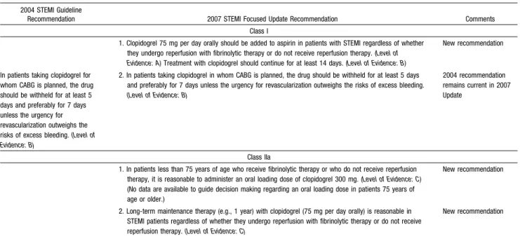
Table 2. Updates to Section 6.3.1.3: Analgesia
2004 STEMI Guideline Recommendation 2007 STEMI Focused Update Recommendation Comments Class I
Morphine sulfate (2 to 4 mg IV with increments of 2 to 8 mg IV repeated at 5- to 15-minute intervals) is the analgesic of choice for management of pain associated with STEMI. (Level of Evidence: C)
1. Morphine sulfate (2 to 4 mg IV with increments of 2 to 8 mg IV repeated at 5- to 15-minute intervals) is the analgesic of choice for management of pain associated with STEMI. (Level of Evidence: C)
2004 recommendation remains current in 2007 Update
2. Patients routinely taking NSAIDs (except for aspirin), both nonselective as well as COX-2 selective agents, before STEMI should have those agents discontinued at the time of
presentation with STEMI because of the increased risk of mortality, reinfarction, hypertension, heart failure, and myocardial rupture associated with their use. (Level of Evidence: C)
New recommendation
Class III
1. NSAIDs (except for aspirin), both nonselective as well as COX-2 selective agents, should not be administered during hospitalization for STEMI because of the increased risk of mortality, reinfarction, hypertension, heart failure, and myocardial rupture associated with their use. (Level of Evidence:C)
New recommendation
COX-2 indicates cyclooxygenase-2; IV, intravenous/intravenously; NSAIDs, nonsteroidal anti-inflammatory drugs; and STEMI, ST-elevation myocardial infarction.
by guest on March 29, 2018
http://circ.ahajournals.org/
or cardiac arrest and death from any cause during the scheduled treatment period.
Neither of the co-primary study end points was signifi-cantly reduced by allocation to metoprolol. For every 1000 patients treated, allocation to metoprolol was associated with 5 fewer episodes of reinfarction, 5 fewer episodes of ventric-ular defibrillation, but 11 more episodes of cardiogenic shock. The excess of cardiogenic shock was seen chiefly from Days 0 to 1 after hospitalization, whereas the reductions in rein-farction and ventricular fibrillation appeared from Day 2 onward.
Allocation to metoprolol produced an average relative in-crease in cardiogenic shock of 30%, with higher rates for those greater than 70 years of age, or with systolic blood pressure less than 120 mm Hg, or with presenting heart rate greater than 110 bpm, or with Killip class greater than 1. On average across the whole study population, the absolute reduction in arrhythmia-related deaths and the absolute increase in cardiogenic shock– related deaths were of similar magnitude. No apparent difference was noted between the 2 treatment groups in the other attributed causes of death, either individually or in aggregate. Metoprolol allocation was associated with significantly more persistent hypotension and more cases of bradycardia.
Though patients at high or low risk could be identified, the authors noted that they were not able to identify any sub-groups in which the benefits clearly outweighed the risks.
3.2. Conclusion
This focused update expands on the concepts introduced in the 2004 STEMI Guidelines, underscoring the potential risk of administering IV beta blockers to patients with severe heart failure or cardiogenic shock. There are several circumstances in which it can be useful (Class IIa) to administer an IV beta blocker acutely to a STEMI patient (Table 3), and these situations are discussed below. It is reasonable to administer IV beta-blocker therapy on Days 0 to 1 of hospitalization for STEMI when hypertension is present and the patient is not at an increased risk of cardiogenic shock on the basis of the risk factors defined above. Patients with sinus tachycardia or atrial fibrillation should have left ventricular (LV) function rapidly evalu-ated before administration of IV beta blockers (or other negative inotropes, such as non-dihydropyridine calcium channel blockers). From Day 2 onward, when beneficial effects on reinfarction and ventricular fibrillation are seen, administration of 200 mg of controlled-release oral meto-prolol daily appears to be safe in hemodynamically stable patients with STEMI who are free of contraindications. It is prudent to initiate a dose of 50 mg of metoprolol orally every 6 hours, transitioning to a dose equivalent to 200 mg per day orally or the maximum tolerated dose. It should be noted that long-term use of oral beta blockers is strongly recommended (Class I, Level of Evidence: A) for second-Table 3. Updates to Section 6.3.1.5: Beta Blockers
2004 STEMI Guideline Recommendation 2007 STEMI Focused Update Recommendation Comments Class I
Oral beta-blocker therapy should be administered promptly to those patients without a contraindication, irrespective of concomitant fibrinolytic therapy or performance of primary PCI.(Level of Evidence: A)
1. Oral beta-blocker therapy should be initiated in the first 24 hours for patients who do not have any of the following: 1) signs of heart failure, 2) evidence of a low output state, 3) increased risk* for cardiogenic shock, or 4) other relative contraindications to beta blockade (PR interval greater than 0.24 seconds, second- or third-degree heart block, active asthma, or reactive airway disease).(Level of Evidence: B)
Modified recommendation (changed LOE and text)
Patients with early contraindications within the first 24 hours of STEMI should be reevaluated for candidacy for beta-blocker therapy as secondary prevention.(Level of Evidence: C)
2. Patients with early contraindications within the first 24 hours of STEMI should be reevaluated for candidacy for beta-blocker therapy as secondary prevention.(Level of Evidence: C)
2004 recommendation remains current in 2007 Update
Patients with moderate or severe LV failure should receive beta-blocker therapy as secondary prevention with a gradual titration scheme.(Level of Evidence: B)
3. Patients with moderate or severe LV failure should receive beta-blocker therapy as secondary prevention with a gradual titration scheme.(Level of Evidence: B)
2004 recommendation remains current in 2007 Update
Class IIa It is reasonable to administer IV beta blockers promptly
to STEMI patients without contraindications, especially if a tachyarrhythmia or hypertension is present.(Level of Evidence: B)
1. It is reasonable to administer an IV beta blocker at the time of presentation to STEMI patients who are hypertensive and who do not have any of the following: 1) signs of heart failure, 2) evidence of a low output state, 3) increased risk* for cardiogenic shock, or 4) other relative contraindications to beta blockade (PR interval greater than 0.24 seconds, second- or third-degree heart block, active asthma, or reactive airway disease).(Level of Evidence: B)
Modified recommendation (changed text)
Class III
1. IV beta blockers should not be administered to STEMI patients who have any of the following: 1) signs of heart failure, 2) evidence of a low output state, 3) increased risk* for cardiogenic shock, or 4) other relative contraindications to beta blockade (PR interval greater than 0.24 seconds, second- or third-degree heart block, active asthma, or reactive airway disease).(Level of Evidence: A)
New recommendation
*Risk factors for cardiogenic shock (the greater the number of risk factors present, the higher the risk of developing cardiogenic shock) are age greater than 70 years, systolic blood pressure less than 120 mm Hg, sinus tachycardia greater than 110 bpm or heart rate less than 60 bpm, and increased time since onset of symptoms of STEMI.
IV indicates intravenous; LOE, level of evidence; LV, left ventricular; PCI, percutaneous coronary intervention; and STEMI, ST-elevation myocardial infarction.
by guest on March 29, 2018
http://circ.ahajournals.org/
ary prevention in patients at highest risk, such as those with low ejection fraction, heart failure, or postshock, once they have stabilized, with gradual dose titration27(see the 2004 STEMI Guidelines, Sections 7.4.1 and 7.12.7).15
The results of the COMMIT-CCS 2 trial raise questions about the safety of early use of IV beta blockers, particularly in high-risk populations, and led the writing group to reex-amine the overall evidence base for beta-blocker therapy. The evidence base for this therapy was developed more than 25 years ago in a treatment environment that differs from contemporary practice. Moreover, no study included an oral beta blocker– only arm. The writing group consensus, how-ever, was not to change the classification of the current early oral beta-blocker recommendation but to restrict it to patients who are not at high risk for complications. In addition, because of the absence of a study that specifically evaluated oral therapy alone, the Level of Evidence has been changed from A to B. Nevertheless, early (within 24 hours) oral beta-blocker therapy remains a Class I recommendation for those patients who are not at high risk for complications. Whether this change should affect current performance measures is beyond the scope of this document. The findings of potential risk of beta-blocker therapy in COMMIT emphasize the importance of continually mon-itoring these patients throughout hospitalization for signs and symptoms of complications of therapy, as noted in other sections of the original guidelines (Sections 6.3.1.5, 7.4.1, and 7.12.7). Because of the uncertainty about the benefit of oral beta blockers early on (e.g., in COMMIT-CCS 2, Days 0 to 1), the writing group recommends further research and additional examination at the time of the next revision to the STEMI Guidelines.
4. Reperfusion
4.1. Logistics of Care
Regardless of the mode of reperfusion, the overarching concept is to minimize total ischemic time, which is defined as the time from onset of symptoms of STEMI to initiation of reperfusion therapy. It is increasingly clear that 2 types of hospital systems provide reperfusion therapy: those with percutaneous coronary intervention (PCI) capability and those without PCI capability. When PCI capability is avail-able, the best outcomes are achieved by offering this strategy 24 hours per day, 7 days per week.28The systems goal should
be a first medical contact–to-balloon time within 90 minutes (Table 4,Figure 1). There should be an ongoing program of outcomes analysis and periodic case review to identify process-of-care strategies that will continually improve time to treatment and facilitate rapid and appropriate treatment. A comprehensive effort in this regard is the AHA Mission Lifeline program, a community-based national initiative to improve the quality of care and outcomes of patients with STEMI by improving health care system readiness and response to STEMI.29 The “Door-to-Balloon (D2B): An
Alliance for Quality” campaign (www.d2balliance.org), launched by the ACC in collaboration with many organiza-tions, including the AHA, aims to improve the timeliness of primary PCI. The goal is to increase the percentage of patients who receive timely primary PCI, with an emphasis on having at least 75% of patients treated within 90 minutes of presentation at the hospital, with a recommendation for the use of evidence-based strategies to reduce needless delays.30
The 75% goal was set in recognition that some patients have clinically relevant non–system-based delays that do not rep-resent quality-of-care issues. In hospitals without PCI capa-bility, immediate transfer for primary PCI is a treatment option when the expected door-to-balloon time is within 90 minutes of first medical contact.31,32
It is important to note that the door-to-balloon goal is a systems goal that may not be possible to achieve for an individual patient because of patient variables (uncertainty about diagnosis, evaluation and treatment of other life-threatening conditions, obtaining informed consent, etc.) that delay the patient’s arrival in the interventional cardi-ology laboratory or anatomical challenges (issues of arte-rial, coronary, or lesion access) that prolong the PCI procedure. In the absence of such circumstances, however, reperfusion should be achieved as soon as possible within this time, and many hospitals with refined systems are approaching median door-to-balloon times of 60 to 70 minutes. Discussions about measurement, particularly with respect to inclusion criteria and the appropriate time to end measurement, are beyond the scope of this document and are being considered by groups that are focusing on how to improve the alignment between what is measured and patient outcomes. The focus on measurement should not displace the emphasis on improving processes that will facilitate more rapid treatment that is delivered safely and appropriately. This committee continues to endorse the
Table 4. Updates to Section 6.3.1.6: Reperfusion
2004 STEMI Guideline Recommendation 2007 STEMI Focused Update Recommendation Comments Class I
Primary PCI should be performed as quickly as possible with the goal of a medical contact–to-balloon or door-to-balloon interval of within 90 minutes.(Level of Evidence: B)
1. STEMI patients presenting to a hospital with PCI capability should be treated with primary PCI within 90 minutes of first medical contact (see Figure 1) as a systems goal. (Level of Evidence: A)
Modified recommendation (changed LOE and text)
STEMI patients presenting to a facility without the capability for expert, prompt intervention with primary PCI within 90 minutes of first medical contact should undergo fibrinolytic therapy unless contraindicated. (Level of Evidence: A)
2. STEMI patients presenting to a hospital without PCI capability and who cannot be transferred to a PCI center and undergo PCI within 90 minutes of first medical contact (see Figure 1) should be treated with fibrinolytic therapy within 30 minutes of hospital presentation as a systems goal unless fibrinolytic therapy is contraindicated. (Level of Evidence: B)
Modified recommendation (changed LOE and text)
PCI indicates primary coronary intervention; LOE, level of evidence; and STEMI, ST-elevation myocardial infarction.
by guest on March 29, 2018
http://circ.ahajournals.org/
concept that faster times to reperfusion and better systems of care are associated with important reduc-tions in morbidity and mortality rates in patients with STEMI. An underutilized but effective strategy for improving systems of care for STEMI patients is to expand the use of prehospital 12-lead electrocardiogra-phy programs by emergency medical systems (EMS) that provide advanced life support.33,34
The emphasis on primary PCI should not obscure the importance of fibrinolytic therapy. Many hospital systems in
North America do not have the capability of meeting the time goal for primary PCI.35 Therefore, because of the critical
importance of time to treatment from onset of symptoms of STEMI in reducing morbidity and mortality, fibrinolytic therapy is preferred. In these settings, transfer protocols need to be in place for arranging rescue PCI when clinically indicated.36
For fibrinolytic therapy, the system goal is to deliver the drug within 30 minutes of the time that the patient presents to the hospital (Table 4). The focus for primary PCI is from first Figure 1. Options for Transportation of STEMI Patients and Initial Reperfusion Treatment Goals
Reperfusion in patients with STEMI can be accomplished by pharmacological (fibrinolysis) or catheter-based (primary PCI) approaches. The overarching goal is tokeep total ischemic time within 120 minutes(ideally within 60 minutes) from symptom onset to initiation of reperfusion treatment. Within this context, the following are goals for the medical system* based on the mode of patient transportation and the capabilities of the receiving hospital:
Medical System Goals: EMS Transport (Recommended):
•If EMS has fibrinolytic capability and the patient qualifies for therapy, prehospital fibrinolysis should be started within 30 minutes of arrival of EMS on the scene.
•If EMS is not capable of administering prehospital fibrinolysis and the patient is transported to anon–PCI-capable hospital, thedoor-to-needletime should be within 30 minutes for patients for whom fibrinolysis is indicated.
•If EMS is not capable of administering prehospital fibrinolysis and the patient is transported to a PCI-capable hospital, theEMS arrival-to-balloontime should be within 90 minutes.
•If EMS takes the patient to anon–PCI-capable hospital, it is appropriate to consider emergencyinterhospital transferof the patient to a PCI-capable hospital for mechanical revascularization if
XThere is a contraindication to fibrinolysis.
XPCI can be initiated promptly within 90 minutesfrom EMS arrival-to-balloon time at the PCI-capable hospital.†
XFibrinolysis is administered and is unsuccessful (i.e., “rescue PCI”).
Patient Self-Transport (Discouraged):
•If the patient arrives at a non–PCI-capable hospital, thedoor-to-needletime should be within 30 minutes of arrival at the emergency department.
•If the patient arrives at a PCI-capable hospital, thedoor-to-balloontime should be within 90 minutes.
•If the patient presents to a non–PCI-capable hospital, it is appropriate to consider emergencyinterhospital transferof the patient to a PCI-capable hospital if
XThere is a contraindication to fibrinolysis.
XPCI can be initiated within 90 minutes after the patient presented to the initial receiving hospital or within 60 minutes compared with when fibrinolysis with a fibrin-specific agent could be initiated at the initial receiving hospital.
XFibrinolysis is administered and is unsuccessful (i.e., “rescue PCI”).
*The medical system goal is to facilitate rapid recognition and treatment of patients with STEMI so thatdoor-to-needle(ormedical contact-to-needle) for initiation of fibrinolytic therapy can be achieved within 30 minutes ordoor-to-balloon(ormedical contact-to-balloon) for PCI can be achieved within 90 minutes. These goals should not be understood as “ideal” times but rather the longest times that should be considered acceptable for a given system. Systems that are able to achieve even more rapid times for treatment of patients with STEMI should be encouraged. Note“medical contact”
is defined as “time of EMS arrival on scene” after the patient calls EMS/9-1-1 or “time of arrival at the emergency department door” (whether PCI-capable or non–PCI-capable hospital) when the patient transports himself/herself to the hospital.
†EMS Arrival¡Transport to non–PCI-capable hospital¡Arrival at non–PCI-capable hospital to transfer to PCI-capable hospital¡Arrival at PCI-capablehospital-to-balloontime⫽90 minutes. EMS indicates emergency medical system; PCI, percutaneous coronary intervention; and STEMI, ST-elevation myocardial infarction.
Modified with permission.15,90
by guest on March 29, 2018
http://circ.ahajournals.org/
medical contact because in regionalization strategies, extra time may be taken to transport patients to a center that performs the procedure. Consequently, it is important to consider the time from first medical contact. The writing group does believe that every effort should be made to reduce the time from first medical contact to fibrinolytic therapy when that is considered the appropriate reperfusion strategy.
5. Facilitated PCI
Facilitated PCI refers to a strategy of planned immediate PCI after administration of an initial pharmacological regimen intended to improve coronary patency before the procedure. These regimens have included high-dose heparin, platelet glycoprotein (GP) IIb/IIIa inhibitors, full-dose or reduced-dose fibrinolytic therapy, and the combination of a GP IIb/IIIa inhibitor with a reduced-dose fibrinolytic agent (e.g., fibrino-lytic dose typically reduced 50%). Facilitated PCI should be differentiated from primary PCI without fibrinolytic therapy, from primary PCI with a GP IIb/IIIa inhibitor started at the time of PCI, from early or delayed PCI after successful fibrinolytic therapy, and from rescue PCI after unsuccessful fibrinolytic therapy. Potential advantages of facilitated PCI include earlier time to reperfusion, smaller infarct size, improved patient stability, lower infarct artery thrombus burden, greater procedural success rates, higher TIMI (Thrombolysis in Myocardial Infarction trial) flow rates, and improved survival rates. Potential risks include increased bleeding complications, especially in older patients. Potential limitations include additional cost.37
Despite the potential advantages, clinical trials of facili-tated PCI have not demonstrated any benefit in reducing infarct size or improving outcomes. The largest of these was the ASSENT-4 PCI (Assessment of the Safety and Efficacy of a New Treatment Strategy with Percutaneous Coronary In-tervention) trial,5in which 1667 patients were randomized to
receive full-dose tenecteplase and PCI versus primary PCI. The trial was terminated prematurely because of a higher in-hospital mortality rate in the facilitated PCI group (6% vs. 3%; p⫽0.01). The primary end point, a composite of death, shock, and congestive heart failure within 90 days, was significantly higher with facilitated PCI than with primary PCI (18.6% vs. 13.4%; p⫽0.0045), and there was a trend toward a higher 90-day mortality rate (6.7% vs. 4.9%;
p⫽0.14). Defenders of the facilitated PCI strategy point out that the absence of an infusion of heparin after bolus administration and the absence of a loading dose of clopi-dogrel, plus prohibition of GP IIb/IIIa inhibitors except in bail-out situations, made adjunctive antithrombotic therapy suboptimal for the facilitated PCI group. Moreover, the median treatment delay between administration of tenect-eplase and PCI was only 104 minutes, and mortality rates were higher in PCI centers. The evidence on whether earlier (prehospital) administration of fibrinolytic therapy, better antithrombotic therapy, longer delays to PCI, or selective use of PCI as a rescue strategy would make the facilitated PCI strategy beneficial is unclear. These issues require further study. On the basis of these data, however, facilitated PCI offered no clinical benefit.
Keeley and coworkers performed a quantitative review of 17 trials that compared facilitated PCI with primary PCI38 (Figure 2). Nine trials involved GP IIb/IIIa inhibitors alone (n⫽1148), 6 trials with fibrinolytic therapy (including ASSENT-4 PCI) (n⫽2953), and 2 trials with a fibrinolytic agent plus a GP IIb/IIIa inhibitor (n⫽399). Facilitated PCI with fibrinolytic therapy had significantly higher rates of mortality, nonfatal reinfarction, urgent target-vessel revascu-larization, total and hemorrhagic stroke, and major bleeding compared with primary PCI. There were no differences in efficacy or safety when facilitated PCI with a GP IIb/IIIa inhibitor was compared with primary PCI.
A planned reperfusion strategy using full-dose fibrino-lytic therapy followed by immediate PCI may be harmful (Table 5). Nevertheless, selective use of the facilitated strategy with regimens other than full-dose fibrinolytic therapy in subgroups of patients at high risk (large MI or hemodynamic or electrical instability) with low risk of bleeding who present to hospitals without PCI capability might be performed when transfer delays for primary PCI are anticipated. Although quantitative analysis showed no advantage for pretreatment with a GP IIb/IIIa inhibitor, it did not document any major disadvantage either. The use of GP IIb/IIIa inhibitors, particularly abciximab, during primary PCI is well established.55 Further trials of
reduced-dose fibrinolytic therapy, with or without GP IIb/IIIa inhibitors, are in progress and may yield different efficacy and/or safety results.
Table 5. Updates to Section 6.3.1.6.4.4: Facilitated PCI
2004 STEMI Guideline Recommendation 2007 STEMI Focused Update Recommendation Comments Class IIb
Facilitated PCI might be performed as a reperfusion strategy in higher-risk patients when PCI is not immediately available and bleeding risk is low.(Level of Evidence: B)
1. Facilitated PCI using regimens other than full-dose fibrinolytic therapy might be considered as a reperfusion strategy when all of the following are present:
a. Patients are at high risk, b. PCI is not immediately available within 90 minutes, and c. Bleeding risk is low (younger age, absence of poorly controlled hypertension, normal body weight).(Level of Evidence: C)
Modified recommendation (changed LOE and text)
Class III
1. A planned reperfusion strategy using full-dose fibrinolytic therapy followed by immediate PCI may be harmful.(Level of Evidence: B)
New recommendation
LOE indicates level of evidence; PCI, percutaneous coronary intervention, and STEMI, ST-elevation myocardial infarction.
by guest on March 29, 2018
http://circ.ahajournals.org/
6. Immediate or Emergency Invasive Strategy
and Rescue PCI
Pharmacological reperfusion with full-dose fibrinolysis is not uniformly successful in restoring antegrade flow in the infarct artery. In such situations, a strategy of prompt coronary angiography with intent to perform PCI is fre-quently contemplated. In certain patients, such as those with cardiogenic shock (especially those less than 75 years of age), severe congestive heart failure/pulmonary edema, or hemodynamically compromising ventricular arrhyth-mias (regardless of age), a strategy of coronary angiogra-phy with intent to perform PCI is a useful approach regardless of the time since initiation of fibrinolytic therapy, provided further invasive management is not considered futile or unsuitable given the clinical circum-stances (Table 6). Further discussion of the management of such patients may be found in the 2004 STEMI Guidelines (see Section 6.3.1.6.4.6, as well as Sections 7.6.3 through 7.6.6).15 These sections have not been updated in this document.
In other patients who do not exhibit the clinical instability noted above, PCI may also be reasonable if there is clinical suspicion of failure of fibrinolysis. This is referred to as rescue PCI. Critical to the success of rescue PCI is the initial clinical identification of patients who are suspected of having failed reperfusion with full-dose fibrinolysis. Because the presence or absence of ischemic discomfort may be unreli-able for identifying failed reperfusion, clinicians should search for evidence of inadequate ST-segment resolution on the 12-lead electrocardiogram (ECG). Operationally, the
12-lead ECG should be scrutinized after adequate time has elapsed before it is decided that fibrinolytic therapy has not been effective. Although earlier times have been used in some studies, the writing committee believed that 90 minutes after initiation of fibrinolysis was the best time point for evaluating the need for rescue PCI; hence, if there is less than 50% ST resolution in the lead showing the greatest degree of ST-segment elevation at presentation, fibrinolytic therapy has likely failed to produce reperfusion.
The 2004 STEMI Guidelines recommendations for rescue PCI were based on observational data and the results of 2 small randomized clinical trials (n⫽179) from the early 1990s.56,57
More recently, MERLIN (Middlesbrough Early Revasculariza-tion to Limit INfarcRevasculariza-tion) (n⫽307), REACT (Rescue Angioplasty versus Conservative Treatment or Repeat Thrombolysis) (n⫽427), and 3 meta-analyses have refocused attention on rescue PCI.58 – 62This subject has been studied with fewer than
1000 patients enrolled in randomized trials.
In the period between trials studying rescue PCI, there was a transition between angiographic and electrocardiographic diagnosis to detect failed reperfusion. Importantly, in the earlier studies, rescue PCI was performed in infarct arteries with TIMI 0/1 flow, often after a protocol-mandated 90-minute angiogram. In MERLIN and REACT, however, pa-tients were randomized if they had less than 50% ST-segment elevation resolution at 60 or 90 minutes, respectively. Many patients had patent infarct arteries on angiography; only 54% of patients in MERLIN and 74% of patients in REACT (which required less than TIMI grade 3 flow for PCI) actually underwent PCI. From a procedural standpoint, stents have Figure 2. Short-Term Death in Patients Treated With Facilitated or Primary PCI. Trials were classified by facilitated regimen. Diamonds and squares indicate odds ratios. Lines indicate 95% confidence intervals. Reprinted with permission.38
by guest on March 29, 2018
http://circ.ahajournals.org/
replaced balloon angioplasty, antiplatelet therapy has im-proved with the addition of a thienopyridine agent and often a GP IIb/IIIa receptor antagonist, and procedural success rates are higher.
Despite these historical differences, recent data support the initial observation that rescue PCI decreases adverse clinical events compared with medical therapy. In the Wijeysundera meta-analysis62(Figure 3), there was a trend toward reduced mortality rates with rescue PCI from 10.4% to 7.3% (RR 0.69 [95% confidence interval (CI) 0.46 to 1.05]; p⫽0.09), re-duced reinfarction rates from 10.7% to 6.1% (RR 0.58 [95% CI 0.35 to 0.97]; p⫽0.04), and reduced heart failure rates from 17.8% to 12.7% (RR 0.73 [95% CI 0.54 to 1.00]; p⫽0.05). These event rates suggest that high-risk patients were selected for enrollment, so these data do not inform the
clinical community about the role of rescue PCI in lower-risk patients. Also, the benefits of rescue PCI need to be balanced against the risk. There was an excess occurrence of stroke in 2 trials (10 events vs. 2 events), but the majority of the strokes were thromboembolic rather than hemorrhagic, and the sam-ple size was small, so more data are needed to define this risk. There also was an increase in absolute risk of bleeding of 13%, suggesting that adjustments in antithrombotic medica-tion dosing are needed to improve safety. It should be noted that the majority of patients who underwent rescue PCI received fibrinolytic therapy with streptokinase.
Given the association between bleeding events and subse-quent ischemic events,63 it might be reasonable to select
moderate- and high-risk patients for PCI after fibrinolysis and to treat low-risk patients with medical therapy. As noted above, Table 6. Updates to Section 6.3.1.6.4.5: Immediate (or Emergency) Invasive Strategy and Rescue PCI
2004 STEMI Guideline Recommendation 2007 STEMI Focused Update Recommendation Comments Class I
Rescue PCI should be performed in patients less than 75 years old with ST elevation or left bundle-branch block who develop shock within 36 hours of MI and are suitable for revascularization that can be performed within 18 hours of shock unless further support is futile because of the patient’s wishes or
contraindications/unsuitability for further invasive care.(Level of Evidence: B)
1. A strategy of coronary angiography with intent to perform PCI (or emergency CABG) is recommended for patients who have received fibrinolytic therapy and have any of the following:
a. Cardiogenic shock in patients less than 75 years who are suitable candidates for revascularization (Level of Evidence: B)
b. Severe congestive heart failure and/or pulmonary edema (Killip class III) (Level of Evidence: B)
c. Hemodynamically compromising ventricular arrhythmias (Level of Evidence: C)
Modified recommendation (changed LOE and text)
Class IIa Rescue PCI is reasonable for selected patients 75 years or older
with ST elevation or left bundle-branch block or who develop shock within 36 hours of MI and who are suitable for revascularization that can be performed within 18 hours of shock. Patients with good prior functional status who are suitable for revascularization and who agree to invasive care may be selected for such an invasive strategy.(Level of Evidence: B)
1. A strategy of coronary angiography with intent to perform PCI (or emergency CABG) is reasonable in patients 75 years of age or older who have received fibrinolytic therapy, and are in cardiogenic shock, provided that they are suitable candidates for revascularization. (Level of Evidence: B)
Modified recommendation (changed text)
It is reasonable to perform rescue PCI for patients with 1 or more of the following:
a. Hemodynamic or electrical instability.(Level of Evidence: C) b. Persistent ischemic symptoms.(Level of Evidence: C)
2. It is reasonable to perform rescue PCI for patients with 1 or more of the following:
a. Hemodynamic or electrical instability.(Level of Evidence: C) b. Persistent ischemic symptoms.(Level of Evidence: C)
2004 recommendation remains current in 2007 Update
3. A strategy of coronary angiography with intent to perform rescue PCI is reasonable for patients in whom fibrinolytic therapy has failed (ST-segment elevation less than 50% resolved after 90 minutes following initiation of fibrinolytic therapy in the lead showing the worst initial elevation) and a moderate or large area of myocardium at risk (anterior MI, inferior MI with right ventricular involvement or precordial ST-segment depression).(Level of Evidence: B)
New recommendation
Class IIb Rescue PCI in the absence of 1 or more of the above Class I or
IIa indications is not recommended.(Level of Evidence: C)
1. A strategy of coronary angiography with intent to perform PCI in the absence of one or more of the above Class I or IIa indications might be reasonable in moderate- and high-risk patients, but its benefits and risks are not well established. The benefits of rescue PCI are greater the earlier it is initiated after the onset of ischemic discomfort.(Level of Evidence: C)
Modified recommendation (changed COR from III to IIb and changed text)
Class III
1. A strategy of coronary angiography with intent to perform PCI (or emergency CABG) is not recommended in patients who have received fibrinolytic therapy if further invasive management is contraindicated or the patient or designee does not wish further invasive care.(Level of Evidence: C)
New recommendation
CABG indicates coronary artery bypass graft; COR, class of recommendation; LOE, level of evidence; MI, myocardial infarction; PCI, percutaneous coronary intervention; and STEMI, ST-elevation myocardial infarction.
by guest on March 29, 2018
http://circ.ahajournals.org/
patients with cardiogenic shock, severe heart failure, or hemo-dynamically compromising ventricular arrhythmias are excellent candidates. An ECG estimate of potential infarct size in patients with persistent ST-segment elevation (less than 50% resolution at 90 minutes following initiation of fibrinolytic therapy in the lead showing the worst initial evaluation) and ongoing ischemic pain is useful for selecting other patients for rescue PCI. Anterior MI or inferior MI with right ventricular involvement or precor-dial ST-segment depression usually predicts increased risk.64 Conversely, patients with symptom resolution, improving ST-segment elevation (less than 50% resolution), or inferior MI localized to 3 ECG leads probably should not be referred for angiography. Likewise, it is doubtful that PCI of a branch artery (diagonal or obtuse marginal branch) will change prognosis in the absence of high-risk criteria noted above.
7. PCI After Fibrinolysis or for Patients Not
Undergoing Primary Reperfusion
As described in the 2004 STEMI Guidelines, PCI has been performed immediately after successful fibrinolytic therapy,
hours to days after successful fibrinolytic therapy, and days to weeks after successful fibrinolytic therapy.15 With the
in-crease in use of an invasive strategy, consideration is now also given to PCI in patients who did not undergo fibrinolysis, and this concept is reflected in the decision of the writing committee to rename this section to reflect considerations for PCI both after fibrinolytic therapy and in STEMI patients who do not undergo primary reperfusion. See the 2004 STEMI Guidelines, Section 6.3.1.6, and updates herein to Sections 6.3.1.6.4.4 and 6.3.1.6.4.5 for additional discussions bearing on PCI after fibrinolysis.
7.1. The Late Open Artery Hypothesis: Clinical Outcomes
The open artery hypothesis suggested that late patency of an infarct artery is associated with improved LV function, increased electrical stability, and provision of collateral vessels to other coronary beds for protection against future events. The OAT (Occluded Artery Trial)12,13 tested the
hypothesis that routine PCI for total occlusion 3 to 28 days after MI would reduce the composite of death, reinfarction, or Figure 3. Efficacy End Points for Rescue PCI Versus Conservative Therapy. CI indicates confidence interval; MERLIN, Middlesbrough Early Revascularization to Limit Infarction trial; NNT, number needed to treat; PCI, percutaneous coronary intervention; REACT, Rescue Angioplasty versus Conservative Treatment or Repeat Thrombolysis trial; RESCUE, Randomized Comparison of Rescue Angioplasty with Conservative Management of Patients with Early Failure of Thrombolysis for Acute Anterior Myocardial Infarction trial; RR, relative risk; and TAMI, Thrombolysis and Angioplasty in Myocardial Infarction study. Reprinted with permission.62
by guest on March 29, 2018
http://circ.ahajournals.org/
Class IV heart failure. Stable patients (n⫽2166) with an occluded infarct artery after MI (about 20% of whom re-ceived fibrinolytic therapy for the index event) were random-ized to optimal medical therapy and PCI with stenting or optimal medical therapy alone. The qualifying period of 3 to 28 days was based on calendar days; thus, the minimal time from symptom onset to angiography was just over 24 hours. Inclusion criteria included total occlusion of the infarct-related artery with TIMI grade 0 or 1 antegrade flow and left ventricular ejection fraction (LVEF) less than 50% or prox-imal occlusion of a major epicardial artery with a large risk region. Exclusion criteria included NYHA Class III or IV heart failure, rest angina, serum creatinine greater than 2.5 mg per dL, left main or 3-vessel disease, clinical instabil-ity, or severe inducible ischemia on stress testing if the infarct zone was not akinetic or dyskinetic.12The 4-year
cumulative end point was 17.2% in the PCI group and 15.6% in the medical therapy group (HR 1.16 [95% CI 0.92 to 1.45]; p⫽0.2).13 Reinfarction rates tended to be
higher in the PCI group, which may have attenuated any benefit in LV remodeling. There was no interaction be-tween treatment effect and any subgroup variable.
7.2. The Late Open Artery Hypothesis: Angiographic Outcomes
Preclinical studies have suggested that late opening of an occluded infarct artery may reduce adverse LV remodeling and preserve LV volumes. However, 5 previous clinical studies in 363 patients have demonstrated inconsistent improvement in LVEF or LV systolic and end-diastolic volumes after PCI. The largest of these, the DECOPI (DEsobstruction COronaire en Post-Infarctus) trial, found a higher LVEF at 6 months with PCI.65 TOSCA-2 (Total Occlusion Study of Canada)14
enrolled 381 stable patients in a mechanistic ancillary study of OAT and had the same eligibility criteria.12,13The
PCI procedure success rate was 92% and the complication rate was 3%, although 9% had periprocedural MI as measured by cardiac biomarkers. At 1 year, patency rates (n⫽332) were higher with PCI (83% vs. 25%; p less than 0.0001), but each group (n⫽286) had equivalent improve-ment in LVEF (4.2% vs. 3.5%; p⫽0.47). There was modest benefit of PCI in preventing LV dilation over 1 year in a multivariate model, but only 42% had paired volume determinations, so it is unclear whether this finding ex-tends to the whole cohort. The potential benefit of PCI in
attenuating remodeling may have been decreased by periprocedural MI and the high rate of beta blocker and angiotensin-converting enzyme (ACE) inhibitor use. There was no significant interaction between treatment effect and time, infarct artery, or infarct size.
7.3. Conclusion
These studies demonstrate that elective PCI of an occluded infarct artery 1 to 28 days after MI in stable patients had no incremental benefit beyond optimal medical therapy with aspirin, beta blockers, ACE inhibitors, and statins in preserv-ing LV function and preventpreserv-ing subsequent cardiovascular events (Table 7).
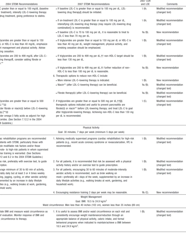
8. Ancillary Therapy
2004 STEMI Guidelines—Section 6.3.1.6.8.1. Anticoagulants as Ancillary Therapy to Reperfusion Therapy
Since publication of the 2004 STEMI Guidelines,15 a
number of studies have provided data that inform the recom-mendations on ancillary therapy to support reperfusion ther-apy for STEMI. In recognition that many agents capable of inhibiting the coagulation cascade may inhibit proteins other than thrombin, the writing group decided to change the nomenclature for this section. Therefore, the term anticoagu-lants is used in place of the prior term antithrombins. Also, although the material discussed below crosses several sub-sections in the 2004 STEMI Guidelines (Sections 6.3.1.6.8.1.1 and 6.3.1.6.8.1.2), because of a number of common issues, the writing group has elected to describe the updates on anticoagulant therapy collectively in this section. Unfractionated heparin (UFH) is commonly administered to patients receiving fibrinolytic therapy. With limited evi-dence supporting the benefits of prolonged infusions of UFH and because of the progressive increase in the risk of heparin-induced thrombocytopenia (both rapid- and delayed-onset presentations),66,67the 2004 STEMI Guidelines
recom-mended that infusions of UFH be given routinely for 48 hours but be given for a longer period only in patients with an ongoing indication for anticoagulation.15,68,69 Although no
new trials specifically focusing on UFH in STEMI were reported, a number of studies have compared alternative anticoagulant regimens with UFH or placebo. Importantly, each study tested a strategy that involved administering the new regimen (reviparin, fondaparinux, or enoxaparin) for the duration of the index hospitalization; that is, longer than Table 7. Updates to Section 6.3.1.6.4.7: PCI After Successful Fibrinolysis or for Patients Not Undergoing Primary Reperfusion
2004 STEMI Guideline Recommendation 2007 STEMI Focused Update Recommendation Comments Class IIb
Routine PCI might be considered as part of an invasive strategy after fibrinolytic therapy. (Level of Evidence: B)
1. PCI of a hemodynamically significant stenosis in a patent infarct artery greater than 24 hours after STEMI may be considered as part of an invasive strategy. (Level of Evidence: B)
Modified recommendation (changed text)
Class III
1. PCI of a totally occluded infarct artery greater than 24 hours after STEMI is not recommended in asymptomatic patients with one- or two-vessel disease if they are hemodynamically and electrically stable and do not have evidence of severe ischemia. (Level of Evidence: B)
New recommendation
PCI indicates percutaneous coronary intervention, and STEMI, ST-elevation myocardial infarction.
by guest on March 29, 2018
http://circ.ahajournals.org/
current practice and longer than recommended in the 2004 STEMI Guidelines. In addition, some of the new anticoagu-lant regimens used dosing schemes that were based on patient weight, age, or both. With the exception of reviparin, the details of the dosing schemes are noted in the recommenda-tions above; the text below refers simply to the name of the anticoagulant regimen. Major efficacy and safety observa-tions from the main trial and important subgroups reported to date are shown inTable 9.
The CREATE (Cardiovascular risk Reduction by Early Anemia Treatment with Epoetin beta) trial was a randomized, double-blind comparison of a strategy of low-molecular-weight heparin (LMWH) reviparin versus placebo in 15 570 patients with STEMI enrolled in China and India70Although
reviparin is not available for clinical use in North America, the writing group felt that the data from the CREATE trial were informative to clinicians and supported the data from the trials discussed subsequently. The dosing regimen for reviparin was as follows: for patients weighing less than 50 kg, subcutaneous injections of 3436 IU Ph Eur anti-Xa units every 12 hours; for patients weighing 50 to 75 kg, subcuta-neous injections of 5153 IU Ph Eur anti-Xa units every 12
hours; and for patients weighing more than 75 kg, subcuta-neous injections of 6871 IU Ph Eur anti-Xa units every 12 hours. Reviparin was continued for the duration of the index hospitalization, up to 1 week. Fibrinolytic therapy (predom-inantly non–fibrin-specific agents) was administered to 73% of the CREATE trial population, and it was recommended that the study drugs be started within 15 minutes of initiation of fibrinolysis. A total of 76% of the trial population received blinded study therapy for 7 days (seeTable 9).
The OASIS-6 (Organization for the Assessment of Strate-gies for Ischemic Syndromes) trial was an international, randomized, double-blind comparison of fondaparinux, a synthetic factor Xa inhibitor, versus control therapy (either placebo or UFH) in 12 092 patients enrolled in 41 countries.7
Patients for whom the treating physician thought UFH was not indicated (e.g., non–fibrin-specific fibrinolytic adminis-tered) were enrolled in stratum I and received placebo in the control arm; patients for whom the treating physician thought UFH was indicated (e.g., fibrin-specific fibrinolytic adminis-tered or primary PCI performed) were enrolled in stratum II and received UFH in the control arm. The median duration of fondaparinux therapy was 8 days in stratum I and 7 days in
Table 8. Updates to Section 6.3.1.6.8.1: Anticoagulants as Ancillary Therapy to Reperfusion Therapy
2004 STEMI Guideline
Recommendation 2007 STEMI Focused Update Recommendation Comments
Class I
1. Patients undergoing reperfusion with fibrinolytics should receive anticoagulant therapy for a minimum of 48 hours (Level of Evidence: C) and preferably for the duration of the index hospitalization, up to 8 days (regimens other than UFH are recommended if anticoagulant therapy is given for more than 48 hours because of the risk of heparin-induced thrombocytopenia with prolonged UFH treatment). (Level of Evidence: A)
Anticoagulant regimens with established efficacy include:
New recommendation
a. UFH (initial intravenous bolus 60 U per kg [maximum 4000 U]) followed by an intravenous infusion of 12 U per kg per hour (maximum 1000 U per hour) initially, adjusted to maintain the activated partial thromboplastin time at 1.5 to 2.0 times control (approximately 50 to 70 seconds) (Level of Evidence: C). (Note: the available data do not suggest a benefit of prolonging the duration of the infusion of UFH beyond 48 hours in the absence of ongoing indications for anticoagulation; more prolonged infusions of UFH increase the risk of development of heparin-induced thrombocytopenia.)
b. Enoxaparin (provided the serum creatinine is less than 2.5 mg per dL in men and 2.0 mg per dL in women): for patients less than 75 years of age, an initial 30 mg intravenous bolus is given, followed 15 minutes later by subcutaneous injections of 1.0 mg per kg every 12 hours; for patients at least 75 years of age, the initial intravenous bolus is eliminated and the subcutaneous dose is reduced to 0.75 mg per kg every 12 hours. Regardless of age, if the creatinine clearance (using the Cockroft-Gault formula) during the course of treatment is estimated to be less than 30 mL per minute, the subcutaneous regimen is 1.0 mg per kg every 24 hours. Maintenance dosing with enoxaparin should be continued for the duration of the index hospitalization, up to 8 days. (Level of Evidence: A)
c. Fondaparinux (provided the serum creatinine is less than 3.0 mg per dL): initial dose 2.5 mg intravenously; subsequently subcutaneous injections of 2.5 mg once daily. Maintenance dosing with fondaparinux should be continued for the duration of the index hospitalization, up to 8 days. (Level of Evidence: B)
2. For patients undergoing PCI after having received an anticoagulant regimen, the following dosing recommendations should be followed:
New recommendation
a. For prior treatment with UFH, administer additional boluses of UFH as needed to support the procedure, taking into account whether GP IIb/IIIa receptor antagonists have been administered.(Level of Evidence: C)Bivalirudin may also be used in patients treated previously with UFH. (Level of Evidence: C)
b. For prior treatment with enoxaparin, if the last subcutaneous dose was administered within the prior 8 hours, no additional enoxaparin should be given; if the last subcutaneous dose was administered at least 8 to 12 hours earlier, an intravenous dose of 0.3 mg per kg of enoxaparin should be given. (Level of Evidence: B)
c. For prior treatment with fondaparinux, administer additional intravenous treatment with an anticoagulant possessing anti-IIa activity taking into account whether GP IIb/IIIa receptor antagonists have been administered. (Level of Evidence: C)
Class III
1. Because of the risk of catheter thrombosis, fondaparinux should not be used as the sole anticoagulant to support PCI. An additional anticoagulant with anti-IIa activity should be administered. (Level of Evidence: C)
New recommendation
GP indicates glycoprotein; PCI, percutaneous coronary intervention; STEMI, ST-elevation myocardial infarction; U, units; and UFH, unfractionated heparin.
by guest on March 29, 2018
http://circ.ahajournals.org/