Acknowledgements
First and foremost, I extend my gratitude to the members of my committee. I have completed this thesis with continuous support from my faculty advisor, Dr. Kevin C. Slep, as well as my in-house advisor, Dr. Charlotte A. Boettiger. They have followed my progress, made themselves available for any questions or concerns I had, and fostered in me a spirit of research and further investigation. I am especially thankful for Dr. Slep’s confidence in me and brilliant suggestions for the directions of my project. In addition to taking the time to participate in my defense, Dr. Vicki W. Chanon has provided me the guidance and direction over the years to pursue an honors project and delve deeper into my study of Psychology and Neuroscience.
Second, I thank all of the Slep lab members, past and current, for their many contributions to my thesis work. Throughout the entire process, Rebecca Adikes, Thomas Lane, Alakananda Das, and Amy Byrnes have been instrumental in providing me with guidance and direction, feedback, and continuous encouragement. I am grateful for the knowledge they have imparted on me. Additionally, I thank my fellow undergraduate lab mate, Angela Xue, for her authentic self. I can always count on her for help, advice, a good laugh, and an occasional cry. I would also like to acknowledge April Hammer, Claudia Szlek, Christian Long, and Juliana Pascual for being our lab’s very own “cytoskeleton”, working to maintain the shape and organization of the lab and to keep daily experiments running smoothly. To the past and current Slep lab members, you have all spent many long hours with me in the lab working to advance the field and have become esteemed leaders and confidants in my life, thank you.
Abstract
Crescerin: Growing the Understanding of Microtubule Dynamics in Neural Primary Cilia Neural Primary Cilia
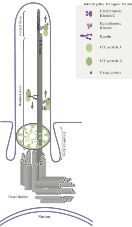
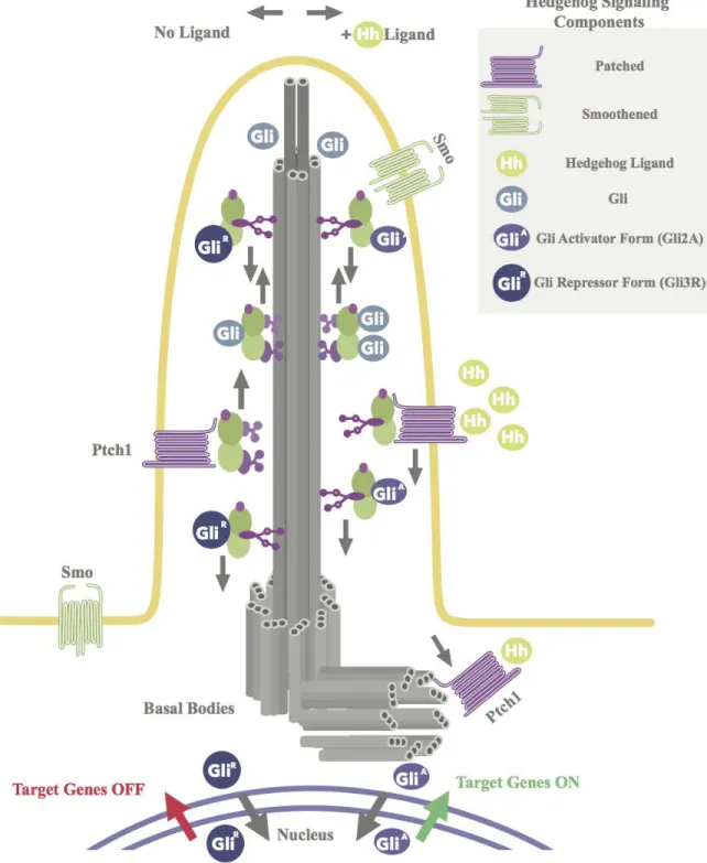
Figure 1. Schematic Representation of the Ultrastructure of the Primary Cilium and Hedgehog Signaling
plasma membrane. Each microtubule doublet is composed of fused A and B tubules. Both A and B tubules extend to the middle zone of the axoneme. As the middle zone transitions into the distal zone of the axoneme, B tubules terminate and singlet A tubules extend to the plus ends of the microtubules (ciliary tip). Kinesin motors move the IFT-B complex and its cargo (i.e. Patched (Ptch), Smoothened (Smo), and Gli) toward the plus ends of the microtubules. Dynein motors move the IFT-A complex and its cargo toward the minus end of microtubules (cell body).
Primary Cilia and Hedgehog Signaling Pathway
Hedgehog signaling (Hh) is an evolutionarily conserved pathway essential for normal development and function. It is currently the pathway most strongly associated with the primary cilium (Fuccillo et al., 2006). Hedgehog signaling utilizes the primary cilium to transduce signals that direct central nervous system development and function (see Figure 1B) (Goetz & Anderson, 2010). Inside and outside the neural tube, the signaling pathway regulates brain development. In the forebrain, Hedgehog signaling divides the eye field and forms the lateral, medial, and central ganglionic eminences (Ericson et al., 1997; Fuccillo et al., 2006; Roelink et al., 1995). While the lateral ganglionic eminence and medial ganglionic eminence generate the striatum and globus pallidus, the medial ganglionic eminence and central ganglionic eminence give rise to GABAergic interneurons, which migrate dorsolaterally into the cerebral cortex (Fuccillo et al., 2006). Hedgehog signaling also maintains progenitor niches in the subgranular zone in the hippocampus and the subventricular zone of the lateral ventricle. While the subgranular zone supplies new neurons to the hippocampal dentate gyrus throughout life, the subventricular zone gives rise to neurons only until about 18 months after birth, but it contributes significantly to the early prefrontal cortex (Fuccillo et al., 2006).
Figure 1. Schematic Representation of the Ultrastructure of the Primary Cilium and Hedgehog Signaling
Smoothened moves into the cilium, where it promotes formation of the activator form of Gli, Gli2A. Once activated, the Gli2A transcription factor is transported out of the primary cilium and into the nucleus, where it activates Hedgehog target genes.
For proper downstream Hedgehog signaling, the primary cilium requires an intact microtubule ultrastructure and intact intraflagellar transport (see Figure 1B) (Huangfu et al., 2003). As mentioned, the collective architecture of the microtubules is termed the axoneme. Dependent on the axoneme is the intraflagellar transport system, which is a trafficking system that is a characteristic of the primary cilium. Since the primary cilium lacks vesicles, it utilizes motor proteins to transport microtubule subunits and select proteins along the axoneme. To transport microtubule subunits and select proteins along the axoneme in the anterograde direction (from the base of the cilium toward the tip), the system uses IFT-B complex and kinesin motors. However, to transport turnover molecules in the retrograde direction, the system employs IFT-A complex and dynein motors (see Figure 1A) (Pederson & Rosenbaum, 2008; Prevo et al., 2015 Scholey, 2003, 2008). This bidirectional transport system is required for the structural integrity of the primary cilium because without it the organelle lacks the microtubule subunits and specific proteins necessary for its own assembly and maintenance (Marshall & Rosenbaum, 2008; Pederson & Rosenbaum, 2008; Scholey, 2003, 2008; Snow et al., 2004).
Growing knowledge of intraflagellar transport has led to the discovery of ciliary genes required for ciliogenesis and maintenance. Mutations of IFT-B complex proteins such as Ift172, Ift88, Ift52, and Ift57 compromise formation of the primary cilium. Furthermore, some
(Gorivodsky et al., 2009; Huangfu, 2003; Huangfu & Anderson, 2005; Liu et al., 2005; May et al., 2005). These findings link disrupted Hedgehog signaling to mutations in intraflagellar transport components, which points out that the primary cilium requires intact intraflagellar transport for correct Hedgehog signaling. Given that intraflagellar transport is dependent on the axoneme, this link also points out that the primary cilium requires intact microtubule ultrastructure for correct Hedgehog signaling. Altogether, the findings from these studies point to a relationship between ciliary microtubule ultrastructure, intraflagellar transport, and Hedgehog signaling. However, they do not confirm whether dysfunctional ciliary microtubule ultrastructure, intraflagellar transport, and Hedgehog signaling result in abnormal development and function of the central nervous system. The findings do not confirm the pathogenic nature of the relationship or reveal the pathogenic mechanisms underlying dysfunctional primary cilia structure and function and the subsequent emergence of neural abnormalities. It is thought that structural perturbations of the axoneme or mutations of intraflagellar transport components disrupt the tightly controlled identities and concentrations of Hedgehog signaling molecules (Fliegauf et al., 2007; Lancaster & Gleeson, 2009; Sharma et al., 2008; Veland et al., 2009). Dysfunctional Hedgehog signaling then results in abnormal central nervous system architectures and intellectual deficits, which are common clinical features shared by most ciliopathies and ciliopathy-related disorders (Gitten et al., 1998; Goetz & Anderson, 2010).
Ciliopathies
and Bardet-Biedl syndrome (BBS) (Tobin & Beales, 2009). Ciliopathy patients have, or may possibly have, genetic mutations in more than one ciliary gene (Tory et al., 2007). Though primary cilia in these patients may still form, the morphology of the organelle may be altered. A network of ciliary genes and proteins that alter the morphology of the primary cilium and mediate the emergence of ciliopathies has been discovered (Bergmann et al., 2008; Brancati et al., 2009; Frank et al., 2008). The existence of this network supports the importance of differentiating between ciliary genes and studying proteins that have role in assembling and maintaining primary cilia structure and function.
In the past, the categorization of ciliopathies has been based on clinical features. However, the prevalence of a range of features has blurred the distinctions between ciliopathies and led to the conceptualization of ciliopathies as spectrum disorders. (Waters & Beales, 2011). Despite the range of clinical features, most ciliopathies and ciliopathy-like disorders share abnormal central nervous system architecture and intellectual deficits as common clinical features (Gitten et al., 1998). Of the distinguished ciliopathies, Joubert syndrome, Bardet-Biedl syndrome, and Alström syndrome have the strongest associations with neural defects (Louvi & Grove, 2011).
syndrome (Cantagrel et al., 2008). In mice, mutations of ARL13B resulted in defects in primary cilia structure, Hedgehog signaling, and neural tube patterning. The neural tube patterning defects were discovered to be due to the coupled effects of compromised primary cilia structure and Hedgehog signaling (Cantagrel et al., 2008; Caspary et al., 2007). Here, the findings from previous studies, once again, suggest a relationship between ciliary microtubule ultrastructure, intraflagellar transport, and Hedgehog signaling. Although these particular findings do not reveal the pathogenic mechanisms underlying dysfunctional primary cilia structure and function and the emergence of neural abnormalities, they confirm the pathogenic nature of the relationship. They confirm that dysfunctional ciliary microtubule ultrastructure, intraflagellar transport, and Hedgehog signaling result in abnormal development of the central nervous system (Fliegauf et al., 2007; Lancaster & Gleeson, 2009; Sloboda & Rosenbaum, 2007; Veland et al., 2009).
intellectual disability (Ikeuchi et al., 2013). Therefore, it is thought that dysfunctional primary cilia structure and function to affect cortical connectivity and normal intellectual function.
Until recently, the study of neural primary cilia has not been a major research topic, but with the advent of high-throughput sequencing, the genetic basis for a number of ciliopathies and related disorders has been identified. The identification of ciliopathies and ciliopathy-related disorders and their genetic bases, along with the strong association between Hedgehog signaling and the neural primary cilium, highlights the critical role of the organelle. To better understand the role of the long-neglected neural primary cilium in brain development and for diverse aspects of brain function, such as neural signaling, neurogenesis, homeostasis, and the emergence of ciliopathies and ciliopathy-related disorders, the primary cilium in model organisms and in non-neural cells must be investigated. C. elegans is an example of a model organism that is easy to access, genetically manipulate and track using new CRISPR/Cas9 technology, and image (Dickinson et al., 2013). To study the molecular basis of neural architecture, signaling, and behavior, C. elegans serve as prime models due to their well-defined nervous system and the conservation of genes and neurobiological processes from C. elegans to higher organisms (i.e. humans) (Inglis et al., 2006; Sengupta & Aravinthan, 2009). Differentiating between ciliary genes and studying proteins that are essential for primary cilia structure and function in model organisms provide insight not only into the biology of the specialized organelle, but also into the role of neural primary cilia in central nervous system development and function. These studies provide insight into the role of particular proteins in altering the morphology of the primary cilium and mediating the emergence of neural abnormalities.
Tubulin is the building block of microtubules and thereby the building block of the axoneme. To assemble the core of the primary cilium, tubulin is added to the plus ends of the microtubules. Tubulin first enters the ciliary compartment from the cytosol and is then transported by the intraflagellar transport system to the distal tip of the axoneme (see Figure 1A). For conventional primary cilia function, the structure and length of the organelle is actively maintained through a balance of polymerization and depolymerization at the distal tip of the axoneme. Therefore, within primary cilia, there is constant tubulin turnover (Hao et al., 2011; Ishikawa & Marshall, 2011).
Microtubule Dynamics and Microtubule Associated Proteins
Microtubules undergo dynamic instability, a term that reflects a microtubule’s ability to oscillate between periods of polymerization and depolymerization. Different zones along the axoneme vary in their degree of dynamic instability. While doublet microtubules in the middle zone are stable and resistant to depolymerization, singlet microtubules in the distal zone are dynamic and change their length (see Figure 1A) (Marshall & Rosenbaum, 2001; Mukhopadhyay et al., 2008; Tilney & Gibbins, 1968). This dynamicity at the distal zone is critical because it maintains appropriate axoneme structure and length in response to changing extracellular environments and stimuli (Mukhopadhyay et al., 2008; Rich & Clark, 2012). Microtubule associated proteins (MAPs) are involved in regulating microtubule organization and dynamics (Popov et al., 2001).
microtubule dynamics. While high eukaryotic ch-TOG family members use a pentameric TOG domain array to promote cytoplasmic microtubule polymerization, CLASP family members use a trimeric TOG domain array to stabilize cytoplasmic microtubules and promote cytoplasmic microtubule pause (see Figure 2) (Al-Bassam et al., 2007; Al-Bassam & Chang, 2011; Brittle & Ohkura, 2005; Slep, 2009; Sousa et al., 2007; Widlund et al., 2011).
Crescerin
Though cytoplasm microtubule associated proteins, such as ch-TOG and CLASP, and their TOG domains have been well studied, the identities and mechanisms of ciliary microtubule associated protein are not well known or understood. Recently, Crescerin was identified as a cilia-specific microtubule associated protein family that regulates ciliary microtubules using arrayed tubulin-binding TOG domains. This newly identified protein is conserved in ciliated/flagellated eukaryotes but is absent in non-ciliated eukaryotes, such as yeast and Dictyostelium (see Figure 3) (Das et al., 2015). Thus far, the only Crescerin protein family members characterized are the C. elegans member, CHE-12, and the mouse member, mCHE-12 (Bacaj et al., 2008; Das et al., 2015).
Mammals contain two closely related Crescerin family genes, FAM179A and FAM179B. Sequence analysis and secondary structure predictions of mouse FAM179B reveal four conserved domains: an N-terminal pair and a C-terminal pair connected by a long central linker. Each of the four domains are thought to exhibit TOG-like features. They contain 12 predicted α-helices that are proposed to form six tandem HEAT repeats (HRs) with intra-HEAT loops displaying similarity to the intra-HEAT loop tubulin-binding determinants in ch-TOG and CLASP TOG domains (Slep, 2009). Crescerin family members that contain TOG-like domains similar to mammalian FAM179B TOG1 or TOG2 are classified as Crescerin1 while those that only contain domains similar to FAM179B TOG3 and TOG 4-like domains are classified as Crescerin2 (Das et al., 2015).
Figure 3. The Crescerin1 Protein Has Conserved TOG domains in Ciliated/Flagellated Eukaryotes
coded according to the Crescerin1 TOG domain that they are most similar to in primary sequence.
The work of Bacaj et al. (2008) and Das et al. (2015) revealed that the C. elegans Crescerin1 protein family member, CHE-12, localizes to the primary cilium in C. elegans amphid and phasmid neurons (a subset of sensory neurons in the head of C. elegans) as well as in mammalian cells. Additionally, research by Bacaj et al. (2008) and Das et al. (2015) uncovered that CHE-12 is necessary for the structure and function of primary cilia in C. elegans amphid neurons. Deleting CHE-12 or mutating a conserved hydrophobic residue to glutamate in the HEAT repeat A intra-HEAT loop of each of the protein’s TOG domains resulted in shorter, poorly defined, and more disorganized primary cilia. Deleting CHE-12 in C. elegans altered the ability of the worms’ primary cilia to take up dye, an assay used to examine the integrity of the structure and function of primary cilia (Bacaj et al., 2008; Das et al., 2015). It also altered the ability of the worm to sense its environment and chemotax along a NaCl gradient. Interestingly, mutating CHE-12’s TOG domains had the same effect as deleting CHE-12; it resulted in shorter cilia with aberrant axoneme architecture and compromised the worm’s ability to take up dye and chemotax (Bacaj et al., 2008; Das et al., 2015). These results suggest that, through use of its arrayed tubulin-binding
TOG domains, Crescerin1 promotes proper cilia structure and function. Given that C. elegans is a prime model organism to better understanding the nervous system in higher organisms, these results imply that Crescerin1 and its TOG domains’ role for formation and function of primary cilia and for proper neurophysiological and behavioral responses are as essential in humans as they are in C. elegans.
similar to other TOG array-containing protein families, employs arrayed TOG domains that contribute differentially but work collectively to regulate microtubule dynamics.
Figure 4. Structure of TOG Domains
Structure of Crescerin1 TOG 2, a bona fide TOG domain with a unique C-terminal– hairpin. TOG domains consist of six pairs of antiparallel helices that are connected by short loops. The antiparallel helices, also known as HEAT repeats, configure themselves so that all intra-HEAT loops lie exposed on one side of the protein while all inter-HEAT loops lie exposed on the opposite side of the domain. It is common for different TOG domains to show variation in their overall primary sequence across protein families and even within the same protein. Many surface-exposed residues in the intra-HEAT loops are conserved since this is the face that binds to tubulin. Each HEAT repeat is shown in a different color and labeled A-F.
Crescerin1 TOG 4
also identified to engage the straight tubulin conformation found in polymerized microtubules and to aide in microtubule lattice association. Cytoplasmic microtubule association was ablated when a tubulin-binding HEAT repeat A intra-HEAT loop residue of overexpressed Crescerin1 TOG 4 was mutated to glutamate (F1559E). Additionally, cytoplasmic microtubule association was ablated when the same conserved residue in the HEAT repeat A intra-HEAT loop of an overexpressed N-terminal Crescerin1 construct containing the central linker and TOG domains 3 and 4 (L-TOG-34, residues 577–1776) was mutated to glutamate (Das et al., 2015). These findings suggest that Crescerin1 TOG 4 has a unique role within the context of the larger Crescerin1 protein for regulating ciliary microtubule dynamics.
In the present study, we ask: does Crescerin1 contain an array of four TOG domains? Is Crescerin1 TOG 4 domain a bona fide TOG domain? Structurally, is Crescerin1 TOG 4 domain distinct from Crescerin1 TOG 2 domain? Is Crescerin1 TOG array structurally and functionally polarized such that each domain contributes differentially to regulate microtubule dynamics? We hypothesize that Crescerin1 contains an array of four different TOG domains and that TOG 4 domain is a bona fide TOG domain possessing characteristic TOG domain features. We speculate that Crescerin1 TOG 4 domain is structurally distinct from Crescerin1 TOG 2 domain and contributes differentially to regulate microtubule dynamics, such that TOG 4 stabilizes microtubules while TOG 2 promotes microtubule polymerization.
Method Materials and Procedure
Crescerin1 TOG 4 Domain Sequence Analysis
Homologs of Crescerin1 from various organisms in the NCBI database were identified by Alakanandas Das using the BLASTP algorithm (Altschul et al., 1990) and used to compare Crescerin1 TOG 4 in a sequence alignment (see Figure 5). To create the sequence alignment and analyze the amino acid conservation across species, the CLUSTAL Omega server was used (Sievers et al., 2011). Crescerin1 TOG 4 secondary structure predictions were acquired using the Phyre 2 server (Kelley & Sternberg, 2009).
Mouse Crescerin1 TOG 4 (1533-1776) Cloning
The sequence alignment and secondary structure predictions were used to identify the presence of mouse Crescerin1 TOG 4 domain. Mouse TOG 4 (1533-1776) was cloned into pET28a vectors for Isopropyl β-ᴅ-1-thiogalactopyranoside (IPTG) inducible expression in BL21 (DE3) E.
-NTA affinity chromatography, the construct was designed to have a thrombin-cleavable N-terminal 6X-His tag.
Mouse Crescerin1 TOG 4 (1533-1776) Expression and Purification
DNA encoding the mouse TOG 4 (1533-1776) in pET28a vector was transformed into BL21 (DE3) cells. These transformed cells were added to a Kanamycin-containing agar plate. To grow transformed Kanamycin-resistant bacterial colonies, the plate was incubated at 37˚C overnight.
After 24 hours, a single bacterial colony was chosen out of the several colonies that had grown on the plate. The single colony was added to 60 mL of pre-autoclaved Luria Broth (LB) media and allowed to grow at 37˚C overnight. The following day, 12 mL of this bacterial growth was added per 1 L pre-autoclaved LB media (6 flasks of 1 L cultures total) each containing Kanamycin at a final concentration of 50 μg/mL. The 1 L cultures were allowed to grow in the shaker/incubator at 37˚C and 200 rpm for roughly 4 hours. After 4 hours, the optical density of the cultures was observed to be between 0.6 and 0.8 at 600 nm wavelength. At this OD600 the bacteria growth rate is ideal, therefore 100 μM IPTG was added to each flask to induce protein expression. Once the bacterial cells were induced, the temperature of the shaker/incubator was brought down to 18˚C, the rotor speed was reduced to 180 rpm, and the cells were left to express protein overnight. The following day, the culture flasks were removed from the incubator and, using a centrifuge and a Fiberlite F10 rotor at 3500 rpm, the cultures were spun down for 10 minutes at 4˚C. Once all the cells were spun down, the pellets were resuspended in 50 mL Ni2+-A buffer (ingredients for buffer found in appendix) and stored at -20˚C.
Phenylmethylsulfonyl fluoride (PMSF) was added as a protease inhibitor. To lyse the bacterial cells, the sample was sonicated while sitting on ice. After five cycles of sonication, the lysate was spun down for 40 minutes using a TLA SS-34 rotor at 15,000 rpm and 4˚C. The supernatant containing soluble protein was poured into a beaker and the pellet was disposed of.
Using a gravity chromatography technique at 4˚C, the supernatant with soluble protein was loaded onto a 10 mL Ni2+-NTA column (Qiagen), washed with 200 mL Ni2+-A buffer, and batch eluted with 100 mL of Ni2+-B buffer (ingredients for buffer found in appendix). The eluted protein was collected in fractions.
To cleave off the N-terminal 6X-His tag on the chosen fractions of eluted protein, the fractions were treated with 0.1 mg of bovine alpha thrombin and 2.5 mM CaCl2. The protein mix sat at 4°C for 24 hours until thrombin digestion was complete as signified by the 2kDa shift of the Crescerin1 TOG 4 band on an SDS-PAGE gel (see Figure 7). The digested protein was placed into a 3,500 MWCO sized dialysis bag and placed into a large volume of dialysis buffer (ingredients for buffer found in appendix). The solutions reached equilibrium within 5 hours while sitting at 4°C. To deactivate the thrombin and remove undigested protein, the digested protein was gravity filtered through an 0.5 mL Benzamadine sepharose column and through a 10mL Ni2+-NTA column, respectively.
The protein was exchanged into storage buffer (ingredients for buffer found in appendix) by dialysis for 24 hours. Using a Millipore Ultrafree 10,000 MWCO concentrator, the protein was concentrated to 16.02 mg/mL (Supplemental Table 1). The protein was aliquoted into collection tubes, frozen in liquid nitrogen, and stored at -80°C.
Mouse TOG 4 (1533-1776) was crystallized using the hanging drop vapor diffusion method. In this method, 1μL of protein at 10 mg/mL was added to 2μL of well solution to form vapor diffusion drops of protein and well solution (1:2 ratio) that were incubated at 20°C (Supplemental Table 2). Initial crystallization of mouse TOG 4 (1533-1776) was attempted using the PEG-Ion screen (Hampton Research). After attaining crystal hits from the second reagent (0.2 M postassium fluoride, 20% PEG 3350, pH 7.3) in the PEG-Ion screen, further screenings were done in search of more ideal crystallization conditions. Several trials varying multiple variables revealed ideal crystallization conditions (Supplemental Table 2). After growing mouse TOG 4 (1533-1776) crystals to optimal dimensions, the crystals were looped, transferred to Fomblin oil (Hampton Research), flash frozen in liquid nitrogen, and shipped for diffraction analysis (see Figure 11).
Additional Mouse Crescerin1 TOG 4 (1533-1776) Crystallization Techniques
To slow down and control nucleation, the hanging drop vapor diffusion method was done on ice at 4°C. To induce crystallization, micro-seeding and streak seeding techniques were used. For micro-seeding, crude crystals were collected, broken by vortexing, and diluted. In this technique, 1μL of the broken crystal solution was added into the vapor diffusion drops. For streak seeding, crude crystals were also collected, broken by vortexing, and diluted. However, in this technique, a cat whisper was dipped into the broken crystal solution and streaked across the vapor diffusion drops.
New Constructs Expression, Purification, and Crystallization
similar manner as the previously stated protocol. For variations of each specific protocol refer to the appendix.
Results Crescerin1 TOG 4 Domain Alignment
Crescerin is a TOG domain-containing protein family that is conserved in ciliated/flagellated eukaryotes but is absent in non-ciliated eukaryotes, such as yeast and Dictyostelium (see Figure 5). To analyze the conservation and relationship between Crescerin1
Figure 5. Crescerin Family TOG 4 Sequence Alignment
domain in mouse and C. elegans Crescerin1 are shown as grey boxes and black lines, respectively.
Mouse Crescerin1 TOG 4 Expression and Purification
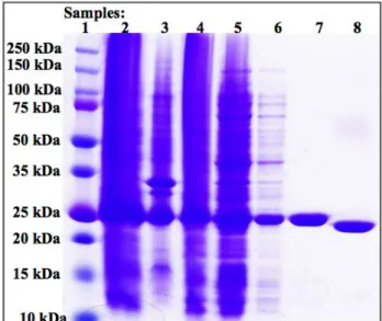
Figure 6. Mouse Crescerin1 TOG 4 (1528-1776) Successfully Expressed and Purified
SDS-PAGE gel showing the purification steps for mouse TOG 4 (1528-1776) using an Ni2+-NTA column. The samples are: 1) protein molecular weight marker (protein ladder) 2) lysate 3) pellet 4) supernatant 5) Ni2+-NTA column flow through 6) Ni2+-NTA column wash 7) eluted protein/pre-cleavage 8) post-cleavage. Mouse TOG 4 (1528-1776) construct expressed soluble Crescerin1 TOG 4 as implied by the supernatant sample. The band at approximately 25kDa across the figure suggests that Crescerin1 TOG 4 remained present and was not lost throughout the purification process. The protein was purified to a high level of purity as pointed out by the eluted protein sample, which displays no contaminants. The pre-cleavage and post-cleavage samples display the expected size shift accompanying the cleavage of the N-terminal 6X-His tag by thrombin digestion.
Figure 7. Mouse Crescerin1 TOG 4 (1533-1776) Successfully Expressed and Purified
Mouse Crescerin1 TOG 4 Crystallization
Initial crystal hits of mouse TOG 4 (1533-1776) were obtained (PEG-Ion screen reagent 2: 0.2 M potassium fluoride, 20% PEG 3350, pH 7.3). After several trials of screening for the best conditions to crystallize mouse TOG 4 (1533-1776), it was determined, that varying the molarity of potassium fluoride had a better effect than varying the percentage of different types of PEG and varying the temperature at which the crystal trays were set up. Through an iterative process with multiple variables aimed at slowing down and controlling nucleation, it was noted that PEG 3350 was better for crystallizing mouse TOG 4 (1533-1776) than PEG 4000 or PEG 8000 and that setting crystal trays up at 4˚C did not slow down nucleation or generate thicker needle-like crystals. Moreover, it was established that an additive screen caused the drops to precipitate out of solution almost immediately and that neither microseeding nor streak seeding (different techniques in which small crystals are used to seed and form much larger crystals) significantly improved the quantity or quality of the mouse TOG 4 (1533-1776) crystals. The crystals were best grown in conditions 0.205 M-0.22 M potassium fluoride, 19.5%-22% PEG 3350, and pH 7.2 (Supplemental Table 2). Vapor diffusion drops varied with 2-20 nucleation points (see Figure 8). Figure 8 depicts the optimal mouse TOG 4 (1533-1776) crystals that were grown. The crystals had a thin needle-like morphology and grew in a clumped orientation within 1 hour of setting up the crystallization tray. Though the crystals reached maximum size within 24 hours, they started to degrade past 48 hours (Supplemental Table 2).
robust Crescerin1 TOG 4 crystals by cloning, expressing, purifying, and crystalizing C. elegans and Tetrahymena Crescerin1 TOG 4 constructs.
Figure 8. Optimal Mouse Crescerin1 TOG 4 (1533-1776) Crystals
A) Mouse TOG 4 (1533-1776) crystallized. Initial crystal hits of mouse TOG 4 (1533-1776) were obtained (0.2 M potassium fluoride, 20% PEG 3350, pH 7.3). Screening revealed that the crystals were best grown in conditions 0.205 M-0.22 M potassium fluoride, 19.5%-22% PEG 3350, and pH 7.2. Vapor diffusion drops varied with 2-20 nucleation points. The crystals had a thin needle-like morphology and grew in a clumped orientation within one hour of setting up the crystallization tray. Though the crystals reached maximum size within 24 hours, they started to degrade past 48 hours. Diffraction data for mouse TOG 4 (1533-1776) crystals was obtained to 2.6 Å and the pattern depicted smeared reflections (spots). After about 60 frames of data collection, the crystals progressively degraded.
B) The clumped needle-like crystals were separated into individual crystals for looping and diffraction analysis.
C. elegans and Tetrahymena Crescerin1 TOG 4 Cloning
repeats. The domain of both species is also implied to contain key differences from mouse TOG 4, including different lengths of intra-HEAT loops and shorter linkers between HEAT repeats 5-6. Additionally, according to secondary structure predictions, C. elegans and Tetrahymena TOG 4 each possess N-terminal alpha helices while mouse TOG 4 does not. Thus, we were directed to clone C. elegans and Tetrahymena TOG 4 with the hypothesis that these different features may provide the domain greater stability, and consequently enhance its ability to crystallize robustly. Interestingly, the C. elegans TOG 4 (1052-1282) construct, which does not include the proposed N-terminal alpha helix, was cloned successfully, while the C. elegans TOG 4 (1044-1282) construct, which begins immediately after the predicted N-terminal alpha helix ends, was not. In contrast, the three Tetrahymena TOG 4 (1853-2132, 1889-2132, 1911-2132) constructs, which respectively include two, one, and none of the proposed N-terminal alpha helices, were all cloned successfully.
C. elegans and Tetrahymena Crescerin1 TOG 4 Expression and Purification
columns, is shown in Figures 9, 10, 11, and 12. The SDS-PAGE gels display the expected size shift accompanying the cleavage of the N-terminal 6X-His tag by protease in the four purifications.
Figure 9. C. elegans Crescerin1 TOG 4 (1052-1282) Successfully Expressed and Purified
Figure 10. Tetrahymena Crescerin1 TOG 4 (1853-2132) Successfully Expressed and Purified
Figure 11. Tetrahymena Crescerin1 TOG 4 (1889-2132) Successfully Expressed and Purified
Figure 12. Tetrahymena Crescerin1 TOG 4 (1911-2132) Successfully Expressed and Purified
SDS-PAGE gel showing the purification steps for Tetrahymena TOG 4 (1911-2132) using an Ni2+-NTA column. The samples are: 1) protein molecular weight marker (protein ladder) 2) lysate 3) pellet 4) supernatant 5) Ni2+-NTA column flow through 6) Ni2+-NTA column wash 7) eluted protein/pre-cleavage 8) post-cleavage. Tetrahymena TOG 4 (1911-2132) construct expressed soluble Crescerin1 TOG 4 as implied by the supernatant sample. The band at approximately 25kDa across the figure suggests that Crescerin1 TOG 4 remained present and was not lost throughout the purification process. The protein was purified to a high level of purity as pointed out by the eluted protein sample, which displays no contaminants. The pre-cleavage and post-cleavage samples display the expected size shift accompanying the cleavage of the N-terminal 6X-His tag by protease digestion.
C. elegans and Tetrahymena Crescerin1 TOG 4 Crystallization
13). Figure 11 shows the optimal crystals that were grown. The crystals had a thick rod-like morphology and grew 72 hours after setting up the crystallization tray and reached maximum dimension after one week (Supplemental Table 2). However, diffraction data for Tetrahymena TOG 4 (1853-2132) was not obtained. Diffraction data displayed ice rings suggesting that an inappropriate cryoprotectant or cryoprotectant concentration resulted in ice formation and damage to the crystal (Supplemental Table 1). Therefore, screening for optimal reagents for crystallization and selecting a suitable cryoprotectant and cryoprotectant concentration continues. Once robust crystals are grown and frozen properly, an adequate diffraction dataset will be obtained and used to solve Crescerin1 TOG 4’s crystal structure.
Figure 13. Optimal Tetrahymena Crescerin1 TOG 4 (1853-2132) Crystals
diffraction data displayed ice rings suggesting that an inappropriate cryoprotectant or cryoprotectant concentration resulted in ice formation and damage to the crystal.
Discussion
To answer our research questions, the first step is to solve the structure of Crescerin1 TOG 4 domain. Of the six purified Crescerin1 TOG 4 constructs, mouse TOG 4 (1533-1776) and Tetrahymena TOG 4 (1853-2132) were the only constructs with crystal hits (Supplemental Table
1). The best conditions to grow mouse TOG 4 (1533-1776) crystals are 0.205 M-0.22 M potassium fluoride, 19.5%-22% PEG 3350, and pH 7.2. These crystals were thin and needle-like. They clumped together in a starburst shape and crystallized quickly, emerging one hour after setting up the crystallization tray and reaching optimal dimensions after 24 hours (Supplemental Table 2). In contrast, the best conditions to crystallize Tetrahymena TOG 4 (1853-2132) are 0.195 M-0.21 M sodium phosphate dibasic dehydrate, 19%-21% PEG 3350, and pH 9.1. These crystals were rod-like and crystallized slowly, emerging 72 hours after setting up the crystallization tray and reaching maximum size after one week (Supplemental Table 2). Of the two types of crystals, the Tetrahymena TOG 4 (1853-2132) crystals were more robust than the mouse TOG 4 (1533-1776)
crystals. Despite this, we were unable to obtain a working diffraction dataset from either of the two types of crystals to understand Crescerin1 TOG 4 at the atomic level. Therefore, we can neither confirm nor disprove our original hypotheses. However, having reviewed the literature and used our knowledge of TOG domains across TOG domain-containing protein families, we have developed predictions of the architecture and structural mechanisms of Crescerin1 TOG 4 in regulating ciliary microtubule dynamics.
predictions, Crescerin1 is thought to contain four arrayed TOG domains. Crescerin1 TOG 4 is postulated to be a bona fide TOG domain because sequence analyses and secondary structure predictions suggest that the domain possesses characteristic TOG domain features–12 helices that pair into six tandem HEAT REPEATs (A–F) with intra-HEAT loops that are conserved and similar in composition and basic electrostatic charge to the tubulin-binding intra-HEAT loops of TOG domains in ch-TOG and CLASP protein families (Leano.et al., 2013; Slep, 2009; Slep & Vale, 2007). The primary sequence of Crescerin1's four TOG domains bear low sequence identity to one another and to TOG domains from ch-TOG and CLASP families. This aligns with previous research which uncovered that TOG domains within an array and across TOG domain-containing proteins show relatively low primary sequence conservations to one another and have slight variations in architecture and tubulin-binding surface conformations (De la Mora-Rey et al., 2013; Fox et al., 2014; Leano et al., 2013). Therefore, Crescerin1 TOG 4 is predicted to be structurally distinct from Crescerin1 TOG 2, which was ascertained to be a bona fide TOG domain with a 21-residue C-terminal β-hairpin. The β-hairpin is a stabilizing feature of TOG 2. It is located alongside the second HEAT repeat triad and forms multiple hydrophobic interactions and hydrogen bonds with conserved residues (Das et al., 2015). Since, sequence analyses and secondary structure predictions do not propose a similar feature in Crescerin1 TOG 4, this indicates that TOG 4 is architecturally unique from Crescerin1 TOG 2.
contrast, Msps TOG 1 and TOG 2 domains do not show microtubule lattice association. Rather, these domains were discovered to interact with free tubulin heterodimers over gel filtration, an activity that Msps TOG 3 and TOG 4 domains lack (Fox et al., 2014; Slep & Vale, 2007). Of interest, Crescerin1 TOG 4 and central linker-TOG (3+4) constructs result in lattice association to cytoplasmic microtubules when overexpressed. This lattice association to cytoplasmic microtubules is ablated when a residue in the TOG 4 HEAT repeat A-loop of the two over-expressed Crescerin1 constructs (TOG 4 and central linker-TOG (3+4)) is mutated to glutamate (Das et al., 2015). These results lead us to hypothesize that Crescerin1 TOG domains, similar to Msps TOG domains, recognize and bind tubulin of specific states, either free tubulin or polymerized tubulin. Despite lattice association to cytoplasmic microtubules occurring only upon over-expression, these findings are important because, by knowing the state and curvature of tubulin that TOG domains recognize and bind to, we can postulate TOG domains’ role in regulating microtubule dynamics (Maki et al., 2015). Therefore, these results prompt us to propose that Crescerin1 TOG 4 binds straight polymerized tubulin and contributes to ciliary microtubule lattice association.
Fox et al., 2014; Slep and Vale, 2007). In contrast, CLASP studies uncovered that an individual TOG domain (TOG2 from human CLASP1) is sufficient to promote tubulin polymerization in vitro (Leano et al., 2013). Of interest, Crescerin1 TOG 2 and TOG 4 domains, similar to human
CLASP1 TOG 2, individually and sufficiently increase microtubule polymerization rates in vitro, indicating that the domains either stabilize a conformation of tubulin that is more prone to polymerize or aide in lattice contact through a direct or allosteric mechanism (Das et al., 2015; Leano et al., 2013). These findings prompt us to hypothesize that the Crescerin1 four TOG domain-containing array, like the pentameric TOG domain array of the ch-TOG, may be polarized, such that the N-terminal TOG domains bind and promote tubulin incorporation into growing ciliary microtubules, while the C-terminal TOG domains enable ciliary microtubule association. Thus, these results direct us to speculate that Crescerin1 TOG 4 regulates ciliary microtubule dynamics, such as promoting microtubule polymerization, by binding straight polymerized tubulin and aiding in ciliary microtubule lattice association.
specific states and curvatures and bind tubulin in different modes. It will serve as the foundation for subsequently deducing the structural mechanisms underlying the ability of TOG array-containing protein families to use arrayed TOG domains that contribute differentially but work collectively to regulate microtubule dynamics.
maintained rely on the movement of tubulin to and from the cilium tip and on ciliary microtubule dynamics (Ludington et al., 2015). Little is known about the availability, recognition, and binding of cilia-specific tubulin isotypes, their movements to and from the cilium tip, and their interactions with ciliary tip macromolecules. It is thought that Crescerin is one of the macromolecule required, as part of a larger complex, to generate and maintain appropriate cilia length and establish proper function.
Developing a model of the atomic arrangement of Crescerin1 TOG 4 domain will shed light on the differences in microtubule dynamics in the distal cilium tip versus the cytoplasm. It will provide insight into cilia-specific tubulin isotype availability and TOG-tubulin isotype recognition and binding. It will also provide insight into cilia-specific tubulin isotype incorporation into ciliary microtubules and turn over. Solving and studying the structure of Crescerin1 TOG 4 will bring us closer to deducing whether Crescerin, like ch-TOG and CLASP, interacts with other regulators of ciliary microtubule dynamics and is a necessary part of a larger complex that generates and maintains right cilia structure and function.
patients with inoperative or mutated Crescerin1 to examine if and how protein knockout or mutations of Crescerin1 TOG domains result in primary cilia defects and subsequently affects the emergence of ciliopathies and ciliopathy-related disorders. The strong association between neural primary cilia and the formation and function of the cerebral cortex also warrants exploration. Further behavioral investigations should examine the intellectual deficits shared by most ciliopathies and ciliopathy-related disorders by comparing deficits, for example in a set of memory tasks, between mice with ablated primary cilia, mice with knockout Crescerin1, and mice with selective mutations of individual or a subset of Crescerin1 TOG domains.
4 is important because the absence or mutation of Crescerin1 and its TOG domains may result in the emergence of neurodevelopmental and behavioral abnormalities.
Continuing to employ our molecular structure-function investigation approach to develop a model of the atomic arrangement of Crescerin1 TOG 4 is further underscored by human disease case studies. In the NCBI ClinVar database for human disease, there are seven case studies that report duplicate chromosome 14 and the Crescerin1 gene (FAM179B). In one of these studies, a shorter fragment spanning six genes are duplicated, which includes the first exon of Crescerin1. It is possible that this results in the expression of a truncated Crescerin1 protein containing TOG 1, TOG 2, and a short region of the central linker (first N-terminal 682 amino acids). This is interesting because the case study reports that the human subject exhibits developmental deficits. Therefore, it is possible that the absence of Crescerin1 C-terminal TOG domains, which includes TOG 4, may explain the developmental deficits observed in the patient. Since the type of developmental deficits are not reported, the case study demands further research to investigate if and how the developmental deficits observed in the patient are nervous system- and Crescerin1 TOG 4-related. Perhaps the absence of Crescerin1 TOG 4 in the human subject may have affected neural ciliogenesis and intraflagellar transport, neural signaling, neurogenesis, and/or homeostasis, which may have subsequently resulted in neurodevelopmental deficits. Despite calling for additional investigation, the case study emphasizes the real life value in continuing our work.
purification column. Doing additive screens and using microseeding and streak seeding techniques to improve the quantity and quality of the C. elegans and Tetrahymena TOG 4 crystals. Cloning new mouse, C. elegans, and Tetrahymena Crescerin1 TOG 4 constructs or Crescerin1 TOG 4 constructs of different ciliated/flagellated eukaryotes organisms, such as T. adhaerens, to include linkers, loops, and N-terminal or C-terminal regions that reduce domain flexibility. Future directions in the laboratory also include using an appropriate cryoprotectant and cryoprotectant concentration to freeze Tetrahymena TOG 4 (1853-2132) crystals properly and prevent ice formation and damage to the crystals. Such future directions would remove contaminants, increase domain stability, induce proper domain folding and freezing, and overall enhance Crescerin1 TOG 4 domain assembly into a highly structured crystal lattice.
References
Adams, N. A., Awadein, A., & Toma, H. S. (2007). The retinal ciliopathies. Ophthalmic genetics, 28(3), 113-125. doi: 10.1080/138168107001537424
Al-Bassam, J., & Chang, F. (2011). Regulation of microtubule dynamics by TOG-domain proteins XMAP215/Dis1 and CLASP. Trends in cell biology, 21(10), 604-614. doi: 10.1016/j.tcb.2011.06.007
Al-Bassam, J., Larsen, N. A., Hyman, A. A., & Harrison, S. C. (2007). Crystal structure of a TOG domain: conserved features of XMAP215/Dis1-family TOG domains and implications for tubulin binding. Structure, 15(3), 355-362. doi: 10.1016/j.str.2007.01.012
Altschul, S. F., Madden, T. L., Schäffer, A. A., Zhang, J., Zhang, Z., Miller, W., & Lipman, D. J. (1997). Gapped BLAST and PSI-BLAST: a new generation of protein database search programs. Nucleic acids research, 25(17), 3389-3402. doi: 10.1016/S0022-2836(05)80360-2
Arellano, J. I., Guadiana, S. M., Breunig, J. J., Rakic, P., & Sarkisian, M. R. (2012). Development and distribution of neuronal cilia in mouse neocortex. Journal of Comparative Neurology, 520(4), 848-873. doi: 10.1002/cne.22793.
Avasthi, P., & Marshall, W. F. (2012). Stages of ciliogenesis and regulation of ciliary length. Differentiation, 83(2), S30-S42. doi: 10.1016/j.diff.2011.11.015
Bacaj, T., Lu, Y., & Shaham, S. (2008). The conserved proteins CHE-12 and DYF-11 are required for sensory cilium function in Caenorhabditis elegans. Genetics, 178(2), 989-1002. doi: 10.1534/genetics.107.082453
Badano, J. L., Mitsuma, N., Beales, P. L., & Katsanis, N. (2006). The ciliopathies: an emerging class of human genetic disorders. Annu. Rev. Genomics Hum. Genet., 7, 125-148. doi: 10.1146/annurev.genom.7.080505.115610
Baudoin, J. P., Viou, L., Launay, P. S., Luccardini, C., Gil, S. E., Kiyasova, V., ... & Lechaire, J. P. (2012). Tangentially migrating neurons assemble a primary cilium that promotes their reorientation to the cortical plate. neuron, 76(6), 1108-1122. doi: 10.1016/j.neuron.2012.10.027
Bergmann, C., Fliegauf, M., Brüchle, N. O., Frank, V., Olbrich, H., Kirschner, J., ... & Nürnberg, G. (2008). Loss of nephrocystin-3 function can cause embryonic lethality, Meckel-Gruber-like syndrome, situs inversus, and renal-hepatic-pancreatic dysplasia. The American Journal of Human Genetics, 82(4), 959-970. doi: 10.1016/j.ajhg.2008.02.017
Besse, L., Neti, M., Anselme, I., Gerhardt, C., Rüther, U., Laclef, C., & Schneider-Maunoury, S. (2011). Primary cilia control telencephalic patterning and morphogenesis via Gli3 proteolytic processing. Development, 138(10), 2079-2088. doi: 10.1242/dev.059808 Bhogaraju, S., Cajanek, L., Fort, C., Blisnick, T., Weber, K., Taschner, M., ... & Lorentzen, E.
(2013). Molecular basis of tubulin transport within the cilium by IFT74 and IFT81. Science, 341(6149), 1009-1012. doi: 10.1126/science.1240985
Syndrome related disorder with liver involvement. Human mutation, 30(2). doi: 10.1002/humu.20924
Breunig, J. J., Sarkisian, M. R., Arellano, J. I., Morozov, Y. M., Ayoub, A. E., Sojitra, S., ... & Town, T. (2008). Primary cilia regulate hippocampal neurogenesis by mediating sonic hedgehog signaling. Proceedings of the National Academy of Sciences, 105(35), 13127-13132. doi: 10.1073/pnas.0804558105
Brittle, A. L., & Ohkura, H. (2005). Mini spindles, the XMAP215 homologue, suppresses pausing of interphase microtubules in Drosophila. The EMBO journal, 24(7), 1387-1396. doi: 10.1038/sj.emboj.7600629
Cantagrel, V., Silhavy, J. L., Bielas, S. L., Swistun, D., Marsh, S. E., Bertrand, J. Y., ... & Traver, D. (2008). Mutations in the cilia gene ARL13B lead to the classical form of Joubert syndrome. The American Journal of Human Genetics, 83(2), 170-179. doi: 10.1016/j.ajhg.2008.06.023.
Caspary, T., Larkins, C. E., & Anderson, K. V. (2007). The graded response to Sonic Hedgehog depends on cilia architecture. Developmental cell, 12(5), 767-778. doi: 10.1016/j.devcel.2007.03.004
Cassimeris, L., & Spittle, C. (2001). Regulation of microtubule-associated proteins. In International review of cytology (Vol. 210, pp. 163-226). Academic Press. doi: 10.1016/S0074-7696(01)10006-9
Corbit, K. C., Aanstad, P., Singla, V., Norman, A. R., Stainier, D. Y., & Reiter, J. F. (2005). Vertebrate Smoothened functions at the primary cilium. Nature, 437(7061), 1018. doi: 10.1038/nature04117
Craft, J. M., Harris, J. A., Hyman, S., Kner, P., & Lechtreck, K. F. (2015). Tubulin transport by IFT is upregulated during ciliary growth by a cilium-autonomous mechanism. J Cell Biol, 208(2), 223-237. doi: 10.1083/jcb.201409036.
Currie, J. D., Stewman, S., Schimizzi, G., Slep, K. C., Ma, A., & Rogers, S. L. (2011). The microtubule lattice and plus-end association of Drosophila Mini spindles is spatially regulated to fine-tune microtubule dynamics. Molecular biology of the cell, 22(22), 4343-4361. doi: 10.1091/mbc.E11-06-0520
Das, A., Dickinson, D. J., Wood, C. C., Goldstein, B., & Slep, K. C. (2015). Crescerin uses a TOG domain array to regulate microtubules in the primary cilium. Molecular biology of the cell, 26(23), 4248-4264. doi: 10.1091/mbc.E15-08-0603
Dickinson, D. J., Ward, J. D., Reiner, D. J., & Goldstein, B. (2013). Engineering the Caenorhabditis elegans genome using Cas9-triggered homologous recombination. Nature methods, 10(10), 1028. doi: 10.1038/nmeth.2641
Doherty, D. (2009, September). Joubert syndrome: insights into brain development, cilium biology, and complex disease. In Seminars in pediatric neurology (Vol. 16, No. 3, pp. 143-154). Elsevier. doi: 10.1016/j.spen.2009.06.002
Emmer, B. T., Maric, D., & Engman, D. M. (2010). Molecular mechanisms of protein and lipid targeting to ciliary membranes. J Cell Sci, 123(4), 529-536. doi: 10.1242/jcs.062968 Ericson, J., Rashbass, P., Schedl, A., Brenner-Morton, S. K. A. W. A. K. A. M. I., Kawakami, A.,
neuronal fate in response to graded Shh signaling. Cell, 90(1), 169-180. doi: 10.1016/S0092-8674(00)80323-2
Fliegauf, M., Benzing, T., & Omran, H. (2007). When cilia go bad: cilia defects and ciliopathies. Nature reviews Molecular cell biology, 8(11), 880. doi: 10.1038/nrm2278 Fox, J. C., Howard, A. E., Currie, J. D., Rogers, S. L., & Slep, K. C. (2014). The XMAP215 family
drives microtubule polymerization using a structurally diverse TOG array. Molecular biology of the cell, 25(16), 2375-2392. doi: 10.1091/mbc.E13-08-0501
Frank, V., den Hollander, A. I., Brüchle, N. O., Zonneveld, M. N., Nürnberg, G., Becker, C., ... & Nürnberg, P. (2008). Mutations of the CEP290 gene encoding a centrosomal protein cause Meckel‐Gruber syndrome. Human mutation, 29(1), 45-52. doi: 10.1002/humu.20614 Fuccillo, M., Joyner, A. L., & Fishell, G. (2006). Morphogen to mitogen: the multiple roles of
hedgehog signalling in vertebrate neural development. Nature Reviews Neuroscience, 7(10), 772. doi: 10.1038/nrn1990
Gard, D. L., & Kirschner, M. W. (1987). A microtubule-associated protein from Xenopus eggs that specifically promotes assembly at the plus-end. The Journal of cell biology, 105(5), 2203-2215. doi: 10.1083/jcb.105.5.2203
Gitten, J., Dede, D., Fennell, E., Quisling, R., & Maria, B. L. (1998). Neurobehavioral development in Joubert syndrome. Journal of child neurology, 13(8), 391-397. doi: 10.1177/088307389801300806
Goetz, S. C., & Anderson, K. V. (2010). The primary cilium: a signalling centre during vertebrate development. Nature Reviews Genetics, 11(5), 331-344. doi: 10.1038/nrg2774
formation and plays a vital role in patterning the mammalian brain. Developmental biology, 325(1), 24-32. doi: 10.1016/j.ydbio.2008.09.019
Guadiana, S. M., Semple-Rowland, S., Daroszewski, D., Madorsky, I., Breunig, J. J., Mykytyn, K., & Sarkisian, M. R. (2013). Arborization of dendrites by developing neocortical neurons is dependent on primary cilia and type 3 adenylyl cyclase. Journal of Neuroscience, 33(6), 2626-2638. doi: 10.1523/JNEUROSCI.2906-12.2013.
Hao, L., Thein, M., Brust-Mascher, I., Civelekoglu-Scholey, G., Lu, Y., Acar, S., ... & Scholey, J. M. (2011). Intraflagellar transport delivers tubulin isotypes to sensory cilium middle and distal segments. Nature cell biology, 13(7), 790. doi: 10.1038/ncb2268
Haycraft, C. J., Banizs, B., Aydin-Son, Y., Zhang, Q., Michaud, E. J., & Yoder, B. K. (2005). Gli2 and Gli3 localize to cilia and require the intraflagellar transport protein polaris for processing and function. PLoS genetics, 1(4), e53. doi: 10.1371/journal.pgen.0010053 Higginbotham, H., Eom, T. Y., Mariani, L. E., Bachleda, A., Hirt, J., Gukassyan, V., ... & Anton,
E. S. (2012). Arl13b in primary cilia regulates the migration and placement of interneurons in the developing cerebral cortex. Developmental cell, 23(5), 925-938. doi: 10.1016/j.devcel.2012.09.019
Huangfu, D., & Anderson, K. V. (2005). Cilia and Hedgehog responsiveness in the mouse. Proceedings of the National Academy of Sciences of the United States of America, 102(32), 11325-11330. doi: 10.1073/pnas.0505328102
Hurd, D. D., Miller, R. M., Núñez, L., & Portman, D. S. (2010). Specific α-and β-tubulin isotypes optimize the functions of sensory cilia in Caenorhabditis elegans. Genetics, 185(3), 883-896. doi: 10.1534/genetics.110.116996
Ikeuchi, Y., de la Torre-Ubieta, L., Matsuda, T., Steen, H., Okazawa, H., & Bonni, A. (2013). The XLID protein PQBP1 and the GTPase Dynamin 2 define a signaling link that orchestrates ciliary morphogenesis in postmitotic neurons. Cell reports, 4(5), 879-889. doi: 10.1016/j.celrep.2013.07.042
Inglis, P. N., Ou, G., Leroux, M. R., & Scholey, J. M. (2005). The sensory cilia of Caenorhabditis elegans.
Ishikawa, H., Thompson, J., Yates, J. R., & Marshall, W. F. (2012). Proteomic analysis of mammalian primary cilia. Current Biology, 22(5), 414-419. doi: 10.1016/j.cub.2012.01.031
Jensen‐Smith, H. C., Ludueña, R. F., & Hallworth, R. (2003). Requirement for the βI and βIV tubulin isotypes in mammalian cilia. Cytoskeleton, 55(3), 213-220. doi: 10.1002/cm.10122
Kee, H. L., Dishinger, J. F., Blasius, T. L., Liu, C. J., Margolis, B., & Verhey, K. J. (2012). A size-exclusion permeability barrier and nucleoporins characterize a ciliary pore complex that regulates transport into cilia. Nature cell biology, 14(4), 431. doi: 10.1038/ncb2450 Kee, H. L., & Verhey, K. J. (2013). Molecular connections between nuclear and ciliary import
processes. Cilia, 2(1), 11. doi: 10.1186/2046-2530-2-11
Kim, J., Kato, M., & Beachy, P. A. (2009). Gli2 trafficking links Hedgehog-dependent activation of Smoothened in the primary cilium to transcriptional activation in the nucleus. Proceedings of the National Academy of Sciences, 106(51), 21666-21671. doi: 10.1073/pnas.0912180106
Kumamoto, N., Gu, Y., Wang, J., Janoschka, S., Takemaru, K. I., Levine, J., & Ge, S. (2012). A role for primary cilia in glutamatergic synaptic integration of adult-born neurons. Nature neuroscience, 15(3), 399. doi: 10.1038/nn.3042
La Mora-Rey, D., Guenther, B. D., & Finzel, B. C. (2013). The structure of the TOG-like domain of Drosophila melanogaster Mast/Orbit. Acta Crystallographica Section F: Structural
Biology and Crystallization Communications, 69(7), 723-729. doi:
10.1107/S1744309113015182
Lancaster, M. A., & Gleeson, J. G. (2009). The primary cilium as a cellular signaling center: lessons from disease. Current opinion in genetics & development, 19(3), 220-229. doi: 10.1016/j.gde.2009.04.008
Leano, J. B., Rogers, S. L., & Slep, K. C. (2013). A cryptic TOG domain with a distinct architecture underlies CLASP-dependent bipolar spindle formation. Structure, 21(6), 939-950. doi: 10.1016/j.str.2013.04.018
Lee, J. E., & Gleeson, J. G. (2011). Cilia in the nervous system: linking cilia function and neurodevelopmental disorders. Current opinion in neurology, 24(2), 98. doi: 10.1097/WCO.0b013e3283444d05
spindle organization. The EMBO Journal, 19(14), 3668-3682. doi: 10.1093/emboj/19.14.3668
Liu, A., Wang, B., & Niswander, L. A. (2005). Mouse intraflagellar transport proteins regulate both the activator and repressor functions of Gli transcription factors. Development, 132(13), 3103-3111. doi: 10.1242/dev.01894
Louvi, A., & Grove, E. A. (2011). Cilia in the CNS: the quiet organelle claims center stage. Neuron, 69(6), 1046-1060. doi: 10.1016/j.neuron.2011.03.002
Ludington, W. B., Ishikawa, H., Serebrenik, Y. V., Ritter, A., Hernandez-Lopez, R. A., Gunzenhauser, J., ... & Marshall, W. F. (2015). A systematic comparison of mathematical models for inherent measurement of ciliary length: how a cell can measure length and volume. Biophysical journal, 108(6), 1361-1379. doi: 10.1016/j.bpj.2014.12.051.
Maki, T., Grimaldi, A. D., Fuchigami, S., Kaverina, I., & Hayashi, I. (2015). CLASP2 has two distinct TOG domains that contribute differently to microtubule dynamics. Journal of molecular biology, 427(14), 2379-2395. doi: 10.1016/j.jmb.2015.05.012
Mariani, L. E., & Caspary, T. (2013). Primary cilia, sonic hedgehog signaling, and spinal cord development. In Cilia and Nervous System Development and Function (pp. 55-82). Springer Netherlands.
Marshall, W. F. (2008). The cell biological basis of ciliary disease. The Journal of cell biology, 180(1), 17-21. doi: 10.1083/jcb.200710085
Marshall, W. F., & Rosenbaum, J. L. (2001). Intraflagellar transport balances continuous turnover of outer doublet microtubules. J Cell Biol, 155(3), 405-414. doi: 10.1083/jcb.200106141 May, S. R., Ashique, A. M., Karlen, M., Wang, B., Shen, Y., Zarbalis, K., ... & Peterson, A. S.
prevents the expression of both activator and repressor functions of Gli. Developmental biology, 287(2), 378-389. doi: 10.1016/j.ydbio.2005.08.050
Mukhopadhyay, S., Lu, Y., Shaham, S., & Sengupta, P. (2008). Sensory signaling-dependent remodeling of olfactory cilia architecture in C. elegans. Developmental cell, 14(5), 762-774. doi: 10.1016/j.devcel.2008.03.002
Ounjai, P., Kim, K. D., Liu, H., Dong, M., Tauscher, A. N., Witkowska, H. E., & Downing, K. H. (2013). Architectural insights into a ciliary partition. Current Biology, 23(4), 339-344. doi: 10.1016/j.cub.2013.01.029
Parisi, M. A. (2009, November). Clinical and molecular features of Joubert syndrome and related disorders. In American Journal of Medical Genetics Part C: Seminars in Medical Genetics (Vol. 151, No. 4, pp. 326-340). Wiley Subscription Services, Inc., A Wiley
Company. doi: 10.1002/ajmg.c.30229
Pedersen, L. B., & Rosenbaum, J. L. (2008). Chapter two intraflagellar transport (IFT): role in ciliary assembly, resorption and signalling. Current topics in developmental biology, 85, 23-61. doi: 10.1016/S0070-2153(08)00802-8
Péchart, I., Kann, M. L., Levilliers, N., Bre, M. H., & Fouquet, J. P. (1999). Composition and organization of tubulin isoforms reveals a variety of axonemal models. Biology of the Cell, 91(9), 685-697. doi: 10.1016/S0248-4900(00)88532-9
Prevo, B., Mangeol, P., Oswald, F., Scholey, J. M., & Peterman, E. J. (2015). Functional differentiation of cooperating kinesin-2 motors orchestrates cargo import and transport in C. elegans cilia. Nature cell biology, 17(12), 1536. doi: 10.1038/ncb3263
Rash, B. G., & Grove, E. A. (2007). Patterning the dorsal telencephalon: a role for sonic hedgehog?. Journal of Neuroscience, 27(43), 11595-11603. doi: 10.1523/JNEUROSCI.3204-07.2007
Rich, D. R., & Clark, A. L. (2012). Chondrocyte primary cilia shorten in response to osmotic challenge and are sites for endocytosis. Osteoarthritis and Cartilage, 20(8), 923-930. doi: 10.1016/j.joca.2012.04.017
Roelink, H., Porter, J. A., Chiang, C., Tanabe, Y., Chang, D. T., Beachy, P. A., & Jessell, T. M. (1995). Floor plate and motor neuron induction by different concentrations of the amino-terminal cleavage product of sonic hedgehog autoproteolysis. Cell, 81(3), 445-455. doi: 10.1016/0092-8674(95)90397-6
Rohatgi, R., Milenkovic, L., & Scott, M. P. (2007). Patched1 regulates hedgehog signaling at the primary cilium. Science, 317(5836), 372-376. doi: 10.1126/science.1139740
Rosenbaum, J. L., & Witman, G. B. (2002). Intraflagellar transport. Nature reviews Molecular cell biology, 3(11), 813. doi: 10.1038/nrm952
Satir, P., & Christensen, S. T. (2008). Structure and function of mammalian cilia. Histochemistry and cell biology, 129(6), 687-693. doi: 10.1007/s00418-008-0416-9
Scholey, J. M. (2003). Intraflagellar transport. Annual review of cell and developmental biology, 19(1), 423-443. doi: 10.1146/annurev.cellbio.19.111401.091318
Sengupta, P., & Samuel, A. D. (2009). Caenorhabditis elegans: a model system for systems neuroscience. Current opinion in neurobiology, 19(6), 637-643. doi: 10.1016/j.conb.2009.09.009
Sharma, N., Berbari, N. F., & Yoder, B. K. (2008). Ciliary dysfunction in developmental abnormalities and diseases. Current topics in developmental biology, 85, 371-427. doi: 10.1016/S0070-2153(08)00813-2
Sievers, F., Wilm, A., Dineen, D., Gibson, T. J., Karplus, K., Li, W., ... & Thompson, J. D. (2011). Fast, scalable generation of high‐quality protein multiple sequence alignments using Clustal Omega. Molecular systems biology, 7(1), 539. doi: 10.1038/msb.2011.75
Slep, K. C. (2009). The role of TOG domains in microtubule plus end dynamics. doi: 10.1042/BST0371002
Slep, K. C., & Vale, R. D. (2007). Structural basis of microtubule plus end tracking by XMAP215, CLIP-170, and EB1. Molecular cell, 27(6), 976-991. doi: 10.1016/j.molcel.2007.07.023 Sloboda, R. D., & Rosenbaum, J. L. (2007). Making sense of cilia and flagella. doi:
10.1083/jcb.200709039
Snow, J. J., Ou, G., Gunnarson, A. L., Walker, M. R. S., Zhou, H. M., Brust-Mascher, I., & Scholey, J. M. (2004). Two anterograde intraflagellar transport motors cooperate to build sensory cilia on C. elegans neurons. Nature cell biology, 6(11), 1109. doi: 10.1038/ncb1186
Sousa, A., Reis, R., Sampaio, P., & Sunkel, C. E. (2007). The Drosophila CLASP homologue, Mast/Orbit regulates the dynamic behavior of interphase microtubules by promoting the pause state. Cytoskeleton, 64(8), 605-620. doi: 10.1002/cm.20208
Taschner, M., Bhogaraju, S., & Lorentzen, E. (2012). Architecture and function of IFT complex proteins in ciliogenesis. Differentiation, 83(2), S12-S22. doi: 10.1016/j.diff.2011.11.001 Ten Donkelaar, H. J., Hoevenaars, F., & Wesseling, P. (2000). A case of Joubert's syndrome with
extensive cerebral malformations. Clinical neuropathology, 19(2), 85-93. doi:
Tilney, L. G., & Gibbins, J. R. (1968). Differential effects of antimitotic agents on the stability and behavior of cytoplasmic and ciliary microtubules. Protoplasma, 65(1), 167-179. doi: 10.1007/BF01666377
Tobin, J. L., & Beales, P. L. (2009). The nonmotile ciliopathies. Genetics in Medicine, 11(6), 386. doi: 10.1097/GIM.0b013e3181a02882.
Tory, K., Lacoste, T., Burglen, L., Morinière, V., Boddaert, N., Macher, M. A., ... & Antignac, C. (2007). High NPHP1 and NPHP6 mutation rate in patients with Joubert syndrome and nephronophthisis: potential epistatic effect of NPHP6 and AHI1 mutations in patients with NPHP1 mutations. Journal of the American Society of Nephrology, 18(5), 1566-1575. doi: 10.1681/ASN.2006101164
Veland, I. R., Awan, A., Pedersen, L. B., Yoder, B. K., & Christensen, S. T. (2009). Primary cilia and signaling pathways in mammalian development, health and disease. Nephron Physiology, 111(3), p39-p53. doi: 10.1159/000208212
Wang, Y., Zhou, Z., Walsh, C. T., & McMahon, A. P. (2009). Selective translocation of intracellular Smoothened to the primary cilium in response to Hedgehog pathway modulation. Proceedings of the National Academy of Sciences, 106(8), 2623-2628. doi: 10.1073/pnas.0812110106
Waters, A. M., & Beales, P. L. (2011). Ciliopathies: an expanding disease spectrum. Pediatric Nephrology, 26(7), 1039-1056. doi: 10.1007/s00467-010-1731-7
Wheatley, D. N., Wang, A. M., Strugnell, G. E. (1996). Expression of primary cilia in mammalian cells. Cell biology international, 20(1), 73-81. doi: 10.1006/cbir.1996.0011
Appendix Purification Buffers
Nickel A- 25 mM Tris (pH 8.0), 300mM NaCl, 10 mM imidazole, 0.1% βME Nickel B-25 mM Tris (pH 8.0), 300mM NaCl, 300 mM imidazole, 0.1% βME S-Sepharose A- 25 mM HEPES (pH 7.0), 0.1% βME
S-Sepharose B- 25 mM HEPES (pH 7.0), 0.1% βME, 1 M NaCl Dialysis Buffer-25 mM HEPES (pH 7.0), 0.1% βME, 150 mM NaCl Storage Buffer
10 mM HEPES pH 7.5, 100mM NaCl Method Variations
Mouse Crescerin1 TOG 4 (1528-1776) Expression, Purification, and Crystallization
The only difference between the mouse TOG 4 (1528-1776) and mouse TOG 4 (1533-1776) procedure was that mouse TOG 4 (1528-(1533-1776) was concentrated to 18.53 mg/mL (Supplemental Table 1). Initial crystallization of mouse TOG 4 (1528-1776) was attempted using the PEG-Ion screen (Hampton Research) however, no crystal hits appeared.
C. elegans Crescerin1 TOG 4 (1052-1282) and Tetrahymena Crescerin1 TOG 4 (1853-2132,
1889-2132, 1911-2132) Expression, Purification, and Crystallization
For C. elegans TOG 4 (1052-1282) and Tetrahymena TOG 4 (1853-2132, 1889-2132,
(1052-1282) protein concentrated to 29 mg/mL, while the Tetrahymena TOG 4 (1853-2132, 1889-2132, 1911-2132) proteins concentrated to 22.5 mg/mL, 64.45 mg/mL, and 75.52 mg/mL, respectively (Supplemental Table 1). Initial crystallizations of C. elegans TOG 4 (1052-1282) and Tetrahymena TOG 4 (1853-2132, 1889-2132, 1911-2132) were attempted using the hanging drop
Supplemental Materials