A DECREASED ENDOGENOUS IFNβ BIOLOGICAL EFFECT MAY CONTRIBUTE TO THE DEVELOPMENT OF THE AUTOIMMUNE RESPONSE IN PATIENTS WITH
RELAPSING-REMITTING MULTIPLE SCLEROSIS
Kinnari Buch
A thesis submitted to the faculty at the University of North Carolina at Chapel Hill in partial fulfillment of the requirements for graduating with Honors in the Department of Biology in the
College of Arts and Sciences.
Chapel Hill 2014
ABSTRACT
Interferon-Beta (IFNβ) is a cytokine, or glycoprotein, which is released by virtually all cell subsets in response to viral and bacterial pathogens. It has been used as a therapy to
suppress multiple sclerosis (MS) disease activity for approximately 20 years. The purpose of this study was to characterize the role of endogenous, or internally produced, IFNβ in the
development of the autoimmune response in this disease by evaluating its biological activity in the serum of patients with relapsing-remitting (RR) MS in comparison to healthy controls (HC). The results of a sensitive cell-based bioassay showed reduced expression of the interferon-inducible genes, myxovirus resistance 1 (MX1) and protein kinase RNA regulated protein (PRKR), in RRMS serum-treated WISH cells, suggesting a deficient endogenous IFNβ
biological activity in RRMS patients in comparison to HCs. This study will enable physicians to identify patients with lower endogenous IFNβ levels who will optimally respond to IFNβ-1a treatment.
INTRODUCTION
In the United States, there are between 250,000 and 350,000 patients with multiple
sclerosis (MS) (National Multiple Sclerosis Society, 2014). A chronic, progressive central
nervous system (CNS) demyelinating autoimmune disease, it typically begins in early adulthood
and has a variable clinical course and response to therapy. The disease is initiated by the
autoimmune response against the CNS myelin proteins, however its pathogenesis is not fully
understood.
There are two kinds of white blood cells that are important to the adaptive immune
the thymus gland, support the B cell response, but also directly attack foreign pathogens and
produce cytokines that help direct responses in other immune cells (NMSS, 2014). T cells can
be divided into three broad categories: helper T cells (CD4+ T cells), regulatory T cells and
cytotoxic T cells. Helper T cells promote the immune response by recognizing foreign antigens,
and subsequently producing antibodies and secreting cytokines to activate other T cells (NMSS,
2014). Th17-cells, a subset of helper T cells that produce IL-17A and IL-17F cytokines, play a critical role in the development of the autoimmune response.
Relapsing-remitting (RR) MS is a particular form of MS characterized by attacks of
worsening neurologic function separated by periods of recovery, during which there is no
apparent progression of the disease (NMSS, 2014). Studies have shown that patients with
RRMS exhibit higher levels of Th17-cells, as well as increased Th17-cell gene expression and
IL-17A protein levels in MS brain lesions. Durelli et al. (2009) have shown that the number of
IL-17A-producing CD4+ T-cells is significantly increased in RRMS patients in comparison to
healthy controls (HC). Consequently, we would expect increased levels of Th17-cell cytokine
secretion into the cerebrospinal fluid (CSF) of RRMS patients; however, studies have yet to
explore this change in expression levels in the CSF.
HCs. Several in vitro studies have demonstrated that IFNβ-1a inhibits Th17-cell differentiation,
thereby reducing disease activity, and therefore, the Markovic-Plese laboratory hypothesizes that deficient endogenous IFNβ secretion and function may contribute to the development of the Th17-cell responses in patients with RRMS. The results of this study may allow physicians to identify those patients who will optimally respond to IFNβ-1a therapy, thereby creating a more personalized approach to MS treatment.
MATERIALS AND METHODS
Patients
To begin the study, 19 RRMS patients and 19 HCs were enrolled upon signing an Institutional
Review Board-approved consent form. Patients were included in the study based on the
following criteria: a RRMS diagnosis, an age range of 18-55, and an expanded disability status
score (EDSS) of 1.5-5.5, and were excluded from the study if they had previously received
immunomodulatory or immunosuppressive therapy (Noseworthy, Lucchinetti, Rodriguez &
Weinshenker, 2000).
Flow cytometry
Fresh peripheral blood mononuclear cells (PBMCs) obtained from 16 RRMS patients and 16
HCs were incubated in serum-free media and stimulated with PMA (50 ng/mL) and Ionomycin
(500 ng/mL) for 2 hours and BFA (1:1000 dilution) for an additional 3 hours for intracellular
cytokine staining. The cells were then harvested, fixed, permeabilized, and stained with
fluorescently-labeled anti-IL-17A, -IFN- and -IL-4 mAbs, as well as with anti-CD4+ mAb for
percentages of cells expressing each molecule in gated CD4+ T-cells were determined by flow
cytometry (FACS).
ELISA
Serum samples were obtained from 23 RRMS patients and 22 HCs, and CSF samples were
obtained from an independent group of 21 RRMS patients and 21 control subjects. Drs. Zhang
and Tao measured the levels of the cytokines IL-17A, IL-17F, IFN- and IL-4 in duplicate using
ELISA. The sample incubation period was extended to 24 hours at 4°C for the measurements in
the CSF and serum samples, and the detection antibody incubation period was prolonged to 2
hours at room temperature (Noseworthy et al., 2000). Supernatants (SNs) from the ex-vivo
magnetic-beads that separated (Miltenyi Biotech, CD4-separation kit) CD4+ lymphocytes from 6
RRMS patients were incubated for 72 hours in the absence or presence of IFNβ-1a (1000 U/mL)
and were stored at 80οC prior to the cytokine measurement.
IFNβ bioactivity assay
The IFNβ bioactivity assay was performed to supplement the measurements obtained using the ELISA. Cells from a human WISH epithelial cell line (Product No. CCL-25; ATCC,
Manassas, VA), chosen because of their expression of the IFNβ receptor, IFNAR, were grown in
minimum essential medium and supplemented with L-glutamine (2mM), HEPES (20mM),
penicillin (100 units/mL), streptomycin (100 μg/mL), and 10% fetal bovine serum at 37°C in an
atmosphere containing 5% CO2 (Noseworthy et al., 2000). To measure the expression of
IFN-inducible genes, myxovirus resistance 1 (MX1) and protein kinase RNA regulated protein
(PRKR), the WISH cells were cultured at a density of 5 x 10⁵/mL in 24-well plates containing
100 μl of medium in the presence of 50% serum samples from 19 RRMS patients and 19 HCs for
To each of the samples, 1mL of Trizol was added and the samples were placed on the
laboratory bench for approximately 5 minutes. In the hood, 200ul of 100x chloroform was added
to each of the samples, and upon shaking, they were incubated at room temperature (RT) for 5
minutes. The samples were spun at 14,000 rpm at 4ο C for 10 minutes. The resulting supernatant
(SN) was transferred into fresh Eppendorf tubes, to which 600 l of isopropanol was added.
After a 10 minute incubation period at RT, the samples were spun for 20 minutes at 14,000 rpm
and 4ο C. The resulting SN was discarded, and 70% ethanol was used to rinse the pellet. The
samples were stored at -20ο C in 100% ethanol.
The RNA from each of these samples was reversely transcribed to cDNA. The ethanol in
which the pellets had been stored was carefully removed from each tube. The pellets were
allowed to air dry for 10 minutes. Each pellet was then dissolved in 35 l of sterile, RT-PCR
water and placed on ice. Using NanoDrop technology, the concentration of RNA was
determined for each sample. Upon reverse transcribing each sample to cDNA, the gene
expression of MX1 and PRKR was measured by qRT-PCR (Hua, Kirou, Lee & Crow, 2006).
Statistics
Statistical analyses of the comparisons between two groups were performed using paired or
unpaired t-tests, and repeated measures ANOVA were used for the comparisons between
multiple groups. Linear correlation was performed using Graphpad InStat software (Graphpad
Software Inc.). A p<0.05 was considered significant.
Blood Samples
RESULTS
Endogenous IFNβbiological activity is decreased in serum samples from RRMS patients in
comparison to HCs.
Initial experiments that members of the Markovic-Plese laboratory conducted to study the
biological effect of endogenous IFNβ involved using ELISA. However, the presence of
immunoreactive proteins in the serum made it difficult to directly and accurately measure IFNβ
levels. In order to detect the extent to which IFN biological activity in serum differs between
RRMS patients and HCs, a sensitive cell-based bioassay was utilized to measure IFNβ
bioactivity and supplement the measurements obtained using ELISA. Serum IFNβ biological
activity was detected at a level of 0.1 pg/mL. The expression of IFNβ-induced MX1 and PRKR
genes was significantly decreased in the RRMS serum-treated WISH cells, suggesting a deficient
endogenous IFNβ biological activity in RRMS patients in comparison to HCs (Figure 1).
Figure 1. RRMS patients have decreased serum endogenous IFN biological activity in comparison to HCs.
performed using an unpaired t-test. A p<0.05 was considered significant. Horizontal lines indicate the mean. P
values were determined by student t-test.
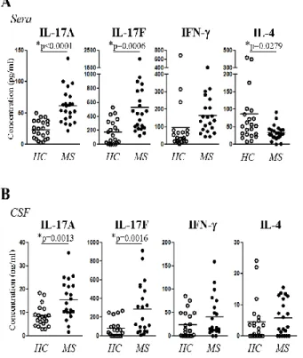
Th17-cell cytokine production is significantly increased in serum and cerebrospinal fluid (CSF)
samples from RRMS patients in comparison to HCs.
Th17-cell cytokines, IL-17A and IL-17F, as well as Th1-cell cytokine IFN-γ, are considered
pro-inflammatory cytokines, while the Th2-cell cytokine IL-4 is anti-pro-inflammatory. In order to
confirm that levels of Th17 cells and their cytokines were in fact, elevated in RRMS patients,
and to begin to visualize a relationship between endogenous IFNβ and Th17 cell differentiation,
the concentrations of these cytokines were measured in both the sera and CSF from a cohort of
RRMS patients (23 and 21 patients, respectively) and control subjects (22 and 21 donors,
respectively). The RRMS patients exhibited a significantly higher production of 17A and
IL-17F cytokines in both sera (Figure 2A) and CSF (Figure 2B), and lower serum IL-4 levels in
Figure 2. RRMS patients exhibit increased levels of IL-17A and IL-17F in sera and CSF samples in comparison
to HCs.(A) Serum samples from 22 RRMS patients and 18 HCs were collected, and the production of 17A,
IL-17F, IFN- and IL-4 was measured using ELISA. (B) CSF samples from 18 RRMS patients and 28 control subjects
were collected, and the production of indicated cytokines was measured using ELISA.
Fresh PBMCs derived from 16 RRMS patients and 16 age-, sex-, and race-matched HCs were
analyzed using intracellular cytokine staining. Flow cytometry showed that the percentages of
ex-vivo detected IL-17A-producing CD4+ T-lymphocytes were significantly increased, and the
Figure 3. The numbers of IL-17A-producing CD4+ cells is increased in RRMS patients. (A) Fresh PBMCs from
16 RRMS patients and 16 age-, sex- and race-matched HCs used. Cells were harvested, fixed, permeabilized, and
stained with fluorescently-labeled anti-IL-17A, -IFN- and -IL-4 mAbs, as well as with anti-CD4 mAb for gating.
Isotype controls were used for determining the background. The percentages of cells expressing each molecule in
gated CD4+ T-cells were determined by flow cytometry (FACS). Statistical analysis was performed using a paired
t-test. (B) The figure presents the representative staining from one RRMS patient and one HC.
DISCUSSION
Only a few studies have addressed the role of endogenous IFNβ in the pathogenesis of
the autoimmune response. Our laboratory initially directly measured levels of IFNβ using
ELISA; however, because of the presence of various immunoreactive serum proteins that
interfere with antibody binding to IFNβ in the ELISA assay, these direct measurements provided false positives at times that were not reproducible after more extensive blocking with fetal calf
gene expression, primarily MX1, whose low levels are associated with relapses in RRMS
patients, in order to indirectly measure serum IFNβ levels. These studies imply that low
endogenous IFNβ levels may be associated with increased disease activity, however no studies of
cytokine regulation by endogenous IFNβ have been reported in MS.
Results from this study indicate a significantly decreased endogenous IFNβ bioactivity in
the serum of RRMS patients in comparison to HCs, based on the reduced IFN-induced MX1 and
PRKR gene expression in the bioassay using IFNβ-responsive WISH cells.
Prior to performing these bioassays, the mechanism by which IFNβ regulates CD4+
T-cell cytokine secretion was characterized through in-vitro experiments. These studies showed
that IFNβ significantly decreased the percentages of IL-17A, IL-17F and IFN--producing CD4+
T-cells, and increased the percentage of IL-4-positive cells (Zhang et al., 2009).
Following the previous results on the IFNβ-mediated inhibition of Th17-cell responses (van der Voort et al., 2009), these new results that identify a deficient endogenous IFNβ
biological effect in patients with RRMS provide, for the first time, an evidence for its role in the development of the autoimmune response in RRMS.
A possible implication of these results is to identify patients that will optimally respond to IFN-1a treatment, as they are expected to have lower endogenous IFNserum levels. This approach to personalizing therapy for MS patients may be valuable in deciding the most effective immunomodulatory therapy before starting it and optimizing the treatment outcome for a large number of patients with RRMS. Predicting the response to individual treatment is even more relevant now, since three new oral therapies have been approved by the FDA over the past three years (finoglimod, teraflunomide and tacfidera). Given a sensitive bioassay employed in
to create a more individualized approach in treating RRMS, which is expected to provide better disease activity control.
Current studies in the laboratory include increasing the serum sample size of both RRMS patients and HCs in order to further validate the results obtained from this study. Additionally, the laboratory would like to expand this study to include patients of Clinically Isolated Syndrome (CIS). CIS describes the first episode of neurologic symptoms that lasts at least 24 hours and is the result of inflammation and demyelination in at least one area of the CNS (NMSS, 2014). Although CIS does not always progress into full-scale MS, physicians are challenged with evaluating each patient’s risk of developing the disease. If a similar trend can be observed for endogenous IFNβ bioactivity in CIS patients, physicians may be able to diagnose MS prior to its onset, allowing for a more specialized treatment plan.
Based on the above studies, we propose that endogenous IFNβ plays an active role in the suppression of the inflammatory responses that give rise to RRMS. Its decreased secretion in RRMS patients may contribute to de-repression of the Th17-mediated autoimmune response. As the field of medicine advances towards a cure for this debilitating disease, these findings may allow for more specialized treatment approaches for patients of RRMS.
ACKNOWLEDGEMENTS
REFERENCES
Durelli, L., L. Conti, M. Clerico, D. Boselli, G. Contessa, P. Ripellino, B. Ferrero, P. Eid & F. Novelli. 2009. T-helper 17 cells expand in multiple sclerosis and are inhibited by interferon-beta. Ann. Neurol. 65: 499-509.
Feng X, Reder N, Yanamandala M, Hill A, Franek B, Niewold T, Reder A & Javed A. "Type I interferon signature is high in lupus and neuromyelitis optica but low in multiple sclerosis." Journal of the Neurological Sciences (2012): 48-53.
Guo B, Chang EY & Cheng G. The type I IFN induction pathway constrains Th17-mediated autoimmune inflammation in mice. J Clin Invest (2008): 118, 1680.
Hua J, Kirou K, Lee C & Crow M. "Functional assay of type I interferon in systemic lupus erythematosus plasma and association with anti–RNA binding protein autoantibodies." Arthritis and Rheumatism (2006): 1906-1916.
National Multiple Sclerosis Society. http://www.nationalmssociety.org/
Noseworthy J, Lucchinetti C, Rodriguez M & Weinshenker B. "Multiple Sclerosis." The New England Journal of Medicine (2000): 938-952.
Tao Y, Zhang X, Chopra M, Kim MJ, Buch K, Jin J, Tang Y, Jewells V & Markovic-Plese S. "The role of endogenous IFNβ in the regulation of Th17 responses in patients with relapsing-remitting multiple sclerosis." (2013).