611
© 2017 by the Serbian Biological Society
Proanthocyanidin monomers and cyanidin 3-o-glucoside accumulation in blood-flesh
peach (
Prunus persica
(l.) Batsch) fruit
Juan Yan, Zhi-xiang Cai, Zhi-jun Shen, Rui-juan Ma and Ming-liang Yu*
Institute of Horticulture, Jiangsu Academy of Agricultural Sciences, Jiangsu Key Laboratory for Horticultural Crop Genetic Improvement, Nanjing 210014, China
*Corresponding author: [email protected]
Received: December 12, 2016; Revised: January 12, 2017; Accepted: February 7, 2017; Published online: March 2, 2017
Abstract: To better understand the characteristics and mechanisms of proanthocyanidin monomers and anthocyanin
syn-thesis in blood-flesh peach (Prunus persica (L.) Batsch), the accumulation of catechin, epicatechin and cyanidin
3-O-glu-coside was determined, and the expression patterns of structural genes associated with biosynthesis of those compounds were investigated in the blood-flesh peach fruit of cultivar “Dahongpao” during fruit development. Our results show that catechin concentration remained low and comparatively stable throughout fruit development. The concentration of epicatechin remained low at the early stages of fruit development and rapidly accumulated during ripening. Cyanidin 3-O-glucoside was not detected in the early stages. Epicatechin started to rapidly accumulate during the ripening period, reaching a maximum at the mature stage. The expressions of the early and common genes, phenylalanine ammonia-lyase and chalcone isomerase, were less associated with proanthocyanidin monomers and cyanidin 3-O-glucoside accumulation. The expression of other flavonoid ‘early’ biosynthetic genes, including chalcone synthase (CHS), flavanone 3-hydroxylase, dihydroflavonol 4-reductase (DFR) and leucoanthocyanidin dioxygenase (LDOX), were partly associated with proanthocy-anidin monomers and cyproanthocy-anidin 3-O-glucoside levels, with expression quantities peaking synchronously at the mature stage. Leucoanthocyanidin reductase and anthocyanidin reductase, which were the key genes for proanthocyanidin monomer synthesis, correlated during fruit development with catechin and epicatechin accumulation respectively; UDP-glucose: flavonoid 3-O-glucosyltransferase (UGFT), the key gene for anthocyanin synthesis, was correlated with cyanidin 3-O-glu-coside levels. The synchronous accumulation of epicatechin and cyanidin 3-O-glu3-O-glu-coside in blood-flesh peach could not be explained by the current theory of competitive distribution mechanism of common substrate.
Key words: blood-flesh peach (Prunus persica (L.) Batsch); catechin; epicatechin; cyanidin 3-O-glucoside; gene expression
How to cite this article: Yan J, Cai Z, Shen Z, Ma R, Yu M. Proanthocyanidin monomers and cyanidin 3-O-glucoside accumulation in blood-flesh peach (Prunus persica (L.) Batsch) fruit. Arch Biol Sci. 2017;69(4):611-7.
INTRODUCTION
Proanthocyanidin and anthocyanin in fruit are in-creasingly recognized as producing health beneficial effects in humans. At the same time, proanthocyani-din possesses astringency and flavor, and is a major quality factor for fruit, while anthocyanin improves visual appeal and enhances the commercial value of fruit [1,2]. Therefore, data collection and theoretical research on the composition, accumulation and syn-thetic mechanism of proanthocyanidin and anthocy-anin in fruit are of great significance for breeding new cultivars rich in beneficial ingredients.
The biosynthesis of proanthocyanidin and antho-cyanin (Fig. 1), initiated from phenylalanine, is
regulating the synthesis of proanthocyanidin and an-thocyanin are phenylalanine ammonia-lyase (PAL), cinnamate 4-hydroxylase(C4H), 4-coumarate-CoA ligase(4CL), CHS, chalcone isomerase (CHI), flava-none 3-hydroxylase (F3H), F3'H, DFR, anthocyanins synthetase (ANS)and LDOX. UFGT is a key gene for the synthesis of cyanidin 3-O-glucoside, while LAR and ANR are the key genes for proanthocyanidin monomers [1,6].
Blood-flesh peach (Prunus persica (L.) Batsch) belongs to the subfamily Prunoideae in the Rosaceae family. It is an important type of peach germplasm with red fruit-flesh color, which is rich in proan-thocyanidins and anthocyanins [7-10]. Chlorogenic acid, neochlorogenic acid, catechin, epicatechin, ru-tin, querceru-tin, gallic acid, ferulic acid, phlorizin and phloretin have been detected in blood-flesh peach, but the proanthocyanidin monomers, including catechin
and epicatechin are the main components [10,11], while the main anthocyanin is cyanidin 3-O-gluco-side [12,13]. Cyanidin 3-O-gluco3-O-gluco-side accumulation and its mechanism have been widely studied [13-17]. However, little is known about the metabolic mecha-nism of proanthocyanidin monomer metabolism in blood-flesh peach because all existing research has been conducted on white-flesh peach [18-20].
In the current paper, the blood-flesh peach culti-var “Dahongpao” was researched to better understand the characteristics and mechanisms of proanthoanidin monomers (catechin and epicatechin) and cy-anidin 3-O-glucoside accumulation. We investigated in detail the accumulation of catechin, epicatechin and cyanidin 3-O-glucoside in blood-flesh peach dur-ing fruit development, and identified the expression of structural genes encoding the proanthocyanidin monomers and cyanidin 3-O-glucoside biosynthe-sis enzymes, including PAL, CHS, CHI, F3H, DFR, LDOX, UFGT, LAR and ANR. We discuss the relation-ship between the accumulation of these proanthocy-anidin monomers and cyproanthocy-anidin 3-O-glucoside. The results of our research could be very useful for further study of the proanthocyanidin and anthocyanin syn-thesis mechanism in peach.
MATERIALS AND METHODS
Plant materials and experimental treatments
Experiments were conducted using seven-year old trees of blood-flesh peach (cultivar “Dahongpao”) maintained at the National Peach Germplasm Re-pository (Nanjing, China) in 2014. Fruit samples were collected from six trees every 7 days, beginning at 30 DAFB (days after full bloom). And each stage consisted 18 fruits with three replicates. Fruit was mechanically peeled and cored and the flesh cut into small sections. Fruit samples at each stage were mixed and immediately frozen in liquid nitrogen and stored at -75°C until use. According to the method of Lom-bardo et al [21], the sampling points were confirmed through the growth curve of a double-S form fitted to fruit weight. Specifically, the sampling points were 30 (S1), 58 (S2), 79 (S3), 93 (S4) and 100 (H) DAFB.
Fig. 1. Proanthocyanidin and anthocyanin biosynthetic pathway in plants [1, 6]. Structural genes for each step are indicated as fol-lows: PAL, phenylalanine ammonia-lyase, C4H, cinnamate 4-hy-droxylase, 4CL, 4-coumarate-CoA ligase, CHS, chalcone synthase,
CHI, chalcone isomerase, F3H, flavanone 3-hydroxylase, F3'H, fla-vanone 3'-hydroxylase, DFR, dihydroflavonol 4-reductase, LDOX, leucoanthocyanidin dioxygenase, ANS,anthocyanins synthetase,
Extraction and measurement of catechin, epicatechin and cyanidin 3-O-glucoside
Catechin and epicatechin were extracted and mea-sured according to Yan et al [11]. Frozen fruit (1 g) was homogenized in 2 mL of methanol containing 0.1% H3PO4, and the extracts were centrifuged at 10000xg for 5 min at 4°C, and filtered through a 0.22-µm filter for analysis in the Agilent 1100 series HPLC system (Agilent, USA). Samples (5 µL of extract) were analyzed using an Agilent ZORBAX SB-C18 column (4.6×250 mm, 5 μm) coupled with a UV detector at 280 nm, with ethanol (0.1% H3PO4) and water (0.1% H3PO4) as solvents: the flow rate was 1.0 mL·min–1
and temperature was 30°C.
Cyanidin 3-O-glucoside was extracted and mea-sured according Yan et al [22]. Frozen fruit (1 g) was homogenized in 2 mL of extracting solution [metha nol:water:phosphate:trifluoroacetic acid = 70:27:2:1 (V/V)], and the extracts were centrifuged at 10000 xg for 5 min at 4°C and filtered through a 0.22-µm filter for analysis in the Agilent 1100 series HPLC system. Samples (5 µL of extract) were analyzed using an Agi-lent ZORBAX SB-C18 column (4.6×250 mm, 5 μm) coupled with a UV detector at 525 nm, with methan ol:phosphate:trifluoroacetic acid = 97:2:1 (V/V) and water:phosphate:trifluoroacetic acid = 97:2:1 (V/V) as solvents: the flow rate was 1.0 mL·min–1 and
tem-perature was 25°C.
Catechin, epicatechin and cyanidin 3-O-gluco-side, and HPLC-grade methanol, acetic acid, phos-phoric acid and trifluoroacetic acid were purchased from Sigma-Aldrich (Shanghai, China). Aqueous so-lutions were prepared using ultra-pure water purified by Milli-Q System (S.A.S. 67120, Millipore, Molsheim,
France). Compounds were quantified by comparing the peak areas and presented as mg of catechin or epicatechin or cyanidin 3-O-glucoside per g of fresh tissue (mg·kg–1 FW).
RNA extraction and quantitative real-time PCR (qRT-PCR) analysis
RNA was extracted from frozen flesh obtained from “Dahongpao” using a modified CTAB method [14]. After the removal of DNA by DNase I, the concentra-tion of total RNA was measured and the first cDNA strand was synthesized from 1 µg of total RNA using Supersmo III M-MuLV Reverse Transcriptase (Bioteke Corporation, Beijing, China) primed with oligo (dT) 18. The cDNA was diluted 50× and 2 µL of the diluted cDNA was used as the template for qRT-PCR analy-sis. We designed qRT-PCR primers with Oligo 7.37 and Primer-Blast (http://www.ncbi.nlm.nih.gov/tools/ primer-blast/) using the obtained sequences of cDNA fragments. RT-PCR analysis was performed using the primers presented in Table 1 [14,18,23]. qRT-PCR was performed on an ABI 7500 System (Applied Biosys-tems, Foster, CA, USA) using the SYBR Premix Ex TaqTM (Takara, Japan). The PCR reaction consisted of
10 μL of SYBR Green PCR Master Mix, 0.8 μL of for-ward and reverse primer (10 μM), 0.4 μL of ROX Ref-erence dye (50x; all from Takara), 6.0 μL of dH2O and 2.0 μL of 1:50-diluted template cDNA in a total volume of 20 μL. The two-step RT-PCR program was initiated with a preliminary step at 95°C for 30 s, followed by 40 cycles at 95°C for 5 s and 60°C for 34 s. The assay included a no-template control for each primer pair. All qRT-PCR reactions were normalized using the Ct value corresponding to actin. Three measurements for each biological replicate sample were performed.
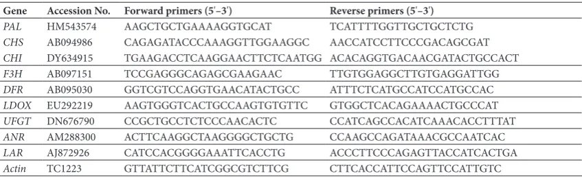
Table 1. Primers for quantitative real-time PCR
Gene Accession No. Forward primers (5'–3') Reverse primers (5'–3')
PAL HM543574 AAGCTGCTGAAAAGGTGCAT TCATTTTGGTTGCTGCTCTG
CHS AB094986 CAGAGATACCCAAAGGTTGGAAGGC AACCATCCTTCCCGACAGCGAT
CHI DY634915 TGAAGACCTCAAGGAACTTCTCAATGG ACACAGGTGACAACGATACTGCCACT
F3H AB097151 TCCGAGGGCAGAGCGAAGAAC TTGTGGAGGCTTGTGAGGATTGG
DFR AB095030 GGTCGTCCAGGTGAACATACTGCC ATTTCTCATGCCATCCATGCCAC
LDOX EU292219 AAGTGGGTCACTGCCAAGTGTGTTC GTGGCTCACAGAAAACTGCCCAT
UFGT DN676790 CCGCTGCCTCTCCCAACACTC CCATCAGCCACATCAAACACCTTTAT
ANR AM288300 ACTTCAAGGCTAAGGGGCTGCTG CCAAGCCAGATAAACGCCAATCAC
LAR AJ872926 CATCCACGGGGAAATTCACCTG ACCCTTCCCAGAGTTACCATCACTGA
Statistical analysis
Figures were drawn with Microsoft Excel 2010 (Mi-crosoft corp., Northampton, MA, USA), and least significant differences were calculated for mean sepa-rations using a t-test of the Data Processing System (DPS, version 14.10, Zhejiang University, Hangzhou, China).
RESULTS
Catechin, epicatechin and cyanidin 3-O-glucoside accumulation during fruit development
In the blood-flesh peach “Dahongpao”, the concen-tration ranges of catechin, epicatechin and cyanidin 3-O-glucoside were 28.00-44.42, 13.45-268.20 and 0-254.42 mg·kg–1 FW, respectively (Fig. 2). This result
pointed to significant differences in proanthocyanidin and anthocyanin concentrations in blood-flesh peach. Moreover, the dynamic change trends of catechin and epicatechin concentrations significantly differed. At the early and middle stages of fruit development, the concentrations of catechin were slightly higher than those of epicatechin, while in the fruit maturation period the concentration of epicatechin was signifi-cantly higher than that of catechin (p<0.01). The cat-echin concentration remained low throughout fruit development, and even declined at stage S2 (p<0.01); it increased during the middle stages (p<0.01) and remained stable in the ripening period, with a slight but non-significant increase. The concentration of
epi-catechin remained low at the early stages of fruit de-velopment, exhibiting rapid accumulation from stage S3 to a peak at stage H, with a concentration of 268.20 mg·kg–1 FW (p<0.01). The dynamic change trend of
cyanidin 3-O-glucoside was only slightly different from that of epicatechin. Cyanidin 3-O-glucoside was not detected at the early stages of fruit development, however, it started to accumulate at stage S3 and rap-idly accumulated from stage S3 to a peak at H, with a concentration of 254.42 mg·kg–1 FW (p<0.01).
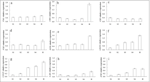
Expression of structural genes during fruit development
There were significant differences in the levels of expression of structural genes during fruit develop-ment in “Dahongpao” (Fig. 3). The upstream region of genes PAL, CHS, CHI, F3H, DFR and LDOX will be discussed first. The expression of PAL and CHI was low and at about equivalent levels at different stages. The level of expression of CHS at the transcriptional level decreased gradually from stage S1 to a minimum that was observed at S3 (p<0.01); it then increased sharply at the ripening stages and attained a maximum at stage H (p<0.01). The level of F3H expression was similar to that of CHS,with a slight decrease at the middle stage and a significant rise at stage H (p<0.01). The level of DFR expression increased slightly and remained stable at the middle stage. As for CHS and F3H, there was a significant rise at stage H (p<0.01). The levels of both LDOX and UFGT expression in-creased gradually from stage S1 and attained a maxi-mum at stage H (p<0.01). The level of LAR
sion did not significantly differ from stage S1 to S3; it slightly decreased at stages S2 and S3 and markedly increased from stage S4 to H (p<0.01). The expression of ANR significantly increased with the stages of fruit development, and reached a stable maximum in the ripening period (p<0.01).
DISCUSSION
The structure, function and synthetic mechanism of plant secondary metabolites have attracted at-tention in recent years. Anthocyanin function and biosynthesis and gene organization, expression and regulation have been investigated, however, there has been little research into proanthocyanidin [6,24]. We determined the accumulation of proanthocyanidin monomers (catechin and epicatechin) and cyanidin 3-O-glucoside in blood-flesh peach “Dahongpao”, and found that the concentration of catechin remained low and almost stable throughout fruit development. The concentration of epicatechin remained low at the early stages of fruit development and it rapidly accumulated to a maximum at the matured stage, while cyanidin
3-O-glucoside was not detected in the early stages of fruit development. As for epicatechin, it accumulated rapidly during the ripening period to a maximum at the mature stage. The developmental profiles of cat-echin and epicatcat-echin accumulation were different from those reported in previous research, in which their accumulation decreased with fruit matura-tion [18,19]. Cyanidin 3-O-glucoside accumulamatura-tion increased with fruit maturation, and the result was consistent with previous research [14].
Until now, there has been no clear definition of the major genes regulating the synthesis of proantho-cyanidins and anthocyanins in peach. Tsuda et al [17] found that CFS and DFR are the key genes regulating the synthesis of anthocyanins in blood-flesh peach. Zhao et al [13] suggested that PAL was the key enzyme for anthocyanin synthesis. Daniela et al [18] estab-lished that only UFGT was weakly correlated with anthocyanin level, while the expression of structural genes CHS, CHI, FSH, DFR, LDOX, UFGT, ANR and LAR correlated with proanthocyanidin accumulation. Jiao et al [14] found that cyanidin 3-O-glucoside accu-mulation in the fruit of blood-flesh peach “Banjintao”
was closely related to the coordinated expression of UFGT and ANS. In white-flesh peach, Daniela et al [18] proposed that the expression of the genes en-coding enzymes of the flavonoid pathway, especially LAR and ANR, correlated with proanthocyanidin concentration. Zhou et al [19] suggested that MYB7 activated transcription of LAR, but not ANR; however, in the same research, the expression of LAR and ANR could not adequately explain the dynamic changes in catechin and epicatechin concentrations during fruit development. In our research on blood-flesh peach “Dahongpao”, the expression of the upstream region of PAL and CHI were less associated with proantho-cyanidin monomers and proantho-cyanidin 3-O-glucoside accumulations, while the expression of other genes, including CHS, F3H, DFR and LDOX, were partly as-sociated with proanthocyanidin monomers. Cyanidin 3-O-glucoside levels and the expression levels peaked synchronously in ripe fruit. Moreover, the expression of UFGT, the key gene for anthocyanin synthesis, was highly correlated with cyanidin 3-O-glucoside levels, while the expression of LAR and ANR, the key genes for proanthocyanidin synthesis, correlated with cate-chin and epicatecate-chin accumulation during fruit devel-opment, respectively. We conclude that LAR, ANR and UFGT are key genes regulating the synthesis of proan-thocyanidin monomers and cyanidin 3-O-glucoside because the levels of their expression correlated with the levels of these compounds. However, the expres-sion of the upstream region of common genes such as CHS, DFR and LDOX, significantly increased with fruit development and maturation. This could give rise to the accumulation of a common substrate used in proanthocyanidin monomer and cyanidin 3-O-glu-coside synthesis. Thus, CHS, DFR and LDOX might be major genes regulating the synthesis of epicatechin and cyanidin 3-O-glucoside in blood-flesh peach.
Interestingly, we found synchronous accumulation of epicatechin and cyanidin 3-O-glucoside during the fruit-ripening period, which was very different from other fruits and could not be explained by the current mechanism of competitive distribution of a common substrate [25]. There were obviously negative corre-lations between the accumulation of epicatechin and cyanidin 3-O-glucoside during fruit development in previous research conducted on colored fruits: grape [26], blueberry [27], bilberry [28], strawberry [29] and blackberry [30]. Cyanidin 3-O-glucoside, which
remained at a low concentration during the early developing stage, increased dramatically as the fruit matured. In contrast, epicatechin exhibited a continu-ously decreasing pattern. Accordingly, the transcript levels of genes specifically controlling either of these two compounds, UFGT anthocyanin and LAR and ANR proanthocyanidin, generally coordinated with the changing patterns of products. These findings im-ply that the mechanism of synchronous accumulation of epicatechin and cyanidin 3-O-glucoside in blood-flesh peach should be researched further.
Acknowledgments: This work was supported by grants from the National Natural Science Foundations of China (31471848) and Jiangsu Agriculture Science and Technology Innovation Fund (CX(14)5014).
Authors’ contribution: Juan Yan and Mingliang Yu conceived and designed the study; Juan Yan performed the experiments; Zhijun Shen and Zhixiang Cai performed the data analysis; Juan Yan wrote the paper; Ruijuan Ma and Mingliang Yu revised the paper. All authors read and approved the final manuscript. Conflict of interest disclosure: The authors have declared that no conflict of interests exists.
REFERENCES
1. Dixon RA, Xie DY, Shashi BS. Proanthocyanidins – a final frontier in flavonoid research? New Phytol. 2005;165:9-28. 2. Sonia DP, Maria TS. Anthocyanins: from plant to health.
Phytochem Rev. 2008;7:281-99.
3. Lepiniec L, Debeaujon I, Routaboul JM, Baudry A, Pourcel L, Nesi N, Caboche M. Genetics and biochemistry of seed flavonoids. Ann Rev Plant Biol. 2006;57:405-30.
4. Tanner GJ, Francki KT, Abrahams S, Watson JM, Lar-kin PJ, Ashton AR. Proanthocyanidin biosynthesis in plants. Purification of legume leucoanthocyanidin reduc-tase and molecular cloning of its cDNA. J Biol Chem. 2003;278:31647-56.
5. Holton TA, Cornish EC. Genetics and biochemistry of anthocyanin biosynthesis. Plant Cell. 1995;7:1071-83. 6. Peng QZ, Yue ZZ, Liu CD, Ke GL, Xie DY. An integrated
approach to demonstrating the ANR pathway of proantho-cyanidin biosynthesis in plants. Planta. 2012;236:901-18. 7. Shen ZJ, Ma RJ, Yu ML, Xu JL, Cai ZX, Ni LJ, Yan SB.
Evalu-ation of antioxidant factors in peach with three types of flesh color. Sci Agric Sin. 2012;45(11):2232-41.
8. Shen ZJ, Confolent C, Lambert PP, Quilot-turion B, Yu ML, MA RJ, Pascal T. Characterization and genetic mapping of a new blood-flesh trait controlled by the single dominant locus DBF in peach. Tree Genet Genomes. 2013; 9:1435-46. 9. Vizzotto M, Cisneros L, Byrne D. Total phenolic, carotenoid,
10. Yan J, Shen ZJ, Cai ZX, Yu ML. Advances of study on pheno-lic compounds in peach fruit. J Fruit Sci. 2014;31(3):477-85. 11. Yan J, Cai ZX, Shen ZJ, Zhang BB, Qian W, Yu ML. Determi-nation and comparison of 10 henolic ompounds in each with hree ypes of lesh olor. Acta Horticul Sin. 2014;41(2):319-28. 12. Francisco AT, María IG, Paedar C, Andrew LW, Betty H,
Adel AK. HPLC-DAD-ESIMS Analysis of phenolic com-pounds in nectarines, peaches, and plums. J Agric Food Chem. 2001;49:4748-60.
13. Zhao Y, Wang LR, Cao K, Zhu GR, Fang WC, Chen CW, Peng FT. Genetic diversity of anthocyanin in peach fruit and the evaluating criterion of red–flesh peach. J Plant Genet. Resour. 2013;14:167-72.
14. Jiao Y, Ma RJ, Shen ZJ, Yan J, Yu ML. Gene regulates antho-cyanin biosynthesis in blood peach (Prunus persica (L.) Batsch) during fruit development. J Zhejiang Univ-Sci. 2014;15(9):809-19.
15. Kataoka I, Beppu K. UV irradiance increases development of red skin color and anthocyanins in ‘Hakuho’ peach. Hort-science. 2004; 39(6):1234-7.
16. Ogendiwin EA, Peace CP, Nicolet CM, Rashbrook VK, Gradziel TM, Bliss FA, Parfitt D, Crisosto CH. Leuco-anthocyanidin dioxygenase gene (PpLDOX): a potential functional marker for cold storage browning in peach. Tree Genet Genomes. 2008;4(3):543-54.
17. Tsuda T, Yamaguchi M, Honda C, Moriguchi T. Expression of anthocyanin biosynthesis genes in the skin of peach and nectarine fruit. J Am Soc Hortic Sci. 2004;129(6):857-62. 18. Daniela R, Richard VE, Rebecca AH, Carlo A, Vanina Z,
Roger PH, Guglielmo C, Andrew CA. Transcriptional regu-lation of flavonoid biosynthesis in nectarine (Prunus persica)
by a set of R2R3 MYB transcription factors. BMC Plant Biol. 2013;13:68.
19. Zhou H, Wang KL, Liao L, Gu C, Lu ZQ, Andrew CA, Han YP. Peach MYB7 activates transcription of the proanthocy-anidin pathway gene encoding leucoanthocyproanthocy-anidin reduc-tase, but not anthocyanidin reductase. Front Plant Sci. 2015;6:908.
20. Zhou J, Chen ZL, Zhang Q, Wang HQ. Effects of bag-ging on accumulation of phenolic acids and flavonoids in peach pericarp during fruit maturity. Acta Horticul Sin. 2009;36(12):1717-24.
21. Lombardo VA, Osorio S. Borsani J, Lauxmann MA, Busta-mante CA, Budde CO, Andreo CS, Lara MV, Fernie AR, Drincovich MF. Metabolic profiling during each fruit development and ripening reveals the metabolic networkst hat underpin each developmental stage. Plant Physiol. 2011;157:1696-710.
22. Yan J, Shen ZJ, Cai ZX, Yu ML, Ma RJ, Qian W, inventors; Beijing Yingke Law Firm, assignee. The method of antho-cyanin extraction and detection with HPLC in blood flesh peach. China patent CN 103,760,289 B. 2015 Jul 22. 23. Tong ZG, Gao ZH, Wang F, Zhou J, Zhang Z. Selection of
reliable reference genes for gene expression studies in peach using real time PCR. BMC Mol Biol. 2009;10:71.
24. Vinterhalter B, NinkoviĆ S, Kozomara B, Vinterhalter D. Carbohydrate nutrition and anthocyanin accumulation in light grown and etiolated shoot cultures of carob (Ceratonia siliqua L.) Arch Biol Sci. 2007;59(1):51-6.
25. Xie DY, Dixon RA. Proanthocyanidin biosynthesis – still more questions than answers? Phytochemistry. 2005;66:2127-44.
26. Kennedy JA, Hayasaka Y, Vidal S, Waters EJ, Jones GP. Com-position of grape skin proanthocyanidins at different stages of berry development. J Agr Food Chem. 2001;49:5348-55. 27. Morazzoni P, Bombardelli E. Vaccinium myrtillus L.
Fitote-rapia. 1996;67:3-29.
28. Jaakola L, Määttä K, Pirttilä AM, Törrönen R, Kärenlampi S, Hohtola A. Expression of genes involved in anthocyanin biosynthesis in relation to anthocyanin, proanthocyanidin and flavonol levels during bilberry fruit development. Plant Physiol. 2002;130(2):729-39.
29. Joao RMA, Eleonora D, Anja P, Fabrizio C, Ric de Vos CH, Bettina D, Fabienne M, Gaetano P, Thilo CF, Arnaud GB, Ste-fan M, Carlo R. Characterization of major enzymes and genes involved in flavonoid and proanthocyanidin biosynthesis during fruit development in strawberry (Fragaria×ananassa). Arch Biochem Biophys. 2007;465:61-71.
![Fig. 1. Proanthocyanidin and anthocyanin biosynthetic pathway in plants [1, 6]. Structural genes for each step are indicated as fol-lows: PAL, phenylalanine ammonia-lyase, C4H, cinnamate 4-hy-droxylase, 4CL, 4-coumarate-CoA ligase, CHS, chalcone synthase, CHI, chalcone isomerase, F3H, flavanone 3-hydroxylase, F3'H, fla-vanone 3'-hydroxylase, DFR, dihydroflavonol 4-reductase, LDOX, leucoanthocyanidin dioxygenase, ANS, anthocyanins synthetase, UFGT, UDP-glucose: flavonoid 3-O-glucosyltransferase, LAR, leu-coanthocyanidin reductase, and ANR, anthocyanidin reductase.](https://thumb-us.123doks.com/thumbv2/123dok_us/7813997.2086542/2.595.56.289.83.346/proanthocyanidin-phenylalanine-dihydroflavonol-leucoanthocyanidin-anthocyanins-glucosyltransferase-coanthocyanidin-anthocyanidin.webp)