EVALUATION OF MULTIPLEX PCR TECHNIQUES FOR
KLEBSIELLA PRODUCING
AMPC-
LACTAMASES IN
CLINICALLY SIGNIFICANT KLEBSILLA ISOLATES
Iman M. A. El-Kholy
[a], M. H. Abul-Aziz
[b], Atef M. Diab
[b]and Mona A. Rezk
[a]*Keywords: Multiplex PCR, cefoxitin resistant Klebsiella-AmpC β-lactamase.
Multiplex PCR for the detection of AmpC genes has proved useful as a rapid screening tool to distinguish cefoxitin resistant non-AmpC producers from cefoxitin resistant AmpC producers. In addition to AmpC gene detection, the data generated from the multiplex PCR method can distinguish which family of AmpC gene is present in the resistant organism thereby distinguishing possible inducible AmpC producers from non-inducible producers of AmpC. The present study was designed to evaluate these issues among cephalosporin-resistant isolates of Klebsiella spp. and to assess the performance characteristics of phenotypic tests, using different inhibitors, compared to the PCR, for their rapid and accurate detection. Fifty eight out of 100 isolates were AmpC producers by PCR. Fifty six out of 58 isolates that were positive by PCR test were resistant to FOX. Thirty out of 58 AmpC producers were ESBL positive by E- test and MDDST in detection of ESBL in the presence of AmpC. While 23 /58 were positive by DDST for detection of ESBL in presence of Amp. This study reveals high prevalence of pAmpC and ESBL enzymes among bacterial isolates from our hospital. ESBL production may mask the phenotypic detection of pAmpC enzymes. Modified 3 dimensional(M3D) is a simple and reliable method for detection of pAmpCs. MDDST serve as reliable confirmatory tests for detection of ESBLs in AmpC-positive isolates.
* Corresponding Authors Fax:
E-Mail: [email protected] [a] Ain Shams University Hospital
[b] Suez Canal University, Faculty of Science
Introduction
Extended-spectrum beta-lactamases (ESBLs) and AmpC beta-lactamases (AmpCs) are important mechanisms of resistance among Enterobacteriaceae. Infections caused by ESBL and /or AmpC- positive bacteria are of particular clinical and epidemiological importance and cause higher patient morbidity and mortality.1,2
ESBLs are typically plasmid-mediated, clavulanate susceptible enzymes that hydrolyze penicillins, extended-spectrum cephalosporins and aztreonam. AmpCs are cephalosporinases that are poorly inhibited by clavulanic acid. They can be differentiated from ESBLs by their ability to hydrolyze cephamycins as well as other extended-spectrum cephalosporins. AmpCs were presumed to be chromosomally mediated but since the late 1980s they have disseminated on plasmids and now represent a substantial clinical threat. The emergence and spread of pAmpCs in Enterobacteriaceae has made their detection clinically relevant, particularly in bacterial species naturally lacking chromosomal cephalosporinase namely in E.coli, Klebsiella spp. and Proteus mirabilis. Detection a pAmpC in a strain with a coexisting ESBL is even more challenging.3. Moreover, AmpCs can interfere with ESBL detection particularly when using the current CLSI ESBL confirmatory tests. Resulting failures to detect ESBLs can endanger patients because false susceptibility to cephalosporins may be reported.4 This warrants the need foran alternative ESBL confirmatory test of greater accuracy for AmpC-producing isolates.5 At present, there are no
guidelines or standardized phenotypic methods recommended for detection of AmpCs, though several methods have been described, including disc synergy assays using Amp-C inhibitors, a specific E test format for AmpC testing and a three-dimensional test. However, these methods are expensive, tedious and have not been systematically compared.6,7 This prospective study was designed and conducted at in the Central Microbiology Laboratory of Ain Shams University Hospital in Egypt to (a) determine the prevalence of ESBLs and AmpC-producers among bacterial isolates with reduced susceptibility to extended spectrum cephalosporines and cephamycins of Klebsiellae, (b) to evaluate the efficacy of the modified double disc synergy test (MDDST) as confirmatory ESBL tests in AmpC–positive isolates and (c) to assess the performance characteristics of phenotypic tests compared to the PCR as a gold standard test for the rapid and accurate detection ofAmpCs in ESBL-positive and ESBL-negative isolates.
Experimental
Bacterial isolates
This study was done on 100 clinical isolates of Klebsiella spp. isolated from different clinical specimens referred to Microbiology Central Laboratory of Ain Shams University Hospitals Cairo, Egypt, for routine culture and sensitivity, from ICU, during a period of 3 months, from September to December 2015.
creating zones of inhibition that have been measured and interpreted according to Ref.8 interpretive criteria for screening of ESBL.
Formation of zone diameters of CPD 17, CAZ ≤ 22 and CTX ≤27 mm indicates ESBL production that is positive screening for ESBL. However, FEP disc is not included by CLSI for ESBL screening and the FEP zone of diameter ≤14 mm indicates resistance which we consider as presumptive ESBL. To perform double disc synergy test using amoxicillin/ clavulanate (20/10 µg) and 3rd generation cephalosporin that is ceftazidime, cefotaxime, cefopodixime and cefepime,9 a disc of AMC (20 μg amoxicillin + 10 μg clavulanic acid) and discs of CAZ, CTX, FEP and CPD were placed around AMC disc with a distance of 20 mm. Clear extension of the edge of the inhibition zone of cephalosporin toward the AMC disc shows positive result and interpreted as positive for ESBL production (Figure ESI 1). Negative result showed that no synergy is present (Figure ESI 2). To detect production of ESBLs by modified double disc synergy using FEP disc and piperacillin/tazobactam disc10 a disc of TZP (piperacillin /tazobactam) (100/10 µg) was placed from FEP (30 μg) disc at a distance of 20 mm from it (centre to centre). Clear extension of the edge of the inhibition zone of FEP disc toward the TZP disc indicates a positive result and was interpreted as positive for ESBL production (Figure ESI3). Negative results showed that there is no synergy (Figure ESI4).
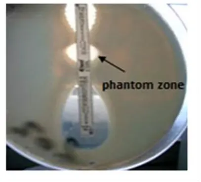
CLSI method using ceftazidime/ ceftazidime-clavulanate (CAZ-CLA) E-test ESBL strips.8 The E-test ESBL strip is a plastic drug-impregnated strip, one end of which generates a stable concentration gradient of ceftazidime (MIC test range, 0.5-32 mg L-1) and the remaining end of which generates a gradient of ceftazidime (MIC test range, 0.064-4 mg L-1) plus 4 mg L-1 clavulanic acid. The MIC value was read from the scale in terms of g mL-1 where the ellipse edge intersects the strip. ESBL production is inferred if the MIC ratio for cephalosporin alone/cephalosporin plus clavulanate MIC is ≥ 8 (Figure ESI5). ESBL production was also identified by the presence of a phantom zone or a deformation of the CAZ inhibition zone independent of the MIC ratios (Figure 1.) A MIC ratio of <8 is indicative of non ESBL production (Figure 2).
Figure 61. TZ = 32, TZL = 0.25 so TZ/TZL = 128, the ratio≥ 8 indicates a clear cut ESBL positive.
Figure 2. TZ = 0.5, TZL = 0.125 so TZ/TZL = 4, the ratio< 8 indicates aclear cut ESBL negative.
When MIC values were above the test device range, interpretation was ‘non-determinable’.Screening for AmpC production by resistance to cefoxitin disc (30 µg).9
The antibiotic disc used FOX (30 µg) was allowed to diffuse into the medium and interact in a plate freshly seeded with the test organisms making zones of inhibition that have been measured and interpreted according to CLSI8 interpretive criteria for screening of AmpC. Isolates showing diameter zone of inhibition ≤ 14mm were considered resistant (Figure ESI6.11 Isolates showing diameter zone of inhibition > 14mm were considered sensitive (Figure ESI7).
Confirmation of production of AmpC was done by modified three-dimensional test.12 This test detects the tested isolate which produce AmpC -lactamase enzyme that isresistant to cefoxitin resulting in distortion of zone of inhibition of cefoxitin which is formed by E. coli ATCC 25922. Enhanced growth of the surface of the organism at the point where the slit intersected the zone of inhibition means a a positive result and is interpreted as evidence for the presence of AmpC β-lactamase (Figure ESI8. Isolates with no distortion are viewed as negative result and are recorded as non-AmpC producers (Figure ESI9).
Real-time PCR
DNA extraction
for 1 min at ≥ 6000 x g (8000 rpm). Flow-through and collection tube were discarded. The DNA Mini spin column was placed in a new 2 mL collection tube. 500 µL Buffer AW2 was added followed by centrifugation for 3 min at 20,000xg (14,000 rpm) to dry the DNA membrane. Flow-through and collection tube were discarded. The DNA Mini spin column was placed in a clean 1.5 or 2 mL micro centrifuge tube, and 100 µL Buffer AE was pipetted directly onto the DNA membrane. Incubation was done at room temperature for 1 min, and then centrifugation for 1min at ≥ 6000 x g (8000 rpm) to elute.
DNA amplification and detection
Reaction set-up
All solutions after thawing were gently vortexed and briefly centrifuged. A reaction master mix was prepared by adding the following components for each 25 μL reaction to a tube at room temperature (Table 1).
Table 1. Components of reaction master mix for each 25 uL reaction.
Reaction component Volume μL Concentration
Maxima® SYBR Green qPCR Master Mix (2X), no ROX
12.5 -
Forward Primer 0.75 (1:10) 0.3 μM Reverse Primer 0.75 (1:10) 0.3 μM Template DNA 5 (1:10) - Water, nuclease-free 6 - Total reaction volume -
The primers used were:
FOXMF: 5´AAC ATG GGG TAT CAG GGA GAT G 3´.
FOXMR: 5´CAA AGC GCG TAA CCG GAT TGG 3´.13
The master mix was mixed thoroughly and the appropriate volumes were dispensed into PCR tubes. The template DNA (≤ 500 ng/reaction) was added to the individual PCR tubes containing the master mix. Then the reaction mixture was mixed gently without creating bubbles (not to be vortexed) then centrifuged briefly (bubbles will interfere with fluorescence detection).
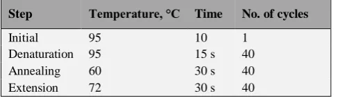
Table 2, Three-step cycling protocol
Step Temperature, °C Time No. of cycles
Initial denaturation
95 10
min 1 Denaturation 95 15 s 40 Annealing 60 30 s 40 Extension 72 30 s 40
The thermal cycler was programmed according to the recommendations below, then the samples were placed in the cycler and we started the program. Data was taken during the extension step. Thermal cycling conditions are given Table 2.14 The amplification program was followed immediately by a melt program consisting of 1 min at 95 °C, 30 sec at 55 °C then again to 95 °C for 30 s.
The greenish horizontal line in the graph (Figure 3) is the threshold line at which the fluorescence begins to be detected (The point at which the amplification plot crosses the threshold is the cycle threshold = Ct).15 The T
m of samples which were identical or close to that of positive control were considered the gene of target as shown in Figure 4.
Figure 3. Results of Syber Green real-time PCR in amplification plot with cycles number on x axis and fluorescence on y axis.
Figure 4. Results of melting curve, average Tm = 77.13 °C
-77.72 °C.
Results
Table 3. Comparison between Cefoxitin screening test and PCR test for AmpC detection.
Fox screening test
R S Total
PCR Positive 56 (96. 6%) 2 (3.4 %) 58 Negative 20 (47.6 %) 22 (52.4 %) 42
Total 76 24 100
Thirty out of 58 AmpC producers were ESBL positive by E-test and MDDST with a sensitivity and specificity of 100 % in detection of ESBL in the presence of AmpC (Table 4). While 23/58 were positive by DDST showing 76.7 % sensitivity and 100 % specificity for detection of ESBL in presence of AmpC with positive predictive value and negative predictive value of (100 % and 80 %) respectively (Table 5). There was substantial agreement between results of both the tests.
Table 4. Comparison between MDDST and E-test when PCR test is positive.
PCR positive MDDST
Positive Negative Total
E-test Positive 30 (100%) 0 (0%) 30 Negative 0 (0%) 28 (100%) 28
Total 30 28 58
Table 5. Comparison between DDST and E-test or MDDST when PCR test is positive.
PCR Positive DDST
Positive Negative Total
E-test or MDDST
Positive 23 (76.7 %)
7 (23.3 %)
30
Negative 0 (0%)
28 (100 %)
28
Total 23 35 58
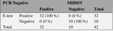
Forty-two out of 100 isolates were negative Ampc by PCR test. Thirty two out of 42 clinical isolates were positive ESBL by both E-test and MDDST showing (100 %) sensitivity and specificity in detection of ESBL in absence of AmpC (Table 6). While, 29/42 (69.05 %) were positive by DDST showing 90.6 % sensitivity and 100% specificity of detection of ESBL in absence of AmpC. The positive predictive value and negative predictive value was (100 % and 76.9 %) respectively (Table 7). There is a substantial agreement between results of both the tests.
Table 6. Comparison between MDDST and E-test when PCR test is negative.
PCR Negative MDDST
Positive Negative Total
E-test Positive 32 (100 %) 0 (0 %) 32 Negative 0 (0 %) 10 (100 %) 10
Total 32 10 42
Discussion
The importance of detecting AmpC-producing isolates is highlighted by data showing high clinical failure rates when AmpC-producing strains of K. pneumoniae are treated with cephalosporin agents or the subsequent development of antibiotic resistance in such strains.16 By antibiotic susceptibility testing it might be difficult to distinguish ESBL-producing organisms from plasmid AmpC-producing organisms because of their similar resistance. Distinguishing between AmpC- and ESBLproducing organisms has epidemiological significance and has therapeutic importance as well.17
Table 7. Comparison between DDST and E-test or MDDST when PCR test is negative.
PCR Negative DDST
Positive Negative Total
E-test or MDDST
Positive 29 (90.6 %) 3 (9.4 %) 32 Negative 0 (0 %) 10 (100 %) 10
Total 29 13 42
In Egypt, one study assessed AmpC production among Enterobacteriaceae,18 however, molecular techniques were not attempted. So, little is known about the genetic background of AmpC-producing isolates in Egypt.
Results of the present study showed that the prevalence of ampC genes in 58/100 isolates of the selected species was 30/58(51.7%) whereas ESBL-producers were detected. AmpC prevalence was lower than that reported by El-Hefnawy18 from Egypt (34 % in 50 K. pneumoniae and E. coli isolates), but it was equivocal with another study from Singapore (26 % in 255 clinical isolates of E. coli, Klebsiella spp. and P. mirabilis). In the first study, molecular techniques were not attempted and they used one phenotypic test, the three dimensional test (TDT), while in the second study both TDT and the multiplex PCR were used.19 These results were much higher than those reported in other parts of the world; in Spain (0.43%),20 in China (2.79 %)21 and in USA (3.3 %).22 This may be due to the differences in the study population and the epidemiological differences in various geographic regions.
In our study 30/58 (51.7%) of the isolates were both AmpC and ESBL-positive by molecular detection methods. This rate was higher than that reported from western parts of the world: 7/117 (6%) in Spain,20 4/81 (4.9 %) in Minnesota in USA.26 Interestingly, 58/62 (93.5 %) of our ESBL-positive Klebsiella spp. was AmpC-positive. This rate is comparable with that reported in India as Vandana and Honnavar27 detected AmpC in 39/52 (75 %) of ESBL-producing isolates of K.pneumoniae. As both ESBL and AmpC β-lactamases may coexist, therefore their detection is difficult because they mask each other. In the present study, all 30 ESBLproducers in AmpC-positive isolates were successfully identified by using the MDDST, in which FEP and TZP were utilized in approximation, implying that the use of this phenotypic method could overcome the masking effect of AmpC on phenotypicdetection of ESBLs. Similar observation has been reported by Khan9 and Fam and El-Damarawy.25
Use of a cefoxitin disk is useful in screening for AmpC but it is non-specific. In our study only 58/64 of cefoxitin-resistant isolates had ampC genes. Several factors may explain resistance to cefoxitin in the AmpC-negative isolates: First, it may arise due to porin channel alterations and mutations as previously reported in Klebsiella spp. isolates.3,16 Second, cefoxitin-resistance phenotype in Klebsiella spp. can result from over expression of the chromosomal ampC gene due to mutations in the promoter or/and attenuator regions resulting in alterations in the permeability of the cell to cefoxitin or a combination of all thesefactors.26 Third, cefoxitin has been demostrated as a substrate to active efflux pump in clinical isolates.27
In conclusion, this study has revealed the occurrence of plasmid mediated AmpC beta-lactamase producing strains in clinically important bacterial isolates for the first time in our region. ESBL production may mask the phenotypic detection of pAmpC enzymes. MDDST is a simple and reliable method serve as reliable confirmatory tests for detection of ESBLs in AmpC positive isolates. Occurrence of a large percentage of multidrug resistant strains has been observed. As AmpC beta-lactamase production is frequently accompanied by multidrug resistance, so conjugative dissemination of this AmpC beta -lactamase encoding plasmids may facilitate the spread of resistance against a wide range of antibiotics among different members of Enterobacteriaceae. Imipenem is superior to other antibiotics for the treatment of serious infections due to AmpC beta-lactamase-producing enterobacteria.
References
1Livermore D. M.,Current Epidemiology and Growing Resistance
of Gram-negative Pathogens, Korean J. Int. Med., 2012,27,
128-142. doi:10.3904/kjim.2012.27.2.128
2Maina D., Revathi G., Kariuki S., Ozwara, H., Genotypes and
cephalosporin susceptibility in extended-spectrum -lactamase producing Enterobacteriaceae in the community. J. Infect. Dev. Ctries, 2012, 6(6), 470-477.DOI: 10.7243/2052-6180-1-5
3Philippon A., Arlet, G., Jacoby, G. A.,Plasmid-determined
AmpC-type β-lactamases. Antimicrob. Agents Chemother., 2002,
46(1), 1–11. doi: 10.1128/AAC.46.1.1-11.2002
4Thomson K. S. Controversies about extended-spectrum and
AmpC -lactamases. Emerg. Inf. Dis.,2001, 7(2), 333-336.
5Munier G. K., Johnson C. L., Snyder J. W. Moland, E. S., Hanson,
N: D., Thomson, K. S., Positive extended-spectrum-lactamase (ESBL) screening results may be due to AmpC β-lactamases more often than to ESBLs. J. Clin. Microbiol. 2010, 48, 1019-1025. doi: 10.1128/JCM.01544-09
6Kanamori, H., Yano, H., Hirakata, Y., Endo, S., Araim K., Ogawa,
M., Shimojima, M., Aoyagi, T., Hatta, M., Yamada, M., Nishimaki, K., Kitagawa, M., Kunishima, H., Kaku, M., Molecular Characteristics of Extended-Spectrum - Lactamases and qnr Determinants in Enterobacter Species from Japan. PLoSONE 2012, 7(6), e37967. DOI:
10.1093/jac/dkr283
7Sabia, C., Gargiulo, R., Sarti, M., Evaluation of a Double Synergy
Differential Test (DSDT) for differential detection of ESBL and AmpC-type β-lactamases in Escherichiac oli, Klebsiella pneumoniae and Proteus mirabilis, New Microbiol., 2012, 35,
221-225.
8CLSI: Clinical laboratory standards institute. Performance
standards for antimicrobial susceptibility testing; nineteenth information supplement. M100–S19. 2009, 9(3), Wayne, Pennsylvania (USA). doi: 10.1371/journal.pone.0079130 9Khan, M. K., Thukral, S. S. and Gaind, R., Evaluation of a
modified double disc synergy test for detection of extended spectrum -lactamases in AmpC -lactamase-producing Proteus mirabilis. Indian J. Med. Microbiol.;2008, 26, 58-61.
10Pitout J. D., Reisbig, M. D., Venter, E. C., Church, D. L. and
Hanson, N. D. Modification of the double disc test for detection of Enterobacteriaceae producing extended spectrum and AmpC β-lactamases. J. Clin. Microbiol., 2003, 41, 33-39.
11Clinical and Laboratory Standards Institute (CLSI,):Performance
standards for antimicrobial susceptibility testing, twenty-second informational supplement. CLSI document M100-S22. Wayne, PA: Clinical and Laboratory Standards Institute.
2012
12Shahid, M., Malik, A., Agrawal, M. and Singhal S., Phenotypic
detection of extended-spectrum and AmpC β-lactamases by a new spot-inoculation method and modified three-dimensional extract test: comparison with the conventional three-dimensional extract test. J. Antimicrob. Chemother. 2004,54, 684-687. DOI:10.1093/jac/dkh389
13Péréz-Péréz, F. J., Hanson, N. D., Detection of plasmid mediated
AmpC β-lactamase genes in clinical isolates by using multiplex PCR. J. Clin. Microbiol., 2002, 40(6), 2153-2162.doi:10.1128/JCM.40.6.2153-2162.2002
14Ergin, A., Koseoglu, Eser, Ö. K., Hascelik, G., Erythromycin and
penicillin resistance mechanisms among viridans group
streptococci isolated from blood cultures of adult patients with underlying diseases. New Microbiol.,2011, 34, 187-193.
15Muldrew K. L., Molecular diagnostics of infectious diseases.
Current Opinion Pediat.; 2009, 21, 102–111.
doi:10.1097/MOP.0b013e328320d87e
16Jacoby G. A., AmpC β-lactamases. Clin. Microbiol. Rev., 2009,
22(1), 161-182. doi: 10.1128/CMR.00036-08
17Park, Y. S., Yoo, S., Seo M., Kim, J. Y., Cho, Y. K., Pai, H.,Risk
factors and clinical features of infections caused by plasmid-mediated AmpC β-lactamase producing Enterobactriacae.
Int. J. Antimicrob. Agents, 2009, 34, 38-43.
doi: 10.1016/j.ijantimicag.2009.01.009
18El-Hefnawy N. N., Phenotypic detectionof AmpC -lactamase in
Gram-negative bacteria. Thesis submitted for partial fulfillment of Master degree in Clinical and Chemistry Pathology, Faculty of Medicine, Ain Shams University.2008. 19Tan T. Y., Ng, S. Y., Teo L., Koh, Y., Teok, C. H.,Detection of
20Mata C., Miro, E., Rivera, A., Mirelis, B., Coll, P., Navarro, F.,
Prevalence of acquired AmpC β-lactamases in
Enterobacteriacae lacking induciblechromosomal ampC
genes at a Spanish hospitalfrom 1999 to 2007. Clin.
Microbiol. Infect., 2009, 16(5), 472-476.
doi.org/10.1111/j.1469-0691.2009.02864.x
21Li Y., Li Q., Du Y. Jiang, X., Tang, J., Wang, J., Li? G., Jiang, Y.,
Prevalence of plasmid-mediated AmpC β-lactamases in a Chinese university hospital from 2003 to 2005: first report of CMY-2-type AmpC β-lactamase resistance in China. J. Clin. Microbiol. 2008, 46, 1317–1321. doi: 10.1128/JCM.00073-07
22Moland E. S., Hanson N. D, Black J. A., Hossain, A., Song, W.,
Thomson, K. S.,Prevalence of newer β-lactamases in Gram-negative clinical isolates collected in the United States from 2001 to 2002. J Clin Microbiol., 2006, 44(9), 3318-3324.
DOI:10.1128/JCM.00756-06
23El-Kholy A., Baseem, H., Hall, G. S., Procop, G. W., Longworth,
D. L.,Antimicrobial resistance in Cairo, Egypt 1999-2000::a survey of five hospitals. J. Antimicrob. Chemother., 2003, 51, 625-630. PMID: 12615864
24Al-Agamy M. H., Ashour M. S., Wiegand I. I.,First description
of CTX-M -lactamase-producing clinical Escherichia coli
isolates from Egypt. Int. J. Antimicrob. Agents, 2006, 27,
545-548. doi: 10.1186/1471-2334-9-84
25Fam N.S. and El-Damarawy M. M., CTX-M-15 extended
spectrum -lactamases detected from intensive care unit of an Egyptian medical research institute. Res. J. Med. Sci., 2008, 3(1), 84-91. doi:10.4172/2329-8731.1000156
26Kohner P. C., Robberts, F. J., Cockerill, III F. R., Patel, R.,
Cephalosporin MIC distribution of extended-spectrum-β-lactamase and pAmpC-producing Escherichia coli and Klebsiella species. J. Clin. Microbiol, .2009, 47(8), 2419-2425.doi: 10.1128/JCM.00508-09
27Vandana K E, Honnavar P., AmpC b-Lactamases among ESBL
Producing Escherichia Coli and Klebsiella Pneumoniae-if you don’t look, you won’t find. J. Clin. Diagn. Res., 2009,
8( 3), 1653-1656.
26Mulvey, M. R., Bryce, E., Boyd, D. A. Ofner-Agostini, M., Land,
A. M., Simor, A. E., Paton, S.,Molecular Characterization of Cefoxitin-Resistant Escherichia coli from Canadian Hospitals. Antimicrob Agents Chemother. 2005, 49(1), 358-365. DOI:10.1128/AAC.49.1.358-365.2005
27Pages, J-M., Lavigne, J-P., Leflon-Guibout, V., Marcon? E., Bert,
F., Noussair, L., Nicolas-Chanoine, M.-H.,Efflux Pump, the masked side of ß-lactam resistance in Klebsiella pneumonia
clinical isolates. PLoS ONE 2009, 4(3), e4817.doi.org/10.1371/journal.pone.0004817