241
© 2018 by the Serbian Biological Society How to cite this article: Kornjača D, Živković V, Krstić D, Čolović M, Đurić M, Stanković S, Mutavdžin S, Jakovljević V, Đurić D. The effects of acute hyperhomocysteinemia induced by DL-homocysteine or DL-homocysteine thiolactone on serum biochemical parameters, plasma antioxidant enzyme and cardiac acetylcholinesterase activities in the rat. Arch Biol Sci. 2018;70(2):241-8.
The effects of acute hyperhomocysteinemia induced by homocysteine or
DL-homocysteine thiolactone on serum biochemical parameters, plasma antioxidant enzyme
and cardiac acetylcholinesterase activities in the rat
Duško Kornjača1, Vladimir Živković2, Danijela Krstić3, Mirjana Čolović4, Marko Đurić5, Sanja Stanković6,
Slavica Mutavdžin7, Vladimir Jakovljević2,8 and Dragan Đurić7,*
1Independent Medical Practice, Novi Sad, Serbia
2University of Kragujevac, Faculty of Medical Sciences, Department of Physiology, Serbia 3Institute of Medical Chemistry, Faculty of Medicine, University of Belgrade, Serbia
4Department of Physical Chemistry, Vinča Institute of Nuclear Sciences, University of Belgrade, Serbia
5Department of Anesthesiology, Reanimation and Intensive Care Medicine, University Clinical Hospital Center
“Dr. Dragiša Mišović – Dedinje”, Belgrade, Serbia
6Centre of Medical Biochemistry, Clinical Centre of Serbia, Belgrade, Serbia
7Institute of Medical Physiology “Richard Burian”, Faculty of Medicine, University of Belgrade, Serbia 8IM Sechenov First Moscow State Medical University, Department of Human Pathology, Moscow, Russia
*Corresponding author: [email protected]; [email protected]
Received: July 31; Revised: October 23, 2017; Accepted: October 23, 2017; Published online: October 30, 2017
Abstract: The aim of this study was to assess the effects of DL-homocysteine (DL-Hcy) and DL-homocysteine thiolactone (DL-Hcy TLHC) on selected serum biochemical parameters, markers of oxidative stress and the activities of antioxidant enzymes (catalase (CAT), glutathione peroxidase (GPx), superoxide dismutase (SOD)) in the plasma, as well as on acetyl-cholinesterase (AChE) activity in the cardiac tissue homogenate in the rat. Male Wistar rats were divided into three groups as follows: control group (1 mL 0.9% NaCl, intraperitoneal (i.p.) injection), DL-Hcy group (8 mmol/kg body mass (b.m.), i.p.) or DL-Hcy TLHC group (8 mmol/kg b.m., i.p.). One hour after administration, the rats were euthanized, whole blood was collected for biochemical analysis, and the heart was excised. Following the i.p. administration of DL-Hcy and DL-Hcy TLHC, the activities of antioxidant enzymes were mostly significantly increased, while plasma malondialdehyde (MDA) was decreased. Administration of DL-Hcy and DL-Hcy TLHC significantly inhibited AChE activity in rat cardiac tissue. Our findings suggest that DL-Hcy and DL-Hcy TLHC exerted prooxidant effects; however, the decrease in MDA points to an inverse response to the increase in antioxidant enzyme activities. While both substances inhibited AChE activity in rat cardiac tissue, DL-Hcy TLHC induced stronger effects than DL-Hcy.
Key words: acetylcholinesterase; antioxidant enzymes; DL-homocysteine; DL-homocysteine thiolactone; rat
INTRODUCTION
Homocysteine (Hcy) is an endogenous sulfhydryl-containing amino acid that is an intermediate product in the normal biosynthesis of the amino acids me-thionine and cysteine [1]. Deficiency of the enzymes and/or vitamins that are involved in the homocysteine metabolic pathway leads to its increased levels in the plasma, referred to as hyperhomocysteinemia (HHcy) [2,3]. Data suggests that metabolic conversion of Hcy to the thioester Hcy-thiolactone contributes to Hcy pathobiology [4].
Literature findings indicate that there is a clear correlation between total serum Hcy and the inci-dence of cardiovascular disease and its complications, such as heart attacks and strokes, thus promoting Hcy as a new and independent risk factor for these dis-eases [5,6]. HHcy leads to endothelial dysfunction, which may further promote the proliferation of vas-cular smooth muscle cells [7] and increase collagen synthesis in vascular smooth muscle cells [8]. There is evidence that HHcy is associated with increased ROS formation, including the superoxide anion radi-cal (O2-) and hydrogen peroxide (H
2O2) [9], thereby
leading to oxidative stress and cell damage [10]. The main antioxidant enzymes responsible for controlling oxygen free radicals are SOD, CAT, and glutathione-dependent enzymes, including GPx, GR and GST [10]. While it has been found that HHcy may impair the glutathione-related antioxidant defense system [11,12], data about its effect on SOD activity are still controversial.
While some studies showed that HHcy decreased erythrocyte SOD activity in patients with cardiovas-cular disease (CVD) [12,13], others have established a positive relationship between SOD activity and Hcy levels in patients with homocystinuria [14]. Acetyl-cholinesterase (AChE) is the serum protease that ter-minates neurotransmission at cholinergic synapses by splitting the acetylcholine into choline and acetate, and it is predominantly distributed in different brain re-gions [15]. Interestingly, elevated homocysteine levels increase AChE activity in the central nervous system [15,16], while homocysteine thiolactone slowly and irreversibly inhibits it [17]. Recent investigations have proved the presence of AChE activity in the heart [18].
Taking into account that the molecular mecha-nisms of Hcy-induced cardiac effects are still poorly understood, this study aimed to examine the effect of DL-homocysteine and its thiolactone metabolite on serum biochemical parameters, markers of oxidative stress, the activities of antioxidant enzymes and car-diac AChE activity in the rat.
MATERIALS AND METHODS
Chemicals
All chemicals were purchased from the Sigma Chemi-cal Co. (USA).
Experimental animals
Male Wistar albino rats (n=36, 12 in each experimen-tal group; 10 weeks old, body mass 250±30 g) were used. The rats were housed under strictly controlled conditions of air temperature (22±1°C), relative hu-midity (50%) and a 12 h light/dark cycle (with the light period commencing at 9 am), with free access to water and standard food. All experimental procedures were performed in accordance with the EU Directive for the Protection of Vertebrate Animals used for Ex-perimental and other Scientific Purposes 86/609/EES.
Experimental protocol
Serum biochemical parameters
The following serum biochemical parameters: Alb, ALT, AMY, AST, Chol, C, Fibr, Gluc, HDL, Hcy, hsT-nT, LDH, LDL, TGL, TP, Urca, Urea, VWF Act, were analyzed using spectrophotometry and commercial kits (Siemens Healthcare Diagnostics Ltd. Frimley, Camberley, UK) on an automatic biochemical analyzer (Dimension Xpand, Siemens). An immunoturbidi-metric commercial assay (Siemens Healthcare GmbH, Marburg, Germany) was used for the determination of fibrinogen and von Willebrand factor activity on a BCS XP coagulation analyzer (Siemens Healthcare GmbH, Marburg, Germany). Serum homocysteine was measured by an automated electrochemiluminescence immunoassay system ADVIA Centaur XP system (Sie-mens Healthcare GmbH, Erlangen, Germany).
Determination of lipid peroxidation products
The MDA content in plasma was determined by the TBARS assay according to the standard procedure [19]. The values of MDA content (nmol of MDA/ mL plasma) were determined on the basis of absorb-ance values and molar absorption coefficient of the malondialdehyde-thiobarbituric acid complex.
Determination of CAT activity
CAT activity was estimated by measuring the degrada-tion of H2O2 [20]. The rate of decomposition of H2O2 by CAT is measured spectrometrically at 230 nm. CAT activity was expressed as U/mL of plasma, with 1 U of enzyme activity defined as 1 μL of spent H2O2/min.
Determination of SOD
SOD activity was measured according to the method of Misra and Fridovich [21], with 1 U of SOD defined as the amount of enzyme that inhibits the rate of the epinephrine oxidation by 50%; enzyme activity was expressed as U/mL of plasma.
Determination of GPx activity
GPx activity was estimated essentially as described [22] and expressed as ΔA/min/mL of plasma.
Determination of AChE activity
AChE activity was determined in cardiac tissue ho-mogenates. Heart tissue was homogenized (20 mg of tissue per 1 mL of phosphate buffer (pH 8.0, 0.1 M)) and centrifuged for 15 min at 10000 x g. The specific activity of AChE in the heart homogenate was deter-mined in vitro by the method of Ellman [23].
Statistics
Statistical analysis of experimental data included the following basic descriptive statistics using the mean val-ue (X)±standard error mean (SEM). Since the sample data do not have a normal distribution, in order to de-termine statistically significant differences between the groups, the Kruskal-Wallis H test (the nonparametric alternative to the one-way ANOVA) was used, as well as multiple comparisons test and Pearson’s chi-square test. P<0.05 was considered to be significant. A database analysis of the results was performed using the software package SPSS 22th (SPSS Inc., Chicago, IL, USA).
RESULTS
Plasma biochemical parameters
Index of lipid peroxidation (MDA)
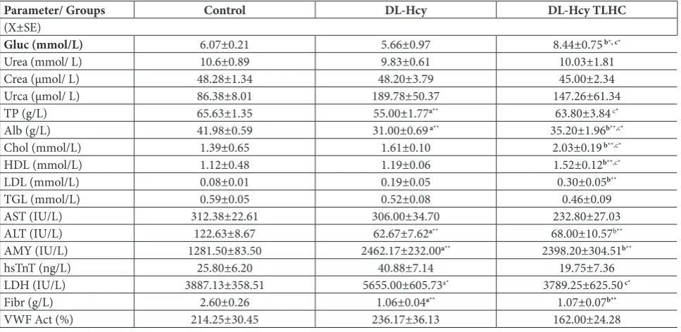
Administration of DL-Hcy (p<0.01) and DL-Hcy TLHC (p<0.05) induced a statistically significant decrease in MDA as compared to the control group (Fig. 1A).
CAT, GPx and SOD activities
Administration of DL-Hcy TLHC induced a statisti-cally significant increase in CAT activity in the plasma as compared to the control group (p<0.01). Further-more, there was significant increase of CAT activity after administration DL-Hcy TLHC when compared to the administration of DL-Hcy (p<0.05) (Fig. 1B).
The same trend in GPx activity was observed after the administration of DL-Hcy and DL-Hcy TLHC. Administration of DL-Hcy TLHC induced a statisti-cally significant increase in GPx activity as compared to the control (p<0.01). There was highly significant increase in GPx activity after DL-Hcy TLHC adminis-tration when compared to DL-Hcy (p<0.01) (Fig. 1C). After DL-Hcy administration, SOD activity was statistically increased (p<0.01). DL-Hcy TLHC ad-ministration decreased SOD activity without statistical
significance. A highly significant increase in SOD ac-tivity was noted after DL-Hcy administration in com-parison to DL-Hcy TLHC administration (p<0.01) (Fig. 1D).
AChE activity
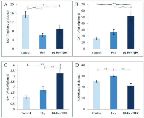
AChE activity was significantly decreased after the ad-ministration of DL-Hcy (p<0.01) and DL-Hcy TLHC (p<0.01) as compared to the control (Fig. 2).
DISCUSSION
Our first objective was to investigate the influence of acute HHcy on different serum biochemical param-eters in order to assess the systemic response to an DL-Hcy load. Our results showed that an acute rise in DL-Hcy and its thiolactone metabolite did not affect the concentrations of most routinely examined bio-chemical markers (Table 1), the only exception being an increase in total cholesterol concentrations and its HDL and LDL fractions in rats treated with DL-Hcy TLHC. The data referring to the effect of increased Hcy on the lipid profile are debatable. A previous study of rats that received DL-Hcy TLHC for 30 days (1 g/kg body weight/day) showed elevated serum
cho-Table 1. Values of measured biochemical parameters in the plasma expressed in standard units (x±SEM).
Parameter/ Groups Control DL-Hcy DL-Hcy TLHC
(X±SE)
Gluc (mmol/L) 6.07±0.21 5.66±0.97 8.44±0.75 b*, c*
Urea (mmol/ L) 10.6±0.89 9.83±0.61 10.03±1.81
Crea (μmol/ L) 48.28±1.34 48.20±3.79 45.00±2.34
Urca (μmol/ L) 86.38±8.01 189.78±50.37 147.26±61.34
TP (g/L) 65.63±1.35 55.00±1.77a** 63.80±3.84 c*
Alb (g/L) 41.98±0.59 31.00±0.69 a** 35.20±1.96b**,c*
Chol (mmol/L) 1.39±0.65 1.61±0.10 2.03±0.19 b**,c*
HDL (mmol/L) 1.12±0.48 1.19±0.06 1.52±0.12b**,c*
LDL (mmol/L) 0.08±0.01 0.19±0.05 0.30±0.05b**
TGL (mmol/L) 0.59±0.05 0.52±0.08 0.46±0.09
AST (IU/L) 312.38±22.61 306.00±34.70 232.80±27.03
ALT (IU/L) 122.63±8.67 62.67±7.62a** 68.00±10.57b**
AMY (IU/L) 1281.50±83.50 2462.17±232.00a** 2398.20±304.51b**
hsTnT (ng/L) 25.80±6.20 40.88±7.14 19.75±7.36
LDH (IU/L) 3887.13±358.51 5655.00±605.73a* 3789.25±625.50 c*
Fibr (g/L) 2.60±0.26 1.06±0.04a** 1.07±0.07b**
VWF Act (%) 214.25±30.45 236.17±36.13 162.00±24.28
lesterol and triglycerides [24], with which our results agree. Interaction between Hcy and lipids is poorly understood, especially with regard to the activity of the enzyme paraoxonase. Nevertheless, it has been proposed that DL-Hcy induces endoplasmic reticulum stress and activates sterol regulatory element-binding proteins (SREBPs), which could increase the expres-sion of genes responsible for cholesterol/triglyceride biosynthesis [25].
The kidneys are important in the removal of Hcy, and they could be affected by elevated plasma DL-Hcy levels. Although a recent investigation on mice suggests that persistently in-creased levels of DL-Hcy induce extra-cellular matrix remodeling and renal fibrosis [26], the parameters of renal function we examined (Urea, Crea and Urca) were not changed following the acute increase in DL-Hcy.
The liver possesses the highest catabolic capacity for DL-Hcy, and in-creases in DL-Hcy or methionine levels can lead to liver oxidative/nitrosative stress [27]. Results linked with hepato-cellular damage (AST and ALT) showed that HHcy is not linked to hepatocel-lular damage; however, the observed lower levels of serum Alb and Fibr in both experimental groups indicate that there is an initial impairment of liver function. Disturbed fibrinogen levels may occur due to an established process of homocysteine vs proteins interactions. Recent studies have demonstrated that treatment with methionine for 8 weeks (100 mg/kg) led to liver dysfunction through increased ROS pro-duction [28].
The observed alterations in serum glucose and AMY concentrations in acute HHcy could be linked with pancreatic dysfunction. Also, the higher glucose concentration in the DL-Hcy TLHC group could be the result of insulin resistance. A previous study on rats has shown that HHcy was associated with a de-crease in the glucose:insulin ratio and an inde-crease in the insulin resistance index [29]. It was also found that a 2-week treatment with DL-Hcy decreased the cerebral expression of GLUT-1 and produced a con-comitant increase in the percentage of damaged cer-ebral vessels in rats [30].
In addition, we observed increased levels of hsTnT (very close to statistical significance) and LDH, as indicators of cardiac and brain damage. The limited metabolic capacity for DL-Hcy in the cardiovascular system might be linked to injury of the cardiovascular system induced by Hcy.
Fig. 2. The effects of DL-Hcy and DL-Hcy TLHC on AChE activ-ity in rat heart. P<0.05 was considered to be significant (*p<0.05; **p<0.01).
Fig. 1. The effects of DL-Hcy and DL-Hcy TLHC on selected markers of oxidative
stress in rat plasma. A – MDA concentration; B – CAT activity; C – GPx activity;
Our study showed that both DL-Hcy and DL-Hcy TLHC decreased MDA, while there was no significant difference between these experimental groups and its metabolite. These results are not in agreement with the results of other studies where it was shown that acute HHcy increased lipid peroxidation in rats [11]. Additionally, it was shown [9] that chronic DL-Hcy ad-ministration also promoted oxidative damage by lipid peroxidation [31]. However, we did not note changes in the index of lipid peroxidation in the isolated rat heart treated with three isoforms of 10 μM homocysteine (DL-Hcy, DL-Hcy TLHC and L-Hcy TLHC) [32]. The discrepancy between our results and the mentioned investigations may be due to the acute application of substances and the resulting lack of time for achieving their negative effects on oxidative stress markers.
Having in mind that GPx-1 may be a key target of the deleterious actions of DL-Hcy [33], it was important to attempt to reveal the complex relationship between HHcy and the activity of this enzyme. We showed that the basic form of DL-Hcy did not significantly affect GPx activity, while DL-Hcy TLHC induced an increase in the activity of this enzyme. One possible explanation for the rise in GPx activity could be the chemical reac-tivity of the toxic thiolactone metabolite, which pos-sesses the ability to cause prominent activation of the glutathione antioxidant system [34]. In contrast, other investigationsfound that in acute HHcy, GPx activ-ity was decreased in a similar experimental model (0.6 μmol DL-Hcy/g body weight) [11]. Nevertheless, the authors used a much lower concentration of DL-Hcy, which may be not have been enough to mobilize this part of the defense system.
On the other hand, we observed completely op-posite results in the case of SOD activity. Namely, the application of DL-Hcy led to a significant increase in SOD, while DL-Hcy-TLHC did not change it. Litera-ture data referring to the DL-Hcy-SOD relation are controversial. Previous studies in rats have shown that 3 weeks of DL-Hcy administration decreased SOD levels in the plasma [9,31]. However, although the time under HHcy was prolonged, its concentration was more than 1000 times lower, which may be the reason for the difference to our investigation. In ad-dition, other investigations found that patients with significantly elevated DL-Hcy have elevated SOD levels [14]. Based on the abovementioned, it would
seem that concentration determines the direction of DL-Hcy toward SOD, i.e. in mild HHcy, SOD is de-creased while in prominent HHcy the activity of this enzyme is strongly activated.
Unlike SOD, CAT values showed a different dy-namic. Thus, only the thiolactone form induced a sig-nificant increase in plasma CAT activity. Recent investi-gations on animal models also observed increased CAT activity 15 min after acute DL-Hcy administration, suggesting that the oxidative status of rats was altered [11]. Taking into consideration the increased activities of all antioxidant enzymes, we suggested that DL-Hcy and DL-Hcy TLHC have prooxidant effects. However, the decrease in MDA levels during the same period could mean that the rise in antioxidant activity was inversed. In contrast, chronic administration of DL-Hcy can decrease CAT levels in the plasma, heart and aorta, which are accompanied by histological changes [9]. This finding indicates that, besides changed con-centrations, the duration of HHcy can also contribute to the effects of DL-Hcy on antioxidative enzyme activity. The potential mechanisms of DL-Hcy-induced changes in antioxidant defense enzymes probably include dif-ferent interactions of ROS and DL-Hcy [9].
As previously described [17], it has been assumed that the harmful effects of elevated homocysteine could in part be due to its actions on cholinesterases. In the available literature, there is almost no information concerning the effects of DL-Hcy on AChE activity in the heart. According to the presented results, both DL-Hcy and DL-Hcy TLHC decreased AChE activity in cardiac tissue homogenates (Fig. 2). However, the thiolactone form induced a more prominent decrease in AChE activity, revealing its more deleterious influence on cardiac tissue than Hcy itself. Investigations on hu-man subjects also found that thiolactone slowly and ir-reversibly inhibited the activity of AChE in plasma [17]. Interestingly, in a recent paper [18] it was suggested that increased cholinergic tone in the heart caused by cholinesterase inhibitors has a positive effect on some cardiovascular disorders such as heart failure.
CONCLUSION
responses in different organs. Following acute i.p. administration of DL-Hcy or DL-Hcy TLHC, the activities of antioxidant enzymes in the plasma were significantly increased. Our findings demonstrate that these amino acids have prooxidant effects. In addition, the obtained results were dependent on the form of acutely applied DL-Hcy.
Acknowledgments: This work was supported by Grant No.175043 from the Ministry of Education, Science and Technical Develop-ment of the Republic of Serbia.
Author contributions: Duško Kornjača performed the experi-ments and the statistical analysis, interpreted the data, analyzed the results and prepared the manuscript; Vladimir Živković per-formed the statistical analysis, interpreted the data, analyzed the results and prepared the manuscript; Danijela Krstić contributed to the study design, performed the biochemical analysis and interpreted the data; Mirjana Čolović contributed to the study design, performed the biochemical analysis and interpreted the data; Marko Đurić performed the experiments; Sanja Stanković performed the biochemical analysis; Slavica Mutavdžin performed the experiments and the statistical analysis, provided the literature review and prepared the graphics; Vladimir Jakovljević contrib-uted to the study design, performed the statistical analysis, inter-preted the data, analyzed the results and prepared the manuscript; Dragan Đurić contributed to the study design, perform the experi-ments, performed the biochemical and statistical analysis, inter-preted the data, analyzed the results and prepared the manuscript. Conflict of interest disclosure: All authors disclose no actual or potential conflicts of interest, including any financial, personal, or other relationships with people or organizations.
REFERENCES
1. Ganguly P, Alam SF. Role of homocysteine in the develop-ment of cardiovascular disease. Nutr J. 2015;14:6.
2. Malinowska J, Kolodziejczyk J, Olas B. The disturbance of hemostasis induced by hyperhomocysteinemia; the role of antioxidants. Acta Biochim Pol. 2012;59(2):185-94.
3. Shenov V, Mehendale V, Prabhu K, Shetty R, Rao P.
Correla-tion of serum homocysteine levels with the severity of coro-nary artery disease. Indian J Clin Biochem. 2014;29(3):339-44. 4. Jakubowski H. The pathophysiological hypothesis of homo-cysteine thiolactone-mediated vascular disease. J Physiol Pharmacol. 2008;59(9):155-67.
5. Schaffer A, Verdoia M, Cassetti E, Marino P, Suryapra-nata H, De Luca G. Novara Atherosclerosis Study Group (NAS). Relationship between homocysteine and coronary artery disease. Results from a large prospective cohort study. Thromb Res. 2014;134(2):288-93.
6. Baszczuk A, Kopczyński Z, Thielemann A. Endothelial dys-function in patients with primary hypertension and hyper-homocysteinemia. Postepy Hig Med Dosw. 2014;68:91-100.
7. Chen C, Halkos ME, Surowiec SM, Conklin BS, Lin PH, Lumsden AB. Effects of homocysteine on smooth muscle cell proliferation in both cell culture and artery perfusion culture models. J Surg Res. 2000;88(1):26-33.
8. Toohey JI. Homocysteine toxicity in connective tissue: theo-ries, old and new. Connect Tissue Res. 2008;49(2):57-61. 9. Derouiche F, Bôle-Feysot C, Naïmi D, Coëffier M.
Hyperho-mocysteinemia-induced oxidative stress differentially alters proteasome composition and activities in heart and aorta. Biochem Biophys Res Commun. 2014;452(3):740-5. 10. Liu HH, Shih TS, Huang HR, Huang SC, Lee LH, Huang YC.
Plasma homocysteine is associated with increased oxidative stress and antioxidant enzyme activity in welders. Sci World J. 2013;e370487.
11. da Cunha AA, Scherer E, da Cunha MJ, Schmitz F, Machado FR, Lima DD, Delwing D, Wyse AT. Acute hyperhomocyste-inemia alters the coagulation system and oxidative status in the blood of rats. Mol Cell Biochem. 2012;360(1-2):205-14. 12. Kerkeni M, Added F, Ben Farhat M, Miled A, Trivin F, Maar-oufi K. Hyperhomocysteinaemia and parameters of anti-oxidative defence in Tunisian patients with coronary heart disease. Ann Clin Biochem. 2008;45(2):193-8.
13. Matté C, Mackedanz V, Stefanello FM, Scherer EB, Andreazza AC, Zanotto C, Moro AM, Garcia SC, Gonçalves CA, Erdtmann B, Salvador M, Wyse AT. Chronic hyperho-mocysteinemia alters antioxidant defenses and increases DNA damage in brain and blood of rats: protective effect of folic acid. Neurochem Int. 2009;54(1):7-13.
14. Wilcken DE, Wang XL, Adachi T, Hara H, Duarte N, Green K, Wilcken B. Relationship between homocysteine and superoxide dismutase in homocystinuria: possible rel-evance to cardiovascular risk. Arterioscler Thromb Vasc Biol. 2000;20(5):1199-202.
15. Hrnčić D, Rašić-Marković A, Stojković T, Velimirović M, Puškaš N, Obrenović R, Macut D, Sušić V, Jakovljević V, Djuric D, Petronijević N, Stanojlović O. Hyperhomocyste-inemia induced by methionine dietary nutritional overload modulates acetylcholinesterase activity in the rat brain. Mol Cell Biochem. 2014;396(1-2):99-105.
16. Scherer EB, Loureiro SO, Vuaden FC, da Cunha AA, Schmitz F, Kolling J, Savio LE, Bogo MR, Bonan CD, Netto CA, Wyse AT. Mild hyperhomocysteinemia increases brain acetylcholinesterase and proinflammatory cytokine levels in different tissues. Mol Neurobiol. 2014;50(2):589-96. 17. Darvesh S, Walsh R, Martin E. Homocysteine
thiolac-tone and human cholinesterases. Cell Mol Neurobiol. 2007;27(1):33-48.
18. Kučera M, Hrabovská A. Cholinergic system of the heart. Ceska Slov Farm. 2015;64(6):254-63.
19. Aruoma OI, Halliwell B, Laughton MJ, Quinlan GJ, Gutter-idge JMC. The mechanism of initiation of lipid peroxidation. Evidence against a requirement for an iron (II)- iron (III) complex. Biochem J. 1989;258:617-20.
20. Beutler E. Red Cell Metabolism: A manual of biochemical
methods. 3rd ed. New York: Grune Startton; 1984. 133 p.
22. Wendel A. Enzymatic basis of detoxication. Vol. 1. New York: Academic Press; 1980. 333 p.
23. Ellman GL, Courtney KD, Andres VJr, Feather-Stone RM. A new and rapid colorimetric determination of acetylcholines-terase activity. Biochem Pharmacol. 1961;7:88-95.
24. de Andrade CR, Tirapelli CR, Haddad R, Eberlin MN, Ramalho LN, Iyomasa MM, Uyemura SA, de Oliveira AM. Hyperhomocysteinemia induced by feeding rats diets rich in DL-homocysteine thiolactone promotes alterations on carotid reactivity independent of arterial structure. Vascul Pharmacol. 2009;51(4):291-8.
25. Werstuck GH, Lentz SR, Dayal S, Hossain GS, Sood SK, Shi YY, Zhou J, Maeda N, Krisans SK, Malinow MR, Austin RC. Homocysteine-induced endoplasmic reticulum stress causes dysregulation of the cholesterol and triglyceride biosynthetic pathways. J Clin Invest. 2001;107(10):1263-73.
26. Pushpakumar S, Kundu S, Narayanan N, Sen U. DNA hyper-methylation in hyperhomocysteinemia contributes to abnor-mal extracellular matrix metabolism in the kidney. FASEB J. 2015;29(11):4713-25.
27. Yamada H, Akahoshi N, Kamata S, Hagiya Y, Hishiki T, Nagahata Y, Matsuura T, Takano N, Mori M, Ishizaki Y, Izumi T, Kumagai Y, Kasahara T, Suematsu M, Ishii M. Methionine excess in diet induces acute lethal hepatitis in mice lacking cystathionine γ-lyase, an animal model of cys-tathioninuria. Free Radic Biol Med. 2012;52(9):1716-26.
28. Mendes RH, Mostarda C, Candido GO, Moraes-Silva IC, D’Almeida V, Belló-Klein A, Irigoyen MC, Rigatto K. Moder-ate hyperhomocysteinemia provokes dysfunction of cardio-vascular autonomic system and liver oxidative stress in rats. Auton Neurosci. 2014;180:43-7.
29. Golbahar J, Aminzadeh MA, Kassab SE, Omrani GR. Hyper-homocysteinemia induces insulin resistance in male Sprague-Dawley rats. Diabetes Res Clin Pract. 2007;76(1):1-5. 30. Lee H, Kim JM, Kim HJ, Lee I, Chang N. Folic acid
supple-mentation can reduce the endothelial damage in rat brain microvasculature due to hyperhomocysteinemia. J Nutr. 2005;135(3):544-8.
31. Kolling J, Scherer EB, da Cunha AA, da Cunha MJ, Wyse AT. Homocysteine induces oxidative-nitrative stress in heart of rats: prevention by folic acid. Cardiovasc Toxicol. 2011;11(1):67-73.
32. Zivkovic V, Jakovljevic V, Djordjevic D, Vuletic M, Barudzic N, Djuric D. The effects of homocysteine-related com-pounds on cardiac contractility, coronary flow, and oxida-tive stress markers in isolated rat heart. Mol Cell Biochem. 2012;370(1-2):59-67.